Article

Investigation of the Effect of Preparation Parameters on the Structural and Mechanical Properties of Gelatin/Elastin/Sodium Hyaluronate Scaffolds Fabricated by the Combined Foaming and Freeze-Drying Techniques

1
Department of Mechanical Engineering, Marquette University, Milwaukee, WI 53233, USA
2
Marquette University School of Dentistry, Milwaukee, WI 53233, USA
3
Institute for Engineering in Medicine, Health, & Human Performance (EnMed), Batten College of Engineering and Technology, Old Dominion University, Norfolk, VA 23529, USA
*
Author to whom correspondence should be addressed.
J. Compos. Sci. 2024, 8(10), 408; https://doi.org/10.3390/jcs8100408
Submission received: 14 May 2024 / Revised: 16 September 2024 / Accepted: 24 September 2024 / Published: 4 October 2024

Abstract

:
This paper aimed to evaluate the effects of different preparation parameters, including agitation speed, agitation time, and chilling temperature, on the structural and mechanical properties of a novel gelatin/elastin/sodium hyaluronate tissue engineering scaffold, recently developed by our research group. Fabricated using a combination of foaming and freeze-drying techniques, the scaffolds were assessed to understand how these parameters influence their morphology, internal microstructure, porosity, mechanical properties, and degradation behavior. The fabrication process used in this study involved preparing a homogeneous aqueous solution containing 8% gelatin, 2% elastin, and 0.5% sodium hyaluronate (w/v), which was then subjected to mechanical agitation at speeds of 500, 1000, and 1500 rpm for durations of 5, 15, and 25 min. This mixture was subsequently frozen at −20 °C and −80 °C, followed by freeze-drying and cross-linking. Morphological analyses using laser microscopy and scanning electron microscopy (SEM) demonstrated that the scaffolds had pore sizes ranging from 100 to 300 µm, which are conducive to effective cell interaction and tissue regeneration. This confirmed the efficacy of the combined foaming and freeze-drying method in creating highly interconnected porous structures. Our findings indicated that chilling temperature slightly influenced pore size. In contrast, higher agitation speeds and longer duration times led to increased porosity and degradation rate but decreased modulus. Mathematical estimators were developed for the porosity and compressive modulus of the scaffolds by statistical analysis of the preparation parameters. The estimators were validated experimentally, with the error between estimated and experimental values being less than 6% for porosity and less than 21% for compressive modulus.

1. Introduction

Scaffolds are essential components in tissue engineering, providing the structural framework to support new tissue formation. They offer mechanical support, facilitate cell attachment and proliferation, and deliver biochemical signals crucial for tissue development [1]. Scaffolds mimic the extracellular matrix (ECM) to create an environment that supports both the structural and biochemical aspects of tissue formation, aiding cells in differentiation, migration, and ECM deposition [2]. For a scaffold to be effective in regenerative medicine, it must integrate well with the host tissue. This integration requires the scaffold to be biocompatible, possess mechanical properties similar to the target tissue, and degrade at a rate that aligns with the formation of new tissue [3]. Therefore, developing scaffolds involves a delicate balance between mimicking the natural ECM and tailoring the scaffold’s properties to meet the specific needs of the engineered tissue.
In engineered scaffolds, careful material selection is crucial to ensure biocompatibility and resemblance to the native ECM while incorporating biological signals that influence cell functions and regenerative outcomes [4]. The chosen materials need to mimic the native ECM as closely as possible, providing a conducive environment for cellular attachment and proliferation. Various polymers and biopolymers have been widely used to fabricate scaffolds. For example, poly(lactic-co-glycolic acid) (PLGA) is popular due to its biodegradability and biocompatibility [5,6,7], chitosan is valued for its natural origin and antibacterial properties [8,9,10,11], and collagen is essential as a primary ECM protein [12,13,14,15,16]. The choice of materials must consider both the mechanical and biochemical properties required for effective tissue regeneration. While synthetic polymers such as PLGA offer robust mechanical support, natural biopolymers such as gelatin, elastin, and sodium hyaluronate are favored for their inherent biocompatibility and close resemblance to the native ECM. Gelatin is a natural biopolymer known for its high biocompatibility and ability to form hydrogels, making it suitable for creating structures that mimic the natural environment of living cells and tissues [17,18]. Elastin provides essential mechanical properties, particularly elasticity, which mimics the characteristics of native tissues [19,20]. It also has a low degradation rate, offering controllability over the biodegradability and release behaviors of the scaffold. Sodium hyaluronate retains water, contributing to hydration and creating a favorable microenvironment for cellular activities. It also provides lubrication, aiding in cellular movement, and supports cellular migration, which is crucial for various biological processes [21,22]. Studies on blends of these biopolymers [23,24,25,26], along with similar studies found in the literature on gelatin-elastin-hyaluronic acid combinations [27,28], have consistently demonstrated highly promising biological properties. These studies show significant biocompatibility, controllable biodegradability, structural similarity to ECM, and ability to promote cell adhesion and migration, creating scaffolds that are both structurally sound and biologically active, closely resembling native tissue, and enabling more effective tissue regeneration.
In addition to the choice of materials, fabrication methods play a critical role in determining the properties of the final scaffolds, ensuring desirable physical characteristics such as porosity and stiffness. Different techniques are utilized to achieve varying levels of porosity, mechanical strength, and interconnected structures. Freeze-drying (lyophilization) is a widely used method that creates highly porous scaffolds by sublimating the solvent from a frozen polymer solution, which is particularly effective for natural biopolymers such as gelatin and elastin [1,29,30,31]. Electrospinning is another versatile technique that produces fibrous structures by applying high voltage to a polymer solution, creating a network that closely resembles the native ECM [8,32,33,34]. 3D printing offers precise control over scaffold architecture by layering biodegradable polymers or hydrogel inks to create complex structures [25,35,36,37,38,39]. Each method contributes to developing scaffolds that meet specific requirements for tissue engineering, providing essential support for effective regeneration.
In addition to fabrication methods, preparation parameters play a pivotal role in determining the physicochemical properties of the resulting scaffolds. These parameters are strongly dependent on the method of fabrication. For example, in electrospinning, critical parameters include the voltage applied, the flow rate of the polymer solution, and the distance between the needle and the collecting plate. These factors influence the fiber diameter, pore size, and overall scaffold morphology, which are crucial for cell attachment and proliferation [40,41,42]. In 3D printing, preparation parameters such as print speed, layer thickness, and nozzle temperature are essential for achieving the desired scaffold architecture and mechanical strength. These parameters affect the resolution, porosity, and interconnectivity of the printed scaffolds, which are important for mimicking the natural extracellular matrix [43].
For freeze-drying, preparation parameters include the concentration of the polymer solution, freezing temperature, and the rate of solvent sublimation. These factors significantly influence the pore structure, mechanical properties, and degradation behavior of the scaffolds. Literature reviews indicate that the polymer concentration affects the scaffold’s density and mechanical strength, where higher concentrations typically result in denser and stronger scaffolds [44]. Additionally, the rate of sublimation during freeze-drying can be adjusted to control the microstructure and porosity of the scaffolds, optimizing them for various tissue engineering applications [45].
Recently, Rasoulianboroujeni et al. [46] combined the freeze-drying method with a mechanical foaming technique to produce large, highly porous gelatin scaffolds with dual-scale porosity that exhibit minimal shrinkage and deformation compared to those created using conventional freeze-drying. The mechanical foaming technique incorporated gas bubbles to form interconnected pores, significantly enhancing scaffold permeability. In this method, an aqueous gelatin solution was agitated at controlled speeds to create a stable foam. The foamy solution was then molded and freeze-dried to form a porous network. This dual-stage process resulted in scaffolds that support enhanced cell infiltration and proliferation due to their unique porosity.
The initial results of this study demonstrated that the combined freeze-drying and foaming technique yields scaffolds with excellent permeability and porosity. Although this method is novel, it shows significant potential. Further evaluation of the preparation parameters could enhance the scaffold properties, making them more suitable for tissue engineering applications. However, the preparation parameters for this combined method differ from those of conventional freeze-drying. In addition to conventional freeze-drying parameters, such as polymer concentration, freezing temperature, and sublimation rate, this method includes parameters related to foaming, including agitation speed and agitation duration time. While the effects of preparation parameters related to conventional freeze-drying have been investigated extensively in the literature [44,45,47], the impact of parameters specific to the foaming process has not been previously explored.
Therefore, the aim of this study was to conduct a detailed investigation into the effects of different preparation parameters, including agitation speed, agitation time, and chilling temperature, on the structural and mechanical properties of the scaffolds. By investigating these parameters, we can develop scaffolds with enhanced mechanical strength, porosity, and degradation rates, ultimately improving their potential for tissue engineering applications.

2. Materials and Methods

2.1. Materials

In the fabrication of scaffolds for this study, the following materials were used: (i) gelatin (type A, derived from porcine skin, bioreagent grade), (ii) soluble elastin (derived from bovine neck ligament, molecular weight 60 kDa), and (iii) sodium hyaluronate (research grade, molecular weight range 500–749 kDa), sourced respectively from Sigma-Aldrich (St. Louis, MO, USA), Elastin Products Company Inc. (Chaska, MN, USA), and Lifecore Biomedical (Ward Hill, MA, USA). Additionally, cross-linking agents N-hydroxysulfosuccinimide (NHS) and 1-Ethyl-3-(3-dimethylaminopropyl) carbodiimide hydrochloride (EDC) were acquired from Alfa Aesar (Haverhill, MA, USA) and employed in the sample preparation process.

2.2. Samples Preparation

As schematically shown in Figure 1, the combination of foaming and freeze-drying methods, previously introduced by our group [46], was used to fabricate the scaffolds in this paper. A composition consisting of 8% gelatin, 2% elastin, and 0.5% sodium hyaluronate (w/v) in an aqueous solution was used to fabricate the samples. This composition was selected based on our previous works [23,24,25,26], which demonstrated that this blend of biopolymers provides a favorable balance between mechanical stability, porosity, and biocompatibility. Notably, while the composition ratios have not been fully optimized for the specific combination of foaming and freeze-drying methods used here, this formulation has been shown to effectively promote cellular functions and maintain scaffold integrity in our earlier research. Therefore, this composition was chosen as a basis to explore the influence of preparation parameters on mechanical properties and porosity, aligning with the core focus of our study, which is centered around preparation techniques rather than chemical composition optimization. In this process, gelatin, elastin, and sodium hyaluronate were dissolved in distilled water, which was then subjected to agitation by a mechanical mixer (IKA, Wilmington, NC, USA) at specified speeds and duration times, as reported in Table A1. The foamed samples were then kept at two different freezing temperatures of −20 °C and −80 °C overnight and subsequently freeze-dried in a manifold freeze-dryer (Labconco, Kansas City, MO, USA) with a condenser temperature of −55 °C under 1 Pa for 48 h. The samples were next crosslinked using 4 mg/mL EDC and 0.5 mg/mL NHS in 90% v/v ethanol at 4 °C overnight. Finally, the samples were thoroughly washed in 1 L of DI water four times (each time for 30 min) to ensure the complete removal of EDC and NHS. This washing procedure was based on previous studies and supported by the references cited, which report acceptable cell viability following this crosslinking method [26,27,48,49]. After washing, the samples were frozen at −20 °C and freeze-dried again following the same protocol to obtain the final products.

2.3. Characterization

The surface morphology of the samples was characterized by LEXT OLS4000 3D laser measuring microscopy (Olympus, Tokyo, Japan). Meanwhile, the microstructure of the scaffolds was examined using scanning electron microscopy (SEM, Tokyo, Japan, JEOL JSM-6510LV) at an acceleration voltage of 15 kV.
The values of total porosity and open porosity of the samples were further measured using Archimedes’ principle at room temperature. Ethanol was selected as the solvent as it infiltrates into the scaffolds’ pores without swelling or shrinking the matrix. The total and open porosities of the scaffolds were calculated using the following set of equations [50,51]:
ρ s a m p l e = m d r y × ρ e t h m w e t m s u s
P t o t a l % = 1 ρ s a m p l e ρ 0 × 100
P o p e n % = m w e t m d r y m w e t m s u s × 100
where ρ s a m p l e , ρ e t h , and ρ 0 are the density of the sample, ethanol, and the base material in g/cm3, respectively. Moreover, P t o t a l is the total porosity percentage and P o p e n denotes the open porosity percentage. Meanwhile, m d r y , m s u s , and m w e t are the respective mass of the dry sample, the mass of the sample suspended in ethanol, and the mass of the sample after soaking in ethanol. Measurements were performed for three randomly selected samples from each group.
To study the weight loss of the samples over time, three samples from each group were randomly selected and immersed in phosphate-buffered saline (PBS) at predetermined intervals of 1, 2, 4, 7, and 14 days. The PBS was regularly exchanged to maintain consistent conditions. The dry weight of each sample on the initial day served as a reference for subsequent measurements. The relative weight of the samples at each time point was normalized by dividing the weight observed on a given day by the initial dry weight. This normalization process facilitated a comparative analysis of degradation rates across different groups. Average weight percentages and standard deviations were calculated for each group at the specified time intervals to quantify the degradation behavior. The degradation tests were conducted at a constant temperature of 37 °C.
The compressive modulus of the samples was measured using a universal testing machine (UTM, AGS-X, Shimadzu, Kyoto, Japan), equipped with a 1 kN load cell. The experiments were conducted by crushing the cylinder samples of ~6 mm height and ~3 mm radius between two flat plates at a constant compression speed of 1 mm/min until failure. Measurements were performed in triplicate, averaged, and reported.

2.4. Statistical Analysis

Quantitative data were analyzed and presented as the mean ± standard deviation, using Microsoft Excel software (Microsoft Excel for Microsoft 365, Version 2408, Microsoft Corporation, Redmond, WA, USA) for all statistical computations. Moreover, in this study, mathematical estimators were developed for scaffolds’ key properties, namely porosity and compressive modulus, based on agitation speeds and duration times for each chilling temperature. To rigorously assess the interactions among these variables, we utilized a second-order multiple regression analysis based on data from a Central Composite Design (CCD) [52]. The CCD is a sophisticated three-level, two-factor fractional factorial method that systematically explores the effect of each variable on scaffold performance. Through this approach, we were able to model the complex dynamics between preparation parameters and scaffold properties. The regression model is expressed as follows:
Y = β 0 + i = 1 n β i X i + i = 1 n β i i X i 2 + i < j β i j X i X j
where Y represents the response variables (compressive modulus and porosity), X i and X j denote the independent variables (agitation speed and duration time), β 0 , β i , β i i , and β i j signify the model constants, linear coefficients, quadratic coefficients, and interaction coefficients, respectively. Regression analysis was performed using MATLAB software (Version 9.13, R2022b, MathWorks, Natick, MA, USA).
In order to validate the obtained estimators, additional samples were fabricated under specific conditions to verify their accuracy. The samples were prepared using the same protocol and composition, with agitation speeds of 800 rpm and 1200 rpm for 10 min at chilling temperatures of −20 °C and −80 °C. For each set of conditions, three randomly selected samples were tested to measure the porosity and compressive modulus, using the same methods employed in the initial experiments. The results were then compared with those predicted by the estimators.

3. Results and Discussion

3.1. Characterization

Figure 2 and Figure 3 depict the SEM images of the typical samples fabricated at chilling temperatures of −20 °C (F0 to F9) and −80 °C (F10 to F19), respectively. These images showed that the pores within the fabricated scaffolds possessed appropriate interconnectivity. The high volume of open porosities observed within the scaffolds fabricated in this study is directly attributed to the innovative application of the foaming and freeze-drying methods during their construction. These processes uniquely contribute to the development of a highly porous structure. The freeze-drying method involves several critical steps. Initially, the sample is frozen, leading to the solidification of the solvent (typically water) within the sample. As the temperature continues to decrease, ice crystals form throughout the scaffold material. During the subsequent sublimation phase, these ice crystals are removed by reducing the pressure, causing the ice to sublimate directly into vapor without transitioning through the liquid phase. This process results in a network of interconnected pores in the locations previously occupied by the ice crystals. With this mechanism, the resulting pore distribution in the samples is typically uniform. Additionally, the foaming technique introduces gas bubbles that expand and create additional porosity within the scaffold matrix. This strategic combination ensures the formation of a scaffold with enhanced porosity and interconnectivity, essential for facilitating cell infiltration, nutrient diffusion, and tissue integration, thereby significantly improving the scaffold’s performance in tissue engineering applications [46].
Figure 4 shows the pore size distribution for different scaffolds fabricated in this study. Our findings indicated that the diameters of the majority of pores present in the scaffolds fell within the range of 100–300 µm, a dimension considered highly beneficial for tissue engineering purposes. Pore sizes within this range are optimal as they allow for effective cell interaction, migration, and nutrient exchange, crucial for tissue regeneration. Perez and Mestres [53] highlight that the ideal pore size for scaffolds should be between 100 and 400 µm to support efficient cell-scaffold interactions, particularly under static seeding conditions. Pore sizes below this optimal range can severely restrict cell activities, potentially leading to the demise of cells adhered to the scaffold. Pores exceeding 500 µm in size impede effective cell engagement with the scaffold, as cells may simply pass through without establishing attachment. Research has outlined specific pore size ranges that are considered optimal for the ingrowth and function of various cell and tissue types, including ranges of 70–120 μm for chondrocyte ingrowth [54], 40–150 μm for fibroblast attachment [55], and 200–350 μm for facilitating osteoconduction [56].
To evaluate the effect of fabrication parameters on the pore size of the scaffolds, a series of one-way ANOVA tests were performed on agitation speed, agitation time, and chilling temperature. Results of the ANOVA test yielded a p-value of ~0.84 and ~0.91 for agitation speed and time, respectively. Both p-values are substantially higher than the conventional α level of 0.05, which is commonly used as a benchmark for statistical significance. This indicates that at the 95% confidence level, neither agitation speed nor agitation time has a statistically significant effect on the mean diameter of the pores. Therefore, variations in these parameters do not contribute to meaningful differences in pore size that could impact the scaffold’s performance in tissue engineering applications.
On the other hand, chilling temperature produced a p-value of ~0.06. While this value is still above the standard threshold of 0.05 for statistical significance, it is marginally close and suggests a potential trend where temperature might influence the mean pore diameter. This near-significant p-value points to a possible effect of chilling temperature on pore size, emphasizing further investigation. Complementing these findings, a careful examination of the SEM images, i.e., Figure 2 and Figure 3, provides qualitative validation of the statistical data. These images suggest that a decrease in chilling temperature correlates with a slight increase in pore size within the scaffold matrix. This morphological trend observed in the SEM analysis resonates with the experimental outcomes reported by O’Brien et al. [57]. Their research, which meticulously explored the effect of varying freezing rates on pore sizes, found that a slower freezing process, typically associated with higher temperatures, leads to larger pore diameters. However, their findings showed that a very rapid freezing approach, such as quenching at −40 °C, unexpectedly yielded larger pores—a finding that aligns with our observations where samples quenched at −80 °C exhibited an increase in pore size. This seeming paradox could be attributed to the complex interplay between freezing dynamics and ice crystal formation, where extremely rapid freezing might lead to less uniform ice crystal formation and thus, larger pore spaces upon sublimation.
Figure 5a,b show the value of the porosity of the scaffolds at different agitation speeds and duration times fabricated at chilling temperatures of −20 °C and −80 °C. By examining the porosity measurements presented in this figure, we observe the interplay between agitation speed, time, and chilling temperature during the freeze-drying process on the resultant porosity of gelatin/elastin/sodium hyaluronate scaffolds. From the analysis of the results, it is clear that scaffolds fabricated at both −20 °C and −80 °C generally exhibit increased porosity with greater agitation speeds and extended agitation times. Statistical analysis confirms that both agitation speed (p-value = 4 × 10⁻¹⁰) and agitation time (p-value = 0.0008) significantly affect porosity. This pattern indicates that increased agitation during preparation may enhance the scaffold’s porosity. Specifically, the act of agitating may lead to the formation of a more open matrix, suggesting a potential refinement of the foam structure as agitation progresses, possibly due to the enhanced distribution of air pockets. The trend of higher porosity with increased agitation speed, particularly at 1500 rpm, may be attributed to the greater energy input, resulting in foam structures with better air incorporation and thus a higher porosity.
When assessing the effect of chilling temperature, scaffolds processed at −80 °C displayed a slight increase in porosity across all agitation parameters compared to those fabricated at −20 °C. Although this difference was not statistically significant according to the ANOVA test (p-value = 0.5287), the observation supports the fact that lower freeze-drying temperatures lead to the formation of larger ice crystals, which, upon sublimation, translate into more significant porosity within the scaffold.
Upon qualitative examination of the porosity trends from the scaffold data at −20 °C and −80 °C, it seemed that agitation speed plays a more pronounced role in influencing porosity, as demonstrated by significant changes across varied speeds. However, the impact of agitation time should not be overlooked, as there is a discernible increase in porosity corresponding to longer agitation periods. This observation underscores the importance of both factors in the scaffold’s structural configuration, with speed seemingly exerting a stronger influence within the experimental scope. The consistency in these trends, regardless of the chilling temperatures, underscores a steadfast correlation between the chosen agitation parameters and the scaffold’s porosity. This insight is crucial for tailoring scaffold architecture to meet the specific demands of biological applications in tissue engineering.
In addition to total porosity, Figure 5c and d show the value of open porosity of the scaffolds at chilling temperatures of −20 °C and −80 °C, respectively. Our findings indicate that at both chilling temperatures, the ratio of open porosity to total porosity exceeds 0.95. The fact that more than 95% of the pores are open indicates high interconnectivity between the pores. This high level of pore interconnectivity is crucial for scaffolds used in tissue engineering, as it facilitates the exchange of nutrients and waste, enhances cell migration, and promotes tissue integration and vascularization.
The combined analysis of total and open porosity provides a comprehensive understanding of the scaffold structure. The high ratio of open porosity not only confirms the effectiveness of the freeze-drying technique in creating interconnected pore networks but also highlights the suitability of these scaffolds for 3D cell culture applications. The enhanced interconnectivity observed supports the scaffold’s potential in facilitating effective tissue regeneration, making them highly suitable for various biomedical applications.
Figure 6 and Figure 7 show the results obtained from the degradation analysis of the samples. The analysis revealed that degradation generally increased with higher porosity, suggesting that scaffolds with greater porosity tend to degrade faster. This observation may be attributed to the larger surface area exposed to the environment, which accelerates the rate of scaffold breakdown. Furthermore, increased porosity often results in reduced mechanical stability, making the scaffold more susceptible to environmental factors. These findings align with previous research indicating that porous structures allow for greater fluid penetration, thus expediting the degradation process [58,59]. This comprehensive degradation study provides valuable insights into the stability and longevity of scaffolds under various conditions, highlighting the importance of carefully selecting processing parameters to optimize scaffold performance for biomedical applications.
The mechanical properties of tissue-engineered scaffolds are one of the most important factors that affect their performance in vivo. To ascertain the compression resistance of gelatin/elastin/sodium hyaluronate scaffolds, compression tests were performed on the fabricated scaffolds. Figure 8 demonstrates the value of the compressive modulus of the scaffolds under different fabrication conditions. Our findings showed that the mechanical properties of the samples were fundamentally influenced by the processing parameters, notably the agitation speed and duration during the pre-freeze mixing phase. The results suggest a complex interplay between these parameters and the resultant mechanical properties, highlighting a trend that suggests an increase in the agitation speed results in scaffolds with lower compressive modulus, pointing towards a less stiff structure. Moreover, prolonged agitation times, particularly at the highest speed of 1500 rpm, displayed a decrease in modulus, potentially implicating a degradation of structural integrity due to excessive porosity. From the findings, generally, increasing the agitation speed and duration leads to an increase in porosity values, subsequently causing a decrease in compressive modulus.
These insights into the mechanical behavior of scaffolds are critical as they directly impact the design and application of tissue-engineered products. These results are important, not only to mimic the native mechanical properties of tissues but also to ensure that the scaffolds can withstand the physiological loads they will encounter post-implantation. In fact, this mechanical characterization study of gelatin/elastin/sodium hyaluronate scaffolds provides a vital link between scaffold design parameters and their functional performance.

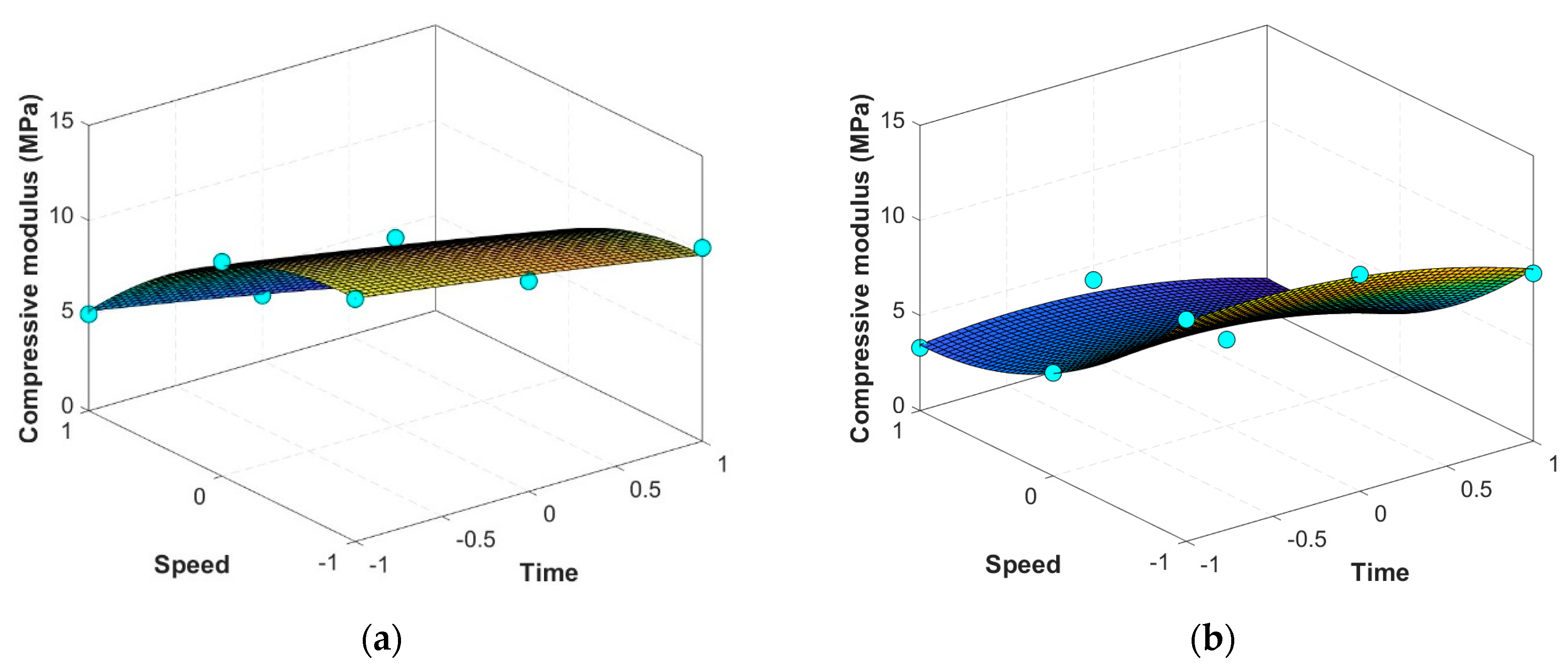
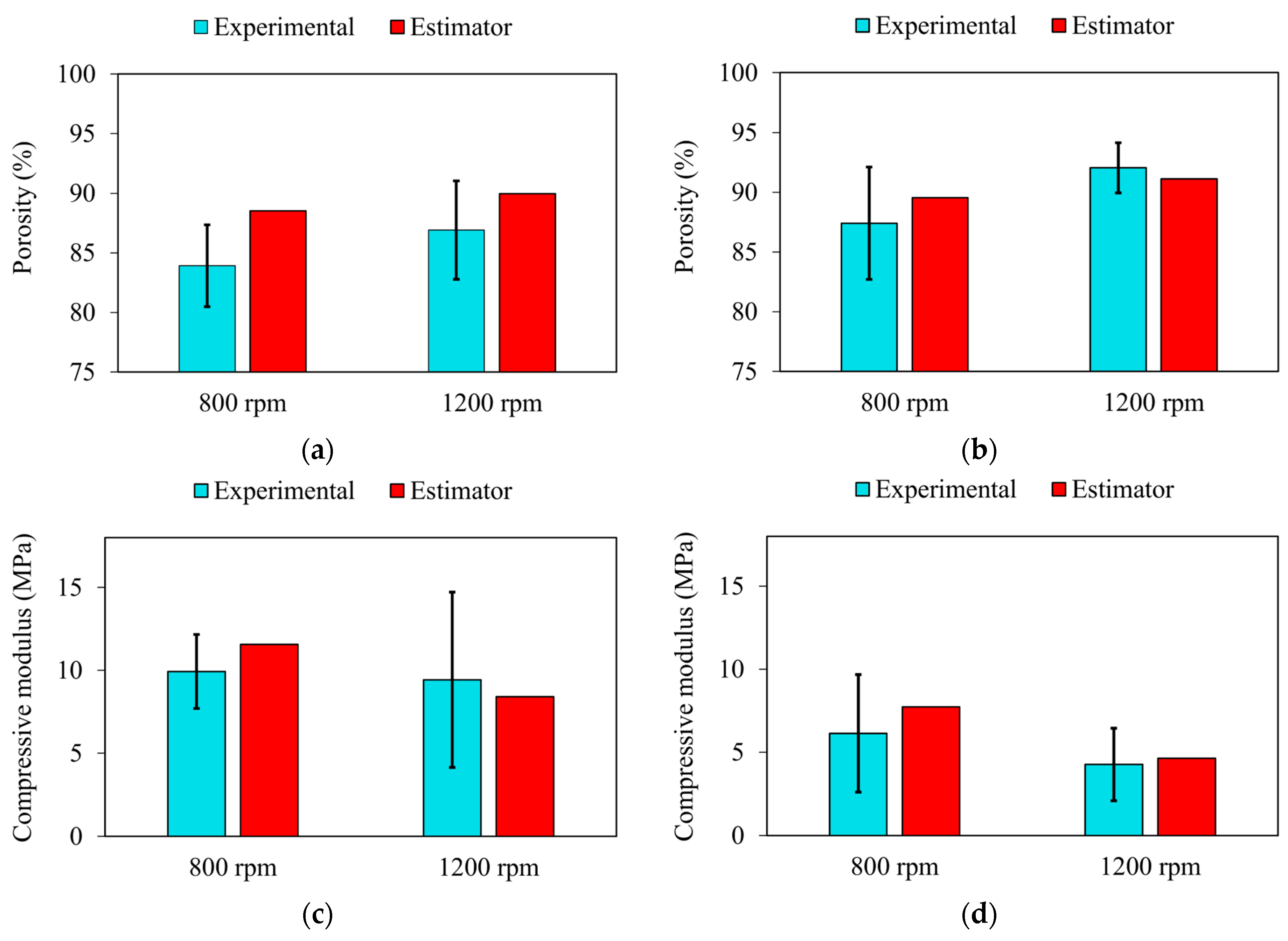
3.2. Mathematical Estimators for Scaffolds’ Properties

Our data set included measurements of compressive modulus and porosity for scaffolds processed at chilling temperatures of −20 °C and −80 °C. By employing regression analysis, we extracted estimators that described the relationship between our independent variables—time and speed of agitation—and our dependent variables—modulus and porosity—at the two different chilling temperatures. Figure 9 and Figure 10 show the results obtained from the regression model for the porosity and compressive modulus, respectively. Moreover, Table 1 reports the values obtained for coefficients calculated through regression analysis. Examination of these coefficients reveals that not all exert a statistically significant influence on the dependent variables, identified by p-values greater than 0.05. Based on these findings, and after discarding coefficients that lack statistical significance, we refined our models to yield the following empirical estimators for compressive modulus and porosity across the two chilling temperatures:
E 20 = 9.47972 1.66895 T 3.9414 V 2.03813 V 2 ,     R 2 = 0.99
E 80 = 5.3079 1.0655 T 3.8576 V + 2.1152 V 2 ,     R 2 = 0.98
20 = 89.67964 + 0.86910 T + 1.82147 V ,     R 2 = 0.95
80 = 90.96153 + 1.27855 T + 1.9665 V ,     R 2 = 0.93
where E 20 and E 80 denote the respective compressive modulus at −20 °C and −80 °C (in MPa), while 20 and 80 represent the porosity (%) at −20 °C and −80 °C, respectively. Additionally, T and V refer to the time and speed of agitation, respectively. It is worth mentioning that these estimators are able to capture the nonlinearities and interactions between the factors, providing a refined model for prediction. For instance, the estimators for modulus at −20 °C and −80 °C revealed that, while both agitation speed and time influence the modulus, the speed has a more pronounced effect at both temperatures. This is crucial for scaffold development, as it emphasizes the need to optimize the mechanical mixing input during fabrication to achieve the desired mechanical strength. Similarly, the porosity estimators highlighted the nuanced effect of both variables on the porosity of the scaffolds. Our models suggest a fine balance is required between agitation speed and time to ensure the scaffold possesses the necessary pore structure to support cell ingrowth and nutrient transport while maintaining its structural integrity.
To verify the accuracy of the obtained estimators, a comparison was made between the estimated values and the experimental values obtained from additional samples fabricated with agitation speeds of 800 rpm and 1200 rpm for 10 min at chilling temperatures of −20 °C and −80 °C. The results are shown in Figure 11. Our findings showed that the error between the estimated values and the mean of the experimental findings for porosity was less than 6% and for compressive modulus, less than 21%. The t-tests performed between the experimental and estimated values showed no statistically significant difference (p > 0.05) for either porosity or compressive modulus across all conditions tested. Therefore, the estimators provide a reasonable prediction of both modulus and porosity, even though the values slightly overestimate in some cases. The slight overestimation of the estimated values in comparison to the experimental results can be attributed to several factors. First, the CCD method used to develop the estimators relies on quadratic models, which may simplify the complex, non-linear relationships between preparation parameters and scaffold properties. This simplification can lead to minor discrepancies between the predicted and actual values. Additionally, fabrication processes such as foaming and freeze-drying introduce inherent variability, including minor inconsistencies in material homogeneity, temperature fluctuations during the preparation, and agitation dynamics, which may result in lower experimental values. Lastly, measurement variability during testing, such as slight differences in sample preparation or testing equipment sensitivity, can further contribute to this difference. Nevertheless, these estimators serve as a reliable method for making initial predictions, providing a strong basis for future refinements. These relationships are particularly useful for obtaining rough estimates and guiding early-stage decision-making in scaffold design and fabrication.

4. Conclusions

This study evaluated the impact of preparation parameters on the properties of gelatin/elastin/sodium hyaluronate scaffolds fabricated using a combination of foaming and freeze-drying techniques. The results underscored the critical role of fabrication parameters in determining scaffold porosity and mechanical stability. Specifically, higher agitation speeds and longer agitation times increased porosity and degradation rates but lowered compressive modulus. For instance, scaffolds fabricated at 1500 rpm and 25 min exhibited the highest porosity but also the lowest compressive modulus, highlighting the trade-off between these properties. The chilling temperature had a lesser impact on scaffold characteristics, with scaffolds processed at −80 °C showing a slight increase in porosity compared to those fabricated at −20 °C, although this difference was not statistically significant (p-value = 0.5287). The findings highlighted the importance of carefully refining preparation parameters to achieve optimal scaffold properties that support cellular activities and mechanical stability.
Our findings indicated that the open porosity to total porosity ratio exceeds 0.95 in our samples, reflecting high interconnectivity. This high ratio is crucial for scaffolds used in tissue engineering, as it facilitates the exchange of nutrients and waste, enhances cell migration, and promotes tissue integration and vascularization. The combined analysis of total and open porosity provided a comprehensive understanding of the scaffold structure, confirming the effectiveness of the freeze-drying technique in creating interconnected pore networks suitable for 3D cell culture applications.
In tissue engineering, developing scaffolds with precise mechanical properties and porosity is a nuanced process that requires meticulous planning and validation. By employing statistical analysis software, we successfully developed mathematical estimators that predict scaffold properties based on preparation parameters. While this study did not include a full optimization framework, these estimators provide a predictive model for determining key properties such as porosity and compressive modulus. For example, the empirical estimators for compressive modulus at −20 °C and −80 °C revealed that agitation speed had a more pronounced effect than agitation time, emphasizing the need to optimize mechanical mixing input during fabrication to achieve the desired mechanical strength. We further validated these estimators by fabricating additional samples and comparing the experimental results with the predicted values. The error between the estimated and experimental values was less than 6% for porosity and less than 21% for compressive modulus. Statistical analysis confirmed that the differences between the estimated and experimental values were not statistically significant (p > 0.05), affirming the reliability of the estimators for scaffold design. These estimators offer a valuable decision-making tool for selecting processing parameters that yield scaffolds within a desired property range. Ultimately, the refined understanding of how preparation parameters influence scaffold characteristics can lead to enhanced tissue engineering applications, enabling the development of scaffolds tailored to specific regenerative medicine needs.

Author Contributions

Conceptualization, S.M.I., M.O. and L.T.; methodology, M.Q., S.M.I. and C.G.; software, M.Q., S.M.I., M.O. and C.G.; validation, M.Q., S.M.I. and L.T.; investigation, M.Q., S.M.I. and M.O.; resources, L.T.; data curation, M.Q., S.M.I. and M.O.; writing—original draft preparation, M.Q. and S.M.I.; writing—review and editing, S.M.I. and L.T.; visualization, S.M.I. and M.O.; supervision, S.M.I., M.O. and L.T.; project administration, L.T.; funding acquisition, L.T. All authors have read and agreed to the published version of the manuscript.

Funding

L.T. acknowledges the support from the National Institutes of Health under award numbers R56 DE029191 and 1R21EY035480-01.

Data Availability Statement

The original contributions presented in the study are included in the article, further inquiries can be directed to the corresponding author.

Conflicts of Interest

The authors declare that they have no known competing financial interests or personal relationships that could have appeared to influence the work reported in this paper.

Appendix A

Table A1. Groups’ specification.
No.Group NameAgitation Speed (rpm)Agitation Duration Time (min)Chilling Temperature (°C)
1F0 (Control)00−20
2F15005−20
3F210005−20
4F315005−20
5F450015−20
6F5100015−20
7F6150015−20
8F750025−20
9F8100025−20
10F9150025−20
11F105005−80
12F1110005−80
13F1215005−80
14F1350015−80
15F14100015−80
16F15150015−80
17F1650025−80
18F17100025−80
19F18150025−80
20F19 (Control)00−80

References

  1. Lu, H.; Hoshiba, T.; Kawazoe, N.; Koda, I.; Song, M.; Chen, G. Cultured cell-derived extracellular matrix scaffolds for tissue engineering. Biomaterials 2011, 32, 9658–9666. [Google Scholar] [CrossRef] [PubMed]
  2. Ge, F.; Lu, Y.; Li, Q.; Zhang, X. Decellularized extracellular matrices for tissue engineering and regeneration. In Biomimicked Biomaterials: Advances in Tissue Engineering and Regenerative Medicine; Springer: Singapore, 2020; pp. 15–31. [Google Scholar]
  3. Badylak, S.F.; Freytes, D.O.; Gilbert, T.W. Extracellular matrix as a biological scaffold material: Structure and function. Acta Biomater. 2009, 5, 1–13. [Google Scholar] [CrossRef] [PubMed]
  4. Causa, F.; Netti, P.A.; Ambrosio, L. A multi-functional scaffold for tissue regeneration: The need to engineer a tissue analogue. Biomaterials 2007, 28, 5093–5099. [Google Scholar] [CrossRef] [PubMed]
  5. Makadia, H.K.; Siegel, S.J. Poly lactic-co-glycolic acid (PLGA) as biodegradable controlled drug delivery carrier. Polymers 2011, 3, 1377–1397. [Google Scholar] [CrossRef]
  6. Clark, A.; Milbrandt, T.A.; Hilt, J.Z.; Puleo, D.A. Mechanical properties and dual drug delivery application of poly(lactic-co-glycolic acid) scaffolds fabricated with a poly(β-amino ester) porogen. Acta Biomater. 2014, 10, 2125–2132. [Google Scholar] [CrossRef] [PubMed]
  7. Pan, Z.; Ding, J. Poly(lactide-co-glycolide) porous scaffolds for tissue engineering and regenerative medicine. Interface Focus 2012, 2, 366–377. [Google Scholar] [CrossRef]
  8. Jayakumar, R.; Prabaharan, M.; Nair, S.; Tamura, H. Novel chitin and chitosan nanofibers in biomedical applications. Biotechnol. Adv. 2009, 28, 142–150. [Google Scholar] [CrossRef]
  9. Osanloo, M.; Noori, F.; Varaa, N.; Tavassoli, A.; Goodarzi, A.; Moghaddam, M.T.; Ebrahimi, L.; Abpeikar, Z.; Farmani, A.R.; Safaei, M.; et al. The wound healing effect of polycaprolactone-chitosan scaffold coated with a gel containing Zataria multiflora Boiss. volatile oil nanoemulsions. BMC Complement. Med. Ther. 2024, 24, 56. [Google Scholar] [CrossRef] [PubMed]
  10. Pahlevanzadeh, F.; Emadi, R.; Kharaziha, M.; Poursamar, S.; Nejatidanesh, F.; Emadi, H.; Aslani, R.; Moroni, L.; Setayeshmehr, M. Amorphous magnesium phosphate-graphene oxide nano particles laden 3D-printed chitosan scaffolds with enhanced osteogenic potential and antibacterial properties. Biomater. Adv. 2024, 158, 213760. [Google Scholar] [CrossRef] [PubMed]
  11. Khoshnood, N.; Frampton, J.P.; Zaree, S.R.A.; Jahanpanah, M.; Heydari, P.; Zamanian, A. The corrosion and biological behavior of 3D-printed polycaprolactone/chitosan scaffolds as protective coating for Mg alloy implants. Surf. Coatings Technol. 2024, 477, 130368. [Google Scholar] [CrossRef]
  12. Glowacki, J.; Mizuno, S. Collagen scaffolds for tissue engineering. Biopolym. Orig. Res. Biomol. 2008, 89, 338–344. [Google Scholar] [CrossRef] [PubMed]
  13. Dewey, M.J.; Collins, A.J.; Tiffany, A.; Barnhouse, V.R.; Lu, C.; Kolliopoulos, V.; Mutreja, I.; Hickok, N.J.; Harley, B.A. Evaluation of bacterial attachment on mineralized collagen scaffolds and addition of manuka honey to increase mesenchymal stem cell osteogenesis. Biomaterials 2023, 294, 122015. [Google Scholar] [CrossRef] [PubMed]
  14. Liu, C.; Nguyen, R.Y.; Pizzurro, G.A.; Zhang, X.; Gong, X.; Martinez, A.R.; Mak, M. Self-assembly of mesoscale collagen architectures and applications in 3D cell migration. Acta Biomater. 2023, 155, 167–181. [Google Scholar] [CrossRef] [PubMed]
  15. Kolliopoulos, V.; Harley, B.A. Mineralized collagen scaffolds for regenerative engineering applications. Curr. Opin. Biotechnol. 2024, 86, 103080. [Google Scholar] [CrossRef]
  16. Megha, K.B.; Syama, S.; Sangeetha, V.P.; Vandana, U.; Oyane, A.; Mohanan, P.V. Development of a 3D multifunctional collagen scaffold impregnated with peptide LL-37 for vascularised bone tissue regeneration. Int. J. Pharm. 2024, 652, 123797. [Google Scholar] [CrossRef] [PubMed]
  17. Echave, M.C.; Burgo, L.S.; Pedraz, J.L.; Orive, G. Gelatin as biomaterial for tissue engineering. Curr. Pharm. Des. 2017, 23, 3567–3584. [Google Scholar] [CrossRef] [PubMed]
  18. Lukin, I.; Erezuma, I.; Maeso, L.; Zarate, J.; Desimone, M.F.; Al-Tel, T.H.; Dolatshahi-Pirouz, A.; Orive, G. Progress in gelatin as biomaterial for tissue engineering. Pharmaceutics 2022, 14, 1177. [Google Scholar] [CrossRef] [PubMed]
  19. Daamen, W.F.; Veerkamp, J.H.; Van Hest, J.C.M.; Van Kuppevelt, T.H. Elastin as a biomaterial for tissue engineering. Biomaterials 2007, 28, 4378–4398. [Google Scholar] [CrossRef] [PubMed]
  20. Audelo, M.L.D.P.; Mendoza-Muñoz, N.; Escutia-Guadarrama, L.; Giraldo-Gomez, D.; González-Torres, M.; Florán, B.; Cortes, H.; Leyva-Gómez, G. Recent advances in elastin-based biomaterial. J. Pharm. Pharm. Sci. 2020, 23, 314–332. [Google Scholar] [CrossRef] [PubMed]
  21. Dovedytis, M.; Liu, Z.J.; Bartlett, S. Hyaluronic acid and its biomedical applications: A review. Eng. Regen. 2020, 1, 102–113. [Google Scholar] [CrossRef]
  22. Guo, Z.-X.; Zhang, Z.; Yan, J.-F.; Xu, H.-Q.; Wang, S.-Y.; Ye, T.; Han, X.-X.; Wang, W.-R.; Wang, Y.; Gao, J.-L.; et al. A biomaterial-based therapy using a sodium hyaluronate/bioglass composite hydrogel for the treatment of oral submucous fibrosis. Acta Biomater. 2023, 157, 639–654. [Google Scholar] [CrossRef] [PubMed]
  23. Tayebi, L.; Cui, Z.; Ye, H. A tri-component knee plug for the 3rd generation of autologous chondrocyte implantation. Sci. Rep. 2020, 10, 17048. [Google Scholar] [CrossRef] [PubMed]
  24. Vahdatinia, F.; Hooshyarfard, A.; Jamshidi, S.; Shojaei, S.; Patel, K.; Moeinifard, E.; Haddadi, R.; Farhadian, M.; Gholami, L.; Tayebi, L. 3D-printed soft membrane for periodontal guided tissue regeneration. Materials 2023, 16, 1364. [Google Scholar] [CrossRef]
  25. Tayebi, L.; Rasoulianboroujeni, M.; Moharamzadeh, K.; Almela, T.K.D.; Cui, Z.; Ye, H. 3D-printed membrane for guided tissue regeneration. Mater. Sci. Eng. C 2018, 84, 148–158. [Google Scholar] [CrossRef] [PubMed]
  26. Dehghani, S.; Rasoulianboroujeni, M.; Ghasemi, H.; Keshel, S.H.; Nozarian, Z.; Hashemian, M.N.; Zarei-Ghanavati, M.; Latifi, G.; Ghaffari, R.; Cui, Z.; et al. 3D-Printed membrane as an alternative to amniotic membrane for ocular surface/conjunctival defect reconstruction: An in vitro & in vivo study. Biomaterials 2018, 174, 95–112. [Google Scholar] [PubMed]
  27. Shokri, A.; Ramezani, K.; Jamalpour, M.R.; Mohammadi, C.; Vahdatinia, F.; Irani, A.D.; Sharifi, E.; Haddadi, R.; Jamshidi, S.; Amirabad, L.M. In vivo efficacy of 3D-printed elastin–gelatin–hyaluronic acid scaffolds for regeneration of nasal septal cartilage defects. J. Biomed. Mater. Res. Part B Appl. Biomater. 2022, 110, 614–624. [Google Scholar] [CrossRef]
  28. Keleshteri, A.R.; Moztarzadeh, F.; Farokhi, M.; Mehrizi, A.A.; Basiri, H.; Mohseni, S.S. Preparation of microfluidic-based pectin microparticles loaded carbon dots conjugated with BMP-2 embedded in gelatin-elastin-hyaluronic acid hydrogel scaffold for bone tissue engineering application. Int. J. Biol. Macromol. 2021, 184, 29–41. [Google Scholar] [CrossRef] [PubMed]
  29. Namini, M.S.; Bayat, N.; Tajerian, R.; Ebrahimi-Barough, S.; Azami, M.; Irani, S.; Jangjoo, S.; Shirian, S.; Ai, J. A comparison study on the behavior of human endometrial stem cell-derived osteoblast cells on PLGA/HA nanocomposite scaffolds fabricated by electrospinning and freeze-drying methods. J. Orthop. Surg. Res. 2018, 13, 63. [Google Scholar] [CrossRef]
  30. Thomas, A.; Agarwal, A.K.; Kashyap, Y.S.; Kumar, I.P.; Bera, J. Quantifying pore characteristics in polymer glass–ceramics composite scaffolds using micro-tomography. J. Mater. Res. 2024, 39, 1258–1272. [Google Scholar] [CrossRef]
  31. Diaz, F.; Zimmermann, L.; Dale, T.P.; Forsyth, N.R.; Boccaccini, A.R. Tuning the properties of all natural polymeric scaffolds for tendon repair with cellulose microfibers. Carbohydr. Polym. Technol. Appl. 2024, 7, 100447. [Google Scholar] [CrossRef]
  32. Hajiabbas, M.; Alemzadeh, I.; Vossoughi, M. A porous hydrogel-electrospun composite scaffold made of oxidized alginate/gelatin/silk fibroin for tissue engineering application. Carbohydr. Polym. 2020, 245, 116465. [Google Scholar] [CrossRef]
  33. Kalluri, L.; Duan, Y.; Janorkar, A.V. Electrospun polymeric nanofibers for dental applications. J. Appl. Polym. Sci. 2024, 141, e55224. [Google Scholar] [CrossRef]
  34. Shadmehri, F.D.; Karimi, E.; Saburi, E. Electrospun PCL/fibrin scaffold as a bone implant improved the differentiation of human adipose-derived mesenchymal stem cells into osteo-like cells. Int. J. Polym. Mater. Polym. Biomater. 2022, 73, 71–78. [Google Scholar] [CrossRef]
  35. Zhu, P.; Yu, Z.; Sun, H.; Zheng, D.; Zheng, Y.; Qian, Y.; Wei, Y.; Lee, J.; Srebnik, S.; Chen, W.; et al. 3D Printed Cellulose Nanofiber Aerogel Scaffold with Hierarchical Porous Structures for Fast Solar-Driven Atmospheric Water Harvesting. Adv. Mater. 2023, 36, e2306653. [Google Scholar] [CrossRef]
  36. Law, A.C.C.; Wang, R.; Chung, J.; Kucukdeger, E.; Liu, Y.; Barron, T.; Johnson, B.N.; Kong, Z. Process parameter optimization for reproducible fabrication of layer porosity quality of 3D-printed tissue scaffold. J. Intell. Manuf. 2023, 35, 1825–1844. [Google Scholar] [CrossRef]
  37. Hanif, M.; Zhang, L.; Shah, A.H.; Chen, Z. Mechanical Analysis and Biodegradation of Oxides-Based Magneto-Responsive Shape Memory Polymers for Material Extrusion 3D Printing of Biomedical Scaffolds. Addit. Manuf. 2024, 86, 104174. [Google Scholar] [CrossRef]
  38. Mattavelli, D.; Verzeletti, V.; Deganello, A.; Fiorentino, A.; Gualtieri, T.; Ferrari, M.; Taboni, S.; Anfuso, W.; Ravanelli, M.; Rampinelli, V.; et al. Computer-aided designed 3D-printed polymeric scaffolds for personalized reconstruction of maxillary and mandibular defects: A proof-of-concept study. Eur. Arch. Oto-Rhino-Laryngol. 2024, 281, 1493–1503. [Google Scholar] [CrossRef] [PubMed]
  39. Wang, C.; Farrag, A.; Jin, Y.; Zhou, Y. Sodium alginate hydrogel scaffolds with internal channels using 3D-printed polyvinyl alcohol (PVA) sacrificial molds. J. Mater. Sci. 2024, 59, 1593–1607. [Google Scholar] [CrossRef]
  40. Seyedmahmoud, R.; Rainer, A.; Mozetic, P.; Giannitelli, S.M.; Trombetta, M.; Traversa, E.; Licoccia, S.; Rinaldi, A. A primer of statistical methods for correlating parameters and properties of electrospun poly(l-lactide) scaffolds for tissue engineering-PART 1: Design of experiments. J. Biomed. Mater. Res. Part A 2014, 103, 91–102. [Google Scholar] [CrossRef] [PubMed]
  41. Szentivanyi, A.; Chakradeo, T.; Zernetsch, H.; Glasmacher, B. Electrospun cellular microenvironments: Understanding controlled release and scaffold structure. Adv. Drug Deliv. Rev. 2010, 63, 209–220. [Google Scholar] [CrossRef] [PubMed]
  42. Virijević, K.; Grujić, J.; Kokanović, M.; Nikolić, D.; Živanović, M.; Filipović, N. Electrospinning and Electrospun Nanofibrous Materials–Promising Scaffolds in Tissue Engineering. In Proceedings of the International Conference on Medical and Biological Engineering, Mostar, Bosnia and Herzegovina, 21–24 April 2021; Springer: Cham, Switzerland, 2021; pp. 726–733. [Google Scholar]
  43. Walker, J.M.; Bodamer, E.; Kleinfehn, A.; Luo, Y.; Becker, M.; Dean, D. Design and mechanical characterization of solid and highly porous 3D printed poly(propylene fumarate) scaffolds. Prog. Addit. Manuf. 2017, 2, 99–108. [Google Scholar] [CrossRef]
  44. Sivashankari, P.R.; Prabaharan, M. Three-dimensional porous scaffolds based on agarose/chitosan/graphene oxide composite for tissue engineering. Int. J. Biol. Macromol. 2020, 146, 222–231. [Google Scholar] [CrossRef] [PubMed]
  45. Barui, S.; Chatterjee, S.; Mandal, S.; Kumar, A.; Basu, B. Microstructure and compression properties of 3D powder printed Ti-6Al-4V scaffolds with designed porosity: Experimental and computational analysis. Mater. Sci. Eng. C 2017, 70, 812–823. [Google Scholar] [CrossRef] [PubMed]
  46. Rasoulianboroujeni, M.; Kiaie, N.; Tabatabaei, F.S.; Yadegari, A.; Fahimipour, F.; Khoshroo, K.; Tayebi, L. Dual porosity protein-based scaffolds with enhanced cell infiltration and proliferation. Sci. Rep. 2018, 8, 14889. [Google Scholar] [CrossRef] [PubMed]
  47. Wu, X.; Liu, Y.; Li, X.; Wen, P.; Zhang, Y.; Long, Y.; Wang, X.; Guo, Y.; Xing, F.; Gao, J. Preparation of aligned porous gelatin scaffolds by unidirectional freeze-drying method. Acta Biomater. 2009, 6, 1167–1177. [Google Scholar] [CrossRef] [PubMed]
  48. Hajiabbas, M.; Alemzadeh, I.; Vossoughi, M.; Shamloo, A. In-situ crosslinking of electrospun gelatin-carbodiimide nanofibers: Fabrication, characterization, and modeling of solution parameters. Chem. Eng. Commun. 2020, 208, 976–992. [Google Scholar] [CrossRef]
  49. Zahra, G.; Gymama, S. Storage stability of electrospun pure gelatin stabilized with EDC/Sulfo-NHS. Biopolymers 2018, 109, e23232. [Google Scholar]
  50. Mohanta, K.; Kumar, A.; Parkash, O.; Kumar, D. Processing and properties of low cost macroporous alumina ceramics with tailored porosity and pore size fabricated using rice husk and sucrose. J. Eur. Ceram. Soc. 2014, 34, 2401–2412. [Google Scholar] [CrossRef]
  51. Hu, L.; Benitez, R.; Basu, S.; Karaman, I.; Radovic, M. Processing and characterization of porous Ti2AlC with controlled porosity and pore size. Acta Mater. 2012, 60, 6266–6277. [Google Scholar] [CrossRef]
  52. Hashemi, M.; Yadegari, A.; Yazdanpanah, G.; Omidi, M.; Jabbehdari, S.; Haghiralsadat, F.; Yazdian, F.; Tayebi, L. Normalization of doxorubicin release from graphene oxide: New approach for optimization of effective parameters on drug loading. Biotechnol. Appl. Biochem. 2016, 64, 433–442. [Google Scholar] [CrossRef]
  53. Perez, R.A.; Mestres, G. Role of pore size and morphology in musculo-skeletal tissue regeneration. Mater. Sci. Eng. C 2016, 61, 922–939. [Google Scholar] [CrossRef] [PubMed]
  54. Oh, S.H.; Kang, S.G.; Kim, E.S.; Cho, S.H.; Lee, J.H. Fabrication and characterization of hydrophilic poly (lactic-co-glycolic acid)/poly (vinyl alcohol) blend cell scaffolds by melt-molding particulate-leaching method. Biomaterials 2003, 24, 4011–4021. [Google Scholar] [CrossRef] [PubMed]
  55. Salem, A.K.; Stevens, R.; Pearson, R.G.; Davies, M.C.; Tendler, S.J.B.; Roberts, C.J.; Williams, P.M.; Shakesheff, K.M. Interactions of 3T3 fibroblasts and endothelial cells with defined pore features. J. Biomed. Mater. Res. Off. J. Soc. Biomater. Jpn. Soc. Biomater. Aust. Soc. Biomater. Korean Soc. Biomater. 2002, 61, 212–217. [Google Scholar] [CrossRef]
  56. Whang, K.; Healy, K.; Elenz, D.; Nam, E.; Tsai, D.; Thomas, C.; Nuber, G.; Glorieux, F.; Travers, R.; Sprague, S. Engineering bone regeneration with bioabsorbable scaffolds with novel microarchitecture. Tissue Eng. 1999, 5, 35–51. [Google Scholar] [CrossRef] [PubMed]
  57. O’Brien, F.J.; Harley, B.A.; Yannas, I.V.; Gibson, L. Influence of freezing rate on pore structure in freeze-dried collagen-GAG scaffolds. Biomaterials 2004, 25, 1077–1086. [Google Scholar] [CrossRef]
  58. Wu, L.; Ding, J. Effects of porosity and pore size on in vitro degradation of three-dimensional porous poly (D,L-lactide-co-glycolide) scaffolds for tissue engineering. J. Biomed. Mater. Res. Part A Off. J. Soc. Biomater. Jpn. Soc. Biomater. Aust. Soc. Biomater. Korean Soc. Biomater. 2005, 75, 767–777. [Google Scholar] [CrossRef]
  59. Zhang, Q.; Jiang, Y.; Zhang, Y.; Ye, Z.; Tan, W.; Lang, M. Effect of porosity on long-term degradation of poly (ε-caprolactone) scaffolds and their cellular response. Polym. Degrad. Stab. 2012, 98, 209–218. [Google Scholar] [CrossRef]
Figure 1. Schematic protocol for the fabrication of the scaffolds.
Jcs 08 00408 g001
Figure 2. SEM images of the typical samples fabricated at the chilling temperature of −20 °C at different agitation speeds (500 rpm, 1000 rpm, 1500 rpm) and duration times (5 min, 15 min, 25 min), along with the control sample without any agitation.
Jcs 08 00408 g002
Figure 3. SEM images of the typical samples fabricated at the chilling temperature of −80 °C at different agitation speeds (500 rpm, 1000 rpm, 1500 rpm) and duration times (5 min, 15 min, 25 min), along with the control sample without any agitation.
Jcs 08 00408 g003
Figure 4. Pore size of the scaffolds fabricated at chilling temperatures of (a) −20 °C and (b) −80 °C, with different agitation speeds (500 rpm, 1000 rpm, 1500 rpm) and duration times (5 min, 15 min, 25 min), along with the control sample without any agitation.
Jcs 08 00408 g004
Figure 5. The value of the porosity of the scaffolds fabricated at chilling temperatures of (a) −20 °C and (b) −80 °C, and the value of the open porosity of the scaffolds fabricated at chilling temperatures of (c) −20 °C and (d) −80 °C, with different agitation speeds (500 rpm, 1000 rpm, 1500 rpm) and duration times (5 min, 15 min, 25 min), along with the control sample without any agitation (* p < 0.05, ** p < 0.01).
Jcs 08 00408 g005
Figure 6. Degradation investigation for the samples fabricated at the chilling temperature of −20 °C at different agitation speeds (500 rpm, 1000 rpm, 1500 rpm) and duration times (5 min, 15 min, 25 min), along with the control sample without any agitation.
Jcs 08 00408 g006
Figure 7. Degradation investigation for the samples fabricated at the chilling temperature of −80 °C at different agitation speeds (500 rpm, 1000 rpm, 1500 rpm) and duration times (5 min, 15 min, 25 min), along with the control sample without any agitation.
Jcs 08 00408 g007
Figure 8. The value of the compressive modulus of the scaffolds fabricated at chilling temperatures of (a) −20 °C and (b) −80 °C, with different agitation speeds (500 rpm, 1000 rpm, 1500 rpm) and duration times (5 min, 15 min, 25 min), along with the control sample without any agitation (* p < 0.05, ** p < 0.01).
Jcs 08 00408 g008
Figure 9. Curve fitting results for the scaffolds’ porosity (%) at chilling temperatures of (a) −20 °C and (b) −80 °C.
Jcs 08 00408 g009
Figure 10. Curve fitting results for the scaffolds’ compressive modulus (MPa) at chilling temperatures of (a) −20 °C and (b) −80 °C.
Jcs 08 00408 g010
Figure 11. Comparison of the experimental results and estimated values for porosity at (a) −20 °C and (b) −80 °C and compressive modulus at (c) −20 °C and (d) −80 °C for samples fabricated with 10 min of agitation at the speeds of 800 rpm and 1200 rpm.
Jcs 08 00408 g011
Table 1. Regression coefficients and p-values (in parentheses) for the prediction of the scaffolds’ compressive modulus (MPa) and porosity (%) at different chilling temperatures.
β 0 β 1 β 2 β 11 β 22 β 12
E 20 9.47972−1.66895−3.94140−0.09458−2.03813−0.17377
(0.000183)(0.005192)(0.000416)(0.82530) (ns)(0.013917)(0.576049) (ns)
E 80 5.3079−1.0655−3.8576−0.98692.11520.1813
(0.00305)(0.04791)(0.00133)(0.18174) (ns)(0.03400)(0.68320) (ns)
20 89.679640.869101.821470.111331.190230.05743
(3.16e−07)(0.04304)(0.00578)(0.81865) (ns)(0.07544) (ns)(0.86687) (ns)
80 90.961531.278551.966500.068550.143600.19497
(7.92e−07)(0.0365)(0.0115)(0.9181) (ns)(0.8300) (ns)(0.6836) (ns)
Note: Values defined by ‘ns’ are not significant (p-value > 0.05).
Disclaimer/Publisher’s Note: The statements, opinions and data contained in all publications are solely those of the individual author(s) and contributor(s) and not of MDPI and/or the editor(s). MDPI and/or the editor(s) disclaim responsibility for any injury to people or property resulting from any ideas, methods, instructions or products referred to in the content.

