
A Short Review on Radiopaque Polyurethanes in Medicine: Physical Principles, Effect of Nanoparticles, Processing, Properties, and Applications


Abstract
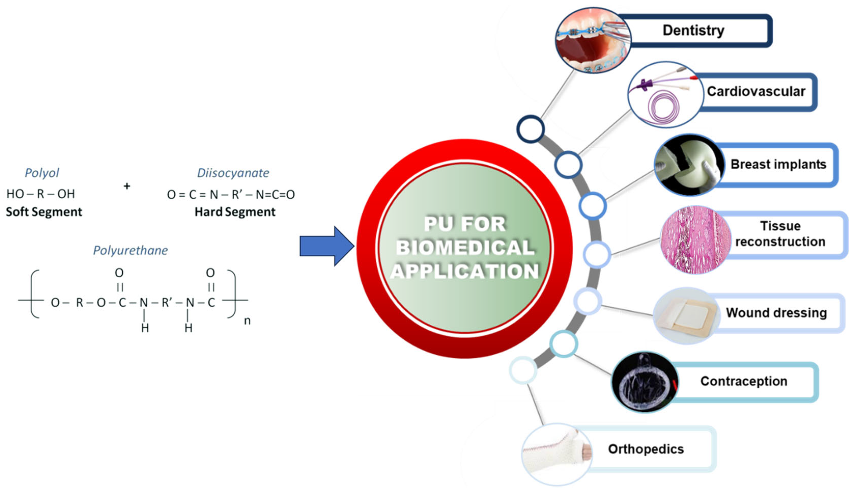
:1. Introduction
2. Radiopacity Measurement
3. Radiopaque Polyurethanes
4. Limitations and Challenges
5. Conclusions
Author Contributions
Funding
Data Availability Statement
Conflicts of Interest
References
- Das, A.; Mahanwar, P. A Brief Discussion on Advances in Polyurethane Applications. Adv. Ind. Eng. Polym. Res. 2020, 3, 93–101. [Google Scholar] [CrossRef]
- Wienen, D.; Gries, T.; Cooper, S.L.; Heath, D.E. An Overview of Polyurethane Biomaterials and Their Use in Drug Delivery. J. Control Release 2023, 363, 376–388. [Google Scholar] [CrossRef]
- Dall Agnol, L.; Ornaghi, H.L., Jr.; Monticeli, F.; Trindade, F.; Bianchi, O. Polyurethanes Synthetized with Polyols of Distinct Molar Masses: Use of the Artificial Neural Network for Prediction of Degree of Polymerization. Polym. Eng. Sci. 2021, 61, 1810–1818. [Google Scholar] [CrossRef]
- Alves, F.C.; Vanessa; Monticeli, F.M.; Ornaghi, H.; Barud, S.; Mulinari, D.R. Efficiency of Castor Oil–Based Polyurethane Foams for Oil Sorption S10 and S500: Influence of Porous Size and Statistical Analysis. Polym. Polym. Compos. 2021, 29, S1063–S1074. [Google Scholar] [CrossRef]
- Ourique, P.A.; Ornaghi, F.G.; Ornaghi, H.L., Jr.; Wanke, C.H.; Bianchi, O. Thermo-oxidative degradation kinetics of renewable hybrid polyurethane-urea obtained from air-oxidized soybean oil. J. Therm. Anal. Calorim. 2019, 137, 1969–1979. [Google Scholar] [CrossRef]
- Dall Agnol, L.; Ornaghi, H.L., Jr.; Neves, R.M.; Monticeli, F.M. Dynamic mechanical and thermogravimetric properties of synthetized polyurethanes. Polym. Bull. 2023, 80, 4181–4194. [Google Scholar]
- Wendels, S.; Avérous, L. Biobased Polyurethanes for Biomedical Applications. Bioact. Mater. 2021, 6, 1083–1106. [Google Scholar] [CrossRef]
- Trhlíková, O.; Vlčková, V.; Abbrent, S.; Valešová, K.; Kanizsová, L.; Skleničková, K.; Paruzel, A.; Bujok, S.; Walterová, Z.; Innemanová, P.; et al. Microbial and Abiotic Degradation of Fully Aliphatic Polyurethane Foam Suitable for Biotechnologies. Polym. Degrad. Stab. 2021, 194, 109764. [Google Scholar] [CrossRef]
- Specialty Chemicals—The Lubrizol Corporation. Lubrizol.com. Available online: https://www.lubrizol.com/ (accessed on 22 January 2024).
- Medical Polyurethanes. @Biomedical. Available online: https://www.dsm.com/biomedical/en_US/biomaterials-solutions/medical-polyurethanes.html (accessed on 22 January 2024).
- Medical Polyurethane Market Research Report: Market Size, Industry Outlook, Market Forecast, Demand Analysis, Market Share, Market Report 2021–2026. Available online: https://www.industryarc.com/Report/18850/medical-polyurethane-market (accessed on 22 January 2024).
- Tian, L.; Lu, L.; Feng, J.; Melancon, M.P. Radiopaque nano and polymeric materials for atherosclerosis imaging, embolization and other catheterization procedures. Acta Pharm. Sin. B 2018, 8, 360–370. [Google Scholar] [CrossRef]
- Yaylaci, A.; Karaarslan, E.S.; Hatirli, H. Evaluation of the radiopacity of restorative materials with different structures and thicknesses using a digital radiography system. Imaging Sci. Dent. 2021, 51, 261–269. [Google Scholar] [CrossRef]
- Ochoa-Rodríguez, V.M.; Homero, J.; Roma, B.; Coaguila-Llerena, H.; Tanomaru-Filho, M.; Gonçalves, A.; Spin-Neto, R.; Faria, G. Radiopacity of Endodontic Materials Using Two Models for Conversion to Millimeters of Aluminum. Braz. Oral. Res. 2020, 34, e080. [Google Scholar] [CrossRef] [PubMed]
- Dantas, R.V.F.; Sarmento, H.R.; Duarte, R.M.; Raso, S.S.M.M.; de Andrade, A.K.M.; Anjos-Pontual, M.L. Radiopacity of restorative composites by conventional radiograph and digital images with different resolutions. Imaging Sci. Dent. 2013, 43, 145–151. [Google Scholar] [CrossRef] [PubMed]
- Valizadeh, S.; Tavakoli, M.A.; Zarabian, T.; Esmaeili, F. Diagnostic Accuracy of Digitized Conventional Radiographs by Camera and Scanner in Detection of Proximal Caries. J. Dent. Res. Dent. Clin. Dent. Prospects 2009, 3, 126–131. [Google Scholar]
- de Paiva, M.A.A.; Mangueira Leite, D.F.B.; Farias, I.A.P.; de Costa, A.P.C.; Sampaio, F.C. Dental Anatomical Features and Caries: A Relationship to Be Investigated. In Dental Anatomy, 1st ed.; Kivanç, B.H., Ed.; Intechopen: London, UK, 2018; pp. 61–84. [Google Scholar]
- Bryant, J.A.; Drage, N.A.; Richmond, S. CT Number Definition. Radiat. Phys. Chem. 2012, 81, 358–361. [Google Scholar] [CrossRef]
- Sneha, K.R.; Sailaja, G.S. Intrinsically Radiopaque Biomaterial Assortments: A Short Review on the Physical Principles, X-ray Imageability, and State-of-the-Art Developments. J. Mater. Chem. B 2021, 9, 8569–8593. [Google Scholar] [CrossRef]
- Lusic, H.; Grinstaff, M.W. X-ray-Computed Tomography Contrast Agents. Chem. Rev. 2012, 113, 1641–1666. [Google Scholar] [CrossRef]
- Berger, M.; Yang, Q.; Maier, A. X-ray Imaging. Lect. Notes Comput. Sci. 2018, 119–145. [Google Scholar]
- Alcântara, A.C.S.; Assis, I.; Prada, D.; Mehle, K.; Schwan, S.; Costa-Paiva, L.; Skaf, M.S.; Wrobel, L.C.; Sollero, P. Patient-Specific Bone Multiscale Modelling, Fracture Simulation and Risk Analysis—A Survey. Materials 2019, 13, 106. [Google Scholar] [CrossRef]
- Emonde, C.K.; Eggers, M.-E.; Wichmann, M.; Hurschler, C.; Ettinger, M.; Denkena, B. Radiopacity Enhancements in Polymeric Implant Biomaterials: A Comprehensive Literature Review. ACS Biomat Sci. Eng. 2024, 10, 1323–1334. [Google Scholar] [CrossRef]
- Kjellson, F.; Almén, T.; Tanner, K.E.; McCarthy, I.D.; Lidgren, L. Bone Cement X-ray Contrast Media: A Clinically Relevant Method of Measuring Their Efficacy. J. Biomed. Mater. Res.-B Appl. Biomater. 2004, 70, 354–361. [Google Scholar] [CrossRef]
- Wang, J.S.; Diaz, J.; Sabokbar, A.; Athanasou, N.; Kjellson, F.; Tanner, K.E.; McCarthy, I.D.; Lidgren, L. In Vitro and in Vivo Biological Responses to a Novel Radiopacifying Agent for Bone Cement. J. R. Soc. Interface 2005, 2, 71–78. [Google Scholar] [CrossRef] [PubMed]
- Manero, J.M.; Ginebra, M.P.; Gil, F.J.; Planell, J.A.; Delgado, J.A.; Morejon, L.; Artola, A.; Gurruchaga, M.; Goñi, I. Propagation of Fatigue Cracks in Acrylic Bone Cements Containing Different Radiopaque Agents. Proc. Inst. Mech. Eng. Part H Proc. Inst. Mech. Eng. H 2004, 218, 167–172. [Google Scholar] [CrossRef] [PubMed]
- Lieberman, I.H.; Togawa, D.; Kayanja, M.M. Vertebroplasty and Kyphoplasty: Filler Materials. Spine J. 2005, 5, S305–S316. [Google Scholar] [CrossRef]
- Pepiol, A.; Teixidor, F.; Saralidze, K.; van der Marel, C.; Willems, P.; Voss, L.; Knetsch, M.L.W.; Vinas, C.; Koole, L.H. A Highly Radiopaque Vertebroplasty Cement Using Tetraiodinated O-Carborane Additive. Biomaterials 2011, 32, 6389–6398. [Google Scholar] [CrossRef]
- Bitsch, R.G.; Kretzer, J.P.; Vogt, S.; Büchner, H.; Thomsen, M.N.; Lehner, B. Increased Antibiotic Release and Equivalent Biomechanics of a Spacer Cement without Hard Radio Contrast Agents. Diagn. Microbiol. Infect. Dis. 2015, 83, 203–209. [Google Scholar] [CrossRef] [PubMed]
- Gillani, R.; Ercan, B.; Qiao, A.; Webster, T.J. Nanofunctionalized Zirconia and Barium Sulfate Particles as Bone Cement Additives. Int. J. Nanomed. 2009, 2, 1–11. [Google Scholar]
- Park, J.-S.; Yim, K.H.; Jeong, S.; Lee, D.H.; Kim, D.G. A Novel High-Visibility Radiopaque Tantalum Marker for Biliary Self-Expandable Metal Stents. Medicine 2019, 13, 366–372. [Google Scholar] [CrossRef]
- Chan, W.A.; Bini, T.B.; Venkatraman, S.S.; Boey, F.Y.C. Effect of Radio-Opaque Filler on Biodegradable Stent Properties. J Biomed. Mater. Res. A 2006, 79A, 47–52. [Google Scholar] [CrossRef]
- Choi, S.Y.; Hur, W.; Kim, B.K.; Shasteen, C.; Kim, M.H.; Choi, L.M.; Lee, S.H.; Park, C.G.; Park, M.; Min, H.S.; et al. Bioabsorbable Bone Fixation Plates for X-ray Imaging Diagnosis by a Radiopaque Layer of Barium Sulfate and Poly(Lactic-Co-Glycolic Acid). J. Biomed. Mater. Res. Part B Appl. Biomater. 2014, 103, 596–607. [Google Scholar] [CrossRef]
- Zaribaf, F.P.; Gill, H.S.; Pegg, E.C. Characterisation of the Physical, Chemical and Mechanical Properties of a Radiopaque Polyethylene. J. Biomater. Appl. 2020, 35, 215–223. [Google Scholar] [CrossRef]
- Wang, Q.; Yu, X.; Chen, X.; Gao, J.; Shi, D.; Shen, Y.; Tang, J.; He, J.; Li, A.; Yu, L.; et al. A Facile Composite Strategy to Prepare a Biodegradable Polymer Based Radiopaque Raw Material for “Visualizable” Biomedical Implants. ACS Appl. Mater. Interfaces 2022, 14, 24197–24212. [Google Scholar] [CrossRef] [PubMed]
- Le Ferrec, M.; Mellier, C.; Boukhechba, F.; Le Corroller, T.; Guenoun, D.; Fayon, F.; Montouillout, V.; Despas, C.; Walcarius, A.; Massiot, D.; et al. Design and Properties of a Novel Radiopaque Injectable Apatitic Calcium Phosphate Cement, Suitable for Image-Guided Implantation. J. Biomed. Mater. Res.-B Appl. 2017, 106, 2786–2795. [Google Scholar] [CrossRef]
- A Novel, Radiopaque, Bioresorbable Tyrosine-Derived Polymer for Cardiovascular Scaffolds. Available online: https://citoday.com/articles/2018-july-aug/a-novel-radiopaque-bioresorbable-tyrosine-derived-polymer-for-cardiovascular-scaffolds (accessed on 15 July 2024).
- Dukic, W. Radiopacity of Composite Luting Cements Using a Digital Technique. J. Prosthodont. 2017, 28, e450–e459. [Google Scholar] [CrossRef] [PubMed]
- Mueller, U.; Reinders, J.; Smith-Romanski, S.; Kretzer, J.P. Wear Performance of Calcium Carbonate-Containing Knee Spacers. Materials 2017, 10, 805. [Google Scholar] [CrossRef]
- Deb, S.; Abdulghani, S.; Behiri, J.C. Radiopacity in Bone Cements Using an Organo-Bismuth Compound. Biomaterials 2002, 23, 3387–3393. [Google Scholar] [CrossRef]
- Roth, A.K.; Boon-Ceelen, K.; Smelt, H.; van Rietbergen, B.; Willems, P.; van Rhijn, L.W.; Arts, J.J. Radiopaque UHMWPE Sublaminar Cables for Spinal Deformity Correction: Preclinical Mechanical and Radiopacifier Leaching Assessment. J. Biomed. Mater. Res. B 2017, 106, 771–779. [Google Scholar] [CrossRef] [PubMed]
- Bogie, R.; Roth, A.K.; de Faber, S.; de Jong, J.J.A.; Welting, T.; Willems, P.; Arts, J.J.; van Rhijn, L.W. Novel Radiopaque Ultrahigh Molecular Weight Polyethylene Sublaminar Wires in a Growth-Guidance System for the Treatment of Early-Onset Scoliosis. Spine 2014, 39, E1503–E1509. [Google Scholar] [CrossRef]
- Kozakiewicz, M.; Olbrzymek, L.; Stefanczyk, L.; Olszycki, M.; Komorowski, P.; Walkowiak, B.; Konieczny, B.; Krasowski, M.; Sokołowski, J. Radio-Opaque Polyethylene for Personalized Craniomaxillofacial Implants. Clin. Oral. Investig. 2016, 21, 1853–1859. [Google Scholar] [CrossRef]
- Chang, W.J.; Pan, Y.H.; Tzeng, J.J.; Wu, T.L.; Fong, T.H.; Feng, S.W.; Huang, H.M. Development and Testing of X-ray Imaging-Enhanced Poly-L-Lactide Bone Screws. PLoS ONE 2015, 10, e0140354. [Google Scholar] [CrossRef]
- Kim, G.B.; Guo, J.; Hu, J.; Shan, D.; Yang, J. Novel Applications of Urethane/Urea Chemistry in the Field of Biomaterials. In Advances in Polyurethane Biomaterials, 1st ed.; Cooper, S.L., Guan, J., Eds.; Woodhead Publishing: Cambridge, UK, 2016; pp. 115–147. [Google Scholar]
- Dawlee, S.; Jayabalan, M. Intrinsically Radiopaque Polyurethanes with Chain Extender 4,4′-Isopropylidenebis [2-(2,6-Diiodophenoxy)Ethanol] for Biomedical Applications. J. Biomaterials Appl. 2014, 29, 1329–1342. [Google Scholar] [CrossRef]
- James, N.; Philip, J.; Jayakrishnan, A. Polyurethanes with Radiopaque Properties. Biomaterials 2006, 27, 160–166. [Google Scholar] [CrossRef] [PubMed]
- Dawlee, S.; Jayabalan, M. Development of Segmented Polyurethane Elastomers with Low Iodine Content Exhibiting Radiopacity and Blood Compatibility. Biomed. Mater. 2011, 6, 055002. [Google Scholar] [CrossRef] [PubMed]
- Kiran, S.; James, N.R.; Joseph, R.; Jayakrishnan, A. Synthesis and Characterization of Iodinated Polyurethane with Inherent Radiopacity. Biomaterials 2009, 30, 5552–5559. [Google Scholar] [CrossRef] [PubMed]
- Qu, W.; Xia, W.; Feng, C.; Tuo, X.; Qiu, T. Synthesis and Characterization of Radiopaque Poly(Ether Urethane) with Iodine-Containing Diol as Chain Extender. J. Polym. Sci. A Polym. Chem. 2011, 49, 2191–2198. [Google Scholar] [CrossRef]
- Kiran, S.; Joseph, R. Synthesis and Characterization of a Noncytotoxic, X-ray Opaque Polyurethane Containing Iodinated Hydroquinone Bis(2-Hydroxyethyl) Ether as Chain Extender for Biomedical Applications. J. Biomed. Mater. Res. A 2013, 102, 3207–3215. [Google Scholar] [CrossRef]
- Kiran, S.; James, N.R.; Jayakrishnan, A.; Joseph, R. Polyurethane Thermoplastic Elastomers with Inherent radiopacity for Biomedical Applications. J. Biomed. Mater. Res. A 2012, 100A, 3472–3479. [Google Scholar] [CrossRef] [PubMed]
- Sang, L.; Wei, Z.; Liu, K.; Wang, X.; Song, K.; Wang, H.; Qi, M. Biodegradable Radiopaque Iodinated Poly(Ester Urethane)S Containing Poly(ε-Caprolactone) Blocks: Synthesis, Characterization, and Biocompatibility. J. Biomed. Mater. Res. A 2013, 102, 1121–1130. [Google Scholar] [CrossRef]
- Liaw, D.-J. The Relative Physical and Thermal Properties of Polyurethane Elastomers: Effect of Chain Extenders of Bisphenols, Diisocyanate, and Polyol Structures. J. Appl. Polym. Sci. 1997, 66, 1251–1265. [Google Scholar] [CrossRef]
- Sang, L.; Luo, D.; Wei, Z.; Qi, M. X-ray Visible and Doxorubicin-Loaded Beads Based on Inherently Radiopaque Poly(Lactic Acid)-Polyurethane for Chemoembolization Therapy. Mater. Sci. Eng. C Biomim. Mater. Sens. Syst. 2017, 75, 1389–1398. [Google Scholar] [CrossRef]
- Shiralizadeh, S.; Nasr-Isfahani, H.; Keivanloo, A.; Bakherad, M.; Yahyaei, B.; Pourali, P. Preparation of radiopaque polyurethane–urea/graphene oxide nanocomposite using 4-(4-iodophenyl)-1,2,4-triazolidine-3,5-dione. J. Mater. Sci. 2018, 53, 9896–9912. [Google Scholar] [CrossRef]
- Wang, W.; Sang, L.; Zhao, Y.; Wei, Z.; Qi, M.; Li, Y. Inherently radiopaque polyurethane beads as potential multifunctional embolic agent in hepatocellular carcinoma therapy. J. Mater. Sci. Tech. 2021, 63, 106–114. [Google Scholar] [CrossRef]
- Egorikhina, M.N.; Bokov, A.E.; Charykova, I.N.; Rubtsova, Y.P.; Linkova, D.D.; Kobyakova, I.I.; Farafontova, E.A.; Kalinina, S.Y.; Kolmogorov, Y.N.; Aleynik, D.Y. Biological Characteristics of Polyurethane-Based Bone-Replacement Materials. Polymers 2023, 15, 831. [Google Scholar] [CrossRef] [PubMed]
- Kiran, S.; Sunny, M.C.; Joseph, R. Inherently X-ray opaque polyurethane microspheres for biomedical applications. Int. J. Polym. Mater. Polym. Biomater. 2016, 66, 213–220. [Google Scholar] [CrossRef]
- Sang, L.; Wei, Z.; Zhai, L.; Wang, H.; Qi, M. Enzymatic degradation and radiopaque attenuation of iodinated poly(ester-urethane)s with inherent radiopacity. J. Mater. Sci. 2014, 49, 7834–7843. [Google Scholar] [CrossRef]
- Attarilar, S.; Yang, J.; Ebrahimi, M.; Wang, Q.; Liu, J.; Tang, Y.; Yang, J. The Toxicity Phenomenon and the Related Occurrence in Metal and Metal Oxide Nanoparticles: A Brief Review from the Biomedical Perspective. Front. Bioeng. Biotechnol. 2020, 8, 822. [Google Scholar] [CrossRef]


| Contrast Agent | Blending Method | Polymer | Application | Content | Reported Effects | Polymer Biodegradable | Biological Response | Ref. |
|---|---|---|---|---|---|---|---|---|
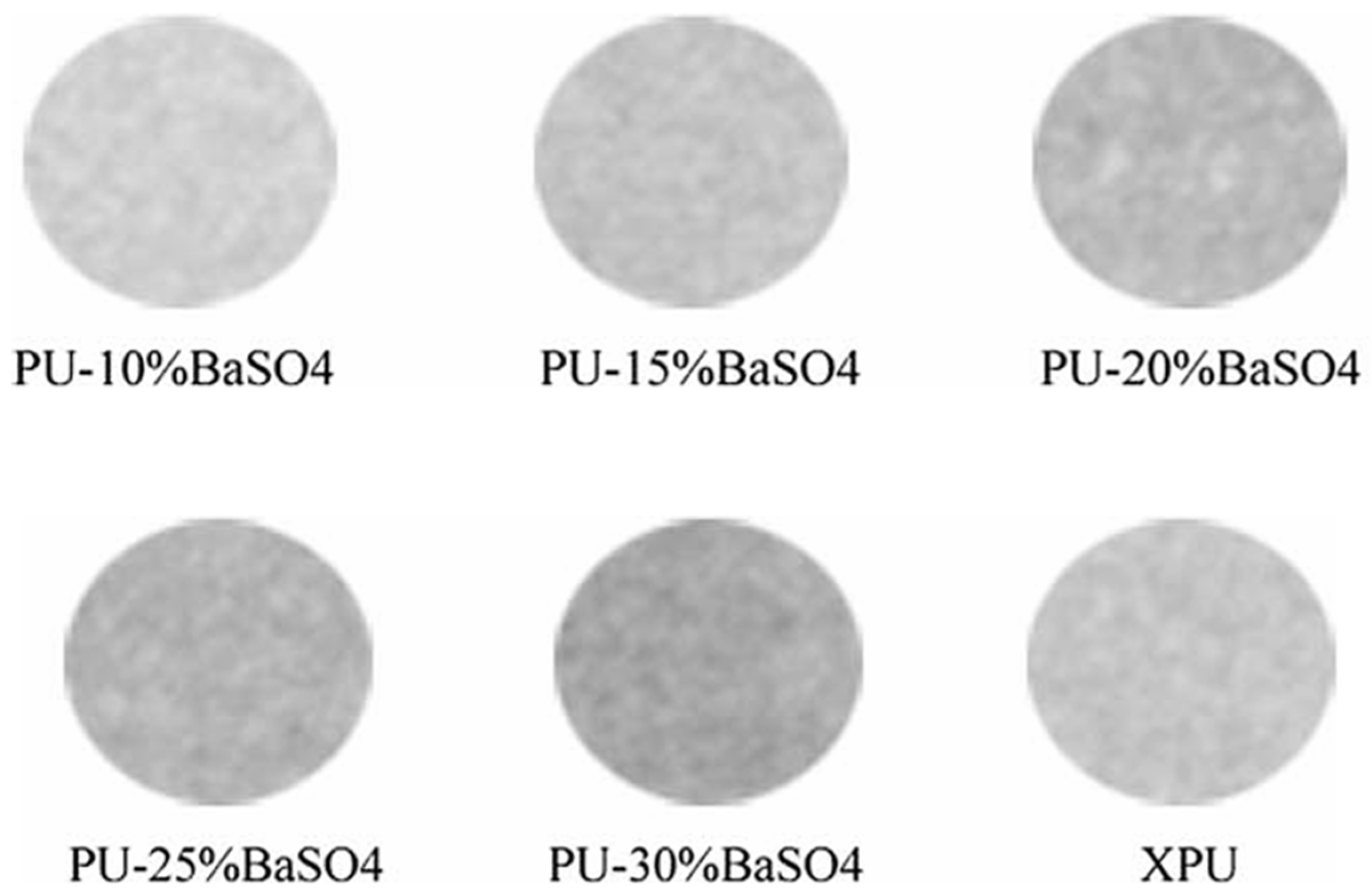
| BaSO4 | Blended in the powder phase | PMMA | Bone cement | 9–15 wt% | Hard particles, third body wear, reduced tensile, and flexural strength | NO | Osteoclast formation | [24] [25] [26] |
| Blended in the powder phase | PMMA | Vertebroplasty cement | 30 wt% | Hard particles, third body wear, lower viscosity | NO | Osteoclast formation | [27] [28] [29] | |
| Twin-screw micro-compounding | PLLA | Bioresorbable stents | 5–20 wt% | Increased tensile modulus and strength, decreased elongation at break and ductility | YES | No adverse effects after 21 days | [30] [31] | |
| Magnetic stirring in organic solvent | PLGA | Bioresorbable stent | 17.9 v/v% | Increased Young’s modulus, reduced elasticity, increased radial strength | YES | Na | [32] | |
| Solution mixing | PLGA | Bone fixation plate | 1:10 and 1:3 w/w PLGA:BaSO4 | Radiopaque up to 56 days, BaSO4 leaching < 0.5 mg/day; insufficient to induce cytotoxicity | YES | No adverse effects | [33] | |
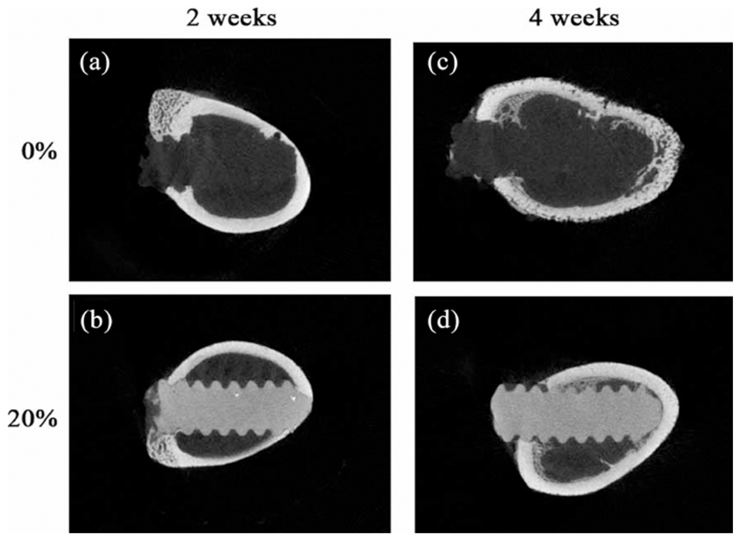
| Lipiodol ultra fluid | Immersion in oil at elevated temperature | UHMWPE | TKA insert | 25 mL | Physical alteration–swelling, 54% reduction in surface radiopacity after 4 weeks | NO | Na | [34] |
| Iohexol (IHX) | Stirring | PLA | Bioresorbable implants | 40 wt% | Reduced tensile strength, elongation at break and increased tensile modulus, enhanced crystallinity, slower polymer degradation | YES | Thin fiber capsule | [35] |
| Blended in the powder phase | PMMA | Bone cement | 10 wt% | Better biocompatibility compared to conventional contrast agents | NO | Osteoclast formation | [26] | |
| Iodixanol (IDX) | Blended in the powder phase | PMMA | Bone cement | 10 wt% | Higher osteoclast formation than IHX | NO | Osteoclast formation | [26] |
| Iobitridol | Dissolved in liquid phase | CPC | Bone cement | 56 mg mL−1 | Rapid release of contrast, no significant change in mechanical properties, no effect on injectability, cohesion, or setting time | YES | No adverse effects | [36] |
| Iodinated diphenol | Polymerization reaction | PLA diol | Coronary stent | <1% of 1 mL of iodine contrast | Increased ultimate tensile strength and elongation at break, long-term radiopacity | YES | No adverse effects | [37] |
| Bismuth salicylate (BS) | Dissolved in liquid phase | PMMA | Vertebroplasty cement | 10 w/w | Reduced compressive and tensile strength, reduced strain, lower setting temperature, increased radiopacity, longer injection time, Better compatibility than BaSO4 | NO | Na | [38] [39] |
| Triphenyl bismuth (TPB) | Dissolved in liquid phase | PMMA | Bone cement | 10 wt% | Increased ultimate tensile strength, Young’s modulus and strain to failure, lower setting temperature, better homogeneity | NO | Na | [40] |
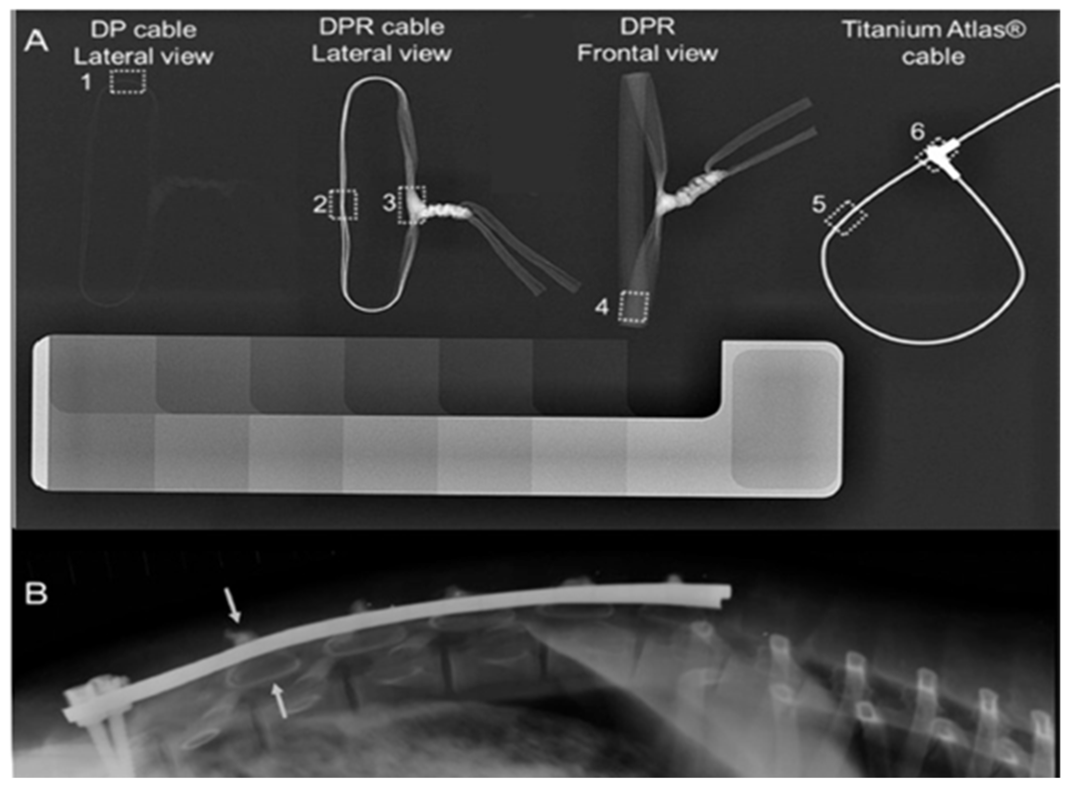
| Bismuth oxide Bi2O3 | Blended into fiber | UHMWPE | Sublaminar cables | 20 wt% | Decreased tensile strength, limited leaching below toxic levels | NO | No adverse effects | [41] [42] |
| Titanium dioxide TiO2 | Blending | PE | Orbital implant | 6% | Slight decrease in tensile strength and modulus, significant decrease in compressive strength and modulus, reduced hardness | NO | No adverse effects | [43] |
| Iron oxide Fe3O4 | Twin-screw extrusion | PLLA | Bone screws | 20 wt% | Reduced flexural strength, increased crystallinity, increased thermal stability | YES | Osteogenic effect, no adverse effects | [44] |
| Reagents | Characterization | Reported Effects | Ref. |
|---|---|---|---|
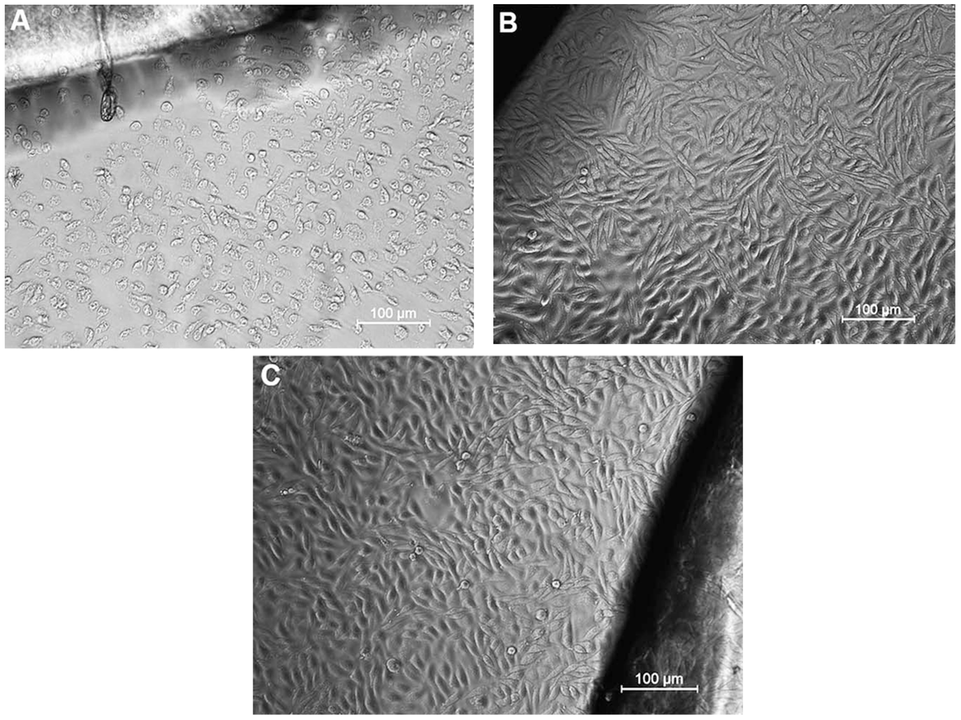
| 2,3-diiodo-2-butene-1,4-diol (I-BOL) (aliphatic chain extender) 4,4′-methylenebis(phenyl isocyanate) (MDI) Hexamethylene diisocyanate (HDI) Polytetramethylene glycol (PTMG) 2-Butyne-1,4-diol, dibutyltin dilaurate (DBTDL) | FTIR X-ray opacity Surface properties under aqueous conditions In vitro cytotoxicity Blood compatibility | HDI-based polyurethane was partially crystalline with a phase-separated surface morphology. MDI-based polyurethane has phase-mixed surface morphology, which undergoes dynamic surface reorganization in aqueous medium to a phase-separated surface morphology. Studies on in vitro cytotoxicity and blood compatibility with MDI-based polyurethane revealed cytocompatibility, blood compatibility, and radiopacity | [48] |
| 4,4′-isopropylidenebis[2-(2,6-diiodophenoxy)ethanol] (IBPA) (chain extender) 4,4′-methylenebis(phenyl isocyanate)(MDI) Poly(tetramethylene glycol) (PTMG) | FTIR TGA DMTA EDX GPC X-radiography In vitro cytotoxicity Radiopaque properties | Highly radiopaque polyurethane with an iodine content of 23% (better than aluminum). In vitro non-cytotoxicity using L929 mouse fibroblast cells, visible even after 12 weeks of implantation | [49] |
| N,N′-Bis(3-hydroxypropxyl)-2,3,5,6-tetraiodoterephthalamide (HPTDP) (chain extender) Poly(tetramethylene glycol) (PTMG) 4,40-Diphenylmethane diisocyanate) (MDI) | NMR Mechanical properties In vitro degradation Cytotoxicity Radiopaque properties | Good radiopacity (compared to aluminum). Radiopacity did not decrease after oxidative degradation treatment. Has good thermal stability and mechanical properties, non-toxicity | [50] |
| 1,6-diisocynatohexane (HDI) Poly(hexamethylene carbonate)diol (PHCD) 2,2′-(2,5-diiodobenzene-1,4-diyl)bis(oxy)diethanol (DBD) Barium sulfate (BaSO4) | FTIR EDX Mechanical properties DMTA X-radiography Opacity Fluoroscopy In vitro cytotoxicity | Melt processable, non-cytotoxic, and blood compatible and possesses a high degree of optical transparency | [51] |
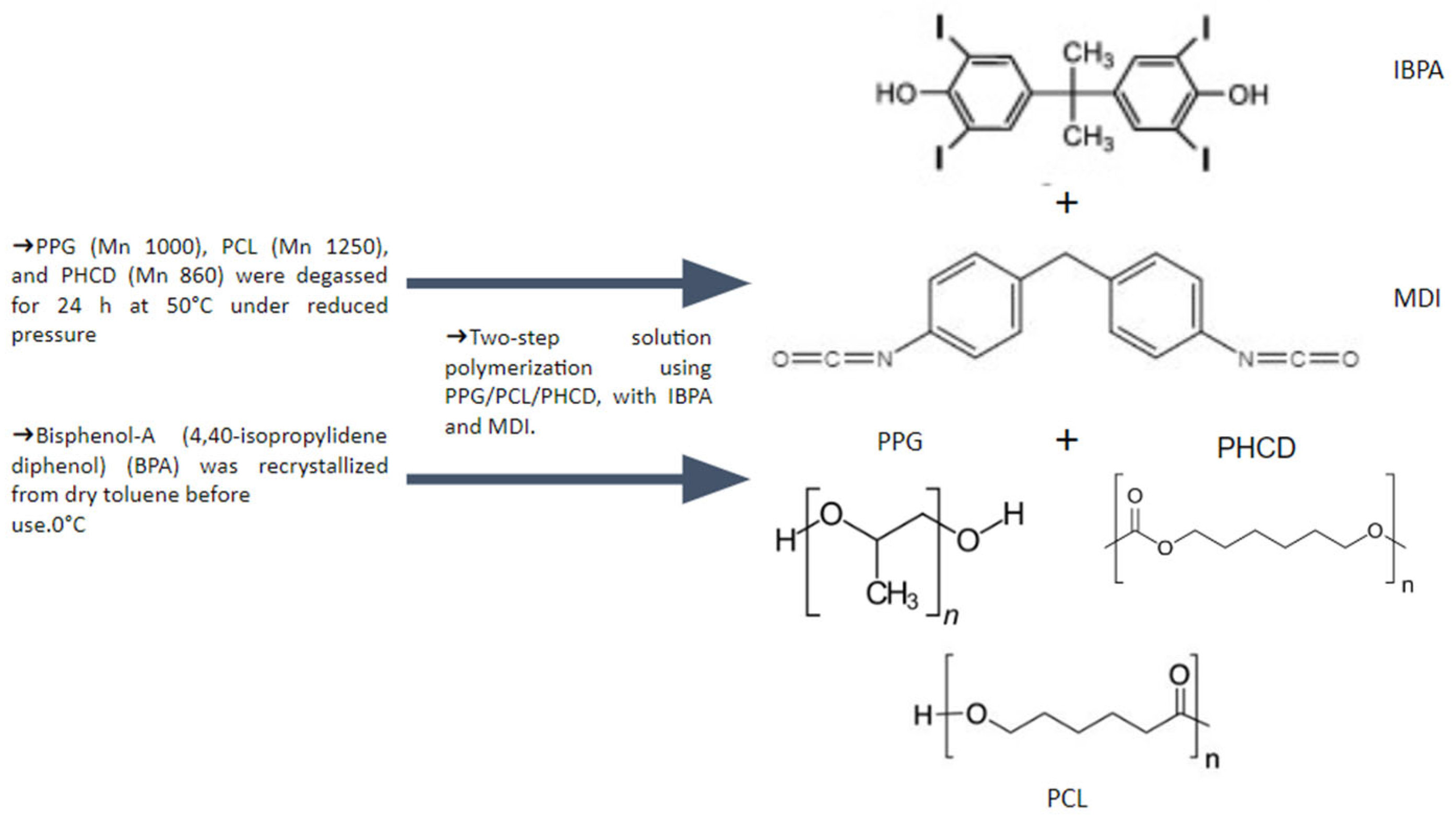
| 4,4′-isopropylidinedi-(2,6-diiodophenol) (IBPA) (chain extender) 4,4′-methylenebis(phenyl isocyanate) (MDI) polypropylene glycol polycaprolactone diol poly(hexamethylene carbonate) diol | FTIR Contact angle GPC Thermal properties DMTA EDX XRF XRD Radiopacity Cytotoxicity | Highly radiopacity compared to aluminum, 18–19% iodine in the polymer matrix, radiopacity equivalent to 20%wt barium sulfate, non-cytotoxicity in L929 cells by direct contact and MTT assay | [52] |
| 4,4′-isopropylidinedi-(2,6-diiodophenol) (IBPA) (chain extender) Poly(e-caprolactone) (PCL) Isophorone diisocyanate (IPDI) | FTIR NMR GPC EDX DSC TGA WAXD X-ray opacity Enzyme-accelerated degradation cytocompatibility | Highly radiopacity, controllable biodegradability, and cell cytocompatibility tracked during the in vitro enzymatic degradation test, non-toxic biomaterials | [53] |
| Polycaprolactone diol polytetramethylene glycol Diphenylmethane 4,4′-diisocyanate (MDI) Dicyclohexyl methane 4,4′-diisocyanate (HMDI) Dibutyl tin dilaurate (DBTDL) Bisphenol-S Biusphenol-AF Bisphenol-A 3,3′,5,5′-Tetrabromo bisphenol-S (TBBPS) 3,3′,5,5′-Tetrabromo bisphenol-A 3,3′,5,5′-Tetrabromo bisphenol-AF | FTIR Hardness Tensile Properties Specific gravity Swelling and sol fraction DMTA DSC TGA XRD LOI Water absorption | The addition of bromine atoms in the polyurethanes markedly decreased their degrees of crystallinity. The brominated polyurethane elastomers have good flame retardancy. All of the unbrominated polyurethanes showed good mechanical properties and high thermal stabilities. Polyurethanes based on bisphenol-S had lower solvent resistance caused by the dipolar nature of sulfonyl groups in the polymer chains | [54] |
| 4,4′-isopropylidinedi-(2,6-diiodophenol) (IBPA) isophorone diisocyanate (IPDI) doxorubicin hydrochloride (DOX) triethylene glycol (TEG) iodinated poly(lactic acid)-polyurethane (I-PLAU) | GPC Thermal properties XRD X-radiography Contact angle Blood compatibility Cell culture Cytotoxicity assay In vivo muscle implantation In vivo drug release Antitumor assay in vitro | Sufficient radiopacity, in vitro non-toxicity, in vivo biocompatibility, release profile controlled by the microstructure, efficient inhibition of tumor cell growth | [55] |
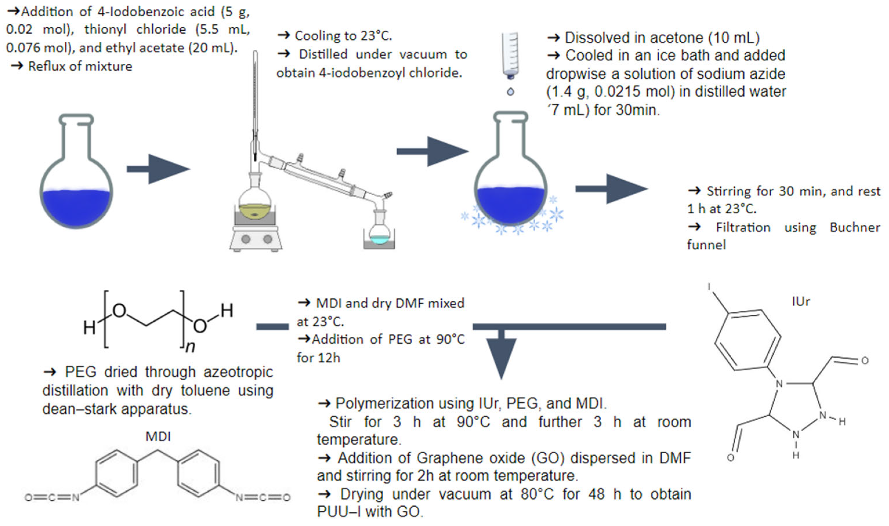
| 4,4′-methylenediphenyl diisocyanate (MDI) polyethylene glycol (PEG) 4-(4-iodophenyl)-1,2,4-triazolidine-3,5-dione (IUr) Graphene oxide (GO) | FTIR NMR XRD CHNO FESEM TGA DMTA | Improved thermal and mechanical properties, heat durability, well dispersed GO in the polymeric matrix, significant contrast. | [56] |
| 4,4′-isopropylidinedi-(2,6-diiodophenol) (IBPA) iodinated polycaprolactone-polyurethanes (I-PCLUs) Doxorubicin (Dox) | Optical microscopy SEM UV-vis spectroscopy In vitro drug release In vitro antitumor assay In vivo experiments (muscle implantation, hepatic embolization, chemoembolization of rabbit hepatic tumor model) | Effective drug-loading, sustained release mode for four weeks, kept well X-ray visibility after drug release, inhibition of the tumor cell proliferation, good histocompatibility, effective chemoembolization, and radiopacity. | [57] |
| zirconium oxide bismuth oxide tantalum pentoxide polypropylene glycol (PPG) 4,4′-methylene diphenyl diisocyanate (MDI) glycerol | Cytotoxicity Culture cells | Cytocompatible and interconnected pores, promising for further development as bases for bone-substituting materials. | [58] |
| 4,4′-isopropylidinedi(2,6-diiodophenol) (IBPA) Dibutyltin dilaurate (DBTL) Polyvinyl alcohol (PVA) Poly(hexamethylene carbonate)diol (PHCD) 4,4′-methylene-bis(cyclohexyl isocyanate) | NMR FTIR TGA GPC ESEM Elemental analysis Cytocompatibility In vitro degradation studies In vivo imaging | Non-toxicity, visible under fluoroscopic conditions, and excellent X-ray opacity. | [59] |
| 4,4′-isopropylidinedi(2,6-diiodophenol) (IBPA) poly(e-caprolactone) diol (PCL) isophorone diisocyanate (IPDI) | Degradation tests TGA X-ray opacity FESEM DSC XRD Contact angle | Poor hydrophilicity, reduced rate of degradation, and no great attenuation of the X-ray opacity after 3-month degradation. | [60] |
Disclaimer/Publisher’s Note: The statements, opinions and data contained in all publications are solely those of the individual author(s) and contributor(s) and not of MDPI and/or the editor(s). MDPI and/or the editor(s) disclaim responsibility for any injury to people or property resulting from any ideas, methods, instructions or products referred to in the content. |
© 2024 by the authors. Licensee MDPI, Basel, Switzerland. This article is an open access article distributed under the terms and conditions of the Creative Commons Attribution (CC BY) license (https://creativecommons.org/licenses/by/4.0/).
Share and Cite
Garavatti, J.; Ornaghi Jr., H.L. A Short Review on Radiopaque Polyurethanes in Medicine: Physical Principles, Effect of Nanoparticles, Processing, Properties, and Applications. J. Compos. Sci. 2024, 8, 409. https://doi.org/10.3390/jcs8100409
Garavatti J, Ornaghi Jr. HL. A Short Review on Radiopaque Polyurethanes in Medicine: Physical Principles, Effect of Nanoparticles, Processing, Properties, and Applications. Journal of Composites Science. 2024; 8(10):409. https://doi.org/10.3390/jcs8100409
Chicago/Turabian StyleGaravatti, Julia, and Heitor Luiz Ornaghi Jr. 2024. "A Short Review on Radiopaque Polyurethanes in Medicine: Physical Principles, Effect of Nanoparticles, Processing, Properties, and Applications" Journal of Composites Science 8, no. 10: 409. https://doi.org/10.3390/jcs8100409