
X-ray Synchrotron Radiation to Look at Pigments in Antiquities: Overview and Examples

 , , , ,
Abstract
:1. Synchrotron for Cultural Heritages and Pigments Studies: Overview
1.1. Synchrotron Beamlines for Cultural Heritages in Europe
1.2. X-ray Synchrotron Techniques for Pigments Studies
1.3. Pigments in Archaeological Artifacts: From Rock Art to Potteries
1.4. Pigments in Glazes
1.5. Ink, Dyes and Organic Colorants
1.6. Paintings: Pigments Characterization and Degradation Products
1.7. Pigments and Radiation Damage
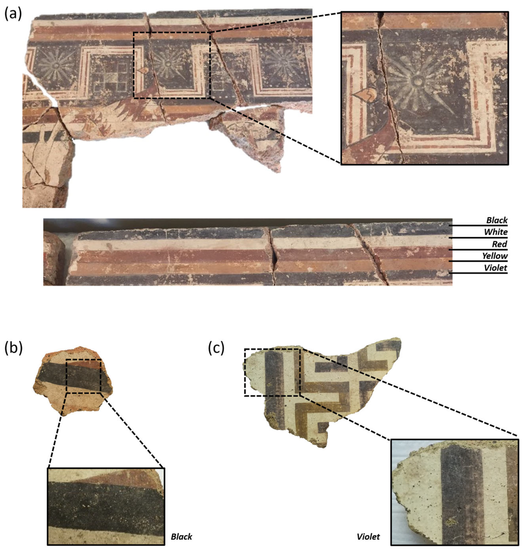
2. Painted Architectural Terracottas: A Meaningful Example of Integrated Approach
2.1. Materials and Methods
2.2. Results and Discussion
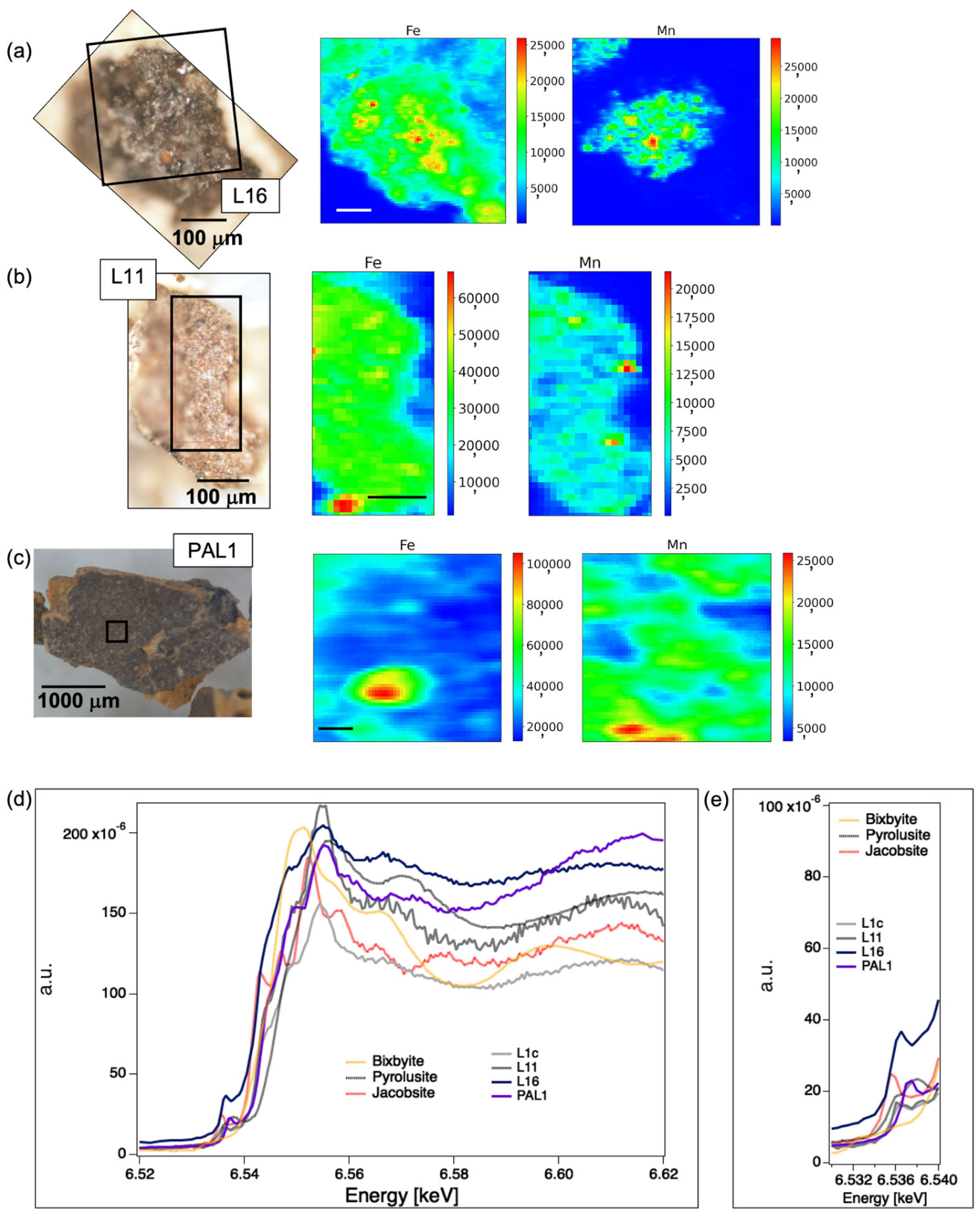
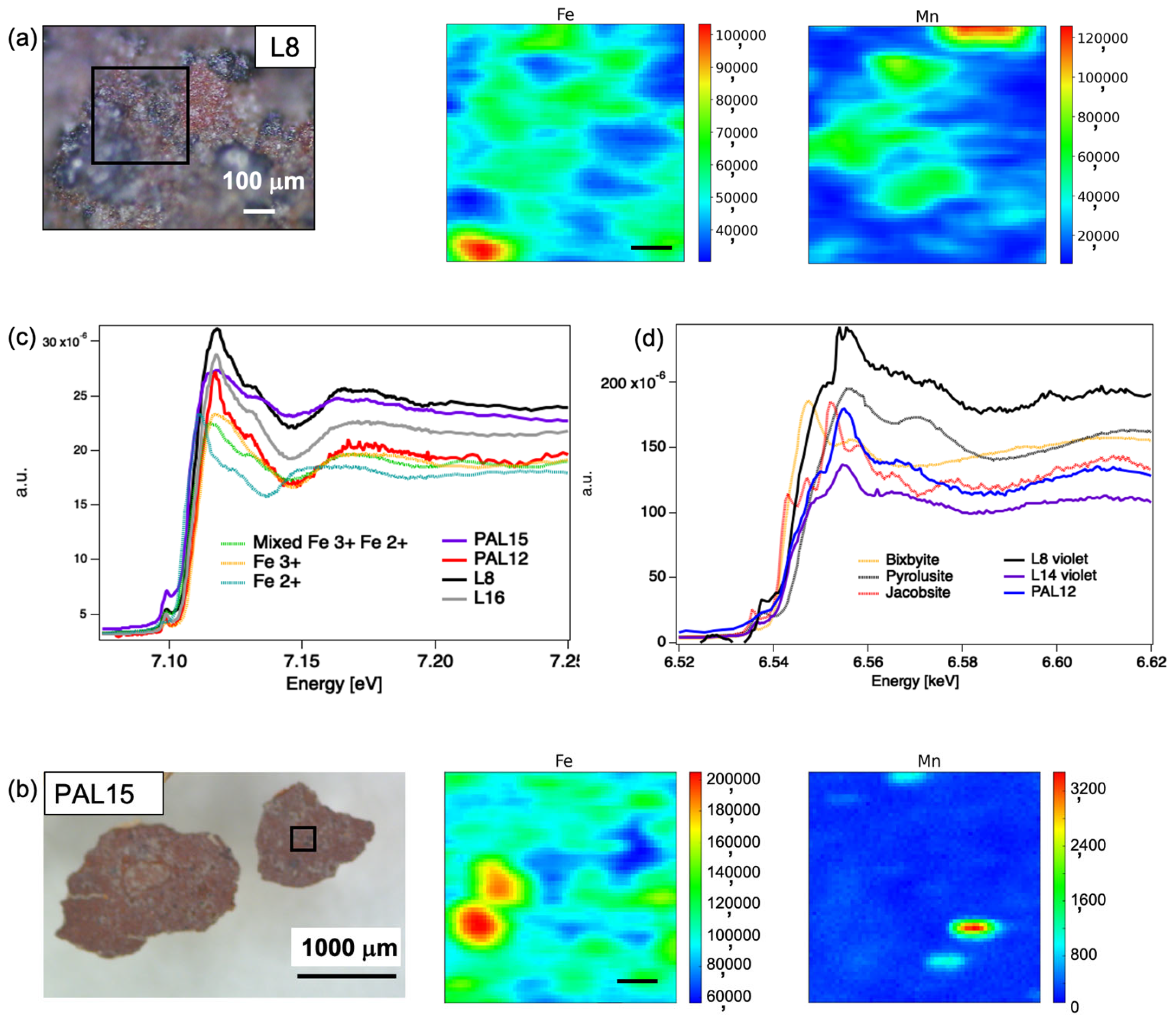
2.2.1. The Case of the Manganese-Black Technique
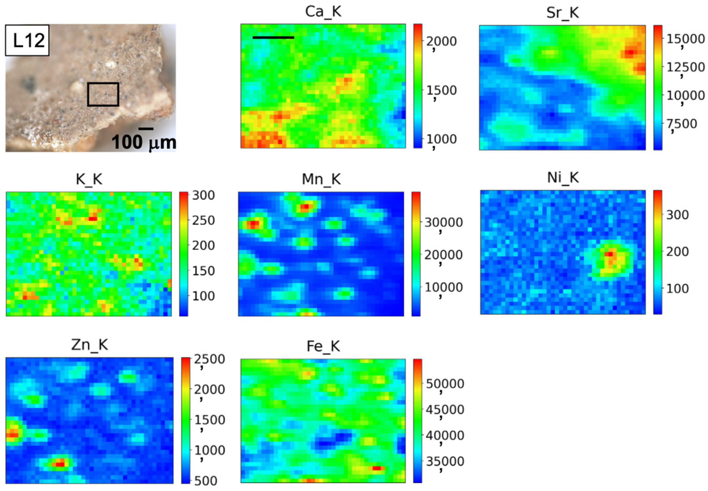
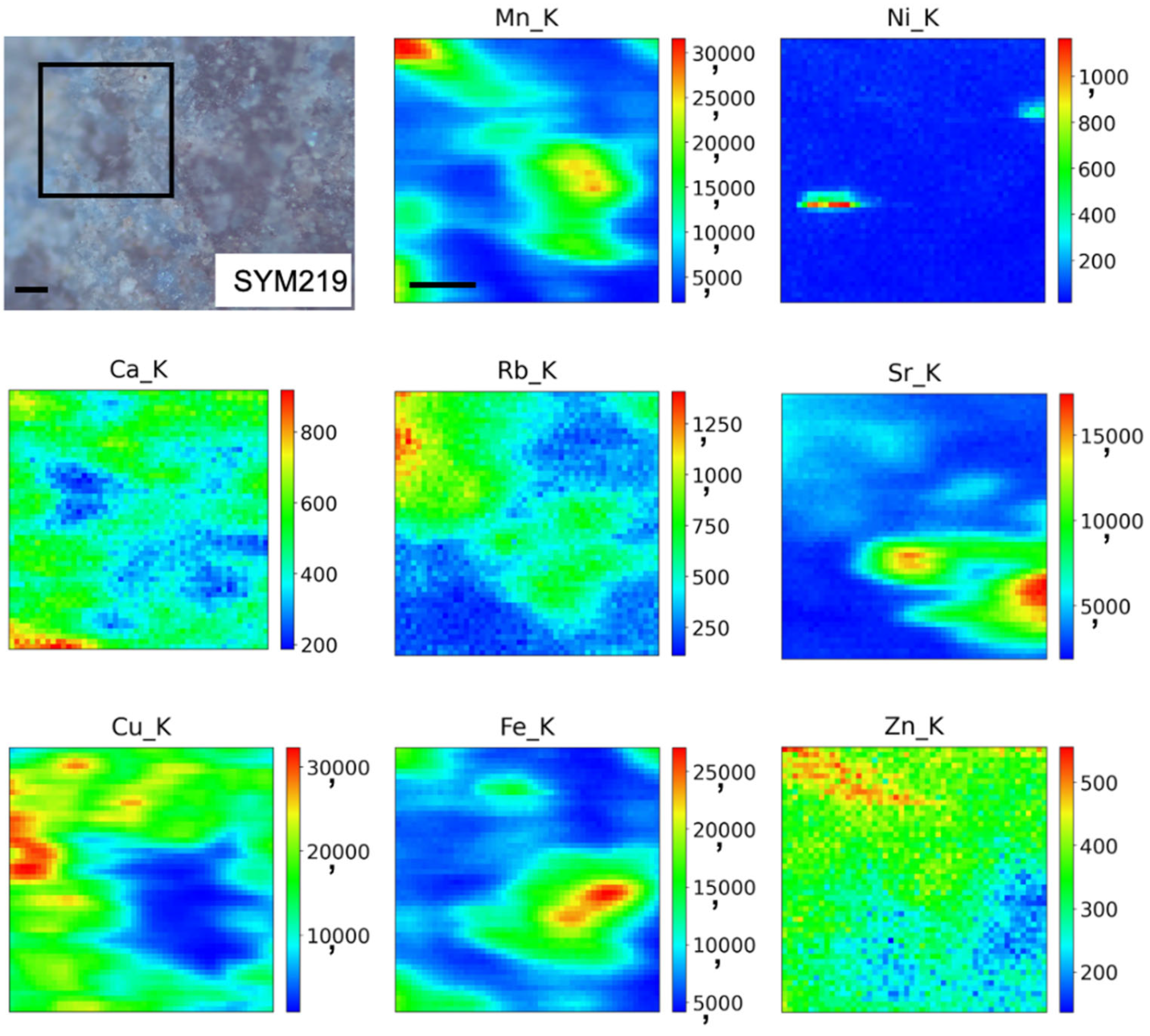
2.2.2. Looking at Pigment Grains: Stratigraphies or Mixtures of Hues?
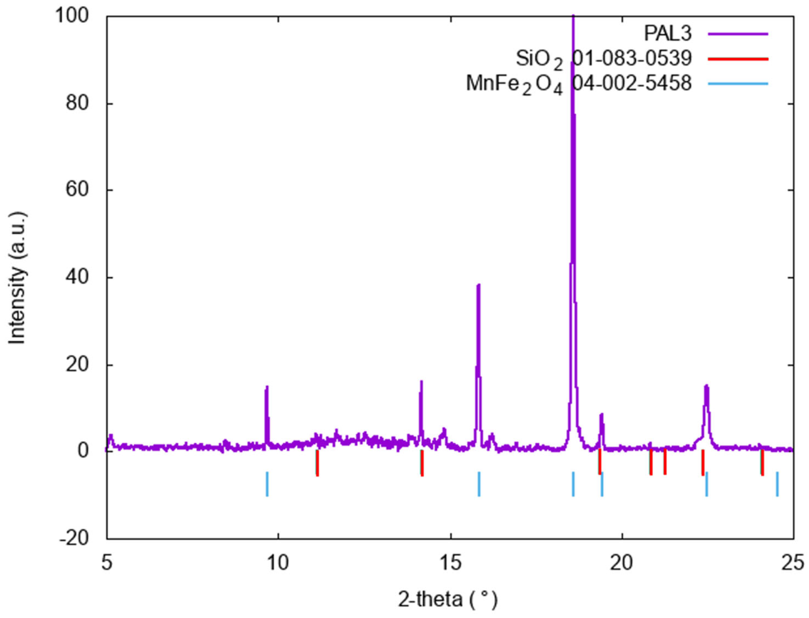
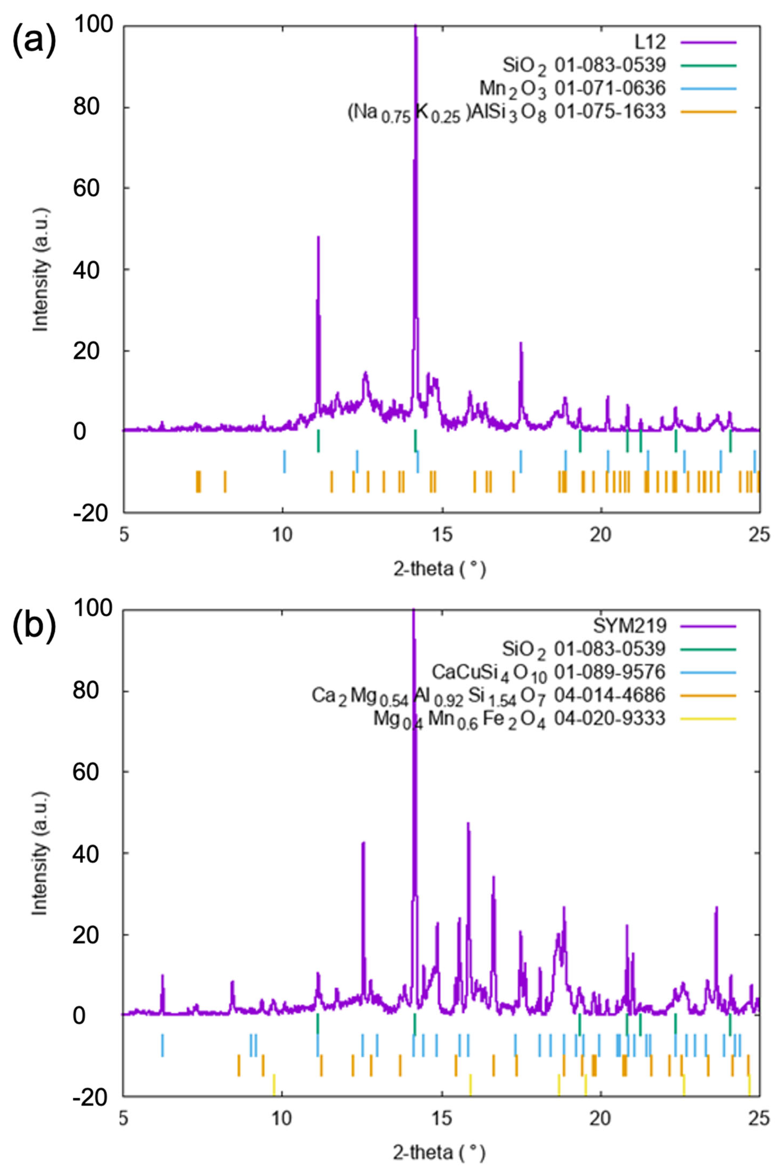
2.2.3. SR-µXRD for Solving Ambiguities: The Case of Blue and Green Pigments
3. Conclusions
Author Contributions
Funding
Data Availability Statement
Acknowledgments
Conflicts of Interest
References
- Cotte, M.; Dollman, K.; Fernandez, V.; Gonzalez, V.; Vanmeert, F.; Monico, L.; Dejoie, C.; Burghammer, M.; Huder, L.; Fisher, S.; et al. New Opportunities Offered by the ESRF to the Cultural and Natural Heritage Communities. Synchrotron Radiat. News 2022, 35, 3–9. [Google Scholar] [CrossRef]
- Reguer, S.; Schöder, S.; Vantelon, D.; Weitkamp, T.; Rueff, J.-P.; Berenguer, F.; King, A.; Jamme, F.; Hunault, M.O.J.Y.; Silly, M.G.; et al. Fifteen Years of Study of Cultural and Natural Heritage Materials at SOLEIL. Synchrotron Radiat. News 2022, 35, 10–20. [Google Scholar] [CrossRef]
- Amati, M.; Gianoncelli, A.; Karantzoulis, E.; Rossi, B.; Vaccari, L.; Zanini, F. Looking at Ancient Objects under a Different Light: Cultural Heritage Science at Elettra. Radiat. Eff. Defects Solids 2022, 177, 1271–1287. [Google Scholar] [CrossRef]
- Cotte, M.; Pouyet, E.; Salomé, M.; Rivard, C.; Nolf, W.D.; Castillo-Michel, H.; Fabris, T.; Monico, L.; Janssens, K.; Wang, T.; et al. The ID21 X-ray and Infrared Microscopy Beamline at the ESRF: Status and Recent Applications to Artistic Materials. J. Anal. At. Spectrom. 2017, 32, 477–493. [Google Scholar] [CrossRef]
- Janssens, K.; Van der Snickt, G.; Vanmeert, F.; Legrand, S.; Nuyts, G.; Alfeld, M.; Monico, L.; Anaf, W.; De Nolf, W.; Vermeulen, M.; et al. Non-Invasive and Non-Destructive Examination of Artistic Pigments, Paints, and Paintings by Means of X-ray Methods. Top. Curr. Chem. 2016, 374, 81. [Google Scholar] [CrossRef] [PubMed]
- Janssens, K.; Cotte, M. Using Synchrotron Radiation for Characterization of Cultural Heritage Materials. In Synchrotron Light Sources and Free-Electron Lasers; Springer: Cham, Switzerland, 2020; pp. 2457–2483. ISBN 978-3-030-23200-9. [Google Scholar]
- Smieska, L.M.; Woll, A.R.; Vanmeert, F.; Janssens, K. Synchrotron-Based High-Energy X-ray MA-XRF and MA-XRD for Art and Archaeology. Synchrotron Radiat. News 2019, 32, 29–33. [Google Scholar] [CrossRef]
- Cotte, M.; Gonzalez, V.; Vanmeert, F.; Monico, L.; Dejoie, C.; Burghammer, M.; Huder, L.; de Nolf, W.; Fisher, S.; Fazlic, I.; et al. The “Historical Materials BAG”: A New Facilitated Access to Synchrotron X-ray Diffraction Analyses for Cultural Heritage Materials at the European Synchrotron Radiation Facility. Molecules 2022, 27, 1997. [Google Scholar] [CrossRef] [PubMed]
- Cotte, M.; Genty-Vincent, A.; Janssens, K.; Susini, J. Applications of Synchrotron X-ray Nano-Probes in the Field of Cultural Heritage. C. R. Phys. 2018, 19, 575–588. [Google Scholar] [CrossRef]
- Dik, J.; Janssens, K.; Van Der Snickt, G.; van der Loeff, L.; Rickers, K.; Cotte, M. Visualization of a Lost Painting by Vincent van Gogh Using Synchrotron Radiation Based X-ray Fluorescence Elemental Mapping. Anal. Chem. 2008, 80, 6436–6442. [Google Scholar] [CrossRef]
- Gianoncelli, A.; Kourousias, G.; Schöder, S.; Santostefano, A.; L’Héronde, M.; Barone, G.; Mazzoleni, P.; Raneri, S. Synchrotron X-ray Microprobes: An Application on Ancient Ceramics. Appl. Sci. 2021, 11, 8052. [Google Scholar] [CrossRef]
- de Boer, D.K.G. Fundamental Parameters for X-ray Fluorescence Analysis. Spectrochim. Acta Part B At. Spectrosc. 1989, 44, 1171–1190. [Google Scholar] [CrossRef]
- De Boer, D.K.G.; Borstrok, J.J.M.; Leenaers, A.J.G.; Van Sprang, H.A.; Brouwer, P.N. How Accurate Is the Fundamental Parameter Approach? XRF Analysis of Bulk and Multilayer Samples. X-ray Spectrom. 1993, 22, 33–38. [Google Scholar] [CrossRef]
- Handbook of X-ray Spectrometry. Available online: https://www.routledge.com/Handbook-of-X-ray-Spectrometry/VanGrieken-Markowicz/p/book/9780824706005 (accessed on 19 March 2024).
- Švarcová, S.; Bezdička, P.; Hradil, D.; Hradilová, J.; Žižak, I. Clay Pigment Structure Characterisation as a Guide for Provenance Determination—A Comparison between Laboratory Powder Micro-XRD and Synchrotron Radiation XRD. Anal. Bioanal. Chem. 2011, 399, 331–336. [Google Scholar] [CrossRef] [PubMed]
- Artioli, G. Science for the Cultural Heritage: The Contribution of X-ray Diffraction. Rend. Fis. Acc. Lincei 2013, 24, 55–62. [Google Scholar] [CrossRef]
- Possenti, E.; Colombo, C.; Conti, C.; Gigli, L.; Merlini, M.; Plaisier, J.R.; Realini, M.; Gatta, G.D. What’s underneath? A Non-Destructive Depth Profile of Painted Stratigraphies by Synchrotron Grazing Incidence X-ray Diffraction. Analyst 2018, 143, 4290–4297. [Google Scholar] [CrossRef] [PubMed]
- Monico, L.; Prati, S.; Sciutto, G.; Catelli, E.; Romani, A.; Balbas, D.Q.; Li, Z.; Meyer, S.D.; Nuyts, G.; Janssens, K.; et al. Development of a Multi-Method Analytical Approach Based on the Combination of Synchrotron Radiation X-ray Micro-Analytical Techniques and Vibrational Micro-Spectroscopy Methods to Unveil the Causes and Mechanism of Darkening of “Fake-Gilded” Decorations in a Cimabue Painting. J. Anal. At. Spectrom. 2022, 37, 114–129. [Google Scholar] [CrossRef]
- Van der Snickt, G.; Janssens, K.; Dik, J.; De Nolf, W.; Vanmeert, F.; Jaroszewicz, J.; Cotte, M.; Falkenberg, G.; Van der Loeff, L. Combined Use of Synchrotron Radiation Based Micro-X-ray Fluorescence, Micro-X-ray Diffraction, Micro-X-ray Absorption Near-Edge, and Micro-Fourier Transform Infrared Spectroscopies for Revealing an Alternative Degradation Pathway of the Pigment Cadmium Yellow in a Painting by Van Gogh. Anal. Chem. 2012, 84, 10221–10228. [Google Scholar] [CrossRef] [PubMed]
- Vanmeert, F.; De Wael, K.; Janssens, K.; Falkenberg, G.; Cotte, M. Using Synchrotron Radiation for Understanding the Spontaneous Degradation of Artists’ Pigments. Synchrotron Radiat. News 2019, 32, 41–47. [Google Scholar] [CrossRef]
- Huntley, J.; Brand, H.; Aubert, M.; Morwood, M. The First Australian Synchrotron Powder Diffraction Analysis of Pigment from a Wandjina Motif in the Kimberley, Western Australia. Aust. Archaeol. 2014, 78, 33. [Google Scholar] [CrossRef]
- Gay, M.; Alfeld, M.; Menu, M.; Laval, E.; Arias, P.; Ontañón, R.; Reiche, I. Palaeolithic Paint Palettes Used at La Garma Cave (Cantabria, Spain) Investigated by Means of Combined in Situ and Synchrotron X-ray Analytical Methods. J. Anal. At. Spectrom. 2015, 30, 767–776. [Google Scholar] [CrossRef]
- Ilmi, M.M.; Nurdini, N.; Maryanti, E.; Saiyasombat, C.; Setiawan, P.; Kadja, G.T.M. Ismunandar Multi-Analytical Characterizations of Prehistoric Rock Art Pigments from Karim Cave, Sangkulirang–Mangkalihat Site, East Kalimantan, Indonesia. Microchem. J. 2020, 155, 104738. [Google Scholar] [CrossRef]
- Maryanti, E.; Ilmi, M.M.; Nurdini, N.; Setiawan, P.; Syah, Y.M.; Saiyasombat, C.; Kadja, G.T.M. Ismunandar Hematite as Unprecedented Black Rock Art Pigment in Jufri Cave, East Kalimantan, Indonesia: The Microscopy, Spectroscopy, and Synchrotron X-ray-Based Investigation. Archaeol. Anthr. Sci. 2022, 14, 122. [Google Scholar] [CrossRef]
- Nurdini, N.; Maryanti, E.; Ilmi, M.M.; Setiawan, P.; Saiyasombat, C.; Kadja, G.T.M. Ismunandar Physicochemical Investigation of Prehistoric Rock Art Pigments in Tewet Cave, Sangkulirang-Mangkalihat Site, East Kalimantan-Indonesia. J. Archaeol. Sci. Rep. 2020, 31, 102345. [Google Scholar] [CrossRef]
- Debastiani, R.; Simon, R.; Batchelor, D.; Dellagustin, G.; Baumbach, T.; Fiederle, M. Synchrotron-Based Scanning Macro-X-ray Fluorescence Applied to Fragments of Roman Mural Paintings. Microchem. J. 2016, 126, 438–445. [Google Scholar] [CrossRef]
- Bugoi, R.; Constantinescu, B.; Pantos, E.; Popovici, D. Investigation of Neolithic Ceramic Pigments Using Synchrotron Radiation X-ray Diffraction. Powder Diffr. 2008, 23, 195–199. [Google Scholar] [CrossRef]
- Jones, R. The Decoration and Firing of Ancient Greek Pottery: A Review of Recent Investigations. Adv. Archaeomater. 2021, 2, 67–127. [Google Scholar] [CrossRef]
- Wang, T.; Zhu, T.Q.; Feng, Z.Y.; Fayard, B.; Pouyet, E.; Cotte, M.; De Nolf, W.; Salomé, M.; Sciau, P. Synchrotron Radiation-Based Multi-Analytical Approach for Studying Underglaze Color: The Microstructure of Chinese Qinghua Blue Decors (Ming Dynasty). Anal. Chim. Acta 2016, 928, 20–31. [Google Scholar] [CrossRef]
- Pradell, T.; Fernandes, R.; Molina, G.; Smith, A.D.; Molera, J.; Climent-Font, A.; Tite, M.S. Technology of Production of Syrian Lustre (11th to 13th Century). J. Eur. Ceram. Soc. 2018, 38, 2716–2727. [Google Scholar] [CrossRef]
- Molera, J.; Colomer, M.; Vallcorba, O.; Pradell, T. Manganese Crystalline Phases Developed in High Lead Glazes during Firing. J. Eur. Ceram. Soc. 2022, 42, 4006–4015. [Google Scholar] [CrossRef]
- Molera, J.; Climent-Font, A.; Garcia, G.; Pradell, T.; Vallcorba, O.; Zucchiatti, A. Experimental Study of Historical Processing of Cobalt Arsenide Ore for Colouring Glazes (15–16th Century Europe). J. Archaeol. Sci. Rep. 2021, 36, 102797. [Google Scholar] [CrossRef]
- Di Febo, R.; Casas, L.; del Campo, Á.A.; Rius, J.; Vallcorba, O.; Melgarejo, J.C.; Capelli, C. Recognizing and Understanding Silica-Polymorph Microcrystals in Ceramic Glazes. J. Eur. Ceram. Soc. 2020, 40, 6188–6199. [Google Scholar] [CrossRef]
- Di Febo, R.; Casas, L.; Rius, J.; Tagliapietra, R.; Melgarejo, J.C. Breaking Preconceptions: Thin Section Petrography For Ceramic Glaze Microstructures. Minerals 2019, 9, 113. [Google Scholar] [CrossRef]
- Febo, R.D.; Molera, J.; Pradell, T.; Vallcorba, O.; Melgarejo, J.C.; Capelli, C. Thin-Section Petrography and SR-µXRD for the Identification of Micro-Crystallites in the Brown Decorations of Ceramic Lead Glazes. Eur. J. Miner. 2017, 29, 861–870. [Google Scholar] [CrossRef]
- Coentro, S.; Alves, L.C.; Relvas, C.; Ferreira, T.; Mirão, J.; Molera, J.; Pradell, T.; Trindade, R.A.A.; Da Silva, R.C.; Muralha, V.S.F. The Glaze Technology of Hispano-Moresque Ceramic Tiles: A Comparison between Portuguese and Spanish Collections. Archaeometry 2017, 59, 667–684. [Google Scholar] [CrossRef]
- Roqué-Rosell, J.; Pinto, A.; Marini, C.; Prieto Burgos, J.; Groenen, J.; Campeny, M.; Sciau, P. Synchrotron XAS Study of Mn and Fe in Chinese Blue-and-White Ming Porcelains from the Second Half of the 15th Century. Ceram. Int. 2021, 47, 2715–2724. [Google Scholar] [CrossRef]
- Zhu, J.; Zhang, J.; Chen, G.; Du, J.; Li, W.; Li, Y. The Application of Confocal Depth-Resolved Micro-X-ray Absorption Spectroscopy to Study Colorant Copper in Ancient Lead-Silica Glassy System at the Beijing Synchrotron Radiation Facility. Optik 2020, 218, 165239. [Google Scholar] [CrossRef]
- Verger, L.; Dargaud, O.; Chassé, M.; Trcera, N.; Rousse, G.; Cormier, L. Synthesis, Properties and Uses of Chromium-Based Pigments from the Manufacture de Sèvres. J. Cult. Herit. 2018, 30, 26–33. [Google Scholar] [CrossRef]
- De Pauw, E.; Tack, P.; Verhaeven, E.; Bauters, S.; Acke, L.; Vekemans, B.; Vincze, L. Microbeam X-ray Fluorescence and X-ray Absorption Spectroscopic Analysis of Chinese Blue-and-White Kraak Porcelain Dating from the Ming Dynasty. Spectrochim. Acta Part B At. Spectrosc. 2018, 149, 190–196. [Google Scholar] [CrossRef]
- Li, Y.; Zhu, J.; Ji, L.; Shan, Y.; Jiang, S.; Chen, G.; Sciau, P.; Wang, W.; Wang, C. Study of Arsenic in Famille Rose Porcelain from the Imperial Palace of Qing Dynasty, Beijing, China. Ceram. Int. 2018, 44, 1627–1632. [Google Scholar] [CrossRef]
- Wen, R.; Wang, D.; Wang, L.; Dang, Y. The Colouring Mechanism of the Brown Glaze Porcelain of the Yaozhou Kiln in the Northern Song Dynasty. Ceram. Int. 2019, 45, 10589–10595. [Google Scholar] [CrossRef]
- Yuan, M.; Hou, J.; Gorni, G.; Crespo, D.; Li, Y.; Pradell, T. Jun Ware Glaze Colours: An X-ray Absorption Spectroscopy Study. J. Eur. Ceram. Soc. 2022, 42, 3015–3022. [Google Scholar] [CrossRef]
- Jia, C.; Li, G.; Guan, M.; Zhao, J.; Zheng, Y.; Wang, G.; Wei, X.; Lei, Y. A Short but Glorious Porcelain Glaze of Early Ming Dynasty: New Finding of Raw Material and Colorants in the Copper Red Glaze. J. Eur. Ceram. Soc. 2021, 41, 3809–3815. [Google Scholar] [CrossRef]
- Gianoncelli, A.; Raneri, S.; Schoeder, S.; Okbinoglu, T.; Barone, G.; Santostefano, A.; Mazzoleni, P. Synchrotron Μ-XRF Imaging and µ-XANES of Black-Glazed Wares at the PUMA Beamline: Insights on Technological Markers for Colonial Productions. Microchem. J. 2020, 154, 104629. [Google Scholar] [CrossRef]
- Dejoie, C.; Sciau, P.; Li, W.; Noé, L.; Mehta, A.; Chen, K.; Luo, H.; Kunz, M.; Tamura, N.; Liu, Z. Learning from the Past: Rare ε-Fe2O3 in the Ancient Black-Glazed Jian (Tenmoku) Wares. Sci. Rep. 2014, 4, 4941. [Google Scholar] [CrossRef]
- Burgio, L. Pigments, Dyes and Inks: Their Analysis on Manuscripts, Scrolls and Papyri. Archaeol. Anthr. Sci. 2021, 13, 194. [Google Scholar] [CrossRef]
- Christiansen, T.; Cotte, M.; Loredo-Portales, R.; Lindelof, P.E.; Mortensen, K.; Ryholt, K.; Larsen, S. The Nature of Ancient Egyptian Copper-Containing Carbon Inks Is Revealed by Synchrotron Radiation Based X-ray Microscopy. Sci. Rep. 2017, 7, 15346. [Google Scholar] [CrossRef] [PubMed]
- Christiansen, T.; Cotte, M.; de Nolf, W.; Mouro, E.; Reyes-Herrera, J.; de Meyer, S.; Vanmeert, F.; Salvadó, N.; Gonzalez, V.; Lindelof, P.E.; et al. Insights into the Composition of Ancient Egyptian Red and Black Inks on Papyri Achieved by Synchrotron-Based Microanalyses. Proc. Natl. Acad. Sci. USA 2020, 117, 27825–27835. [Google Scholar] [CrossRef] [PubMed]
- Autran, P.-O.; Dejoie, C.; Bordet, P.; Hodeau, J.-L.; Dugand, C.; Gervason, M.; Anne, M.; Martinetto, P. Revealing the Nature of Black Pigments Used on Ancient Egyptian Papyri from Champollion Collection. Anal. Chem. 2021, 93, 1135–1142. [Google Scholar] [CrossRef] [PubMed]
- Smieska, L.M.; Mullett, R.; Ferri, L.; Woll, A.R. Trace Elements in Natural Azurite Pigments Found in Illuminated Manuscript Leaves Investigated by Synchrotron X-ray Fluorescence and Diffraction Mapping. Appl. Phys. A 2017, 123, 484. [Google Scholar] [CrossRef]
- Arlt, T.; Mahnke, H.-E.; Siopi, T.; Menei, E.; Aibéo, C.; Pausewein, R.-R.; Reiche, I.; Manke, I.; Lepper, V. Absorption Edge Sensitive Radiography and Tomography of Egyptian Papyri. J. Cult. Herit. 2019, 39, 13–20. [Google Scholar] [CrossRef]
- Pouyet, E.; Devine, S.; Grafakos, T.; Kieckhefer, R.; Salvant, J.; Smieska, L.; Woll, A.; Katsaggelos, A.; Cossairt, O.; Walton, M. Revealing the Biography of a Hidden Medieval Manuscript Using Synchrotron and Conventional Imaging Techniques. Anal. Chim. Acta 2017, 982, 20–30. [Google Scholar] [CrossRef] [PubMed]
- Späth, A.; Meyer, M.; Huthwelker, T.; Borca, C.N.; Meßlinger, K.; Bieber, M.; Barkova, L.L.; Fink, R.H. X-ray Microscopy Reveals the Outstanding Craftsmanship of Siberian Iron Age Textile Dyers. Sci. Rep. 2021, 11, 5141. [Google Scholar] [CrossRef] [PubMed]
- Borg, B.; Dunn, M.; Ang, A.; Villis, C. The Application of State-of-the-Art Technologies to Support Artwork Conservation: Literature Review. J. Cult. Herit. 2020, 44, 239–259. [Google Scholar] [CrossRef]
- Dalecky, L.; Bonaduce, I.; Anheim, É.; Nasa, J.L.; L’héronde, M.; Morel, C.; Catelli, E.; Prati, S.; Li, Z.; Beck, L.; et al. A Typical Postwar Workshop: Insights into Simon Hantaï’s Oil Paint Palette. J. Cult. Herit. 2024, 66, 511–522. [Google Scholar] [CrossRef]
- Gonzalez, V.; Wallez, G.; Calligaro, T.; Cotte, M.; De Nolf, W.; Eveno, M.; Ravaud, E.; Menu, M. Synchrotron-Based High Angle Resolution and High Lateral Resolution X-ray Diffraction: Revealing Lead White Pigment Qualities in Old Masters Paintings. Anal. Chem. 2017, 89, 13203–13211. [Google Scholar] [CrossRef] [PubMed]
- Miliani, C.; Monico, L.; Melo, M.J.; Fantacci, S.; Angelin, E.M.; Romani, A.; Janssens, K. Photochemistry of Artists’ Dyes and Pigments: Towards Better Understanding and Prevention of Colour Change in Works of Art. Angew. Chem. Int. Ed. 2018, 57, 7324–7334. [Google Scholar] [CrossRef] [PubMed]
- Monico, L.; Janssens, K.; Hendriks, E.; Vanmeert, F.; Van der Snickt, G.; Cotte, M.; Falkenberg, G.; Brunetti, B.G.; Miliani, C. Evidence for Degradation of the Chrome Yellows in Van Gogh’s Sunflowers: A Study Using Noninvasive In Situ Methods and Synchrotron-Radiation-Based X-ray Techniques. Angew. Chem. Int. Ed. 2015, 54, 13923–13927. [Google Scholar] [CrossRef] [PubMed]
- Monico, L.; Janssens, K.; Cotte, M.; Sorace, L.; Vanmeert, F.; Brunetti, B.G.; Miliani, C. Chromium Speciation Methods and Infrared Spectroscopy for Studying the Chemical Reactivity of Lead Chromate-Based Pigments in Oil Medium. Microchem. J. 2016, 124, 272–282. [Google Scholar] [CrossRef]
- Monico, L.; Cartechini, L.; Rosi, F.; Chieli, A.; Grazia, C.; De Meyer, S.; Nuyts, G.; Vanmeert, F.; Janssens, K.; Cotte, M.; et al. Probing the Chemistry of CdS Paints in The Scream by in Situ Noninvasive Spectroscopies and Synchrotron Radiation X-ray Techniques. Sci. Adv. 2020, 6, eaay3514. [Google Scholar] [CrossRef]
- Monico, L.; Cotte, M.; Vanmeert, F.; Amidani, L.; Janssens, K.; Nuyts, G.; Garrevoet, J.; Falkenberg, G.; Glatzel, P.; Romani, A.; et al. Damages Induced by Synchrotron Radiation-Based X-ray Microanalysis in Chrome Yellow Paints and Related Cr-Compounds: Assessment, Quantification, and Mitigation Strategies. Anal. Chem. 2020, 92, 14164–14173. [Google Scholar] [CrossRef]
- Pouyet, E.; Fayard, B.; Salomé, M.; Taniguchi, Y.; Sette, F.; Cotte, M. Thin-Sections of Painting Fragments: Opportunities for Combined Synchrotron-Based Micro-Spectroscopic Techniques. Herit. Sci. 2015, 3, 3. [Google Scholar] [CrossRef]
- Bronken, I.A.T.; Boon, J.J.; Corkery, R.; Steindal, C.C. Changing Surface Features, Weeping and Metal Soap Formation in Paintings by Karel Appel and Asger Jorn from 1946–1971. J. Cult. Herit. 2019, 35, 279–287. [Google Scholar] [CrossRef]
- Ghirardello, M.; Gonzalez, V.; Monico, L.; Nevin, A.; MacLennan, D.; Patterson, C.S.; Burghammer, M.; Réfrégiers, M.; Comelli, D.; Cotte, M. Application of Synchrotron Radiation-Based Micro-Analysis on Cadmium Yellows in Pablo Picasso’s Femme. Microsc. Microanal. 2022, 28, 1504–1513. [Google Scholar] [CrossRef]
- Salvadó, N.; Butí, S.; Aranda, M.A.G.; Pradell, T. New Insights on Blue Pigments Used in 15th Century Paintings by Synchrotron Radiation-Based Micro-FTIR and XRD. Anal. Methods 2014, 6, 3610–3621. [Google Scholar] [CrossRef]
- Monico, L.; d’Acapito, F.; Cotte, M.; Janssens, K.; Romani, A.; Ricci, G.; Miliani, C.; Cartechini, L. Total Electron Yield (TEY) Detection Mode Cr K-Edge XANES Spectroscopy as a Direct Method to Probe the Composition of the Surface of Darkened Chrome Yellow (PbCr1-xSxO4) and Potassium Chromate Paints. Nucl. Instrum. Methods Phys. Res. Sect. B Beam Interact. Mater. At. 2023, 539, 141–147. [Google Scholar] [CrossRef]
- Ganio, M.; Pouyet, E.S.; Webb, S.M.; Schmidt Patterson, C.M.; Walton, M.S. From Lapis Lazuli to Ultramarine Blue: Investigating Cennino Cennini’s Recipe Using Sulfur K-Edge XANES. Pure Appl. Chem. 2018, 90, 463–475. [Google Scholar] [CrossRef]
- Cato, E.; Borca, C.; Huthwelker, T.; Ferreira, E.S.B. Aluminium X-ray Absorption near-Edge Spectroscopy Analysis of Discoloured Ultramarine Blue in 20th Century Oil Paintings. Microchem. J. 2016, 126, 18–24. [Google Scholar] [CrossRef]
- Gonzalez, V.; Calligaro, T.; Wallez, G.; Eveno, M.; Toussaint, K.; Menu, M. Composition and microstructure of the lead white pigment in Masters paintings using HR Synchrotron XRD. Microchem. J. 2016, 125, 43–49. [Google Scholar] [CrossRef]
- Vermeulen, M.; Janssens, K.; Sanyova, J.; Rahemi, V.; McGlinchey, C.; De Wael, K. Assessing the Stability of Arsenic Sulfide Pigments and Influence of the Binding Media on Their Degradation by Means of Spectroscopic and Electrochemical Techniques. Microchem. J. 2018, 138, 82–91. [Google Scholar] [CrossRef]
- Fanost, A.; Gimat, A.; de Viguerie, L.; Martinetto, P.; Giot, A.-C.; Clémancey, M.; Blondin, G.; Gaslain, F.; Glanville, H.; Walter, P.; et al. Revisiting the Identification of Commercial and Historical Green Earth Pigments. Colloids Surf. A Physicochem. Eng. Asp. 2020, 584, 124035. [Google Scholar] [CrossRef]
- Dredge, P.; Ives, S.; Howard, D.L.; Spiers, K.M.; Yip, A.; Kenderdine, S. Mapping Henry: Synchrotron-Sourced X-ray Fluorescence Mapping and Ultra-High-Definition Scanning of an Early Tudor Portrait of Henry VIII. Appl. Phys. A 2015, 121, 789–800. [Google Scholar] [CrossRef]
- Pouyet, E.; Lluveras-Tenorio, A.; Nevin, A.; Saviello, D.; Sette, F.; Cotte, M. Preparation of Thin-Sections of Painting Fragments: Classical and Innovative Strategies. Anal. Chim. Acta 2014, 822, 51–59. [Google Scholar] [CrossRef] [PubMed]
- Pereira, M.O.; Felix, V.S.; Oliveira, A.L.; Ferreira, D.S.; Pimenta, A.R.; Carvalho, C.S.; Silva, F.L.; Perez, C.A.; Galante, D.; Freitas, R.P. Investigating Counterfeiting of an Artwork by XRF, SEM-EDS, FTIR and Synchrotron Radiation Induced MA-XRF at LNLS-BRAZIL. Spectrochim. Acta Part A Mol. Biomol. Spectrosc. 2021, 246, 118925. [Google Scholar] [CrossRef] [PubMed]
- Smieska, L.M.; Twilley, J.; Woll, A.R.; Schafer, M.; Marcereau DeGalan, A. Energy-Optimized Synchrotron XRF Mapping of an Obscured Painting beneath Exit from the Theater, Attributed to Honoré Daumier. Microchem. J. 2019, 146, 679–691. [Google Scholar] [CrossRef]
- Oriols, N.; Salvadó, N.; Pradell, T.; Jiménez, N.; Juanhuix, J.; Butí, S. The Three-Tone System in Sant Climent de Taüll Wall Paintings: An Imprint of Medieval Treatises. J. Cult. Herit. 2023, 62, 114–123. [Google Scholar] [CrossRef]
- Alfeld, M.; de Viguerie, L. Recent Developments in Spectroscopic Imaging Techniques for Historical Paintings—A Review. Spectrochim. Acta 2017, 136, 81–105. [Google Scholar] [CrossRef]
- Vandenabeele, P.; Donais, M.K. Mobile Spectroscopic Instrumentation in Archaeometry Research. Appl. Spectrosc. 2016, 70, 27–41. [Google Scholar] [CrossRef] [PubMed]
- Bertrand, L.; Schoeder, S.; Anglos, D.; Breese, M.B.H.; Janssens, K.; Moini, M.; Simon, A. Mitigation Strategies for Radiation Damage in the Analysis of Ancient Materials. Trends Anal. Chem. 2015, 66, 128–145. [Google Scholar] [CrossRef]
- Bertrand, L.; Schöder, S.; Joosten, I.; Webb, S.M.; Thoury, M.; Calligaro, T.; Anheim, É.; Simon, A. Practical Advances towards Safer Analysis of Heritage Samples and Objects. TrAC Trends Anal. Chem. 2023, 164, 117078. [Google Scholar] [CrossRef]
- Godet, M.; Binet, L.; Schöder, S.; Brunel-Duverger, L.; Thoury, M.; Bertrand, L. X-ray Irradiation Effects on Egyptian Blue and Green Pigments. J. Anal. At. Spectrom. 2022, 37, 1265–1272. [Google Scholar] [CrossRef]
- Gervais, C.; Thoury, M.; Réguer, S.; Gueriau, P.; Mass, J. Radiation Damages during Synchrotron X-ray Micro-Analyses of Prussian Blue and Zinc White Historic Paintings: Detection, Mitigation and Integration. Appl. Phys. A 2015, 121, 949–955. [Google Scholar] [CrossRef]
- Carrasco, E.; Oujja, M.; Sanz, M.; Marco, J.F.; Castillejo, M. X-ray and Ion Irradiation Effects on Azurite, Malachite and Alizarin Pictorial Models. Microchem. J. 2018, 137, 381–391. [Google Scholar] [CrossRef]
- Barone, G.; Fugazzotto, M.; Mazzoleni, P.; Raneri, S.; Russo, A. Color and Painting Techniques in Etruscan Architectural Slabs. Dyes Pigment. 2019, 171, 107766. [Google Scholar] [CrossRef]
- Fugazzotto, M.; Stroscio, A.; Mazzoleni, P.; Panella, C.; Russo, A.; Raneri, S.; Barone, G. Ceramic Technology and Paintings of Archaic Architectural Slabs, Louteria and Antefixes from the Palatine Hill in Rome (Italy). Archaeometry 2022, 64, 118–133. [Google Scholar] [CrossRef]
- Plaisier, J.R.; Nodari, L.; Gigli, L.; Miguel, E.P.R.S.; Bertoncello, R.; Lausi, A. The X-ray Diffraction Beamline MCX at Elettra: A Case Study of Non-Destructive Analysis on Stained Glass. Acta IMEKO 2017, 6, 71–75. [Google Scholar] [CrossRef]
- Cosentino, R.; Russo, A.; Zaccagnini, R. (Eds.) Pittura di Terracotta: Mito e Immagine Nelle Lastre Dipinte di Cerveteri; Gangemi Editore: Rome, Italy, 2018; ISBN 978-88-492-3632-3. [Google Scholar]
- Barone, G.; Mazzoleni, P.; Cecchini, A.; Russo, A. In Situ Raman and pXRF Spectroscopic Study on the Wall Paintings of Etruscan Tarquinia Tombs. Dyes Pigment. 2018, 150, 390–403. [Google Scholar] [CrossRef]
- Guidi, G.F.; Bellelli, V.; Trojsi, G. Il Guerriero di Ceri: Tecnologie per far Rivivere e Interpretare un Capolavoro della Pittura Etrusca su Terracotta; Guidi, G.F., Bellelli, V., Trojsi, G., Eds.; ENEA: Rome, Italy, 2006; ISBN 978-88-8286-195-7. [Google Scholar]
- Gates-Rector, S.; Blanton, T. The Powder Diffraction File: A Quality Materials Characterization Database. Powder Diffr. 2019, 34, 352–360. [Google Scholar] [CrossRef]
- Schweizer, F.; Rinuy, A. Manganese Black as an Etruscan Pigment. Stud. Conserv. 1982, 27, 118–123. [Google Scholar] [CrossRef]
- Bordignon, F.; Postorino, P.; Dore, P.; Trojsi, G. Raman Identification of Green and Blue Pigments in Etruscan Polychromes on Architectural Terracotta Panels. J. Raman Spectrosc. 2007, 38, 255–259. [Google Scholar] [CrossRef]
- Vermeersch, E.; Pincé, P.; Jehlička, J.; Culka, A.; Rousaki, A.; Vandenabeele, P. Micro-Raman Spectroscopy on Pigments of Painted pre-Islamic Ceramics from the Kur River Basin (Fars Province, Iran): The Case of Manganese Oxides Identification. J. Raman Spectrosc. 2022, 53, 1402–1414. [Google Scholar] [CrossRef]
- Bernardini, S.; Bellatreccia, F.; Casanova Municchia, A.; Della Ventura, G.; Sodo, A. Raman Spectra of Natural Manganese Oxides. J. Raman Spectrosc. 2019, 50, 873–888. [Google Scholar] [CrossRef]
- Bernardini, S.; Bellatreccia, F.; Della Ventura, G.; Sodo, A. A Reliable Method for Determining the Oxidation State of Manganese at the Microscale in Mn Oxides via Raman Spectroscopy. Geostand. Geoanal. Res. 2021, 45, 223–244. [Google Scholar] [CrossRef]
- Jorge-Villar, S.E.; Edwards, H.G.M. Green and Blue Pigments in Roman Wall Paintings: A Challenge for Raman Spectroscopy. J. Raman Spectrosc. 2021, 52, 2190–2203. [Google Scholar] [CrossRef]
- Wiggins, M.B.; Alcántara-García, J.; Booksh, K.S. Characterization of Copper-Based Pigment Preparation and Alteration Products. MRS Adv. 2017, 2, 3973–3981. [Google Scholar] [CrossRef]
- Coccato, A.; Bersani, D.; Coudray, A.; Sanyova, J.; Moens, L.; Vandenabeele, P. Raman Spectroscopy of Green Minerals and Reaction Products with an Application in Cultural Heritage Research. J. Raman Spectrosc. 2016, 47, 1429–1443. [Google Scholar] [CrossRef]
- Hunt, A.M.W.; Speakman, R.J. Portable XRF Analysis of Archaeological Sediments and Ceramics. J. Archaeol. Sci. 2015, 53, 626–638. [Google Scholar] [CrossRef]
- Anderson, E.; Almond, M.J.; Matthews, W.; Cinque, G.; Frogley, M.D. Analysis of Red Pigments from the Neolithic Sites of Çatalhöyük in Turkey and Sheikh-e Abad in Iran. Spectrochim. Acta Part A Mol. Biomol. Spectrosc. 2014, 131, 373–383. [Google Scholar] [CrossRef] [PubMed]
- Sabatini, F.; Eis, E.; Degano, I.; Thoury, M.; Bonaduce, I.; Lluveras-Tenorio, A. The Issue of Eosin Fading: A Combined Spectroscopic and Mass Spectrometric Approach Applied to Historical Lakes. Dyes Pigment. 2020, 180, 108436. [Google Scholar] [CrossRef]
- Pronti, L.; Romani, M.; Viviani, G.; Stani, C.; Gioia, P.; Cestelli-Guidi, M. Advanced Methods for the Analysis of Roman Wall Paintings: Elemental and Molecular Detection by Means of Synchrotron FT-IR and SEM Micro-Imaging Spectroscopy. Rend. Fis. Acc. Lincei 2020, 31, 485–493. [Google Scholar] [CrossRef]
- Beltran, V.; Salvadó, N.; Butí, S.; Cinque, G. Micro Infrared Spectroscopy Discrimination Capability of Compounds in Complex Matrices of Thin Layers in Real Sample Coatings from Artworks. Microchem. J. 2015, 118, 115–123. [Google Scholar] [CrossRef]
- Crupi, V.; Allodi, V.; Bottari, C.; D’Amico, F.; Galli, G.; Gessini, A.; Russa, M.F.L.; Longo, F.; Majolino, D.; Mariotto, G.; et al. Spectroscopic Investigation of Roman Decorated Plasters by Combining FT-IR, Micro-Raman and UV-Raman Analyses. Vib. Spectrosc. 2016, 83, 78–84. [Google Scholar] [CrossRef]


| Beamline | Facility | Country | Energy Range [keV] | Available Techniques | Beam Size [µm] | Sample Environment | Applications |
|---|---|---|---|---|---|---|---|
| PUMA | SOLEIL | France | 7–22 | µXRF, µXANES, µXRD | 5 × 7 | Air | CH (70%), other (30%) |
| LUCIA | SOLEIL | France | 0.8–8 | µXRF, µXANES | 2 × 3 | Vacuum | Life sciences, materials sciences, CH |
| Nanoscopium | SOLEIL | France | 5–20 | µXRF, µXANES | 0.03–1 | Air | Life sciences, materials sciences, environmental science, geobiology, CH |
| Psiche | SOLEIL | France | 15–100 | µCT | 1 × 1 * | Air | Geosciences, physics, chemistry, biology. CH |
| ID21 | ESRF | France | 2–11 | µXRF, µXANES, µXRD | 0.03 × 0.07 | Vacuum | Life sciences, materials sciences, CH |
| ID16B | ESRF | France | 6–65 | µXRF, µXANES, µXRD | 0.05 × 0.05 | Air | Life sciences, materials sciences, CH |
| ID22 | ESRF | France | 6–80 | XRD, µXRD | 50 × 50–2000 × 1000 | Air | Chemistry, physics, engineering, CH |
| ID13 | ESRF | France | 7–30 | µXRF, µXRD, SAXS | 0.1 × 0.1–20 × 20 | Air | Materials sciences, physics, engineering, CH |
| TwinMic | Elettra | Italy | 0.2–2.2 | µXRF, µXANES, STXM | circular, diameter from 0.1 to 2.5 | Vacuum | Life sciences, materials sciences, CH |
| X-ray Fluorescence | Elettra | Italy | 2–15 | µXRF, µXANES, | 100–200 | Vacuum | Life sciences, materials sciences, CH |
| MCX | Elettra | Italy | 6–20 | µXRD | 300–2000 | Air | Life sciences, materials sciences, CH |
| Esca Microscopy | Elettra | Italy | 90–1200 | SPEM, XPS | 0.1–0.2 | Vacuum | Life sciences, materials sciences, CH |
| XAFS | Elettra | Italy | 2.4–25 | XAS, XANES | 2000 | Air | Life sciences, materials sciences, CH |
| SYRMEP | Elettra | Italy | 9–40 | µCT | 0.9 * | Air | Life sciences, materials sciences, CH |
| I08 | Diamond | UK | 0.2–4.2 | µXRF, µXANES, STXM | circular, diameter from 0.1 to 2 | Vacuum, cryostat | Life sciences, materials sciences, CH |
| I13 | Diamond | UK | 8–30 | µCT | 0.05 | Air | Life sciences, materials sciences, CH |
| I14 | Diamond | UK | 5–23 | XRF, XRD, XANES mapping, ptychography | 0.05 | Air | Life sciences, materials sciences, CH |
| I18 | Diamond | UK | 2–20.7 | XAS-CT, XRD-CT, XAS/XRF, XRD/XRF | 2 × 2 | Air or helium enclosure, He/N2 cryostat, sample heating | Life sciences, materials sciences, CH |
| B18 | Diamond | UK | 2–35 | XAS/XRD, XAS/DRIFTSXAS/Raman, XAS/UV-Vis | 200 × 250 | Air | Life sciences, materials sciences, CH |
| NanoMAX | MAXIV | Sweden | 6–28 | µXRF, µXANES, ptychography | 0.05 to 0.2 | Air | Life sciences, materials sciences, CH |
| SoftiMAX | MAXIV | Sweden | 0.275–2.5 | µXRF, µXANES, STXM | 0.01 to 0.1 | Vacuum | Life sciences, materials sciences, CH |
| P06 | DESY | Germany | 8–30 | nanoXRF, XAS, XRD, ptychography | 0.05–0.1 | Air | Life sciences, materials sciences, CH |
| P05 | DESY | Germany | 5–50 | µCT | ≤0.1 * | Air | Life sciences, materials sciences, CH |
| Sample ID | Mineralogical Phases (in Order of Relative Abundance) |
|---|---|
| L1c | Quartz, anorthite (Na-bearing, disordered), melilite, Mn-Fe spinel (jacobsite; [MnFe]2O4)), clay minerals (illite, ?). |
| L9 | Quartz, anorthite (Na-bearing), hematite, Mn-Fe spinel (jacobsite; [MnFe]2O4), bixbyite (Mn2O3). |
| Pal1 | Quartz, Mn-Fe spinel (jacobsite; [MnFe]2O4). |
| Pal3 | Mn-Fe spinel (jacobsite; [MnFe]2O4)), quartz. |
| Pal13 | Quartz, calcite, Fe-Mn spinel (jacobsite; [MnFe]2O4), gehlenite. |
| Sample ID | Mineralogical Phases (in Order of Relative Abundance) |
|---|---|
| L14 | Quartz, gypsum, illite, hematite, pyrolusite. |
| L16 | Albite (Ca-bearing), quartz, gehlenite, Fe-Mn spinel. |
| Pal12 | Calcite, Fe-Mn spinel, melilite. |
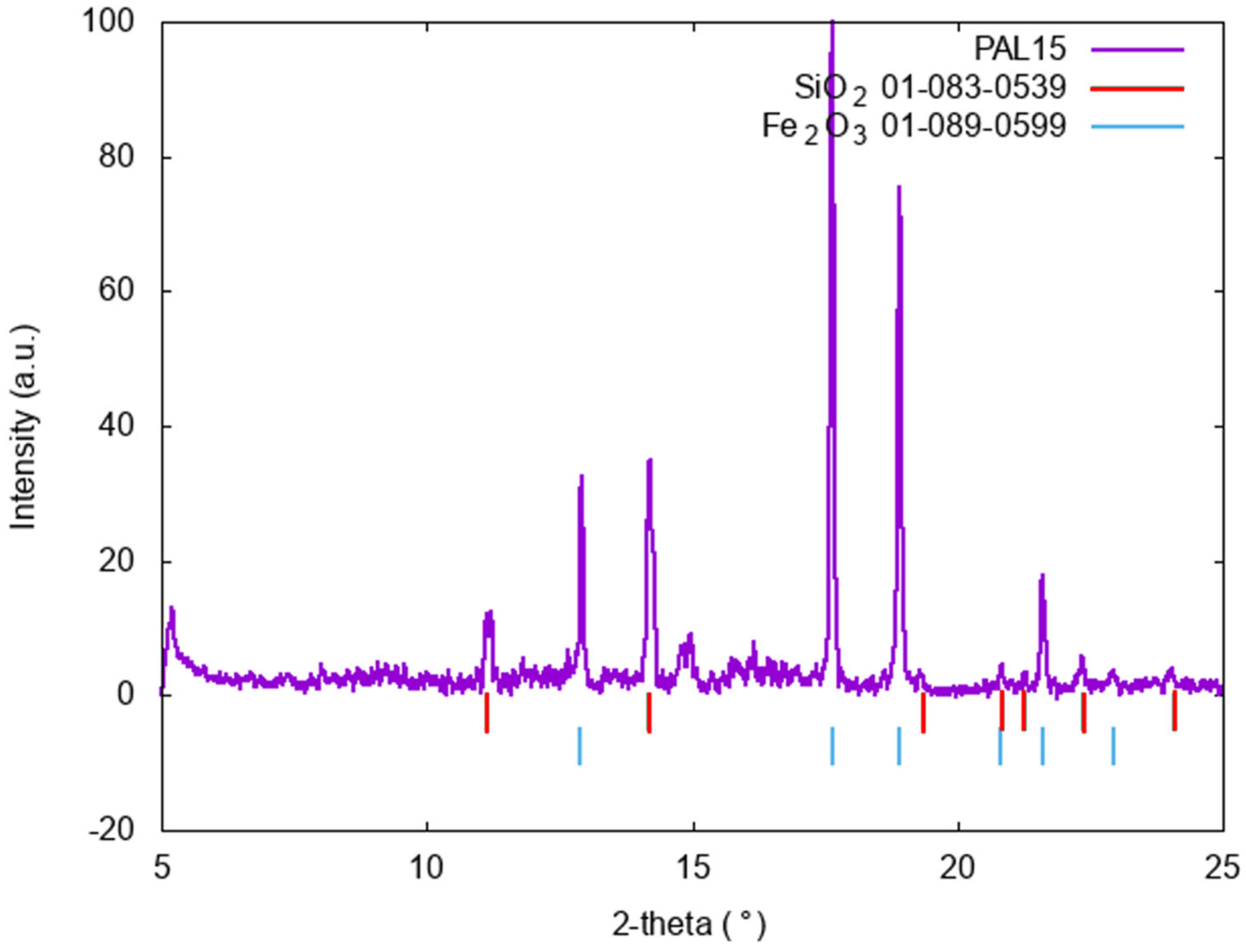
| Pal15 | Hematite, MnOx (pyrolusite, MnO2, ?). |
Disclaimer/Publisher’s Note: The statements, opinions and data contained in all publications are solely those of the individual author(s) and contributor(s) and not of MDPI and/or the editor(s). MDPI and/or the editor(s) disclaim responsibility for any injury to people or property resulting from any ideas, methods, instructions or products referred to in the content. |
© 2024 by the authors. Licensee MDPI, Basel, Switzerland. This article is an open access article distributed under the terms and conditions of the Creative Commons Attribution (CC BY) license (https://creativecommons.org/licenses/by/4.0/).
Share and Cite
Gianoncelli, A.; Schöder, S.; Plaisier, J.R.; Fugazzotto, M.; Barone, G.; Russo, A.; Mazzoleni, P.; Raneri, S. X-ray Synchrotron Radiation to Look at Pigments in Antiquities: Overview and Examples. Heritage 2024, 7, 2118-2137. https://doi.org/10.3390/heritage7040100
Gianoncelli A, Schöder S, Plaisier JR, Fugazzotto M, Barone G, Russo A, Mazzoleni P, Raneri S. X-ray Synchrotron Radiation to Look at Pigments in Antiquities: Overview and Examples. Heritage. 2024; 7(4):2118-2137. https://doi.org/10.3390/heritage7040100
Chicago/Turabian StyleGianoncelli, Alessandra, Sebastian Schöder, Jasper R. Plaisier, Maura Fugazzotto, Germana Barone, Alfonsina Russo, Paolo Mazzoleni, and Simona Raneri. 2024. "X-ray Synchrotron Radiation to Look at Pigments in Antiquities: Overview and Examples" Heritage 7, no. 4: 2118-2137. https://doi.org/10.3390/heritage7040100
APA StyleGianoncelli, A., Schöder, S., Plaisier, J. R., Fugazzotto, M., Barone, G., Russo, A., Mazzoleni, P., & Raneri, S. (2024). X-ray Synchrotron Radiation to Look at Pigments in Antiquities: Overview and Examples. Heritage, 7(4), 2118-2137. https://doi.org/10.3390/heritage7040100