Review

Nano-Encapsulation and Conjugation Applied in the Development of Lipid Nanoparticles Delivering Nucleic Acid Materials to Enable Gene Therapies

Division of Pharmaceutical Sciences, James L. Winkle College of Pharmacy, University of Cincinnati, Cincinnati, OH 45229, USA
*
Author to whom correspondence should be addressed.
Appl. Nano 2024, 5(3), 143-161; https://doi.org/10.3390/applnano5030011
Submission received: 25 July 2024 / Revised: 23 August 2024 / Accepted: 27 August 2024 / Published: 29 August 2024
(This article belongs to the Collection Review Papers for Applied Nano Science and Technology)

Abstract

:
Nano-encapsulation and conjugation are the main strategies employed for drug delivery. Nanoparticles help improve encapsulation and targeting efficiency, thus optimizing therapeutic efficacy. Through nanoparticle technology, replacement of a defective gene or delivery of a new gene into a patient’s genome has become possible. Lipid nanoparticles (LNPs) loaded with genetic materials are designed to be delivered to specific target sites to enable gene therapy. The lipid shells protect the fragile genetic materials from degradation, then successfully release the payload inside of the cells, where it can integrate into the patient’s genome and subsequently express the protein of interest. This review focuses on the development of LNPs and nano-pharmaceutical techniques for improving the potency of gene therapies, reducing toxicities, targeting specific cells, and releasing genetic materials to achieve therapeutic effects. In addition, we discuss preparation techniques, encapsulation efficiency, and the effects of conjugation on the efficacy of LNPs in delivering nucleic acid materials.

1. Introduction

The purpose of gene therapy is to treat or prevent diseases through the modification of the host genome. Inserting a gene into a patient’s cells and/or replacing a defective gene with a healthy one has proven to be an effective therapy for numerous diseases. Through the process of transcription, genetic information stored in genomic sequences of DNA are transcribed into RNA; then, through translation, the RNA is used to produce functional proteins. Thus, there are a wide range of gene-therapy drugs utilizing nucleic acid polymers such as gene-editing complexes, product proteins, plasmid DNAs (pDNAs), messenger ribonucleic acids (mRNAs), and short interfering RNAs (siRNAs). They have the potential to treat a range of diseases by targeting the root cause as opposed to blocking downstream signaling pathways or treating symptoms. In addition, the development of Clustered Regularly Interspaced Short Palindromic Repeats (CRISPR), a gene-editing platform, has facilitated the replacement of dysfunctional genes with the wild-type allele in a straightforward manner, potentially enabling the correction of hereditary disorders. The recent discovery of Fanzor proteins, which work like CRISPR but are much smaller [1], sparked the idea to encapsulate or conjugate Fanzor proteins into LNPs to better facilitate its delivery into cells compared to CRISPR. Despite the possibilities for their wide clinical applications, the delivery of these compounds remains a challenge, especially due to the lack of evidence of effective intracellular delivery into target tissues in vivo [2]. Most naked genetic materials cannot survive in an extracellular environment rich in serum nucleases [3]. Furthermore, they are subject to degradation through hepatic metabolism, innate immunity, and renal filtration [4]. Naked functional pDNA was reported to have an in vitro half-life of approximately 10 min in whole blood in mouse models [5] and even shorter than that in vivo. Some modified synthetic nucleic acids, which could persist through these external barriers, can only achieve marginal bioavailability [6]. Electrostatic repulsion resulting from the negatively charged nucleic acid backbone leads to poor size uniformity and distribution of the nucleic acid payload and therefore to impaired delivery. Additionally, these negatively charged nucleic acids can interact with positively charged macromolecules within the body, further compounding this issue [7]. Various challenges have complicated the development of effective gene therapies [2,3,4,5,6,7]. For many years, researchers have been striving to overcome these obstacles by finding the proper vectors for these materials. There are many concerns with the use of viral vectors, including immunogenicity and difficult manufacturing processes, whereas many non-viral vectors have been reported to be toxic and harmful to cells [8].
In 2020, under the enormous pressure of the global pandemic caused by the SARS-CoV-2 coronavirus, a remarkable breakthrough was made in the therapeutic application of gene mRNA vaccines were authorized for use against the coronavirus disease. In comparison with available viral vector vaccines, non-viral novel mRNA vaccines are preferred because they have shown impressively high rates of efficacy [9]. These mRNA vaccines serve as proof of concept for an entirely new way to deliver genetic materials and enable gene therapies: lipid nanoparticles (LNPs). LNPs (Figure 1) are the most extensively studied non-viral vector for gene therapies [8]. They have been employed in vaccines, monoclonal antibodies, immunomodulatory drugs, and CAR T-cell therapies [9]. LNPs are being explored for the delivery of corrective genes to treat cystic fibrosis and various types of cancer. Despite their fragility, genetic materials can be protected from degradation in the extracellular environment and then transported into cells by LNPs [10]. In addition, their therapeutic value, ability to infiltrate cells, and stability can be improved by encapsulating them into advanced LNP-based delivery systems [11]. If nucleotide materials are transported by these lipid-based carriers, the complexes can be sustained in the circulation for a longer time, thus facilitating better cellular uptake. LNPs were reported to increase passive cell targeting by enhancing cell permeability and retention [12]. By using specific ligands, LNPs can also actively target cells through interactions with cell-surface receptors.
The clinical application of LNPs has been hindered by several challenges, especially delivery efficiency. LNPs must be delivered to the appropriate tissues or organs and taken up by target cells. Table 1 lists major issues in LNP delivery, such as delivery efficiency, intracellular stability, and endosome escape, along with their potential solutions. LNPs can escape from the injection site, enter the systemic circulation, and accumulate in the liver even after administration via local injection routes such as intramuscular and intratumoral injection, thus limiting the localized site-specific protein expression [13,14]. Unwanted liver accumulation of mRNA-loaded LNPs can be minimized by reticuloendothelial-system-blockade strategies such as stealth coating of liver sinusoids using two-arm-PEG-Oligo(l-Lysine). These RES-blockade strategies improved selective organ targeting of nucleic acid therapeutics by dramatically decreasing accumulation in the liver [15]. Another significant hurdle to the widespread utility of LNPs is their indispensable component: ionizable lipids. These lipids are not only highly inflammatory but also generate electrophilic impurities. These impurities can give rise to reactive moieties, including reactive oxygenated species, which can hydrolyze tertiary amines, rendering the mRNA untranslatable and leading to a loss of protein expression [16,17]. Moreover, delivering LNPs to targeted organs is often challenging due to obstacles like the blood-brain barrier and the immune system. Addressing the immune response triggered by LNPs remains a significant challenge. Stealth coatings and immunomodulators can help mitigate this issue.
Nanotechnology is a multidisciplinary science that uses fundamental aspects of physics, chemistry, and mathematics to design and manipulate atoms and molecules at the nanoscale to produce structures 1–1000 nm in size. Because of their small size and high surface area, the use of nanoparticles has resulted in improved drug solubility and enhanced bioavailability [18]. Over the last few decades, there have been major nanotechnological advances in the use of LNP medicine for gene and immunotherapy. In this article, we will focus on the development of LNPs through different nano-pharmaceutical techniques as well as on the strategies that may be employed to encapsulate bio-therapeutic drugs that enable gene therapies.

2. Nanoencapsulation

Nanoencapsulation is defined as the packaging of a drug core within a nanomaterial matrix shell [19]. Nanospheres are formed through the dispersion of drug molecules into polymeric materials, also known as carriers. Encapsulating genetic materials into carriers offers a key advantage: protection from degradation. A major issue with mRNA delivery is the rapid degradation of mRNA both outside and inside the cell by extracellular and intracellular ribonucleases, which reduce the effectiveness and duration of protein expression from the mRNA. Packaging nucleic acid therapeutics dramatically improves cargo stability and protects against extracellular and intracellular ribonucleases.
Common polymers in drug-delivery systems are polyethylene glycol (PEG), poloxamers, polyvinyl alcohol (PVA), polyesters, and polysaccharides [20]. In nanoencapsulation, polymers form physical barriers between the inner and outer phases of the particles and the environment, preventing drug degradation and thus facilitating more efficient drug delivery. Various polysaccharides (alginate, chitosan, carrageenan, etc.) mainly serve as a barrier against external conditions [21]. Encapsulation in polymers allows for the controlled release of the enclosed active ingredient under certain conditions. The polymer properties influence the physical and mechanical properties of the nanoparticle, which ultimately impact the biodistribution. Among polymeric nanomaterials, poly (lactic-co-glycolic acid) (PLGA) is frequently used to prepare micro- and nanospheres. PLGA can be synthesized in various molecular weights and lactide-to-glycolide ratios, where an increase in the ratio corresponds to a slower rate of polymer degradation and drug release [22,23]. There are challenges associated with micro- and nano-spheres, including controlling the particle size and distribution, as well as the presence of residual solvents and their toxicity. Particle size varies depending on the preparation technique used, but most manufacturing techniques result in microspheres. The size and distribution depend heavily on polymer and surfactant type and concentration, the dispersed-phase volume fraction, the stirring rate, and the temperature.
LNPs and lipid−polymer hybrid nanoparticles have several advantages over polymeric nanoparticles, including biocompatibility and versatility [24]. LNPs with uniform lipid bilayers or solid lipid cores can entrap hydrophobic and hydrophilic drugs with higher drug loading. However, differences in the size, density, and flexibility of the particle all play a role in the final fate of LNPs in vivo. Vaidya et al. were the first to demonstrate the application of density gradient-based ultracentrifugation, which relies on varying degrees of heterogeneity in mRNA-LNPs [25].

2.1. The Evolution of Genetic Materials for LNP Encapsulation

Over the past decade, several lipid-based formulations of mRNAs, siRNAs, and other genetic materials have been developed, and these are currently undergoing evaluation in vitro and in vivo for the treatment of various diseases. Currently, cationic LNPs, which are stable complexes between cationic lipids and anionic nucleic acids, are the most widely used non-viral delivery system for nucleic acid drugs. While the field of LNP technology has been evolving steadily, pivotal periods, key events, and significant strides in increasing encapsulation efficiency and the effective delivery of various types of genetic materials are described below. Available FDA-approved LNP products for nucleic therapeutics are listed in the Supplementary Materials section (Table S1).

2.1.1. LNPs for mRNA Delivery

The concept of mRNA as a therapeutic agent was officially recognized in 1990, when Wolff et al. injected naked RNA into mouse muscles [26]. Then, in 1992, Jirikowski et al. used mRNA to transiently reverse diabetes insipidus in Brattleboro rats that did not produce the hormone vasopressin [27]. mRNA strands are large, negatively charged macromolecules that cannot transverse the protective lipid membranes of cells [28]. Therefore, an emerging strategy is to incorporate mRNA within LNPs, which are spherical vesicles made of positively charged lipids at low pH. Cationic lipids can pair with anionic RNA to form an RNA-LNP complex. Although the concept is simple, it is not easy to identify an optimized ionizable cationic lipid and incorporate it with a genetic candidate. Previous studies have demonstrated that mRNA-loaded LNPs boosted immune responses to Zika virus, human immunodeficiency virus (HIV), herpes simplex virus, Ebola virus, and influenza virus, among others [29,30,31,32,33,34]. Recently, the success of using an LNP formulation of mRNA to prevent COVID-19 proved that LNPs are safe and effective non-viral vectors for delivering genetic materials. In onco-immunotherapy, several studies reported the successful application of LNP formulations for anticancer mRNA-based vaccines [35,36]. In work by Saad et al., cationic liposomes were produced from positively charged 1,2-dioleoyl-3-trimethylammonium-propane (DOTAP) lipid using an ethanol-injection method for the co-delivery of doxorubicin, BCL2 mRNA, and MRP1-targeted siRNA. The mixture led to the formation of large complexes with a size of approximately 500 nm and showed exceptionally high cytotoxicity to multidrug-resistant cancer [37].

2.1.2. LNPs for siRNA Delivery

Before the historic development of the Moderna and Pfizer BioTech COVID-19 vaccines, an LNP formulation of siRNA to treat transthyretin (TTR)-induced amyloidosis was considered the first successful systemic siRNA treatment for patients. Patisiran, sold under the brand name ONPATTRO®, is generated by the incorporation of siRNA with an optimized ionizable cationic lipid: heptatriaconta-6,9,28,31-tetraen-19-yl 4-(dimethylamino) butanoate (DLin-MC3-DMA or MC3) [38]. The incorporation of MC3 in LNP-siRNA systems can result in gene silencing in the liver at dose levels as low as 5 μg siRNA/kg in murine models [39]. The concept behind RNAi therapeutics is simple: prevent the production of disease-causing proteins by intercepting mRNAs with corresponding siRNAs. mRNA utilizes the protein-synthesis machinery of cells, while siRNA relies on the RNA-interference pathway. Although both mRNAs and siRNAs are delivered using LNPs, their mechanism of action and cellular uptake differ significantly. Like mRNAs, LNP-encapsulated siRNAs are typically taken up by cells through endocytosis. However, siRNAs can also be delivered through other pathways due to the double-stranded molecules being only 20 to 25 nucleotides in length. Conventionally, liposomes containing siRNAs have been prepared using the lipid-film method [40], but this method requires a post-processing treatment such as extrusion or sonication. For example, cationic lipoplexes produced using thin-film hydration and subsequent bath sonication yielded particles with a size of 72–291 nm [41]. A 2′OMe-modified siRNA targeting apolipoprotein B was successfully encapsulated inside 100 to 130 nm liposomes with an entrapment efficiency of 90–95% through the stepwise ethanol-dilution method [42]. Although siRNAs have been exploited for use in gene therapy over the years, extending siRNAs’ half-life in the circulation remains a challenge. Biological obstacles such as opsonin proteins and premature nuclease disintegration shorten siRNAs’ half-life; therefore, siRNAs may not enter the targeted cells while they are in circulation [43]. siRNAs diffuse poorly through the extracellular matrix. In the event that an siRNA reaches the target tissue, additional cell-level barriers impact cell entrance and stability [44]. The critical problem is the negative charge of siRNAs, which produces repulsion from the negatively charged cell membrane, resulting in poor uptake. If an siRNA successfully enters the cell, it must escape the endosome and withstand the hostile acidic environment of the cytosol. For those reasons, improved delivery methods are necessary to overcome the biological obstacles to siRNA delivery to the target cell and increase the gene-silencing effect.

2.1.3. LNPs for DNA and Protein Product Delivery

Carrillo et al. first reported lipoplexes formed between pDNA and cationic lipids, then moved forward to develop cationic solid LNPs for DNA delivery [45]. DNA and pDNA−LNP complexations have been successfully formulated and developed [46,47,48]. DNA−LNPs can be stabilized under conditions in which lipids and DNA are conjugated and then solubilized into micelles.
Strategies for gene delivery can be adapted for protein delivery, despite several challenges limiting efficient protein delivery [49,50]. Unsaturated hydrophobic nucleotide tails can be integrated into nanoparticle lipids to facilitate the delivery of large genetic materials such as pDNA and mRNA by improving intracellular pDNA and mRNA release. The release is triggered by intracellular glutathione, which can degrade disulfide bonds, resulting in endosome escape [51]. A chemically modified protein combined with CRISPR-associated protein 9 Cas9-single-guided RNA (sgRNA) ribonucleoproteins formed a protein/lipid nanocomplex with synthetic lipids and was delivered to cells through the endocytosis pathway instead of through the lipid-assisted cell-membrane-permeation pathway [52,53,54]. Although none of the complexes can induce significant cell toxicity, the study indicates that the electrostatic and hydrophobic interactions between lipids and proteins are critical. This was further confirmed by varying the lipid compositions of the nanoparticles.
Table 2 includes the interactions between nucleic acid materials (mRNAs, siRNAs, and pDNAs) and LNPs. The interaction between these nucleic acids and LNPs is crucial for their formation, encapsulation, stability, and subsequent release. siRNAs are typically encapsulated within the LNP core due to their small size. mRNAs can also be encapsulated within the LNP core. On the other hand, pDNAs can form complex structures with LNPs and are often conjugated to the LNP surface. Interestingly, the hydrophobic interaction between LNPs and their nucleic acids is important in LNP formation. This interaction helps to drive the encapsulation of mRNAs within the LNP core, which can be stronger if the mRNA structures are highly hydrophobic [55]. mRNAs with regions that have higher levels of secondary structure, such as helices, may not be fully encapsulated.

2.2. The Development of LNPs for the Encapsulation of Genetic Materials
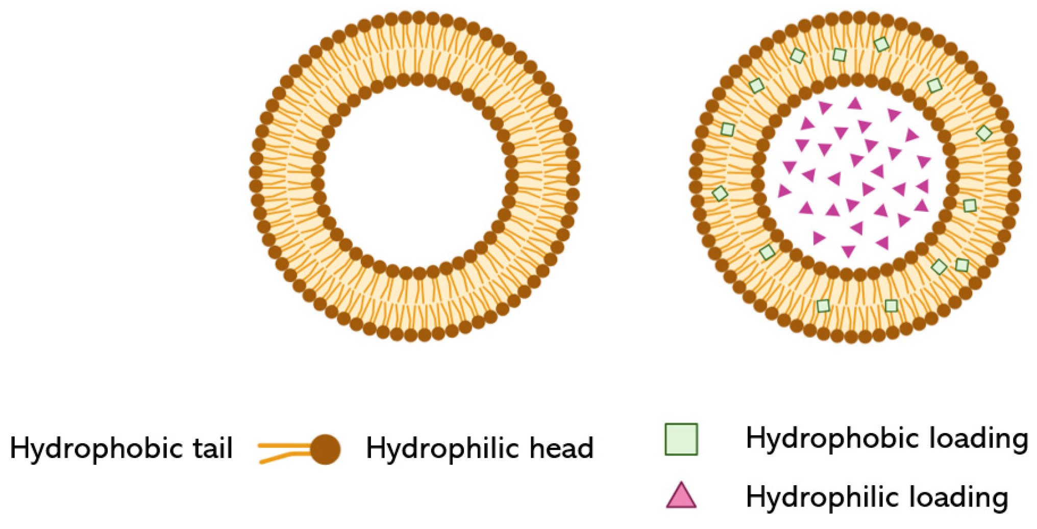
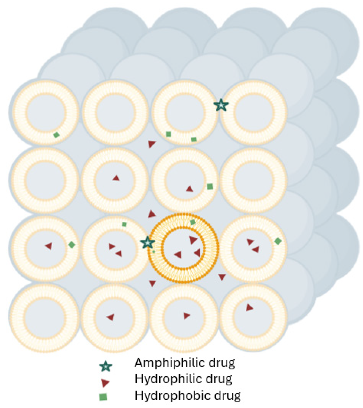
In conjunction with enhancing mRNA stability, scientists have worked on improving RNA delivery, notably through the application of LNP vesicles.

2.2.1. Liposomes

Looking back at their development, LNP formulations evolved from their earliest version: liposomes. Liposomes are closed bilayer structures spontaneously formed by hydrated phospholipids (Figure 2). Even before the 90s, in 1978, the first liposomes were utilized for the delivery of mRNA to eukaryotic cells [56], and a decade after that, a cationic liposome mRNA delivery system, 1,2-di-O-octadecenyl-3-trimethylammonium propane (DOTMA), was commercialized [57]. Many liposomal formulations have been approved for use as drugs and vaccines; here, we previously mentioned the ONPATTRO® story, because not only was it the first commercially available LNP formulation of siRNA, but also because it was a liposomal formulation.

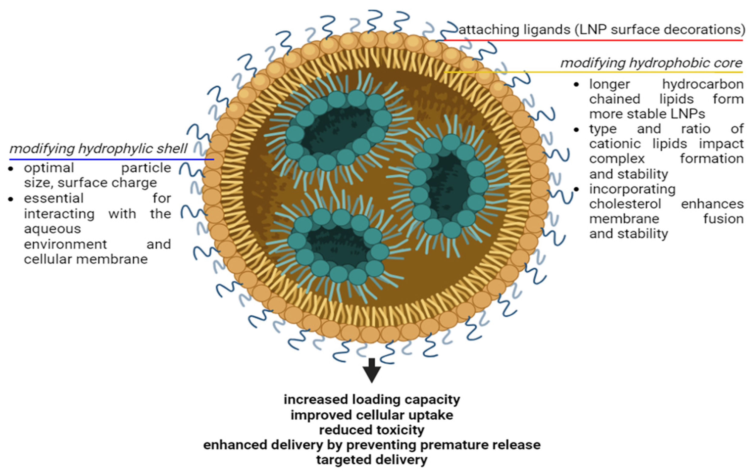
2.2.2. Solid Lipid Nanoparticles

Solid LNPs and nanostructured lipid carriers were first introduced by Müller et al. as micro/nano-sized spherical particles made of lipids that are solids at room temperature [58]. While phospholipid-based liposomes require a complex preparation method, solid LNPs are lipid-based emulsions constructed of a solid matrix that allows the controlled release of the drug. Compared to liposome structures, solid LNPs have higher loading capacities, are produced more easily, and are more cost-effective on a large scale. Biologics including peptides, proteins, and genes are the main targeting moieties. mRNA-based-solid LNPs were produced using three different techniques: solvent evaporation/emulsification, hot-melt emulsification, and coacervation to induce interleukin 10 synthesis; all were used in corneal cells to treat corneal inflammation [59]. There, solid LNPs facilitated a transfection efficiency higher than that seen with naked mRNA, but there was no correlation observed between in vitro and in vivo performance. Another problem associated with using solid LNPs in the encapsulation of genetic materials is crystallization, which can trigger an immune response, cause stability issues, and reduce bioavailability. When crystallization occurs during long-term storage, the LNPs can lyse, thus exposing the incorporated drugs to the surrounding environment. The exact mechanism of nucleic acid sequestration and subsequent crystallization within solid LNPs is not fully understood, but it is believed to involve nucleic acids and nucleic acid−solid lipid aggregations, which may form crystals within the lipid matrix. Strategies to minimize nucleic acid crystallization in solid LNPs include reduction of the nucleic acid concentration, lyophilization, lipid optimization, and excipients that can inhibit crystallization. The delivery of biological drugs encapsulated into solid LNPs may still be possible through the exploration of new therapeutic strategies, such as interference with bacterial transcription processes through the delivery of DNA molecules [60].

2.2.3. Lyotropic Liquid Crystal (LLC)

A lyotropic liquid crystal (LLC) is formed by dissolving an amphiphilic mesogen in a suitable solvent under appropriate conditions of concentration, temperature, and pressure [61]. When many amphiphilic molecules are present, the self-assembly of amphiphilic lipids due to hydrophobic interactions could potentially lead to the formation of well-defined, thermodynamically stable structures such as lamellar, hexagonal, and bicontinuous cubic phases. Lyotropic liquid crystalline drug-delivery systems have become one of the more advanced systems in the field of colloidal dispersions. Among the different types of LLCs, cubosomes exhibit many distinct advantages. While LNPs are often spherical, non-spherical shapes like cubosomes can offer advantages in terms of loading capacity and sustained release [62,63]. The cubic phase, which is made of bicontinuous lipid bilayers, develops a 3D network separating two distinct, continuous, non-intersecting hydrophilic, with hydrophobic components placed within [62] (Figure 3). There are two primary ways to encapsulate genetic material into cubosomes: direct incorporation and post-formation loading. The principle of direct incorporation involves mixing genetic materials with lipid components during cubosome formation. However, the lipid mixture is required to be heated above its phase-transition temperature, directly affecting the stability of the loaded genetic material and making uniform distribution of the genetic material within the cubic phase challenging. Double-stranded DNA fragments were encapsulated and released within monoolein-based cationic lipid phases of cationic cubic phases and their dispersed cationic cubosomes [63]. Meanwhile, loading the genetic materials into preformed cubosomes involves incorporating the gene through adsorption and complexation, which is not limited to encapsulation. An improved understanding of the mechanism of drug loading and release will assist in the design of novel lipid nanovectors for gene delivery. Besides, a variety of strategies for engineering cubosomes should be developed to render these liquid crystalline structures suitable for advanced drug delivery.
Table 3 summarizes the pros and cons of using different lipid-based nanocarriers for the encapsulation of nucleic acids.

3. Nanoconjugation

Nanoconjugation between hydrophilic polymeric shells and hydrophobic drug cores, as well as between hydrophilic drugs in self-assembled amphiphiles, have been studied for loading drugs into nanoparticles [13]. In the application of nano-encapsulation and conjugation to delivering nucleic acid materials, nanoconjugation happens between nucleic acids and cationic materials.

3.1. Lipid-Mediated Delivery System (Lipoplex)

A lipid-mediated delivery system (lipoplex), which is usually assembled from mixtures of cationic liposomes and DNA, was first reported in 1987 by Felgner et al. [64]. In the present day, it has become one of the most common strategies for nonviral gene therapy. Lipoplex laid the first stone for the development of cationic LNPs.
Lipoplexes form between genetic materials and LNPs because of the electrostatic interaction between the negatively charged nucleic acids and the positively charged lipids [37,38,41,45,46,47,48]. Other genetic materials were also employed with the lipoplex protocol or variations of it. Jin et al. successfully created an siRNA−polyethylene glycol−solid nanoparticle, which was proven to be able to cross the blood−brain barrier (BBB) to reach brain tumors with no apparent toxicity [65]. In another study, Kranz et al. reported in vivo experiments using intravenously injected mRNA−lipoplexes based on DOTMA/phospholipid dioleoylphosphatidylethanolamine (DOPE) and/or DOTAP/DOPE formulations; by optimizing the mRNA/cationic lipid ratio, then self-assembled LNPs were obtained [66]. Following the development of DOTAP, ionic interactions between the core and the shell were reported; these interaction drastically affected their entrapment efficiencies and capacity for intracellular delivery [66]. Toxicity issues resulting from the cationic LNPs’ permanent positive charge and non-biodegradable nature, plagued these initial lipoplex-like formulations. LNPs are neutral at physiological pH, thus reducing potential toxic effects compared with positively charged liposomes. In contrast, the effectiveness of lipoplexes for targeted gene delivery in vivo has been disappointing. It is known that their high cationic-charge density aids their rapid clearance from circulation by the reticuloendothelial system and that their tendency to form large aggregates enhances their accumulation in the microvasculature of ‘first pass’ organs such as the spleen, the liver, and particularly the lungs. The ability to manipulate the composition and structure of LNPs is crucial for optimizing gene delivery. LNP formation influences the amount of nucleic acid material encapsulated, the release rate, and ultimately the transfection efficiency. Figure 4 shows that modifying lipid composition can affect LNP stability and efficiency. For example, modifying the head groups of phospholipids can influence the particle size and its interaction with the external environment. Smaller particles often exhibit better cellular uptake, resulting in a higher transfection rate [67]. Another example is increasing the proportion of cationic lipids in a formulation, thus enhancing the binding of nucleic acids and loading capacity. However, an excessive amount of cationic lipids may reduce transfection efficiency due to aggregation.

3.2. LNP Surface Decorations

Due to the PEG-transformable end –OH- group, which can transform into various active functional groups, the uses of PEG and PEGylated lipids for LPNs have been one of the most efficient chemical modifications enabling conjunction for target delivery of micro- and nanoparticles [68]. Moreover, PEG can be used as a surfactant that promotes the formation of the hydrated layer of LNPs in solution, thus preventing aggregation of LNPs and enhancing LNP stability [69]. A typical LNP formulation contains four ingredients (Figure 1): ionizable lipids, PEGylated lipids, phospholipids, and cholesterol, in which the ionizable lipids are protonated to encapsulate negatively charged nucleic acids by electrostatic interactions. PEGylated lipids, phospholipids, and cholesterol contribute to the LNPs’ structure and stability [70]. Upon administration of LNPs, PEGylated lipids disrupt the equilibrium of surface-lipid compositions, promoting the adsorption of LNPs to serum proteins [69,70,71,72]. For the delivery of nucleic acid materials for gene therapies, LNP surfaces with PEGylated lipids are often decorated with targeting ligands. Lipid-DNA nanoparticles (Genospheres™) were made to effectively target specific cells by the addition of an antibody−lipopolymer (anti-HER2 scFv (F5)-PEG-DSPE) conjugate into the highly PEGylated lipid particles. It was reported that Genospheres could be stably stored under a variety of conditions and were able to achieve a high degree of transfection activity [73].
Ligands can be polymer or lipid molecules, drug-lipid conjugates, and protein− or peptide−lipid conjugates. They are often synthesized and incorporated into LNPs by the “in-lipid mixing” or the “post-insertion” methods. Notably, Dilliard and coworkers reported a strategy termed Selective ORgan Targeting (SORT) for tissue-specific mRNA delivery using LNP-SORT molecule conjugates that could direct LNPs to the lungs and the spleen. The SORT lipids of choice were mixed with other LNP lipid components. The factors that define their organ-targeting properties include the chemical nature of the SORT molecule and the biodistribution, pKa, and serum-protein interactions of the SORT nanoparticles. Moreover, the SORT organ-targeting mechanism was proven to function via PEG−lipid, protein−SORT LNP surfaces, and protein−receptor conjugates [74]. The post-insertion technique using ligand−PEG−lipid conjugates was initially developed to produce liposomes for immunotherapy but was later adapted to produce LNPs for targeted delivery [75]. Kasiewicz et al. reported the preparation of N-acetylgalactosamine (GalNAc)-LNPs that can deliver CRISPR-based gene-editing therapy to the liver [76]. Interestingly, GalNAc-siRNA conjugates have played a major role in vaccines and gene therapies and have been used to help cure several life-threatening diseases [6,76]. Sward et al. described a robust and simple “post-insertion” method for the preparation of targeted or fluorescently labeled siRNA LNPs using a copper-free strain-promoted azide-alkyne cycloaddition [77]. The “post-insertion” method is superior to direct surface modification because it preserves the physicochemical parameters of the LNPs. Dibenzocyclooctyne (DBCO)-azide copper-free reaction-based conjugation, along with other reaction-based conjugationsis termed “in situ conjugation”; the ligands are not pre-synthesized [77]. It is very important to remove the unattached molecules and the excess ligands after “in situ conjugation” to yield only surface-decorated LNPs. Figure 5 is a schematic representation of different ligand-targeted LNP decorations.

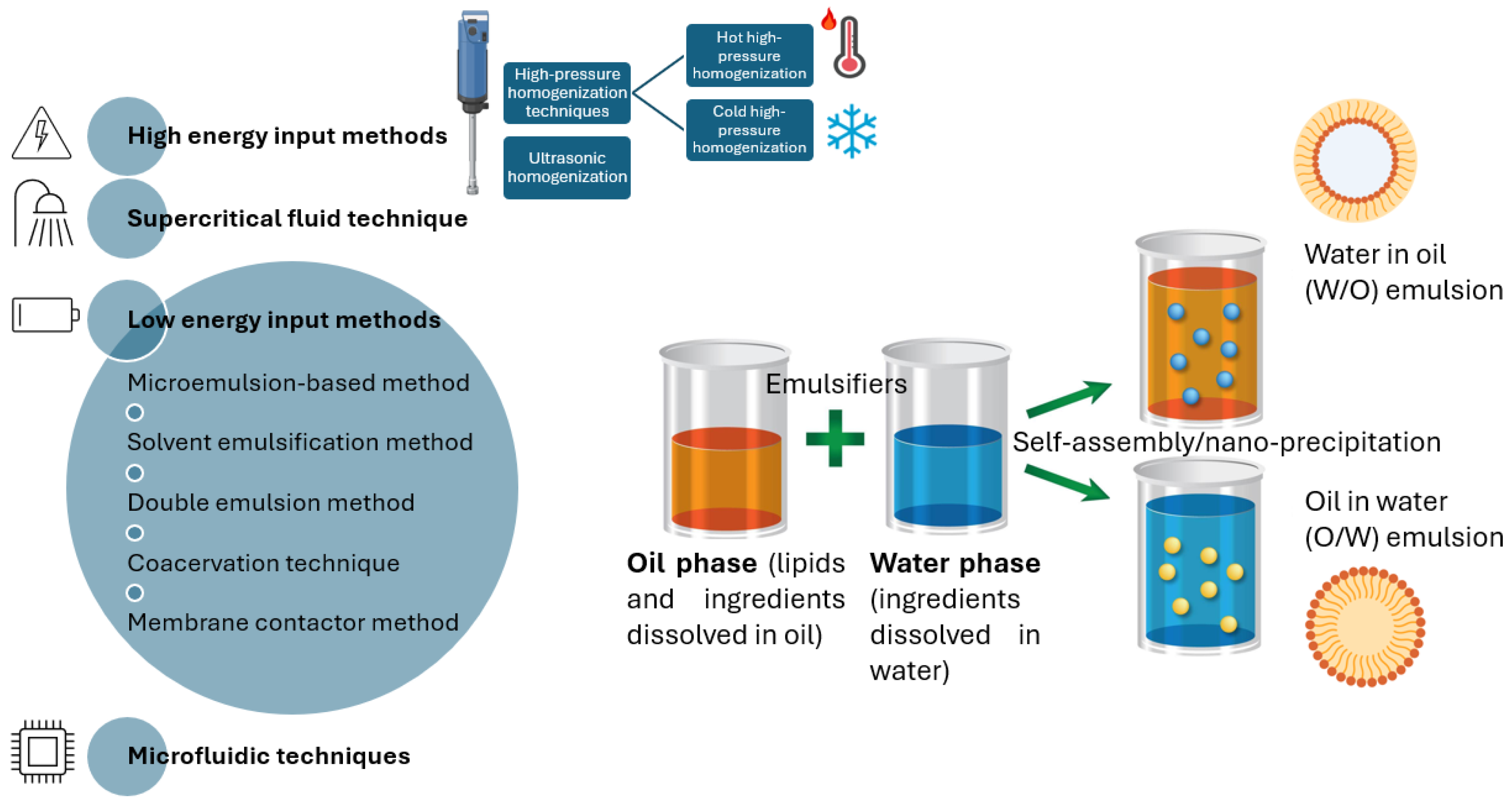
4. Preparation Methods of LNPs for the Encapsulation of Genetic Materials

A variety of techniques are available for the synthesis of LNPs (Figure 6). Several traditional physical methods with reasonable modifications to their methodologies have been optimized for the controlled synthesis of LNPs. LNP-preparation methods can be classified by energy input and solvent usage. The concentration ranges of the organic solvent used to prepare genetic-material-loaded LNPs were chosen based on the ability of the aqueous/organic solvent monophase to independently solubilize the nucleic acids and the lipid components before their combination. Notably, the use of organic solvents is sometimes unfavorable due to the possible interaction of organic solvents with shell and core materials and the extra production steps needed to remove the organic solvent. This extra step is crucial, as the leftover solvent can be a source of toxicity.

4.1. High-Energy-Input Methods

Although homogenization, ultrasonication techniques, and supercritical-fluid technology are very dispersive, these methods might not be compatible with delicate genetic materials. In applications for gene delivery, these methods can get involved with the process of making cationic lipid particles before the formation of a complex with the genetic materials.

4.1.1. Homogenization

Homogenization remains one of the most common, scalable, and cost-effective methods used to produce LNPs. High-pressure homogenization is a very dispersive technique, but the intense energy input and heat generated during the process might harm the thermolabile pharmaceutical ingredients. Further modifications of homogenization techniques, such as hot homogenization and cold homogenization, were eventually introduced. Hot homogenization causes bubbles to form due to the sudden decrease in pressure. In addition, thermal exposure also induces drug degradation. Cold-homogenization techniques are considered more advanced as they are more suitable for hydrophilic drugs and avoid the drug degradation associated with high processing temperatures. The disadvantages of cold homogenization have been reported to be large particle sizes ranging from 50 to 100 µm and a broad size distribution [78].
Ultrasonication, high-speed homogenization, or high-speed stirring methods can also be used in LNP preparation. Still, the application of these methods is often restricted due to unavoidable metal contamination, which results in large particle sizes.

4.1.2. The Supercritical-Fluid Technique

The supercritical-fluid technique can produce solvent-free products under mild temperature and pressure conditions. This newer, greener technique has the significant advantage of using CO2 as a solvent for LNP production and has thus drawn a lot of attention from researchers [79]. Several polymer−lipid hybrid formulations modified by DNA/RNA aptamers and antibodies have been developed for cancer treatment [80]. Various biomolecules including proteins, nucleic acids, peptides, and phospholipids were successfully encapsulated in or conjugated to polymer−lipid hybrid nanoparticles. Ge et al. presented a general method using microemulsion precipitation in supercritical CO2 for producing biodegradable nanoparticles that can be loaded with nucleic acids. The advantages of this method are ease of scalability, lack of residual organic solvents, and the formation of dry nanoparticles that can be placed in long-term storage [81]. However, questions regarding this method remain, including how to increase the loading capacity of the nanoparticles, how to facilitate nanoparticle entry to the target cell, and how to target specific cell types.
Another LNP-preparation method that is derived from the application of supercritical fluid is gas-assisted melting atomization. In this method, the lipids or protein/lipid mixtures are loaded into a mixing chamber, where they are melted with supercritical CO2 under proper temperature and pressure conditions. Then, the lipid-saturated fluid is forced through a nozzle by opening a valve at the bottom of a chamber to produce micron and sub-micron particles. It is claimed that this method produces fine and non-agglomerated low-density powders. It is also suitable for protein-loaded-lipid submicron-particle preparation, which is used to obtain solid colloidal formulations with high protein-loading efficiency and a controlled release profile [82].

4.2. Low-Energy-Input Methods

The original design of LNPs was based on liposomes and emulsions. A wide range of emulsion-based techniques has been applied to the preparation of LNPs, including microemulsion and different types of solvent-emulsification methods, each of which possesses advantages and disadvantages.

4.2.1. Microemulsion-Based Method

The microemulsion method was first proposed in 1943 by Hoar and Shulman. The mechanism of the microemulsion method is based on the achievement of spontaneous reduction in interfacial tension via the use of surfactants. This technique requires low energy input and results in a thermodynamically stable emulsion [83]. The microemulsion cooling technique, in which emulsification is followed by cooling, leads to particle precipitation and was perfected based on the same principle. The microemulsion method is reproducible, simple, and easily scalable and can result in solvent-free nanoparticle formation. This is one of the most traditional, yet still one of the most popular, novel carrier systems for encapsulating active moieties and has many applications for gene therapies. pDNA−nanoparticles have been synthesized by encapsulating hydrophobized pDNA from oil-in-water (o/w) microemulsion precursors [84]. In the work of Carrillo et al. mentioned above, solid LNPs were obtained by dispersing the hot microemulsion in cold water [45]. McAllister et al. were the pioneers in the application of microemulsion methods for non-viral gene-based formulations. They developed a cationic nanogel−DNA monodispersed complex that exhibited improved deposition of oligonucleotides in the culture medium.
Some microgel and nanogel formulations were developed via microemulsion polymerization [85]. In cancer immunotherapy, as proof of crossing paths between gene therapy and chemotherapy, some hydrogel systems loaded with genetic materials and chemicals were prepared by microemulsion [86,87]. However, after many attempts, it is still quite difficult to obtain an adequate concentration of LNPs in the resulting emulsion. This might be due to the dilution of the particle suspension during the emulsification process.

4.2.2. Solvent-Emulsification Methods

Solvent-emulsification methods such as the solvent-emulsification−evaporation method, solvent-emulsification−diffusion method, and solvent-injection/solvent-displacement method normally result in LNPs with a narrow size distribution. The solvent-emulsification method is widely used because it is appropriate for thermolabile drugs. In solvent-emulsification methods, lipids are first dissolved in an aqueous immiscible solvent, forming a low-viscosity system. An additional step such as ultrafiltration, lyophilization, or evaporation is required to eliminate the organic solvent used in the earlier step. The organic solvent may remain in the final preparation, which can cause toxicity. A double-emulsion−solvent-evaporation protocol for the preparation of siRNA-loaded lipidoid−polymer hybrid nanoparticles for delivery to the cytosol was described by Thanki et al. [88]. Physicochemical characterization was performed, and the gene-silencing effect was confirmed at the mRNA level by reverse transcription polymerase chain reaction.
Phase-inversion techniques are often used for the spontaneous inversion of o/w emulsions to w/o emulsions via an increase in temperature, with the assistance of additional molecules that influence the inversion phenomenon. Previous studies have shown that LNPs can be formed using the phase-inversion technique [89]. Solid LNPs and LLCs produced using this method were suitable for the topical application of bioactive ingredients. It was suggested that they may be suitable colloidal delivery systems for transporting lipophilic bioactive agents across the blood−brain barrier (BBB). A reported disadvantage of this method is emulsion instability.
Examples of commonly used solvents are water, ethanol, methanol, and acetone. The ethanol-injection method is one of the most notable solvent-injection methods. The ethanol-injection method was developed as an improved alternative to the thin-film hydration method, after the use of which sonication is necessary to produce siRNA-containing liposomes [90]. In contrast, in the ethanol-injection method, a solution of lipids in ethanol is directly injected through a syringe into a solution. This method is quick and easy to grasp. When ethanol is quickly diluted in aqueous buffer, lipid vesicles self-assemble due to a rise in solvent polarity.

4.2.3. Double-Emulsion Method

The double-emulsion method is an emulsion-based method for the preparation of LNPs. It can also be considered a novel method based on solvent emulsification−evaporation. Both hydrophilic and hydrophobic drugs, but mainly hydrophilic drugs, are dissolved in an aqueous solution and then emulsified in melted lipid. A stabilizer is incorporated to increase emulsion stability and protect against phase separation. Then, the stabilized primary emulsion is dispersed in an aqueous phase containing hydrophilic emulsifiers. Thereafter, the double emulsion is stirred and isolated by filtration. Xie et al. prepared a hydrophilic protein-loaded solid LNP by w/o/w double emulsion followed by solvent evaporation. The results showed that PLGA was essential for the primary w/o emulsification [91]. In addition, the stability of the w/o emulsion, the encapsulation efficiency, and the loading capacity of the nanoparticles were enhanced by an increase in PLGA concentration. Furthermore, increasing the PLGA concentration decreased zeta potential significantly without influencing particle size. In vitro release studies demonstrated that PLGA significantly affected the initial burst release, i.e., the higher the content of PLGA, the lower the burst release.

4.2.4. Coacervation Technique

A decrease in the pH of a micellar solution of alkaline salts of fatty acids by acidification in the presence of a polymeric stabilizer causes proton exchange, thus resulting in lipid precipitation (coacervation). This technique is suitable for use with lipophilic drugs (by solubilizing the drug in the micellar solution after coacervation) and with hydrophobic ion pairs of hydrophilic drugs. Despite being a simple, solvent-free technique that results in monodispersed nanoparticles, the coacervation technique is not appropriate for use with pH-sensitive active pharmaceutical ingredients.

4.2.5. Membrane-Contactor Method

In this method, a membrane contactor is used to prepare LNPs, and lipid materials are pressed through a porous membrane at a temperature above their melting point. Water circulated beyond the pores flows alongside the produced droplets of melted lipid which are then cooled to room temperature. The advantages of this method are the ability to control particle size by the choice of membrane and the scalability. Due to limits in the pore size of commercially available membranes, this method is more commonly employed in making micro-sized particles and has not yet been utilized to create nanoparticles.

4.3. Microfluidic Methods

Following the revelation of the relationship between the size of nanomedicines and their penetration efficiency in tumor tissues, controlling the size of LNPs has proven to be another challenge in the development of efficacious LNPs [92]. The particle size distribution of LNPs can be controlled using manufacturing methods such as extrusion, sonication, and homogenization. More recently, microfluidic methods have been successfully used for LNP manufacture and size control [93]. Microfluidics refers to the precise manipulation of fluids on a small scale at which volumetric forces are dominated by surface forces. Therefore, microfluidic systems represent the optimal method for producing size-controlled LNPs, as the microfluidics technique offers a simple, scalable, high-throughput continuous-flow process.
Mitchell and colleagues engineered a high-throughput screening transwell platform for the BBB specialized for mRNA−LNPs [94,95]. The same principles that apply to the conventional method are used for LNP production using a microfluidic device. The lipid phase (stream 1), including cationic components, is dissolved in ethanol (solvent) and the genetic material solution (stream 2) is prepared using an acid buffer. The LNPs are then self-assembled in the mixing chamber at the liquid−liquid interface where the two streams meet. Microfluidic devices for LNP production can be classified into different types depending on the flow type (Y- or T-shaped, chaotic mixer device).
Scaled-up RNA encapsulation can be accomplished via impingement jets mixing technology, in which two streams containing lipid and mRNA solution collide at high velocity in a jet mixing chamber, as shown in Figure 7. Table 4 lists the advantages and challenges of microfluidic applications for LNP production.

5. Future Scopes and Prospects

In this section, we discuss strategies for the use of LNPs and opportunities associated with their use.
Nano-encapsulation and conjugation are the main strategies employed for drug delivery. In this review, we focused on the application of nano-encapsulation and conjugation to LNP-based delivery of nucleic acid materials to enable gene therapies. LNPs and LNP-production technologies allow the encapsulation and targeted delivery of a variety of genetic materials, including but not limited to siRNAs, mRNAs, and DNAs, as well as undruggable proteins [96], growth factors promoting tissue regeneration, and other bioactive molecules.
There are a variety of available techniques for LNP production, and each has its advantages and disadvantages. Compared with conventional LNP-production methods, microfluidic approaches can produce homogeneous-sized LNPs with high encapsulation efficiency and high reproducibility. Additionally, optimized approaches to generating LNP-encapsulated genetic materials with surface decorations suggest that LNPs have accelerated the development of targeted delivery and, ultimately, of further gene therapies. Multifunctional LNPs can be developed by incorporating multiple functionalities such as imaging agents or therapeutic molecules into LNPs. LNP production should be improved so that techniques can be expanded, and scalable manufacturing processes enabled. The next generation of RNA, cell, and gene therapies and combinations thereof, delivered via LNPs, will become essential in personalized nanomedicine for both diagnostic and therapeutic purposes.

Supplementary Materials

The following supporting information can be downloaded at: https://www.mdpi.com/article/10.3390/applnano5030011/s1, Table S1: Available LNP products for nucleic therapeutics.

Author Contributions

Conceptualization, L.D.; resources, B.Y.; writing—original draft preparation, L.D.; writing—review and editing, L.D. and L.M.; visualization, L.D.; supervision, B.Y.; project administration, B.Y.; funding acquisition, B.Y. All authors have read and agreed to the published version of the manuscript.

Funding

This research was supported by the National Institutes of Health (Grants: R01 AI172959).

Acknowledgments

Figures were created with BioRender.com (accessed on 22 August 2024). Thanks go to Briana Simms, University of Cincinnati for her technical discussions and contributions to the manuscript revision. The first author especially thanks Sung-Joo Hwang, Yonsei University for his useful comments on an earlier draft of this manuscript.

Conflicts of Interest

The authors declare no conflicts of interest.

References

  1. Saito, M.; Xu, P.; Faure, G.; Maguire, S.; Kannan, S.; Altae-Tran, H.; Vo, S.; Desimone, A.; Macrae, R.K.; Zhang, F. Fanzor is a eukaryotic programmable RNA-guided endonuclease. Nature 2023, 620, 660–668. [Google Scholar] [CrossRef]
  2. Sioson, V.A.; Kim, M.; Joo, J. Challenges in delivery systems for CRISPR-based genome editing and opportunities of nanomedicine. Biomed. Eng. Lett. 2021, 11, 217–233. [Google Scholar] [CrossRef]
  3. Dirisala, A.; Uchida, S.; Tockary, T.A.; Yoshinaga, N.; Li, J.; Osawa, S.; Gorantla, L.; Fukushima, S.; Osada, K.; Kataoka, K. Precise tuning of disulphide crosslinking in mRNA polyplex micelles for optimising extracellular and intracellular nuclease tolerability. J. Drug Target. 2019, 27, 670–680. [Google Scholar] [CrossRef]
  4. Roberts, T.C.; Langer, R.; Wood, M.J.A. Advances in oligonucleotide drug delivery. Nat. Rev. Drug Discov. 2020, 19, 673–694. [Google Scholar] [CrossRef]
  5. Kawabata, K.; Takakura, Y.; Hashida, M. The fate of plasmid DNA after intravenous injection in mice: Involvement of scavenger receptors in its hepatic uptake. Pharm. Res. 1995, 12, 825–830. [Google Scholar] [CrossRef]
  6. Thangamani, L.; Balasubramanian, B.; Easwaran, M.; Natarajan, J.; Pushparaj, K.; Meyyazhagan, A.; Piramanayagam, S. GalNAc-siRNA conjugates: Prospective tools on the frontier of anti-viral therapeutics. Pharmacol. Res. 2021, 173, 105864. [Google Scholar] [CrossRef]
  7. Mendes, B.B.; Conniot, J.; Avital, A.; Yao, D.; Jiang, X.; Zhou, X.; Sharf-Pauker, N.; Xiao, Y.; Adir, O.; Liang, H.; et al. Nanodelivery of nucleic acids. Nat. Rev. Methods Primers 2022, 2, 24. [Google Scholar] [CrossRef]
  8. Zu, H.; Gao, D. Non-viral Vectors in Gene Therapy: Recent Development, Challenges, and Prospects. AAPS J. 2021, 23, 78. [Google Scholar] [CrossRef]
  9. Wang, Y.-S.; Kumari, M.; Chen, G.H.; Hong, M.-H.; Yuan, J.P.-Y.; Tsai, J.-L.; Wu, H.-C. mRNA-based vaccines and therapeutics: An in-depth survey of current and upcoming clinical applications. J. Biomed. Sci. 2023, 30, 84. [Google Scholar] [CrossRef]
  10. Yang, L.; Gong, L.; Wang, P.; Zhao, X.; Zhao, F.; Zhang, Z.; Li, Y.; Huang, W. Recent Advances in Lipid Nanoparticles for Delivery of mRNA. Pharmaceutics 2022, 14, 2682. [Google Scholar] [CrossRef]
  11. Jung, H.N.; Lee, S.Y.; Lee, S.; Youn, H.; Im, H.J. Lipid nanoparticles for delivery of RNA therapeutics: Current status and the role of in vivo imaging. Theranostics 2022, 12, 7509–7531. [Google Scholar] [CrossRef]
  12. Maeki, M.; Fujishima, Y.; Sato, Y.; Yasui, T.; Kaji, N.; Ishida, A.; Tani, H.; Baba, Y.; Harashima, H.; Tokeshi, M. Understanding the formation mechanism of lipid nanoparticles in microfluidic devices with chaotic micromixers. PLoS ONE 2017, 12, e0187962. [Google Scholar] [CrossRef]
  13. Yuan, M.; Han, Z.; Liang, Y.; Sun, Y.; He, B.; Chen, W.; Li, F. mRNA nanodelivery systems: Targeting strategies and administration routes. Biomater. Res. 2023, 27, 90. [Google Scholar] [CrossRef]
  14. Pardi, N.; Tuyishime, S.; Muramatsu, H.; Kariko, K.; Mui, B.L.; Tam, Y.K.; Madden, T.D.; Hope, M.J.; Weissman, D. Expression kinetics of nucleoside-modified mRNA delivered in lipid nanoparticles to mice by various routes. J. Control. Release 2015, 217, 345–351. [Google Scholar] [CrossRef]
  15. Dirisala, A.; Uchida, S.; Toh, K.; Li, J.; Osawa, S.; Tockary, T.A.; Liu, X.; Abbasi, S.; Hayashi, K.; Mochida, Y.; et al. Transient stealth coating of liver sinusoidal wall by anchoring two-armed PEG for retargeting nanomedicines. Sci. Adv. 2020, 6, eabb8133. [Google Scholar] [CrossRef]
  16. Ndeupen, S.; Qin, Z.; Jacobsen, S.; Bouteau, A.; Estanbouli, H.; Igyártó, B.Z. The mRNA-LNP platform’s lipid nanoparticle component used in preclinical vaccine studies is highly inflammatory. iScience 2021, 24, 103479. [Google Scholar] [CrossRef]
  17. Packer, M.; Gyawali, D.; Yerabolu, R.; Schariter, J.; White, P. A novel mechanism for the loss of mRNA activity in lipid nanoparticle delivery systems. Nat. Commun. 2021, 12, 6777. [Google Scholar] [CrossRef]
  18. Tran, T.T.D.; Tran, P.H.L. Nanoconjugation and Encapsulation Strategies for Improving Drug Delivery and Therapeutic Efficacy of Poorly Water-Soluble Drugs. Pharmaceutics 2019, 11, 325. [Google Scholar] [CrossRef]
  19. Cano-Sarabia, M.; Maspoch, D. Nanoencapsulation. In Encyclopedia of Nanotechnology; Bhushan, B., Ed.; Springer: Dordrecht, The Netherlands, 2012; pp. 1518–1530. [Google Scholar] [CrossRef]
  20. Deng, S.; Gigliobianco, M.R.; Censi, R.; Di Martino, P. Polymeric Nanocapsules as Nanotechnological Alternative for Drug Delivery System: Current Status, Challenges and Opportunities. Nanomaterials 2020, 10, 847. [Google Scholar] [CrossRef]
  21. Aboudzadeh, M.A.; Hamzehlou, S. Special Issue on “Function of Polymers in Encapsulation Process”. Polymers 2022, 14, 1178. [Google Scholar] [CrossRef]
  22. Makadia, H.K.; Siegel, S.J. Poly Lactic-co-Glycolic Acid (PLGA) as Biodegradable Controlled Drug Delivery Carrier. Polymers 2011, 3, 1377–1397. [Google Scholar] [CrossRef] [PubMed]
  23. Butreddy, A.; Gaddam, R.P.; Kommineni, N.; Dudhipala, N.; Voshavar, C. PLGA/PLA-Based Long-Acting Injectable Depot Microspheres in Clinical Use: Production and Characterization Overview for Protein/Peptide Delivery. Int. J. Mol. Sci. 2021, 22, 8884. [Google Scholar] [CrossRef]
  24. Lu, H.; Zhang, S.; Wang, J.; Chen, Q. A Review on Polymer and Lipid-Based Nanocarriers and Its Application to Nano-Pharmaceutical and Food-Based Systems. Front. Nutr. 2021, 8, 783831. [Google Scholar] [CrossRef] [PubMed]
  25. Vaidya, A.; Parande, D.; Khadse, N.; Vargas-Montoya, N.; Agarwal, V.; Ortiz, C.; Ellis, G.; Kaushal, N.; Sarode, A.; Karve, S.; et al. Analytical Characterization of Heterogeneities in mRNA-Lipid Nanoparticles Using Sucrose Density Gradient Ultracentrifugation. Anal. Chem. 2024, 96, 5570–5579. [Google Scholar] [CrossRef] [PubMed]
  26. Wolff, J.A.; Malone, R.W.; Williams, P.; Chong, W.; Acsadi, G.; Jani, A.; Felgner, P.L. Direct gene transfer into mouse muscle in vivo. Science 1990, 247, 1465–1468. [Google Scholar] [CrossRef]
  27. Jirikowski, G.F.; Sanna, P.P.; Maciejewski-Lenoir, D.; Bloom, F.E. Reversal of diabetes insipidus in Brattleboro rats: Intrahypothalamic injection of vasopressin mRNA. Science 1992, 255, 996–998. [Google Scholar] [CrossRef]
  28. Dolgin, E. The tangled history of mRNA vaccines. Nature 2021, 597, 318–324. [Google Scholar] [CrossRef]
  29. Pardi, N.; Hogan, M.J.; Pelc, R.S.; Muramatsu, H.; Andersen, H.; DeMaso, C.R.; Dowd, K.A.; Sutherland, L.L.; Scearce, R.M.; Parks, R.; et al. Zika virus protection by a single low-dose nucleoside-modified mRNA vaccination. Nature 2017, 543, 248–251. [Google Scholar] [CrossRef]
  30. Pardi, N.; Secreto, A.J.; Shan, X.; Debonera, F.; Glover, J.; Yi, Y.; Muramatsu, H.; Ni, H.; Mui, B.L.; Tam, Y.K.; et al. Administration of nucleoside-modified mRNA encoding broadly neutralizing antibody protects humanized mice from HIV-1 challenge. Nat. Commun. 2017, 8, 14630. [Google Scholar] [CrossRef]
  31. Awasthi, S.; Hook, L.M.; Pardi, N.; Wang, F.; Myles, A.; Cancro, M.P.; Cohen, G.H.; Weissman, D.; Friedman, H.M. Nucleoside-modified mRNA encoding HSV-2 glycoproteins C, D, and E prevents clinical and subclinical genital herpes. Sci. Immunol. 2019, 4, eaaw7083. [Google Scholar] [CrossRef]
  32. Meyer, M.; Huang, E.; Yuzhakov, O.; Ramanathan, P.; Ciaramella, G.; Bukreyev, A. Modified mRNA-Based Vaccines Elicit Robust Immune Responses and Protect Guinea Pigs from Ebola Virus Disease. J. Infect. Dis. 2018, 217, 451–455. [Google Scholar] [CrossRef] [PubMed]
  33. Bahl, K.; Senn, J.J.; Yuzhakov, O.; Bulychev, A.; Brito, L.A.; Hassett, K.J.; Laska, M.E.; Smith, M.; Almarsson, Ö.; Thompson, J.; et al. Preclinical and Clinical Demonstration of Immunogenicity by mRNA Vaccines against H10N8 and H7N9 Influenza Viruses. Mol. Ther. 2022, 25, 1316–1327. [Google Scholar] [CrossRef]
  34. Sahin, U.; Muik, A.; Derhovanessian, E.; Vogler, I.; Kranz, L.M.; Vormehr, M.; Baum, A.; Pascal, K.; Quandt, J.; Maurus, D.; et al. COVID-19 vaccine BNT162b1 elicits human antibody and TH1 T cell responses. Nature 2020, 586, 594–599. [Google Scholar] [CrossRef]
  35. Chen, J.; Ye, Z.; Huang, C.; Qiu, M.; Song, D.; Li, Y.; Xu, Q. Lipid nanoparticle-mediated lymph node-targeting delivery of mRNA cancer vaccine elicits robust CD8+ T cell response. Proc. Natl. Acad. Sci. USA 2022, 119, e2207841119. [Google Scholar] [CrossRef] [PubMed]
  36. Cafri, G.; Gartner, J.J.; Zaks, T.; Hopson, K.; Levin, N.; Paria, B.C.; Parkhurst, M.R.; Yossef, R.; Lowery, F.J.; Jafferji, M.S.; et al. mRNA vaccine-induced neoantigen-specific T cell immunity in patients with gastrointestinal cancer. J. Clin. Investig. 2020, 130, 5976–5988. [Google Scholar] [CrossRef] [PubMed]
  37. Saad, M.; Garbuzenko, O.B.; Minko, T. Co-delivery of siRNA and an anticancer drug for treatment of multidrug-resistant cancer. Nanomedicine 2008, 3, 761–776. [Google Scholar] [CrossRef] [PubMed]
  38. Yonezawa, S.; Koide, H.; Asai, T. Recent advances in siRNA delivery mediated by lipid-based nanoparticles. Adv. Drug Deliv. Rev. 2020, 154-155, 64–78. [Google Scholar] [CrossRef]
  39. Tam, Y.Y.; Chen, S.; Cullis, P.R. Advances in Lipid Nanoparticles for siRNA Delivery. Pharmaceutics 2013, 5, 498–507. [Google Scholar] [CrossRef]
  40. Alshehri, A.; Grabowska, A.; Stolnik, S. Pathways of cellular internalisation of liposomes delivered siRNA and effects on siRNA engagement with target mRNA and silencing in cancer cells. Sci. Rep. 2018, 8, 3748. [Google Scholar] [CrossRef]
  41. Paecharoenchai, O.; Niyomtham, N.; Apirakaramwong, A.; Ngawhirunpat, T.; Rojanarata, T.; Yingyongnarongkul, B.E.; Opanasopit, P. Structure relationship of cationic lipids on gene transfection mediated by cationic liposomes. AAPS PharmSciTech 2012, 13, 1302–1308. [Google Scholar] [CrossRef]
  42. Judge, A.D.; Bola, G.; Lee, A.C.; MacLachlan, I. Design of noninflammatory synthetic siRNA mediating potent gene silencing in vivo. Mol. Ther. 2006, 13, 494–505. [Google Scholar] [CrossRef]
  43. Babu, A.; Muralidharan, R.; Amreddy, N.; Mehta, M.; Munshi, A.; Ramesh, R. Nanoparticles for siRNA-Based Gene Silencing in Tumor Therapy. IEEE Trans. Nanobiosci. 2016, 15, 849–863. [Google Scholar] [CrossRef]
  44. Wang, T.; Hamilla, S.; Cam, M.; Aranda-Espinoza, H.; Mili, S. Extracellular matrix stiffness and cell contractility control RNA localization to promote cell migration. Nat. Commun. 2017, 8, 896. [Google Scholar] [CrossRef] [PubMed]
  45. Carrillo, C.; Sánchez-Hernández, N.; García-Montoya, E.; Pérez-Lozano, P.; Suñé-Negre, J.M.; Ticó, J.R.; Suñé, C.; Miñarro, M. DNA delivery via cationic solid lipid nanoparticles (SLNs). Eur. J. Pharm. Sci. 2013, 49, 157–165. [Google Scholar] [CrossRef]
  46. Cui, L.; Renzi, S.; Quagliarini, E.; Digiacomo, L.; Amenitsch, H.; Masuelli, L.; Bei, R.; Ferri, G.; Cardarelli, F.; Wang, J.; et al. Efficient Delivery of DNA Using Lipid Nanoparticles. Pharmaceutics 2022, 14, 1698. [Google Scholar] [CrossRef]
  47. Prazeres, P.H.D.M.; Ferreira, H.; Costa, P.A.C.; da Silva, W.; Alves, M.T.; Padilla, M.; Thatte, A.; Santos, A.K.; Lobo, A.O.; Sabino, A.; et al. Delivery of Plasmid DNA by Ionizable Lipid Nanoparticles to Induce CAR Expression in T Cells. Int. J. Nanomed. 2023, 18, 5891–5904. [Google Scholar] [CrossRef]
  48. Kulkarni, J.A.; Myhre, J.L.; Chen, S.; Tam, Y.Y.C.; Danescu, A.; Richman, J.M.; Cullis, P.R. Design of lipid nanoparticles for in vitro and in vivo delivery of plasmid DNA. Nanomedicine 2017, 13, 1377–1387. [Google Scholar] [CrossRef]
  49. Rehman, K.; Hamid Akash, M.S.; Akhtar, B.; Tariq, M.; Mahmood, A.; Ibrahim, M. Delivery of Therapeutic Proteins: Challenges and Strategies. Curr. Drug Targets 2016, 17, 1172–1188. [Google Scholar] [CrossRef]
  50. Wu, J.; Sahoo, J.K.; Li, Y.; Xu, Q.; Kaplan, D.L. Challenges in delivering therapeutic peptides and proteins: A silk-based solution. J. Control. Release 2022, 345, 176–189. [Google Scholar] [CrossRef]
  51. Chang, J.; Chen, X.; Glass, Z.; Gao, F.; Mao, L.; Wang, M.; Xu, Q. Integrating Combinatorial Lipid Nanoparticle and Chemically Modified Protein for Intracellular Delivery and Genome Editing. Acc. Chem. Res. 2019, 52, 665–675. [Google Scholar] [CrossRef]
  52. Im, S.H.; Jang, M.; Park, J.H.; Chung, H.J. Finely tuned ionizable lipid nanoparticles for CRISPR/Cas9 ribonucleoprotein delivery and gene editing. J. Nanobiotechnol. 2024, 22, 175. [Google Scholar] [CrossRef] [PubMed]
  53. Chen, K.; Stahl, E.C.; Kang, M.H.; Xu, B.; Allen, R.; Trinidad, M.; Doudna, J.A. Engineering self-deliverable ribonucleoproteins for genome editing in the brain. bioRxiv 2023. [Google Scholar] [CrossRef] [PubMed]
  54. Kim, D.; Le, Q.V.; Wu, Y.; Park, J.; Oh, Y.K. Nanovesicle-Mediated Delivery Systems for CRISPR/Cas Genome Editing. Pharmaceutics 2020, 12, 1233. [Google Scholar] [CrossRef]
  55. Eygeris, Y.; Gupta, M.; Kim, J.; Sahay, G. Chemistry of Lipid Nanoparticles for RNA Delivery. Acc. Chem. Res. 2022, 55, 2–12. [Google Scholar] [CrossRef] [PubMed]
  56. Dimitriadis, G.J. Translation of rabbit globin mRNA introduced by liposomes into mouse lymphocytes. Nature 1978, 274, 923–924. [Google Scholar] [CrossRef]
  57. Malone, R.W.; Felgner, P.L.; Verma, I.M. Cationic liposome-mediated RNA transfection. Proc. Natl. Acad. Sci. USA 1989, 86, 6077–6081. [Google Scholar] [CrossRef]
  58. Müller, R.H.; Mäder, K.; Gohla, S. Solid lipid nanoparticles (SLN) for controlled drug delivery—A review of the state of the art. Eur. J. Pharm. Biopharm. 2000, 50, 161–177. [Google Scholar] [CrossRef]
  59. Gómez-Aguado, I.; Rodríguez-Castejón, J.; Beraza-Millor, M.; Vicente-Pascual, M.; Rodríguez-Gascón, A.; Garelli, S.; Battaglia, L.; Del Pozo-Rodríguez, A.; Solinís, M.Á. mRNA-Based Nanomedicinal Products to Address Corneal Inflammation by Interleukin-10 Supplementation. Pharmaceutics 2021, 13, 1472. [Google Scholar] [CrossRef]
  60. González-Paredes, A.; Sitia, L.; Ruyra, A.; Morris, C.J.; Wheeler, G.N.; McArthur, M.; Gasco, P. Solid lipid nanoparticles for the delivery of anti-microbial oligonucleotides. Eur. J. Pharm. Biopharm. 2019, 134, 166–177. [Google Scholar] [CrossRef]
  61. Dinh, L.; Yan, B. Oral Drug Delivery via Intestinal Lymphatic Transport Utilizing Lipid-Based Lyotropic Liquid Crystals. Liquids 2023, 3, 456–468. [Google Scholar] [CrossRef]
  62. Dinh, L.; Kim, D.M.; Lee, G.; Yoon, Y.; Han, H.; Oh, D.J.; Lee, J.; Hwang, S.J. Lyotropic liquid crystalline nanoparticles for oral delivery: Formulation and evaluation of sustained-released cromolyn sodium loaded cubosomes. J. Pharm. Investig. 2024, 54, 539–554. [Google Scholar] [CrossRef]
  63. Sarkar, S.; Tran, N.; Soni, S.K.; Conn, C.E.; Drummond, C.J. Size-Dependent Encapsulation and Release of dsDNA from Cationic Lyotropic Liquid Crystalline Cubic Phases. ACS Biomater. Sci. Eng. 2020, 6, 4401–4413. [Google Scholar] [CrossRef]
  64. Felgner, P.L.; Gadek, T.R.; Holm, M.; Roman, R.; Chan, H.W.; Wenz, M.; Northrop, J.P.; Ringold, G.M.; Danielsen, M. Lipofection: A highly efficient, lipid-mediated DNA-transfection procedure. Proc. Natl. Acad. Sci. USA 1987, 84, 7413–7417. [Google Scholar] [CrossRef] [PubMed]
  65. Jin, J.; Bae, K.H.; Yang, H.; Lee, S.J.; Kim, H.; Kim, Y.; Joo, K.M.; Seo, S.W.; Park, T.G.; Nam, D.H. In Vivo specific delivery of c-Met siRNA to glioblastoma using cationic solid lipid nanoparticles. Bioconj. Chem. 2011, 22, 2568–2572. [Google Scholar] [CrossRef] [PubMed]
  66. Kranz, L.M.; Diken, M.; Haas, H.; Kreiter, S.; Loquai, C.; Reuter, K.C.; Meng, M.; Fritz, D.; Vascotto, F.; Hefesha, H.; et al. Systemic RNA delivery to dendritic cells exploits antiviral defence for cancer immunotherapy. Nature 2016, 534, 396–401. [Google Scholar] [CrossRef]
  67. Behzadi, S.; Serpooshan, V.; Tao, W.; Hamaly, M.A.; Alkawareek, M.Y.; Dreaden, E.C.; Brown, D.; Alkilany, A.M.; Farokhzad, O.C.; Mahmoudi, M. Cellular uptake of nanoparticles: Journey inside the cell. Chem. Soc. Rev. 2017, 46, 4218–4244. [Google Scholar] [CrossRef]
  68. Miatmoko, A.; Asmoro, F.H.; Azhari, A.A.; Rosita, N.; Huang, C.S. The effect of 1,2-dioleoyl-3-trimethylammonium propane (DOTAP) Addition on the physical characteristics of β-ionone liposomes. Sci. Rep. 2023, 13, 4324. [Google Scholar] [CrossRef]
  69. Kim, J.; Kim, J.Y.; Kim, H.; Kim, E.; Park, S.; Ryu, K.H.; Lee, E.G. Increasing Transfection Efficiency of Lipoplexes by Modulating Complexation Solution for Transient Gene Expression. Int. J. Mol. Sci. 2021, 22, 12344. [Google Scholar] [CrossRef] [PubMed]
  70. Suk, J.S.; Xu, Q.; Kim, N.; Hanes, J.; Ensign, L.M. PEGylation as a strategy for improving nanoparticle-based drug and gene delivery. Adv. Drug Deliv. Rev. 2016, 99 Pt A, 28–51. [Google Scholar] [CrossRef]
  71. Hald Albertsen, C.; Kulkarni, J.A.; Witzigmann, D.; Lind, M.; Petersson, K.; Simonsen, J.B. The role of lipid components in lipid nanoparticles for vaccines and gene therapy. Adv. Drug Deliv. Rev. 2022, 188, 114416. [Google Scholar] [CrossRef]
  72. Berger, M.; Degey, M.; Leblond Chain, J.; Maquoi, E.; Evrard, B.; Lechanteur, A.; Piel, G. Effect of PEG Anchor and Serum on Lipid Nanoparticles: Development of a Nanoparticles Tracking Method. Pharmaceutics 2023, 15, 597. [Google Scholar] [CrossRef]
  73. Hayes, M.E.; Drummond, D.C.; Kirpotin, D.B.; Zheng, W.W.; Noble, C.O.; Park, J.W.; Marks, J.D.; Benz, C.C.; Hong, K. Genospheres: Self-assembling nucleic acid-lipid nanoparticles suitable for targeted gene delivery. Gene Ther. 2006, 13, 646–651. [Google Scholar] [CrossRef] [PubMed]
  74. Dilliard, S.A.; Cheng, Q.; Siegwart, D.J. On the mechanism of tissue-specific mRNA delivery by selective organ targeting nanoparticles. Proc. Natl. Acad. Sci. USA 2021, 118, e2109256118. [Google Scholar] [CrossRef]
  75. Lin, Y.; Cheng, Q.; Wei, T. Surface engineering of lipid nanoparticles: Targeted nucleic acid delivery and beyond. Biophys. Rep. 2023, 9, 255–278. [Google Scholar] [CrossRef] [PubMed]
  76. Kasiewicz, L.N.; Biswas, S.; Beach, A.; Ren, H.; Dutta, C.; Mazzola, A.M.; Rohde, E.; Chadwick, A.; Cheng, C.; Garcia, S.P.; et al. GalNAc-Lipid nanoparticles enable non-LDLR dependent hepatic delivery of a CRISPR base editing therapy. Nat. Commun. 2023, 14, 2776. [Google Scholar] [CrossRef]
  77. Swart, L.E.; Koekman, C.A.; Seinen, C.W.; Issa, H.; Rasouli, M.; Schiffelers, R.M.; Heidenreich, O. A robust post-insertion method for the preparation of targeted siRNA LNPs. Int. J. Pharm. 2022, 620, 121741. [Google Scholar] [CrossRef] [PubMed]
  78. Ezzati Nazhad Dolatabadi, J.; Valizadeh, H.; Hamishehkar, H. Solid Lipid Nanoparticles as Efficient Drug and Gene Delivery Systems: Recent Breakthroughs. Adv. Pharm. Bull. 2015, 5, 151–159. [Google Scholar] [CrossRef]
  79. Park, H.; Kim, J.S.; Kim, S.; Ha, E.S.; Kim, M.S.; Hwang, S.J. Pharmaceutical Applications of Supercritical Fluid Extraction of Emulsions for Micro-/Nanoparticle Formation. Pharmaceutics 2021, 13, 1928. [Google Scholar] [CrossRef]
  80. Subjakova, V.; Oravczova, V.; Hianik, T. Polymer Nanoparticles and Nanomotors Modified by DNA/RNA Aptamers and Antibodies in Targeted Therapy of Cancer. Polymers 2021, 13, 341. [Google Scholar] [CrossRef]
  81. Ge, J.; Jacobson, G.B.; Lobovkina, T.; Holmberg, K.; Zare, R.N. Sustained release of nucleic acids from polymeric nanoparticles using microemulsion precipitation in supercritical carbon dioxide. Chem. Commun. 2010, 46, 9034–9036. [Google Scholar] [CrossRef]
  82. Salmaso, S.; Elvassore, N.; Bertucco, A.; Caliceti, P. Production of solid lipid submicron particles for protein delivery using a novel supercritical gas-assisted melting atomization process. J. Pharm. Sci. 2009, 98, 640–650. [Google Scholar] [CrossRef] [PubMed]
  83. Pradhan, M.; Singh, D.; Singh, M.R. Novel colloidal carriers for psoriasis: Current issues, mechanistic insight and novel delivery approaches. J. Control. Release 2013, 170, 380–395. [Google Scholar] [CrossRef]
  84. Russell, J.M.; Michael, J.; Lubrizol Life Science Health Inc. Microemulsions as precursors to solid nanoparticles. U.S. Patent No. US7153525B1, 26 December 2006. [Google Scholar]
  85. McAllister, K.; Sazani, P.; Adam, M.; Cho, M.J.; Rubinstein, M.; Samulski, R.J.; DeSimone, J.M. Polymeric nanogels produced via inverse microemulsion polymerization as potential gene and antisense delivery agents. J. Am. Chem. Soc. 2002, 124, 15198–15207. [Google Scholar] [CrossRef]
  86. Morya, V.; Walia, S.; Mandal, B.B.; Ghoroi, C.; Bhatia, D. Functional DNA Based Hydrogels: Development, Properties and Biological Applications. ACS Biomater. Sci. Eng. 2020, 6, 6021–6035. [Google Scholar] [CrossRef]
  87. Zhong, R.; Talebian, S.; Mendes, B.B.; Wallace, G.; Langer, R.; Conde, J.; Shi, J. Hydrogels for RNA delivery. Nat. Mater. 2023, 22, 818–831. [Google Scholar] [CrossRef] [PubMed]
  88. Thanki, K.; Zeng, X.; Foged, C. Preparation, Characterization, and In Vitro Evaluation of Lipidoid-Polymer Hybrid Nanoparticles for siRNA Delivery to the Cytosol. Methods Mol. Biol. 2019, 1943, 141–152. [Google Scholar] [CrossRef] [PubMed]
  89. Pattipeiluhu, R.; Zeng, Y.; Hendrix, M.M.R.M.; Voets, I.K.; Kros, A.; Sharp, T.H. Liquid crystalline inverted lipid phases encapsulating siRNA enhance lipid nanoparticle mediated transfection. Nat. Commun. 2024, 15, 1303. [Google Scholar] [CrossRef]
  90. Hattori, Y.; Saito, H.; Nakamura, K.; Yamanaka, A.; Tang, M.; Ozaki, K. In Vitro and in vivo transfections using siRNA lipoplexes prepared by mixing siRNAs with a lipid-ethanol solution. J. Drug Deliv. Sci. Technol. 2022, 75, 103635. [Google Scholar] [CrossRef]
  91. Xie, S.; Wang, S.; Zhao, B.; Han, C.; Wang, M.; Zhou, W. Effect of PLGA as a polymeric emulsifier on preparation of hydrophilic protein-loaded solid lipid nanoparticles. Colloids Surf. B Biointerfaces 2008, 67, 199–204. [Google Scholar] [CrossRef]
  92. Xu, J.; Song, M.; Fang, Z.; Zheng, L.; Huang, X.; Liu, K. Applications and challenges of ultra-small particle size nanoparticles in tumor therapy. J. Control. Release 2023, 353, 699–712. [Google Scholar] [CrossRef]
  93. Maeki, M.; Uno, S.; Niwa, A.; Okada, Y.; Tokeshi, M. Microfluidic technologies and devices for lipid nanoparticle-based RNA delivery. J. Control. Release 2022, 344, 80–96. [Google Scholar] [CrossRef] [PubMed]
  94. Shepherd, S.J.; Warzecha, C.C.; Yadavali, S.; El-Mayta, R.; Alameh, M.G.; Wang, L.; Weissman, D.; Wilson, J.M.; Issadore, D.; Mitchell, M.J. Scalable mRNA and siRNA Lipid Nanoparticle Production Using a Parallelized Microfluidic Device. Nano Lett. 2021, 21, 5671–5680. [Google Scholar] [CrossRef] [PubMed]
  95. Hamilton, A.G.; Swingle, K.L.; Thatte, A.S.; Mukalel, A.J.; Safford, H.C.; Billingsley, M.M.; El-Mayta, R.D.; Han, X.; Nachod, B.E.; Joseph, R.A.; et al. High-Throughput In Vivo Screening Identifies Differential Influences on mRNA Lipid Nanoparticle Immune Cell Delivery by Administration Route. ACS Nano 2024, 18, 16151–16165. [Google Scholar] [CrossRef] [PubMed]
  96. Chan, A.; Haley, R.M.; Najar, M.A.; Gonzalez-Martinez, D.; Bugaj, L.J.; Burslem, G.M.; Mitchell, M.J.; Tsourkas, A. Lipid-mediated intracellular delivery of recombinant bioPROTACs for the rapid degradation of undruggable proteins. Nat. Commun. 2024, 15, 5808. [Google Scholar] [CrossRef]
Figure 1. A typical LNP for mRNA delivery contains 4 lipid ingredients: ionizable lipids, PEGylated lipids, phospholipids, and cholesterol.
Applnano 05 00011 g001
Figure 2. A liposomal vesicle and a liposome loaded with active compounds.
Applnano 05 00011 g002
Figure 3. A cubosome exhibiting internal and cubic structures with active compounds encapsulated.
Applnano 05 00011 g003
Figure 4. LNP modifications that can influence genetic material loading and transfection.
Applnano 05 00011 g004
Figure 5. Schematic representation of different ligand-targeted LNP decorations.
Applnano 05 00011 g005
Figure 6. Available methods for LNP preparation. Methods based on emulsion and nanoprecipitation principles.
Applnano 05 00011 g006
Figure 7. A schematic representation of flow-based production of LNPs.
Applnano 05 00011 g007
Table 1. Challenges in LNP delivery.
LNP Delivery IssuesPotential Solutions
1. Cellular Uptake
  • Modifying lipid composition, particle size and shape, and surface properties
  • Attaching ligands that bind to specific cell-surface receptors
2. Tissue Distribution
  • PEGylation coatings to reduce immune recognition
  • Incorporating immunomodulatory agents to suppress the immune response
  • Minimizing immunogenicity and toxicity
3. Degradation
  • Coatings
  • Using self-amplifying RNA, increasing nucleic acid concentration
4. Endosomal Trapping
  • Incorporating protonatable lipids or peptides to promote endosomal escape
  • Incorporating pH-sensitive polymers to trigger release at endosomal pH
5. Inflammatory
  • Modifying lipid composition to decrease the concentration of ionizable lipids used
6. Off-Target Effects
  • Reducing particle size to reduce non-specific interactions and improve targeting
  • Employing formulation optimization, coatings, ligand conjugation, and surface decorations to enhance specificity
  • Minimizing immunogenicity and toxicity, dose optimization
Table 2. Interactions between nucleic acid materials (mRNAs, siRNAs, and pDNAs) and LNPs.
Nucleic AcidSizeInteraction with LNP
mRNA a few hundred to several thousand nucleotides in length
  • Electrostatic attraction between the negatively charged phosphate backbone of mRNAs and the positively charged head groups of cationic lipids in LNPs.
  • Hydrophobic interaction between the hydrophobic regions of mRNAs and the hydrophobic cores of LNPs.
siRNA20–25 nucleotides in length
  • Electrostatic attraction
  • Hydrophobic interaction
pDNAa few thousand to several thousand base pairs (one base pair consists of two nucleotides)
  • Electrostatic attraction
  • Hydrophobic interaction
  • Complex formation
Table 3. Advantages and disadvantages of lipid-based nanocarriers.
Lipid-Based NanocarrierAdvantagesDisadvantages
Liposomes
  • Easy preparation
  • Biodegradable
  • Biocompatible
  • Well-established technology
  • Various formulations with approved products on the market
  • Large mRNAs, regions of RNA with higher levels of secondary structures with strong hydrophobic interaction may not be encapsulated
  • Lower encapsulation efficiency for nucleic acids
  • Instability, premature release
  • Limited tissue penetration
LNPs
polymer−lipid hybrid nanoparticles
  • High encapsulation efficiency
  • Efficient cellular uptake
  • Versatile for various nucleic acids
  • Controlled release
  • Increased stability compared to liposomes
  • Off-target effects, limited tissue penetration
Solid LNPs
Nanostructured lipid carriers
  • High protection from degradation
  • High encapsulation efficiency
  • Increased loading capacity
  • Increased stability compared to liposomes
  • Controlled release
  • Aggregation of highly concentrated nucleic acids and crystallization of solid lipids.
  • Off-target effects, limited tissue penetration
Cubosomes
  • Unique cubic structure
  • Increased loading capacity
  • Enhanced stability
  • Sustained release
  • Reduced immune response
  • Challenges in scaling up
  • Off-target effects
  • Limited clinical data
  • Large nucleic acids may not be encapsulated
Table 4. Advantages and challenges of microfluidic applications for LNP production.
AdvantagesChallenges
  • Same principles as an emulsion (conventional) method, including hydration, solvent injection, emulsification, and solvent-evaporation steps in one process
  • Controlled droplet (vesicle) sizes through power (manipulation of liquid flows)
  • Various types of channels and chips.
  • High surface-to-volume ratio
  • Small particle-size distribution.
  • Efficient mixing
  • High encapsulation efficiency
  • Reproducibility
  • Easy set-up
  • Scaled-up production using microfluidic jetting/mixing
  • Microfluidic channel clogs
  • Compatibility between solvent used and channels
  • Specialized equipment
  • Difficult fluid handling with multiple operation parameters
Disclaimer/Publisher’s Note: The statements, opinions and data contained in all publications are solely those of the individual author(s) and contributor(s) and not of MDPI and/or the editor(s). MDPI and/or the editor(s) disclaim responsibility for any injury to people or property resulting from any ideas, methods, instructions or products referred to in the content.

