Article

Biofabrication of Silver Nanoparticles by Azadirachta indica Rhizosphere Bacteria with Enhanced Antibacterial Properties

by
Mashhoor Kattali
1,*,
Keerthana P. Mampett
2,
Hamna Fathima Kodoor
1,
Sreejesh Govindankutty Ponnenkunnathu
3,
Somy Soman
1,
Debarshi Kar Mahapatra
4,
Tomy Muringayil Joseph
5,*,
Józef Haponiuk
5 and
Sabu Thomas
6
1
Department of Biotechnology, EMEA College of Arts and Science, Kondotty 673638, Kerala, India
2
Department of Microbiology, Majlis Arts and Science College, Valanchery 676552, Kerala, India
3
Centre d’Immunologie Marseille-Luminy (CIML), INSERM, 171 Av. de Luminy, 13009 Marseille, France
4
Department of Pharmaceutical Chemistry, J. K. College of Pharmacy, Bilaspur 495550, Chhattisgarh, India
5
Department of Polymer Technology, Faculty of Chemistry, Gdańsk University of Technology, G. Narutowicza 11/12, 80-233 Gdańsk, Poland
6
International and Inter-University Centre for Nanoscience and Nanotechnology, Mahatma Gandhi University, Kottayam 686560, Kerala, India
*
Authors to whom correspondence should be addressed.
Eng 2024, 5(2), 600-613; https://doi.org/10.3390/eng5020034
Submission received: 6 March 2024 / Revised: 7 April 2024 / Accepted: 10 April 2024 / Published: 15 April 2024
(This article belongs to the Section Materials Engineering)

Abstract

:
Microorganisms (MOs) are prominent in ecological functioning and balance. The rhizosphere is considered one of the most diverse ecosystems on Earth and serves as a breeding spot for many MOs. Rhizosphere microbial diversity changes according to plant species, genotype, and the nature of the soil. The current study reports the possible use of bacteria isolated from the rhizosphere of Azadirachta indica for synthesizing silver nanoparticles (AgNPs). The physicochemical characterization and antibacterial activity of these green synthesized AgNPs are also reported. The gene (16S rRNA) sequence of bacteria isolated from the rhizosphere showed a maximum similarity of 99.25% with Bacillus subtilis. After incubation, the colorless reaction mixture transformed to brown, which indicates the formation of AgNPs, and UV-vis spectral analysis also confirmed the biosynthesis of AgNPs. Compared to lower temperatures, the efficiency of AgNP synthesis was high at the higher temperature. The scanning electron microscope image demonstrated spherical-shaped AgNPs with sizes ranging from 18 to 21 nm. Energy-dispersive X-ray analysis established the elemental analysis of synthesized AgNPs. The synthesized AgNPs showed strong bactericidal properties against pathogenic bacteria Klebsiella pneumonia, Pseudomonas aeruginosa, Escherichia coli, and methicillin-resistant Staphylococcus aureus.

1. Introduction

Microorganisms (MOs) exist in almost all parts of the biosphere, and they play a pivotal role in maintaining ecological balance [1,2]. In the past decade, microbial biotechnology has attracted more attention due to the wide application of MOs in different sectors [3,4]. Soil is a kind of bioreactor that provides space for many MOs to carry out countless enzymatic and biochemical processes to degrade pollutants and organic and inorganic ingredients [5]. From a biodiversity perspective, soil is a suitable depository for MOs, the number of MOs per gram of soil is several billion, and hundreds of thousands of bacteria and archaea species reside in the soil [6]. Because of the interaction between plants and MOs, rhizospheres are the most microbe-rich area in the soil, and MOs can act on anything in the environment, including metals.
Advanced synthesis methods in nanotechnology revolutionize applications by allowing for precise control over the size, shape, and composition of nanoparticles (NPs), using their unique physical and chemical properties [7,8,9,10,11]. AgNPs are considered one of the most promising nanostructures, and their physicochemical and biological properties make them suitable for an eclectic array of biomedical applications. In addition, AgNPs have the advantage of being less reactive than Ag+, making them well suited for therapeutic and clinical applications [12,13,14]. AgNPs have a vital role in therapeutics, biomolecular sensors, and catalysis that can be applied to antibacterial applications, composite fibers, sensing materials, transformers, cosmetics, electronic components, etc. [15,16,17,18].
The green synthesis of AgNPs is becoming more attractive nowadays due to its minimal environmental and health risks. The green synthesis of AgNPs is based on a redox reaction, in which Ag+ ions are reduced to stable AgNPs using the components of living things [19]. The green synthesis of AgNPs involves the reduction of Ag+ ions to stable AgNPs through redox reactions facilitated by biological constituents like plant extracts, which utilize electron donors from the metabolism of organisms [20,21,22]. Plant extracts with antioxidant compounds like flavonoids or phenolics can donate electrons to Ag+ ions, while enzymes like laccase or peroxidase catalyze the reduction reaction through redox-active sites. This process transfers electrons or hydrogen atoms from the biological reducing agent to Ag+ ions, forming stable AgNPs. Understanding these biological components is crucial for optimizing NP synthesis and enhancing their properties. Biotic agents such as fungi, bacteria, algae, and plants in nanobiotechnology are generally used to synthesize NPs [23,24,25]. Microbial NP synthesis is environmentally approachable and has substantial advantages over other processes [26], and in microorganism-based synthesis, the shape and size of NPs can also be controlled [27]. The need for ideal conditions and mild temperatures makes microbial-based NPs more commercially feasible [28]. In addition, the occurrence of a biological styling agent of specific MO-based NPs acts as a shielding coating against oxidation, agglomeration, and aggregation, thereby offering more excellent stability to NPs [29].
MOs can synthesize intracellular or extracellular metal NPs. Intracellular synthesis necessitates further steps to release the synthesized NPs, and it requires sonication treatment or reaction with an appropriate detergent [30,31,32]. Fortunately, the extracellular biosynthesis of NPs is inexpensive and necessitates only more straightforward processing. For this reason, various studies have focused on extracellular approaches for synthesizing AgNPs using MOs [33]. Hence, this study reports the extracellular biosynthesis of AgNPs using bacteria isolated from the rhizosphere of Azadirachta indica, commonly known as neem. This study also summarizes the molecular identification of bacteria isolated from the rhizosphere soil, as well as the physicochemical characterization and in vitro antibacterial activity of these green-synthesized AgNPs.

2. Materials and Methods

2.1. Isolation of Bacteria from Rhizosphere

A. indica rhizosphere soil was collected from the campus of EMEA College of Arts and Science, Kondotty, Kerala, India, in a sterile polythene cover. The soil sample was serially diluted, and each dilution was plated on nutrient agar plates. The plates were incubated at 37 ± 1 °C for 24 h, and the morphology of different colonies formed was observed.

2.2. Screening for the AgNPs Synthesizing Bacteria

The bacterial isolates were inoculated into 10 mL sterile nutrient broth (NB), incubated at 37 ± 1 °C for 48 h, and then centrifuged. The culture was kept at 6000 rpm for 5 min to remove the biomass, and the AgNPs were synthesized using the supernatant. To screen the synthesis of AgNPs, 100 µL of supernatant was added to 5 mL of 1 mM silver nitrate solution (AgNO3), and the reaction mixture was incubated for 24 h. The visual observation of NP synthesis was conducted by checking for brown color formation in the reaction mixture. From the isolates, one bacterial isolate that formed the highest color transformation in the reaction mixture was taken for further analysis.

2.3. Green Synthesis of AgNPs

To synthesize AgNPs, the selected bacterial isolate was cultured in 50 mL NB at 37 ± 1 °C for 48 h. Then, the culture was centrifuged at 6000 rpm for 5 min, and the cell-free supernatant was used to synthesize AgNPs. Cell-free supernatant (500 µL) was added to 50 mL 1 mM AgNO3 and incubated in the reaction mixture for 24 h. The color formation in the reaction mixture was observed, and the synthesized AgNPs were separated by centrifugation at 12,000 rpm for 15 min. The resultant pellet was washed three times with double-distilled water. The obtained AgNPs were lyophilized, and for further applicational studies, the lyophilized AgNPs were stored at 4 ± 1 °C. The impact of temperature on AgNP synthesis was studied by incubating the reaction mixture at various temperatures, i.e., room temperature (RT), 40 °C, 50 °C, and 60 °C, for 24 h.

2.4. Molecular Identification of the Bacteria

The gene sequence of 16S rRNA was used to identify selected bacterial isolate. With the help of the NucleoSpin Tissue Kit (Macherey-Nagel), the genomic DNA of the chosen bacteria was extracted using agarose (0.8% agarose) gel electrophoresis, and the purity of the extracted genomic DNA was examined. The 16S rRNA gene was amplified using the extracted genomic DNA as a template by a forward primer with the sequence 5′CAGGCCTAACACATGCAAGTC3′ and a reverse primer with the sequence 5′GGGCGGGTGTACAAGGC3′. The PCR mixture comprised 2.5 μL 10× reaction buffer, 0.20 μL Taq polymerase (5 U/μL), 1 μL (2 ng) DNA template, 2 μL primers (10 μM; 1 μL forward and 1 μL reverse), 2.5 μL (2 mM) dNTPs (dATP, dTTP, dGTP, and dCTP), and 16.8 μL nuclease-free water. The PCR temperature profile comprised initial heating, performed at 95 °C for 3 min. Subsequently, the mixture underwent 35 cycles at 96 °C for 30 s, 55 °C for 40 s, and 1 min at 72 °C, and the reaction was ended with a final stage at 72 °C for 3 min, and the amplified product was checked in 1.2% agarose gels. The purification of the PCR product was performed using an Ultraclean PCR Clean-up Kit (Mo-Bio Laboratories, Inc., Carlsbad, CA, USA), the amplified product was sequenced at the Rajiv Gandhi Centre for Biotechnology, Trivandrum, India, and the quality of sequence was measured using Sequence Scanner Software v.1 (Applied Biosystems, Waltham, MA, USA). The consensus DNA sequence was taken for analysis and similarity searches using BLAST to identify the organism, and the MEGA11 program was used to perform evolutionary analyses.

2.5. Characterization of AgNPs

A Shimadzu UV-1800 spectrophotometer (Shimadzu, Kyoto, Japan) was used to monitor the optical properties of bacteria-synthesized AgNPs by checking the visible spectrum from 350 nm to 600 nm, and 1 mM AgNO3 was measured as the blank. SEM (ZEISS® Gemini SEM instrument-300, Zeiss, Cambridge, UK) operating at 120 kV, which was connected to Octane Plus EDX, was used to reveal the structure of synthesized AgNPs. EDX analysis was performed to check the purity of the synthesized AgNPs.

2.6. Antibacterial Studies

Antibacterial studies were performed against four human infective bacteria, Escherichia coli, Staphylococcus aureus, Pseudomonas aeruginosa, and Klebsiella pneumonia. The antibacterial studies were conducted through the disc diffusion method on nutrient agar (NA) plates. In the lawn culture of bacteria, 10 mm wells were prepared with 20 µL of different concentrations of AgNPs; viz., 10 µg/mL, 20 µg/mL, 40 µg/mL, 80 µg/mL, and 100 µg/mL were added to separate wells. The antibacterial activity was calculated by determining the zone of inhibition after 24 h of incubation.

3. Results

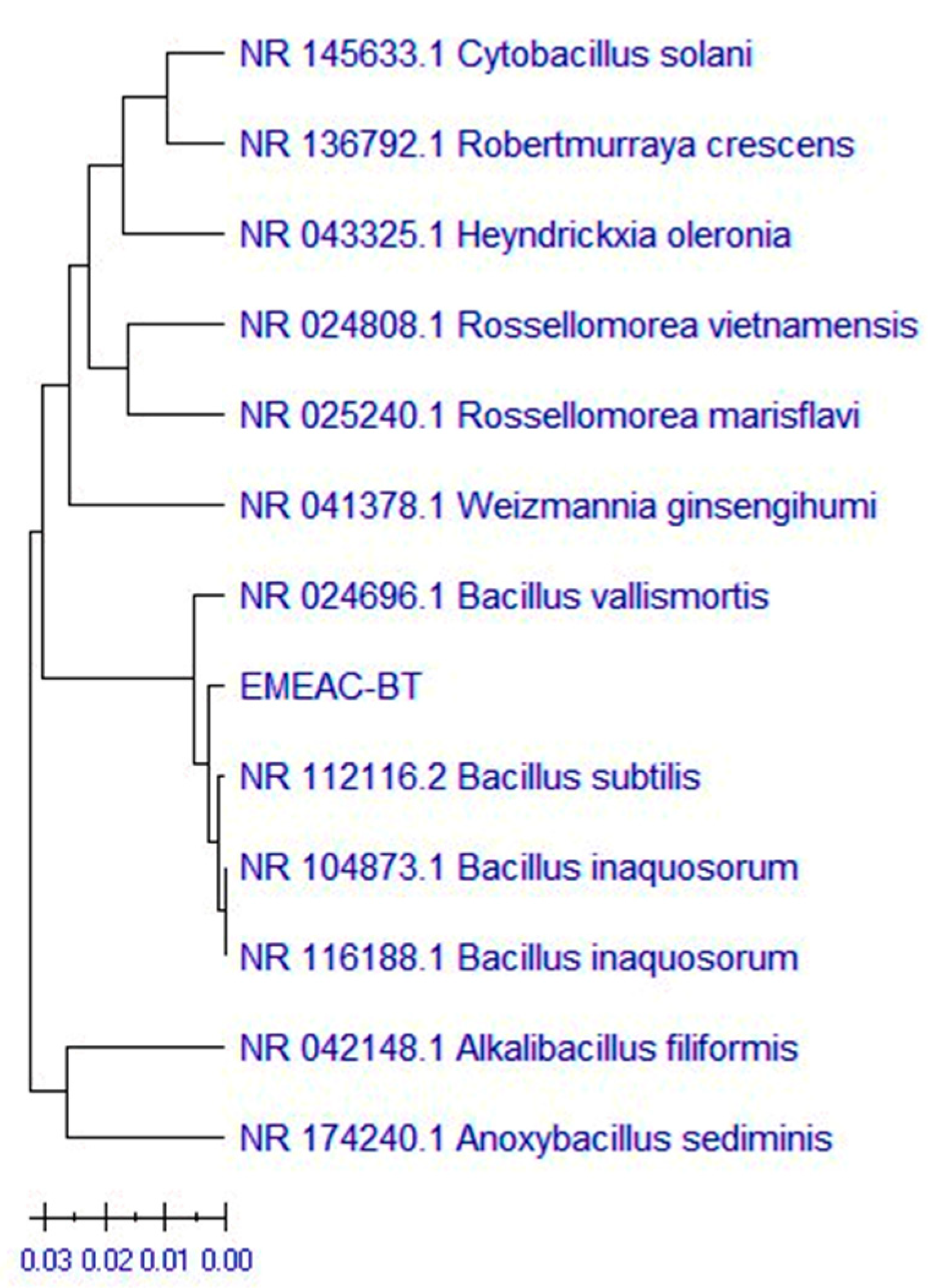
3.1. Isolation and Identification of AgNP-Synthesizing Bacteria

Five morphologically different bacterial colonies (B1–B5) were isolated by serial dilution and the spread plate technique. Screening for AgNP synthesis revealed that all isolates caused the formation of brown color in the reaction mixture, and the color change indicated the formation of AgNPs as a result of the reduction of Ag+ to AgNPs by active metabolites produced by MOs. The isolate B4, which caused a stronger color transformation in the reaction mixture, was taken for further analysis. The chosen bacterial isolate was subject to identification using a 16S rRNA gene sequence. The BLAST similarity search revealed that the selected bacteria have 99.58% identity with Bacillus subtilis. The UPGMA algorithm was used to infer the evolutionary history of the species (Figure 1). The ideal tree is displayed. The phylogenetic tree is inferred using the evolutionary distances used to calculate the branch lengths, and the tree is scaled. The evolutionary distances were calculated using the maximum composite likelihood technique and are expressed in the units of the number of base substitutions per site. A total of 13 different nucleotide sequences were examined in this study. Each sequence pair has all unclear locations eliminated (pairwise deletion option). The final dataset had a total of 473 locations. Many researchers have proved the possibility of using bacteria as bio-nanoreactors to synthesize NPs in an eco-friendly manner [34,35,36]. The identified bacterial gene sequence is submitted in NCBI under the accession OP006732.

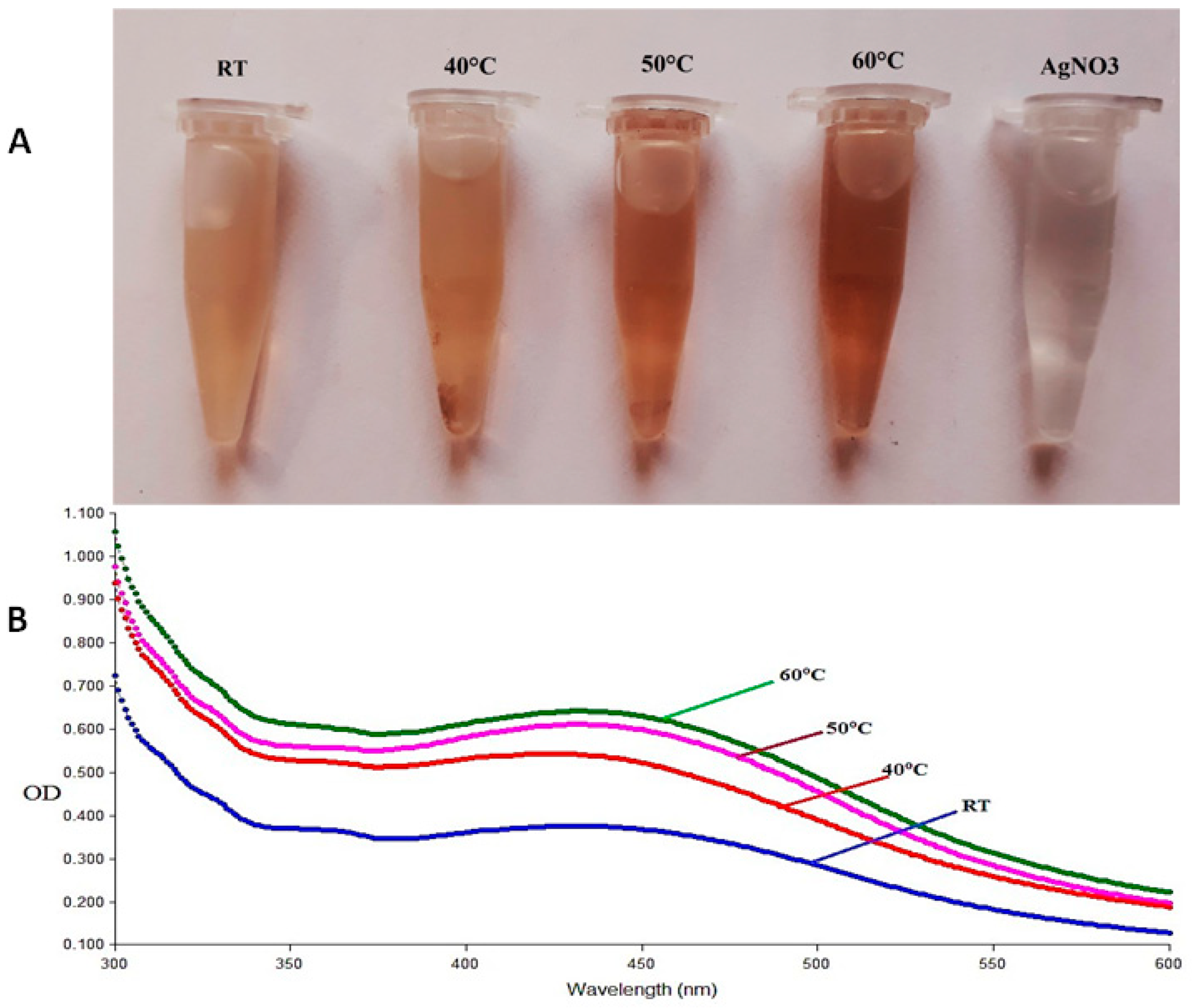
3.2. Synthesis and Characterization of AgNPs

The incubation temperature has a characteristic effect on the efficiency of AgNP synthesis. A browner color was visible in the reaction mixture when kept at 60 ± 1 °C compared to lower temperatures, indicating the temperature dependency of NPs during production (Figure 2A). Studies have defined the role of soil bacteria in synthesizing AgNPs. Soil contains possibly the highest microbial diversity globally, and in all types of soils, prokaryotes including bacteria are the most unrestrained living things found and constitute the primary portion of soil biomass [6]. MOs have inherent systems of converting metallic salt to NPs [37], and MOs like bacteria and fungi that are associated with various parts of plants perform a significant role in green-synthesized AgNPs’ stabilization [33]. Previous studies suggest that bacterial metabolites and interactive pathways are accountable for the bacteria-mediated synthesis of NPs [38] and bacteria can synthesize NPs at a massive scale [39]. The formation of the brown color in the reaction mixture after incubation can be associated with the excitation of the surface plasmon resonance (SPR) vibration of green-synthesized AgNPs [40,41]. The maximum absorbance peak was recorded between 410–450 nm in the UV-vis spectral measurement of the reaction mixture (λmax) incubated at various temperatures (Figure 2B). The UV-vis absorption spectra of synthesized AgNPs also revealed that temperature positively impacts the green synthesis of AgNPs, i.e., the maximum absorbance (0.67) was recorded in the reaction mixture incubated at 60 °C rather than at lower temperatures. The band links to the colloidal AgNPs in 400–450 nm in UV-vis spectral analysis due to the SPR [42]. The result of the temperature impact analysis on the synthesis of AgNPs was in line with previous studies [43,44]. The efficiency of AgNP synthesis will be increased with the rise in temperature [45], and Liu et al. (2020) [46] showed that at high reaction temperatures, the rate of synthesis of AgNPs is found to be significantly increased.
The structural and elemental characteristics of NPs were analyzed using SEM and EDX [47]. SEM analysis revealed that the synthesized AgNPs are uniform in shape and these spherical AgNPs are small in size, ranging from 18 nm to 21 nm with standard deviation ±3.4 nm (Figure 3A). The small-sized AgNPs have several advantages when used for biomedical purposes [48]. They can easily penetrate the cell wall and induce a direct effect on cells, and these small-sized AgNPs can be used as carriers to deliver drugs into the cell. EDX analysis depicted the purity of synthesized AgNPs by expressing a solid signal at 3 KeV (Figure 3B), which indicates the presence of AgNPs [49], and the unveiled strong signal at 3 KeV can be due to the SPR [50].

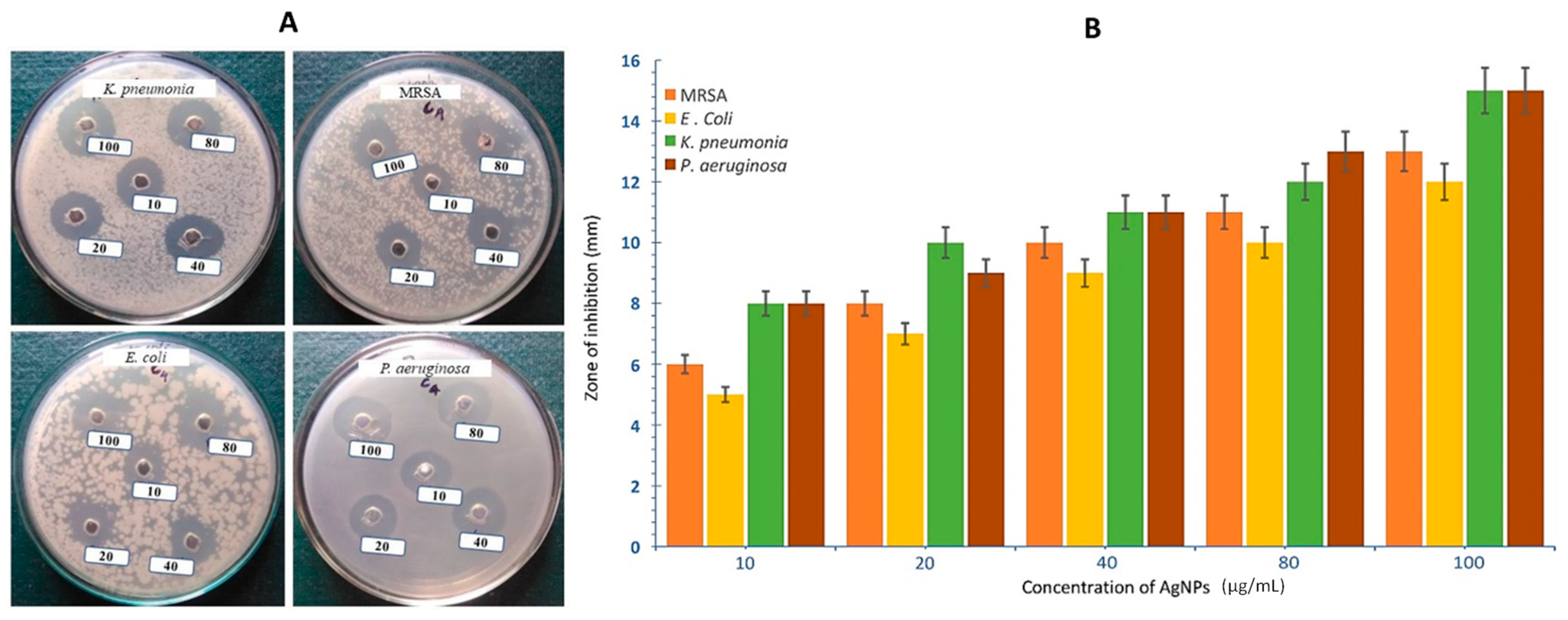
3.3. Antibacterial Activity of AgNPs

The potential antibacterial properties of microorganism-based AgNPs have motivated many researchers to identify bacteria from several environmental sources to synthesize AgNPs. This study also reports that bacteria-mediated synthesized AgNPs exhibited substantial bactericidal properties against Gram-positive and -negative human pathogenic bacteria (Figure 4), particularly against methicillin-resistant S. aureus (MRSA). The highest zone of inhibition, 15 mm, was found at 100 µg/mL of AgNPs against P. aeruginosa and K. pneumonia, followed by 13 mm against MRSA and 12 mm against E. coli. The possibilities of AgNPs as an antibacterial agent were demonstrated against many pathogenic bacteria [36,51,52]. The antibacterial activity of AgNPs depends on several factors. The activity of AgNPs against bacteria was reliant on the concentration and size [53,54]. Agnihotri et al. (2013) [55] proposed that the efficacy of small AgNPs is high compared to large ones, and AgNPs with sizes ranging from 10 to 100 nm expressed more antibacterial activity against both Gram-positive and Gram-negative bacteria [48].

4. Discussion

The present study aligns with numerous prior investigations highlighting the potential of NPs, particularly AgNPs, in controlling the proliferation of various bacteria, including pathogenic strains. As described by Rai et al. (2009), AgNPs possess distinctive characteristics that enhance their interaction with bacterial membranes, facilitating penetration into cells or direct cellular damage [56]. This property is further emphasized by Agnihotri et al. (2014), who demonstrated that the antibacterial activity of AgNPs escalates with decreasing particle size, a phenomenon observed in our synthesized AgNPs as well [57]. Consistent with previous research by Kim et al. (2017), our study reveals a concentration-dependent increase in the antibacterial efficacy of synthesized AgNPs [58]. The mechanism underlying the antibacterial action of AgNPs has garnered significant attention in bionanotechnology. Mendis et al. (2015) proposed that the formation of free radicals by AgNPs plays a pivotal role in disrupting bacterial membranes and inhibiting cellular growth [59].
The antibacterial property of silver has been known for centuries, and with advancements in nanotechnology, researchers have been able to synthesize AgNPs with various sizes, shapes, and surface coatings, enhancing their antibacterial effectiveness. The antibacterial mechanism of AgNPs is multifaceted and not exactly understood [60]. However, some of the key mechanisms through which AgNPs exert their antibacterial effects have been identified. AgNPs can interact with the bacterial cell membrane, leading to structural changes and disruption [61]. This can result in increased permeability, the leakage of cellular components, and eventual cell death. The lipopolysaccharides present on the membrane of the bacteria facilitated the contact of AgNPs with the bacterial membrane [62]. After the penetration of the cell membrane, the AgNPs damage various cell molecules like proteins, DNA, lipids, etc. [63], and they also inhibit different enzymes and cause DNA damage [64].
B. subtilis produces a variety of bacterial metabolites, including enzymes, proteins, peptides, and secondary metabolites, which can act as reducing agents, stabilizing agents, or capping agents in the synthesis of AgNPs [65,66,67]. Enzymes like reductases convert Ag ions into AgNPs, while proteins and peptides secreted by B. subtilis have reducing capabilities [68,69]. Secondary metabolites like surfactants, biosurfactants, and organic acids can act as stabilizing or capping agents in AgNP synthesis [70,71]. Exopolysaccharides (EPS) produced by B. subtilis can also serve as stabilizing agents, preventing AgNP agglomeration and providing stability to the synthesized NPs [72,73,74]. Ghosh et al. suggested that the biological synthesis of NPs by bacteria is achieved through the utilization of their diverse repertoire of biomolecules, including enzymes, proteins, amino acids, DNA, lipids, and carbohydrates, etc. [75]. This mechanism underscores the multifaceted biochemical pathways inherent within bacterial systems, which serve as the foundation for NP synthesis. In NP synthesis, bacterial extracts serve as both reducing and stabilizing agents, with biosurfactants, such as those found in B. subtilis, being extensively explored. Joanna et al. (2018) utilized B. subtilis cultivated on agro-industrial wastes to synthesize biogenic AgNPs, demonstrating enhanced stability and antimicrobial activity due to biosurfactant capping [76]. Similarly, Salazar-Bryam et al. (2021) investigated pH’s effect on AgNPs stabilized by rhamnolipids, showcasing their role in stabilization [77]. Furthermore, Reddy et al. (2009) reported the synthesis of silver and gold NPs using surfactin, a lipopeptide biosurfactant from B. subtilis, affirming the versatility of biosurfactants in NP stabilization [78]. All these findings underscore the potential of bacterial biomolecules, particularly biosurfactants, as effective capping agents in NP synthesis, offering improved stability and tailored properties for various applications.
Capping agents also play a significant role in the antibacterial activity of AgNPs. Biological capping agents, including lipoic acid, chitosan, polyethylene glycol, tannic acid, epigallocatechin, gelatin, and β-cyclodextrin, enhance the antimicrobial activity of AgNPs by stabilizing them and facilitating interaction with bacterial cells [79,80]. Lipoic acid is a potent antioxidant that stabilizes AgNPs and enhances their antimicrobial properties. Chitosan-coated AgNPs exhibit enhanced antimicrobial properties due to the interaction between positively charged amino groups on chitosan and negatively charged bacterial cell membranes [81]. PEG-coated AgNPs are stable and have improved antibacterial activity due to their enhanced dispersibility and prolonged retention on bacterial cell surfaces [82]. Tannic acid, a polyphenolic compound found in plants, provides stability and antimicrobial activity through interactions with bacterial cell components [83]. Gelatin, a collagen-derived protein, enhances AgNPs’ stability and antibacterial efficacy by forming a protective layer around the NPs [84]. β-cyclodextrin, a cyclic oligosaccharide, can also serve as a capping agent for AgNPs, enhancing their stability and antimicrobial activity by forming inclusion complexes with silver ions and facilitating their release at the bacterial cell surface [85]. These biological capping agents offer advantages such as biocompatibility, eco-friendliness, and the targeted delivery of AgNPs to bacterial cells.
When discussing the plausible antibacterial mechanisms of produced nanosilvers, two theories are postulated, where the first way is described as follows: AgNPs are toxic when they come into direct contact with cell culture medium, proteins in the cytoplasm, or the acidic environment of the lysosome; antibacterial activity occurs by inducing the production of reactive oxygen species (ROS). Ag+ and produced AgNPs escape from lysosomes, thereby promoting the amplification of intracellular ROS. AgNPs enter the nucleus via nuclear pore complexes formed by reactive oxygen species (ROS), which induce DNA damage and chromosomal abnormalities directly. The ROS group consists of components that are highly relative, such as hydrogen peroxide, hydroxyl radicals, and superoxide anions. These radicals induce mitochondrial dysfunction by causing oxidative damage to DNA and proteins. Ag+ is liberated in vivo when hydrogen peroxide (H2O2) and AgNPs undergo a chemical reaction. Additionally, Ag+ interacts with the thiol groups of molecules found in cellular components; as a result, lipid peroxide is released, and cell membrane and mitochondrial permeation are increased. It ultimately leads to necrosis by causing the leakage of cytoplasmic contents; the rupture of lysosomal membranes triggers lysosome-mediated apoptosis; and damage to mitochondria disrupts electron transfer, which triggers apoptosis dependent on the mitochondria. It has also been reported that AgNPs upregulate genes associated with oxidative stress and metabolism, thereby increasing ROS production (Figure 5).
AgNPs have been reported to exhibit broad-spectrum antimicrobial effects against various pathogenic bacterial species, namely Pseudomonas aeruginosa, Enterobacter cloacae, Escherichia coli, Streptococcus pyogens, Staphylococcus aureus, Klebsiella pneumoniae, Proteus vulgaris, Salmonella enterica, Aeromonas hydrophilla, Shigella dysenteriae, Salmonella paratyphi, Micrococcus luteus, Shigella sonnei, Proteus mirabilis, Corney bacterium, Bacillus cereus, Vibrio cholerae, Staphylococcus saprophyticus, etc. They are also used against fungal species such as Candida krusei, Candida tropicalis, Candida crusei, Candida kefyr, Aspergillus flavus, Candida tropicalis, Aspergillus niger, Aspergillus fumigates, Fusarium, Curvularia, Rhizopus, etc. From the above reports, a second mechanism can be postulated: Antibacterial inactivation is largely dependent on the reaction between Ag+ ions and the thiol groups of proteins. Reacting with electron-donating groups and producing reactive oxygen species, AgNPs deactivate cellular enzymes, DNA, and proteins within bacterial cells. AgNPs also disrupt membrane permeability or impede enzymes implicated in the respiratory chain by detaching respiratory electron transport from oxidative phosphorylation. Through interaction with the bacterial plasmid, cell wall, plasma membrane, and/or bacterial protein assembly, AgNPs may possess antibacterial properties. The deformation of peptidoglycan structures within the bacterial cell wall is induced by Ag+ ions, which subsequently lyse the cell membrane and impede bacterial growth. Ag+ causes the denaturation of ribosomes at the site of protein assembly, thereby successfully impeding protein synthesis in its entirety. Plasmid growth is impeded when Ag+ ions interact with DNA bases in bacterial plasmids, resulting in DNA condensation and the subsequent cessation of replication (Figure 6).
Studies have noted the ability of AgNPs to produce ROS and free radicals inside the cell, which eventually increases oxidative stress and cell death [62,63]. AgNPs can generate ROS, such as H2O2 and superoxide radicals, within bacterial cells. ROS can cause oxidative damage to cellular components, leading to bacterial cell death [86]. AgNPs can interact with bacterial proteins and enzymes, affecting their structure and function. This interference disrupts essential cellular processes and ultimately leads to bacterial cell death [87]. The unique properties of AgNPs make them effective against a broad spectrum of bacteria, including both Gram-positive and Gram-negative bacteria. They have demonstrated potential in combating antibiotic-resistant bacteria, a significant issue in modern medicine, underscoring their importance in addressing contemporary medical challenges.
Chemically synthesized NPs offer simplicity, high yields, and scalability, while biosynthesis, particularly through green methods, offers environmental sustainability, biocompatibility, cost effectiveness, and precise control over properties. Biosynthesized NPs are stable, easily functionalized, and suitable for various applications, making them a promising approach for healthcare and environmental fields. Biosynthesis is an environmentally friendly method for producing NPs, using biological entities like microorganisms or plants, resulting in reduced toxic chemicals and hazardous waste. It also enhances biocompatibility, crucial for biomedical applications like drug delivery and tissue engineering. Biosynthesis is cost-effective, using inexpensive raw materials like agricultural waste or plant extracts and reducing the need for expensive reagents and energy-intensive processes, leading to long-term cost savings. Biosynthesis offers tunable properties for NPs, allowing for specific applications like drug delivery or targeting. It reduces agglomeration and improves stability due to biomolecules acting as capping agents. Biosynthesized NPs also have the potential for functionalization, as they offer functional groups for further modification, enabling the attachment of targeting ligands, imaging agents, or therapeutic payloads. This enhances the versatility and utility of biosynthesized NPs across biomedical and industrial domains. The composition of ligands on biosynthesized AgNPs is influenced by the biological source and synthesis method, typically proteins, peptides, polysaccharides, and organic compounds, which act as capping agents and enhance NP stability. Proteins, peptides, polysaccharides, and other organic compounds are essential for biomedical applications. Understanding their composition and characteristics is crucial for tailoring biosynthesized AgNPs to diverse applications, such as healthcare and environmental remediation. These ligands facilitate interactions with biological systems, enhancing the stability and functionality of NPs.

5. Conclusions

Antibiotic resistance has prompted a quest for alternative antimicrobial therapy options. Engineered NPs designed for effective penetration into biological systems are becoming more widespread in health and hygiene. The use of microbial proteins and enzymes as potential reducing agents for the production of NPs has rapidly increased compared to physical and chemical methods. It is an economical, eco-friendly, and efficient method. Fungi and bacteria are preferred among biogenic sources because they can produce a higher concentration of the reductase enzyme needed to transform ionic forms into their nanoforms. They are also preferred because it is easier to cultivate them and control the size and morphology of the synthesized NPs, which can significantly lower the cost of large-scale manufacturing. The ability of NPs to limit bacterial growth is enabled by effective penetration through the exopolysaccharides of a biofilm matrix. The rhizosphere soil is a good reservoir of bacteria with potential applications in industrial, food, and biomedical industries. The bacteria isolated from the rhizosphere soil of A. indica showed 99.25% identity with Bacillus subtilis, and it has high biotransformation efficiency to convert Ag+ to AgNPs. The physical, chemical, and biological properties of AgNPs synthesized by bacteria were analyzed. The formation of brown color and UV-vis spectral analysis proved the formation of AgNPs, and this study reports the positive effect of temperature on the efficiency of AgNPs synthesis. Morphological analysis revealed the formation of small uniformly structured spherical AgNPs ranging from 18 to 21 nm. The synthesized AgNPs showed bactericidal properties against both Gram-positive and -negative bacteria, including MRSA. Therefore, this study concludes that the rhizosphere soil of A. indica is a potential source of bacteria for the extracellular synthesis of AgNPs in a green way. These bio-synthesized AgNPs can be applied as antibacterial agents in industrial and biomedical applications.

Author Contributions

Conceptualization, M.K. and T.M.J.; methodology, M.K. and T.M.J.; validation, K.P.M. and S.S.; formal analysis, H.F.K., S.G.P. and S.S.; investigation, M.K., D.K.M. and T.M.J.; resources, D.K.M. and M.K.; data curation, D.K.M., H.F.K. and S.G.P.; writing—original draft preparation, M.K. and T.M.J.; writing—review and editing, M.K. and T.M.J.; visualization, M.K.; supervision, J.H. and S.T.; project administration, J.H. and S.T. All authors have read and agreed to the published version of the manuscript.

Funding

This research received no external funding.

Institutional Review Board Statement

Not applicable.

Informed Consent Statement

Not applicable.

Data Availability Statement

The authors confirm that the data supporting the findings of this study are available within the article.

Conflicts of Interest

The authors declare no conflicts of interest.

References

  1. Gupta, A.; Gupta, R.; Singh, R.L. Microbes and Environment. In Principles and Applications of Environmental Biotechnology for a Sustainable Future; Springer: Singapore, 2016; pp. 43–84. [Google Scholar] [CrossRef]
  2. The Microbial World: Foundation of the Biosphere: This Report Is Based on an American Academy of Microbiology Colloquium Held January 19–21, 1996, in Palm Coast, Florida. The Colloquium Was Supported by the National Science Foundation, the National Oceanic and Atmospheric Administration of the U.S. Department of Commerce, the U.S. Department of Energy, and the American Society for Microbiology; American Academy of Microbiology Colloquia Reports; American Society for Microbiology: Washington, DC, USA, 1997.
  3. de Souza, P.M.; de Oliveira Magalhães, P. Application of Microbial α-Amylase in Industry—A Review. Braz. J. Microbiol. 2010, 41, 850–861. [Google Scholar] [CrossRef] [PubMed]
  4. Timmis, K.; de Lorenzo, V.; Verstraete, W.; Ramos, J.L.; Danchin, A.; Brüssow, H.; Singh, B.K.; Timmis, J.K. The Contribution of Microbial Biotechnology to Economic Growth and Employment Creation. Microb. Biotechnol. 2017, 10, 1137–1144. [Google Scholar] [CrossRef] [PubMed]
  5. Salem, H.M.; Abdel-Salam, A.; Abdel-Salam, M.A.; Seleiman, M.F. Soil Xenobiotics and Their Phyto-Chemical Remediation. In Xenobiotics in the Soil Environment: Monitoring, Toxicity and Management; Hashmi, M.Z., Kumar, V., Varma, A., Eds.; Springer International Publishing: Cham, Switzerland, 2017; pp. 267–280. ISBN 978-3-319-47744-2. [Google Scholar]
  6. Daniel, R. The Metagenomics of Soil. Nat. Rev. Microbiol. 2005, 3, 470–478. [Google Scholar] [CrossRef] [PubMed]
  7. Khan Khanzada, A.; Al-Hazmi, H.E.; Śniatała, B.; Muringayil Joseph, T.; Majtacz, J.; Abdulrahman, S.A.M.; Albaseer, S.S.; Kurniawan, T.A.; Rahimi-Ahar, Z.; Habibzadeh, S.; et al. Hydrochar-Nanoparticle Integration for Arsenic Removal from Wastewater: Challenges, Possible Solutions, and Future Horizon. Environ. Res. 2023, 238, 117164. [Google Scholar] [CrossRef]
  8. Joseph, T.M.; Al-Hazmi, H.E.; Śniatała, B.; Esmaeili, A.; Habibzadeh, S. Nanoparticles and Nanofiltration for Wastewater Treatment: From Polluted to Fresh Water. Environ. Res. 2023, 238, 117114. [Google Scholar] [CrossRef] [PubMed]
  9. Muringayil Joseph, T.; Mariya, H.J.; Haponiuk, J.T.; Thomas, S.; Esmaeili, A.; Sajadi, S.M. Electromagnetic Interference Shielding Effectiveness of Natural and Chlorobutyl Rubber Blend Nanocomposite. J. Compos. Sci. 2022, 6, 240. [Google Scholar] [CrossRef]
  10. Jose Varghese, R.; Vidya, L.; Joseph, T.M.; Gudimalla, A.; Harini Bhuvaneswari, G.; Thomas, S. Potential Applications of XLPE Nanocomposites in the Field of Cable Insulation. In Crosslinkable Polyethylene Based Blends and Nanocomposites; Thomas, J., Thomas, S., Ahmad, Z., Eds.; Materials Horizons: From Nature to Nanomaterials; Springer: Singapore, 2021; pp. 197–213. ISBN 9789811604867. [Google Scholar]
  11. Joseph, T.M.; Pallikkunnel, M.L.; Mahapatra, D.K.; Kallingal, A.; Thomas, S.; Haponiuk, J.T. Polyurethane−Epoxy Composites: Recent Developments and Future Perspectives. In Polyurethane Chemistry: Renewable Polyols and Isocyanates; ACS Symposium Series; American Chemical Society: Washington, DC, USA, 2021; Volume 1380, pp. 257–280. ISBN 978-0-8412-9840-8. [Google Scholar]
  12. Hamouda, R.A.; Hussein, M.H.; Abo-elmagd, R.A.; Bawazir, S.S. Synthesis and Biological Characterization of Silver Nanoparticles Derived from the Cyanobacterium Oscillatoria Limnetica. Sci. Rep. 2019, 9, 13071. [Google Scholar] [CrossRef]
  13. Joseph, T.M.; Kar Mahapatra, D.; Esmaeili, A.; Piszczyk, Ł.; Hasanin, M.S.; Kattali, M.; Haponiuk, J.; Thomas, S. Nanoparticles: Taking a Unique Position in Medicine. Nanomaterials 2023, 13, 574. [Google Scholar] [CrossRef] [PubMed]
  14. Mussin, J.; Robles-Botero, V.; Casañas-Pimentel, R.; Rojas, F.; Angiolella, L.; San Martín-Martínez, E.; Giusiano, G. Antimicrobial and Cytotoxic Activity of Green Synthesis Silver Nanoparticles Targeting Skin and Soft Tissue Infectious Agents. Sci. Rep. 2021, 11, 14566. [Google Scholar] [CrossRef]
  15. Akter, M.; Sikder, M.d.T.; Rahman, M.d.M.; Ullah, A.K.M.A.; Hossain, K.F.B.; Banik, S.; Hosokawa, T.; Saito, T.; Kurasaki, M. A Systematic Review on Silver Nanoparticles-Induced Cytotoxicity: Physicochemical Properties and Perspectives. J. Adv. Res. 2018, 9, 1–16. [Google Scholar] [CrossRef]
  16. Shanmuganathan, R.; MubarakAli, D.; Prabakar, D.; Muthukumar, H.; Thajuddin, N.; Kumar, S.S.; Pugazhendhi, A. An Enhancement of Antimicrobial Efficacy of Biogenic and Ceftriaxone-Conjugated Silver Nanoparticles: Green Approach. Environ. Sci. Pollut. Res. Int. 2018, 25, 10362–10370. [Google Scholar] [CrossRef] [PubMed]
  17. Ratan, Z.A.; Haidere, M.F.; Nurunnabi, M.; Shahriar, S.M.; Ahammad, A.J.S.; Shim, Y.Y.; Reaney, M.J.T.; Cho, J.Y. Green Chemistry Synthesis of Silver Nanoparticles and Their Potential Anticancer Effects. Cancers 2020, 12, 855. [Google Scholar] [CrossRef] [PubMed]
  18. Saravanan, A.; Kumar, P.S.; Karishma, S.; Vo, D.-V.N.; Jeevanantham, S.; Yaashikaa, P.R.; George, C.S. A Review on Biosynthesis of Metal Nanoparticles and Its Environmental Applications. Chemosphere 2021, 264, 128580. [Google Scholar] [CrossRef]
  19. Merga, G.; Wilson, R.; Lynn, G.; Milosavljevic, B.H.; Meisel, D. Redox Catalysis on “Naked” Silver Nanoparticles. J. Phys. Chem. C 2007, 111, 12220–12226. [Google Scholar] [CrossRef]
  20. Zhang, N.; Sun, J.; Yin, L.; Liu, J.; Chen, C. Silver Nanoparticles: From In Vitro Green Synthesis to In Vivo Biological Effects in Plants. Adv. Agrochem. 2023, 2, 313–323. [Google Scholar] [CrossRef]
  21. Asif, M.; Yasmin, R.; Asif, R.; Ambreen, A.; Mustafa, M.; Umbreen, S. Green Synthesis of Silver Nanoparticles (AgNPs), Structural Characterization, and Their Antibacterial Potential. Dose Response 2022, 20, 15593258221088709. [Google Scholar] [CrossRef]
  22. Dhir, R.; Chauhan, S.; Subham, P.; Kumar, S.; Sharma, P.; Shidiki, A.; Kumar, G. Plant-Mediated Synthesis of Silver Nanoparticles: Unlocking Their Pharmacological Potential—A Comprehensive Review. Front. Bioeng. Biotechnol. 2024, 11, 1324805. [Google Scholar] [CrossRef] [PubMed]
  23. El-Naggar, N.E.-A.; Hussein, M.H.; El-Sawah, A.A. Bio-Fabrication of Silver Nanoparticles by Phycocyanin, Characterization, In Vitro Anticancer Activity against Breast Cancer Cell Line and In Vivo Cytotxicity. Sci. Rep. 2017, 7, 10844. [Google Scholar] [CrossRef] [PubMed]
  24. Ferdous, Z.; Nemmar, A. Health Impact of Silver Nanoparticles: A Review of the Biodistribution and Toxicity Following Various Routes of Exposure. Int. J. Mol. Sci. 2020, 21, 2375. [Google Scholar] [CrossRef]
  25. Devanesan, S.; Jayamala, M.; AlSalhi, M.S.; Umamaheshwari, S.; Ranjitsingh, A.J.A. Antimicrobial and Anticancer Properties of Carica Papaya Leaves Derived Di-Methyl Flubendazole Mediated Silver Nanoparticles. J. Infect. Public. Health 2021, 14, 577–587. [Google Scholar] [CrossRef]
  26. Gade, A.; PP, B.; Ingle, A.; Marcato, P.; Duran, N.; Rai, M. Exploitation of Aspergillus Niger for Synthesis of Silver Nanoparticles. J. Biobased Mater. Bioenergy 2008, 2, 243–247. [Google Scholar] [CrossRef]
  27. Narayanan, K.B.; Sakthivel, N. Biological Synthesis of Metal Nanoparticles by Microbes. Adv. Colloid. Interface Sci. 2010, 156, 1–13. [Google Scholar] [CrossRef] [PubMed]
  28. Vaseghi, Z.; Nematollahzadeh, A.; Tavakoli, O. Green Methods for the Synthesis of Metal Nanoparticles Using Biogenic Reducing Agents: A Review. Rev. Chem. Eng. 2018, 34, 529–559. [Google Scholar] [CrossRef]
  29. Durán, N.; Seabra, A.B. Metallic Oxide Nanoparticles: State of the Art in Biogenic Syntheses and Their Mechanisms. Appl. Microbiol. Biotechnol. 2012, 95, 275–288. [Google Scholar] [CrossRef]
  30. Ahmad, A.; Mukherjee, P.; Senapati, S.; Mandal, D.; Khan, M.I.; Kumar, R.; Sastry, M. Extracellular Biosynthesis of Silver Nanoparticles Using the Fungus Fusarium Oxysporum. Colloids Surf. B Biointerfaces 2003, 28, 313–318. [Google Scholar] [CrossRef]
  31. Ahmad, A.; Senapati, S.; Khan, M.I.; Kumar, R.; Ramani, R.; Srinivas, V.; Sastry, M. Intracellular Synthesis of Gold Nanoparticles by a Novel Alkalotolerant Actinomycete, Rhodococcus Species. Nanotechnology 2003, 14, 824–828. [Google Scholar] [CrossRef]
  32. Kalimuthu, K.; Suresh Babu, R.; Venkataraman, D.; Bilal, M.; Gurunathan, S. Biosynthesis of Silver Nanocrystals by Bacillus Licheniformis. Colloids Surf. B Biointerfaces 2008, 65, 150–153. [Google Scholar] [CrossRef] [PubMed]
  33. Durán, N.; Marcato, P.D.; Alves, O.L.; De Souza, G.I.; Esposito, E. Mechanistic Aspects of Biosynthesis of Silver Nanoparticles by Several Fusarium Oxysporum Strains. J. Nanobiotechnol. 2005, 3, 8. [Google Scholar] [CrossRef] [PubMed]
  34. Priyadarshini, S.; Gopinath, V.; Meera Priyadharsshini, N.; MubarakAli, D.; Velusamy, P. Synthesis of Anisotropic Silver Nanoparticles Using Novel Strain, Bacillus Flexus and Its Biomedical Application. Colloids Surf. B Biointerfaces 2013, 102, 232–237. [Google Scholar] [CrossRef]
  35. Shanthi, S.; Jayaseelan, B.D.; Velusamy, P.; Vijayakumar, S.; Chih, C.T.; Vaseeharan, B. Biosynthesis of Silver Nanoparticles Using a Probiotic Bacillus Licheniformis Dahb1 and Their Antibiofilm Activity and Toxicity Effects in Ceriodaphnia Cornuta. Microb. Pathog. 2016, 93, 70–77. [Google Scholar] [CrossRef]
  36. Esmail, R.; Afshar, A.; Morteza, M.; Abolfazl, A.; Akhondi, E. Synthesis of Silver Nanoparticles with High Efficiency and Stability by Culture Supernatant of Bacillus ROM6 Isolated from Zarshouran Gold Mine and Evaluating Its Antibacterial Effects. BMC Microbiol. 2022, 22, 97. [Google Scholar] [CrossRef]
  37. Li, X.; Xu, H.; Chen, Z.-S.; Chen, G. Biosynthesis of Nanoparticles by Microorganisms and Their Applications. J. Nanomater. 2011, 2011, e270974. [Google Scholar] [CrossRef]
  38. Lahiri, D.; Nag, M.; Sheikh, H.I.; Sarkar, T.; Edinur, H.A.; Pati, S.; Ray, R.R. Microbiologically-Synthesized Nanoparticles and Their Role in Silencing the Biofilm Signaling Cascade. Front. Microbiol. 2021, 12, 636588. [Google Scholar] [CrossRef]
  39. Fariq, A.; Khan, T.; Yasmin, A. Microbial Synthesis of Nanoparticles and Their Potential Applications in Biomedicine. J. Appl. Biomed. 2017, 15, 241–248. [Google Scholar] [CrossRef]
  40. Thangaraju, N.; Venkatalakshmi, R.P.; Chinnasamy, A.; Kannaiyan, P. Synthesis of Silver Nanoparticles and the Antibacterial and Anticancer Activities of the Crude Extract of Sargassum Polycystum C. Agardh. Nano Biomed. Eng. 2012, 4, 89–94. [Google Scholar] [CrossRef]
  41. Awwad, A.M.; Salem, N.M.; Abdeen, A.O. Green Synthesis of Silver Nanoparticles Using Carob Leaf Extract and Its Antibacterial Activity. Int. J. Ind. Chem. 2013, 4, 29. [Google Scholar] [CrossRef]
  42. Njagi, E.C.; Huang, H.; Stafford, L.; Genuino, H.; Galindo, H.M.; Collins, J.B.; Hoag, G.E.; Suib, S.L. Biosynthesis of Iron and Silver Nanoparticles at Room Temperature Using Aqueous Sorghum Bran Extracts. Langmuir 2011, 27, 264–271. [Google Scholar] [CrossRef]
  43. Fayaz, M.A.; Balaji, K.; Kalaichelvan, P.T.; Venkatesan, R. Fungal Based Synthesis of Silver Nanoparticles—An Effect of Temperature on the Size of Particles. Colloids Surf. B Biointerfaces 2009, 74, 123–126. [Google Scholar] [CrossRef] [PubMed]
  44. Usha Rani, P.; Rajasekharreddy, P. Green Synthesis of Silver-Protein (Core–Shell) Nanoparticles Using Piper Betle L. Leaf Extract and Its Ecotoxicological Studies on Daphnia Magna. Colloids Surf. A 2011, 389, 188–194. [Google Scholar] [CrossRef]
  45. Sukirtha, R.; Priyanka, K.M.; Antony, J.J.; Kamalakkannan, S.; Thangam, R.; Gunasekaran, P.; Krishnan, M.; Achiraman, S. Cytotoxic Effect of Green Synthesized Silver Nanoparticles Using Melia Azedarach against In Vitro HeLa Cell Lines and Lymphoma Mice Model. Process Biochem. 2012, 47, 273–279. [Google Scholar] [CrossRef]
  46. Liu, H.; Zhang, H.; Wang, J.; Wei, J. Effect of Temperature on the Size of Biosynthesized Silver Nanoparticle: Deep Insight into Microscopic Kinetics Analysis. Arab. J. Chem. 2020, 13, 1011–1019. [Google Scholar] [CrossRef]
  47. Dada, A.O.; Inyinbor, A.A.; Idu, E.I.; Bello, O.M.; Oluyori, A.P.; Adelani-Akande, T.A.; Okunola, A.A.; Dada, O. Effect of Operational Parameters, Characterization and Antibacterial Studies of Green Synthesis of Silver Nanoparticles Using Tithonia Diversifolia. PeerJ 2018, 6, e5865. [Google Scholar] [CrossRef] [PubMed]
  48. Morones, J.R.; Elechiguerra, J.L.; Camacho, A.; Holt, K.; Kouri, J.B.; Ramírez, J.T.; Yacaman, M.J. The Bactericidal Effect of Silver Nanoparticles. Nanotechnology 2005, 16, 2346–2353. [Google Scholar] [CrossRef]
  49. Rautela, A.; Rani, J.; Debnath (Das), M. Green Synthesis of Silver Nanoparticles from Tectona Grandis Seeds Extract: Characterization and Mechanism of Antimicrobial Action on Different Microorganisms. J. Anal. Sci. Technol. 2019, 10, 5. [Google Scholar] [CrossRef]
  50. Das, J.; Paul Das, M.; Velusamy, P. Sesbania Grandiflora Leaf Extract Mediated Green Synthesis of Antibacterial Silver Nanoparticles against Selected Human Pathogens. Spectrochim. Acta Part A Mol. Biomol. Spectrosc. 2013, 104, 265–270. [Google Scholar] [CrossRef]
  51. Ananthi, V.; Siva Prakash, G.; Mohan Rasu, K.; Gangadevi, K.; Boobalan, T.; Raja, R.; Anand, K.; Sudhakar, M.; Chuturgoon, A.; Arun, A. Comparison of Integrated Sustainable Biodiesel and Antibacterial Nano Silver Production by Microalgal and Yeast Isolates. J. Photochem. Photobiol. B 2018, 186, 232–242. [Google Scholar] [CrossRef] [PubMed]
  52. Pirtarighat, S.; Ghannadnia, M.; Baghshahi, S. Green Synthesis of Silver Nanoparticles Using the Plant Extract of Salvia Spinosa Grown In Vitro and Their Antibacterial Activity Assessment. J. Nanostruct. Chem. 2019, 9, 1–9. [Google Scholar] [CrossRef]
  53. Bhattacharya, R.; Mukherjee, P. Biological Properties of “Naked” Metal Nanoparticles. Adv. Drug Deliv. Rev. 2008, 60, 1289–1306. [Google Scholar] [CrossRef] [PubMed]
  54. Rai, M.K.; Deshmukh, S.D.; Ingle, A.P.; Gade, A.K. Silver Nanoparticles: The Powerful Nanoweapon against Multidrug-Resistant Bacteria. J. Appl. Microbiol. 2012, 112, 841–852. [Google Scholar] [CrossRef]
  55. Agnihotri, S.; Mukherji, S.; Mukherji, S. Size-Controlled Silver Nanoparticles Synthesized over the Range 5–100 nm Using the Same Protocol and Their Antibacterial Efficacy. RSC Adv. 2013, 4, 3974–3983. [Google Scholar] [CrossRef]
  56. Rai, M.; Yadav, A.; Gade, A. Silver Nanoparticles as a New Generation of Antimicrobials. Biotechnol. Adv. 2009, 27, 76–83. [Google Scholar] [CrossRef] [PubMed]
  57. Mahapatra, D.K.; Bharti, S.K.; Asati, V. Nature inspired green fabrication technology for silver nanoparticles. Curr. Nanomed. 2017, 7, 5–24. [Google Scholar] [CrossRef]
  58. Kim, D.H.; Park, J.C.; Jeon, G.E.; Kim, C.S.; Seo, J.H. Effect of the Size and Shape of Silver Nanoparticles on Bacterial Growth and Metabolism by Monitoring Optical Density and Fluorescence Intensity. Biotechnol. Bioprocess Eng. 2017, 22, 210–217. [Google Scholar] [CrossRef]
  59. Mendis, E.; Rajapakse, N.; Byun, H.-G.; Kim, S.-K. Investigation of Jumbo Squid (Dosidicus Gigas) Skin Gelatin Peptides for Their In Vitro Antioxidant Effects. Life Sci. 2005, 77, 2166–2178. [Google Scholar] [CrossRef] [PubMed]
  60. More, P.R.; Pandit, S.; Filippis, A.D.; Franci, G.; Mijakovic, I.; Galdiero, M. Silver Nanoparticles: Bactericidal and Mechanistic Approach against Drug Resistant Pathogens. Microorganisms 2023, 11, 369. [Google Scholar] [CrossRef] [PubMed]
  61. Khorrami, S.; Zarrabi, A.; Khaleghi, M.; Danaei, M.; Mozafari, M.R. Selective Cytotoxicity of Green Synthesized Silver Nanoparticles against the MCF-7 Tumor Cell Line and Their Enhanced Antioxidant and Antimicrobial Properties. Int. J. Nanomed. 2018, 13, 8013–8024. [Google Scholar] [CrossRef] [PubMed]
  62. Loo, Y.Y.; Rukayadi, Y.; Nor-Khaizura, M.-A.-R.; Kuan, C.H.; Chieng, B.W.; Nishibuchi, M.; Radu, S. In Vitro Antimicrobial Activity of Green Synthesized Silver Nanoparticles Against Selected Gram-Negative Foodborne Pathogens. Front. Microbiol. 2018, 9, 1555. [Google Scholar] [CrossRef] [PubMed]
  63. Ullah Khan, S.; Saleh, T.A.; Wahab, A.; Khan, M.H.U.; Khan, D.; Ullah Khan, W.; Rahim, A.; Kamal, S.; Ullah Khan, F.; Fahad, S. Nanosilver: New Ageless and Versatile Biomedical Therapeutic Scaffold. Int. J. Nanomed. 2018, 13, 733–762. [Google Scholar] [CrossRef] [PubMed]
  64. Singh, P.; Pandit, S.; Mokkapati, V.; Garnæs, J.; Mijakovic, I. A Sustainable Approach for the Green Synthesis of Silver Nanoparticles from Solibacillus isronensis sp. and Their Application in Biofilm Inhibition. Molecules 2020, 25, 2783. [Google Scholar] [CrossRef]
  65. Dhaka, A.; Chand Mali, S.; Sharma, S.; Trivedi, R. A Review on Biological Synthesis of Silver Nanoparticles and Their Potential Applications. Results Chem. 2023, 6, 101108. [Google Scholar] [CrossRef]
  66. Kulkarni, D.; Sherkar, R.; Shirsathe, C.; Sonwane, R.; Varpe, N.; Shelke, S.; More, M.P.; Pardeshi, S.R.; Dhaneshwar, G.; Junnuthula, V.; et al. Biofabrication of Nanoparticles: Sources, Synthesis, and Biomedical Applications. Front. Bioeng. Biotechnol. 2023, 11, 1159193. [Google Scholar] [CrossRef] [PubMed]
  67. Yu, X.; Li, J.; Mu, D.; Zhang, H.; Liu, Q.; Chen, G. Green Synthesis and Characterizations of Silver Nanoparticles with Enhanced Antibacterial Properties by Secondary Metabolites of Bacillus Subtilis (SDUM301120). Green. Chem. Lett. Rev. 2021, 14, 190–203. [Google Scholar] [CrossRef]
  68. Hsueh, Y.-H.; Lin, K.-S.; Ke, W.-J.; Hsieh, C.-T.; Chiang, C.-L.; Tzou, D.-Y.; Liu, S.-T. The Antimicrobial Properties of Silver Nanoparticles in Bacillus subtilis Are Mediated by Released Ag+ Ions. PLoS ONE 2015, 10, e0144306. [Google Scholar] [CrossRef] [PubMed]
  69. Mikhailova, E.O. Silver Nanoparticles: Mechanism of Action and Probable Bio-Application. J. Funct. Biomater. 2020, 11, 84. [Google Scholar] [CrossRef] [PubMed]
  70. Sidhu, A.K.; Verma, N.; Kaushal, P. Role of Biogenic Capping Agents in the Synthesis of Metallic Nanoparticles and Evaluation of Their Therapeutic Potential. Front. Nanotechnol. 2022, 3, 801620. [Google Scholar] [CrossRef]
  71. Telrandhe, R.; Mahapatra, D.K.; Kamble, M.A. Bombax ceiba thorn extract mediated synthesis of silver nanoparticles: Evaluation of anti-Staphylococcus aureus activity. Int. J. Pharm. Drug Anal. 2017, 5, 376–379. [Google Scholar] [CrossRef]
  72. Khan, S.S.; Mukherjee, A.; Chandrasekaran, N. Impact of Exopolysaccharides on the Stability of Silver Nanoparticles in Water. Water Res. 2011, 45, 5184–5190. [Google Scholar] [CrossRef] [PubMed]
  73. Saravanan, C.; Rajesh, R.; Kaviarasan, T.; Muthukumar, K.; Kavitake, D.; Shetty, P.H. Synthesis of Silver Nanoparticles Using Bacterial Exopolysaccharide and Its Application for Degradation of Azo-Dyes. Biotechnol. Rep. 2017, 15, 33–40. [Google Scholar] [CrossRef] [PubMed]
  74. Escárcega-González, C.E.; Garza-Cervantes, J.A.; Vázquez-Rodríguez, A.; Morones-Ramírez, J.R. Bacterial Exopolysaccharides as Reducing and/or Stabilizing Agents during Synthesis of Metal Nanoparticles with Biomedical Applications. Int. J. Polym. Sci. 2018, 2018, e7045852. [Google Scholar] [CrossRef]
  75. Ghosh, S.; Ahmad, R.; Zeyaullah, M.; Khare, S.K. Microbial Nano-Factories: Synthesis and Biomedical Applications. Front. Chem. 2021, 9, 626834. [Google Scholar] [CrossRef]
  76. Joanna, C.; Marcin, L.; Ewa, K.; Grażyna, P. A Nonspecific Synergistic Effect of Biogenic Silver Nanoparticles and Biosurfactant towards Environmental Bacteria and Fungi. Ecotoxicology 2018, 27, 352–359. [Google Scholar] [CrossRef]
  77. Salazar-Bryam, A.M.; Yoshimura, I.; Santos, L.P.; Moura, C.C.; Santos, C.C.; Silva, V.L.; Lovaglio, R.B.; Costa Marques, R.F.; Jafelicci Junior, M.; Contiero, J. Silver Nanoparticles Stabilized by Ramnolipids: Effect of pH. Colloids Surf. B Biointerfaces 2021, 205, 111883. [Google Scholar] [CrossRef] [PubMed]
  78. Reddy, A.S.; Chen, C.Y.; Baker, S.C.; Chen, C.C.; Jean, J.S.; Fan, C.W.; Chen, H.R.; Wang, J.C. Synthesis of Silver Nanoparticles Using Surfactin: A Biosurfactant as Stabilizing Agent. Mater. Lett. 2009, 63, 1227–1230. [Google Scholar] [CrossRef]
  79. Yin, I.X.; Yu, O.Y.; Zhao, I.S.; Mei, M.L.; Li, Q.-L.; Tang, J.; Chu, C.-H. Developing Biocompatible Silver Nanoparticles Using Epigallocatechin Gallate for Dental Use. Arch. Oral. Biol. 2019, 102, 106–112. [Google Scholar] [CrossRef] [PubMed]
  80. Niska, K.; Knap, N.; Kędzia, A.; Jaskiewicz, M.; Kamysz, W.; Inkielewicz-Stepniak, I. Capping Agent-Dependent Toxicity and Antimicrobial Activity of Silver Nanoparticles: An In Vitro Study. Concerns about Potential Application in Dental Practice. Int. J. Med. Sci. 2016, 13, 772–782. [Google Scholar] [CrossRef] [PubMed]
  81. Yan, D.; Li, Y.; Liu, Y.; Li, N.; Zhang, X.; Yan, C. Antimicrobial Properties of Chitosan and Chitosan Derivatives in the Treatment of Enteric Infections. Molecules 2021, 26, 7136. [Google Scholar] [CrossRef] [PubMed]
  82. Luo, C.; Zhang, Y.; Zeng, X.; Zeng, Y.; Wang, Y. The Role of Poly (Ethylene Glycol) in the Formation of Silver Nanoparticles. J. Colloid. Interface Sci. 2005, 288, 444–448. [Google Scholar] [CrossRef] [PubMed]
  83. Kaczmarek, B. Tannic Acid with Antiviral and Antibacterial Activity as A Promising Component of Biomaterials—A Minireview. Materials 2020, 13, 3224. [Google Scholar] [CrossRef] [PubMed]
  84. Luo, L.-J.; Lin, T.-Y.; Yao, C.-H.; Kuo, P.-Y.; Matsusaki, M.; Harroun, S.G.; Huang, C.-C.; Lai, J.-Y. Dual-Functional Gelatin-Capped Silver Nanoparticles for Antibacterial and Antiangiogenic Treatment of Bacterial Keratitis. J. Colloid. Interface Sci. 2019, 536, 112–126. [Google Scholar] [CrossRef]
  85. Jaiswal, S.; Duffy, B.; Jaiswal, A.K.; Stobie, N.; McHale, P. Enhancement of the Antibacterial Properties of Silver Nanoparticles Using Beta-Cyclodextrin as a Capping Agent. Int. J. Antimicrob. Agents 2010, 36, 280–283. [Google Scholar] [CrossRef]
  86. Dakal, T.C.; Kumar, A.; Majumdar, R.S.; Yadav, V. Mechanistic Basis of Antimicrobial Actions of Silver Nanoparticles. Front. Microbiol. 2016, 7, 1831. [Google Scholar] [CrossRef] [PubMed]
  87. Arif, R.; Uddin, R. A Review on Recent Developments in the Biosynthesis of Silver Nanoparticles and Its Biomedical Applications. Med. Devices Sens. 2021, 4, e10158. [Google Scholar] [CrossRef]
Figure 1. Phylogenetic tree of sequenced organism.
Eng 05 00034 g001
Figure 2. (A) AgNPs synthesized using bacteria at different temperatures. (B) UV-vis absorption spectra of AgNPs synthesized by soil bacteria at different temperatures.
Eng 05 00034 g002
Figure 3. (A) SEM photographs of AgNPs synthesized by bacteria isolated from rhizosphere. (B) EDX analysis of green-synthesized AgNPs by Octane Plus EDX system.
Eng 05 00034 g003
Figure 4. (A) Well diffusion assay for the bactericidal activity of soil-bacteria-synthesized AgNPs (different concentrations) against human pathogenic bacteria. (B) The antibacterial effect of bio-synthesized AgNPs against human pathogenic bacteria.
Eng 05 00034 g004
Figure 5. Mechanisms of cytotoxicity induced by AgNPs: AgNPs exhibit cytotoxicity by DNA damage, impaired ETS, upregulated oxidative stress gene, and increased reactive oxygen species (ROS) production.
Eng 05 00034 g005
Figure 6. Anti-bacterial targets of AgNPs. In a bacterial cell, AgNPs deactivate cellular enzymes, DNA, and proteins by reacting with electron-donating groups and generating ROS.
Eng 05 00034 g006
Disclaimer/Publisher’s Note: The statements, opinions and data contained in all publications are solely those of the individual author(s) and contributor(s) and not of MDPI and/or the editor(s). MDPI and/or the editor(s) disclaim responsibility for any injury to people or property resulting from any ideas, methods, instructions or products referred to in the content.

