Article

Evaluation of Antioxidant, Antibacterial and Enzyme-Inhibitory Properties of Dittany and Thyme Extracts and Their Application in Hydrogel Preparation

by
Stamatia Spyrou
1,
Myrto G. Bellou
1,
Angelos Papanikolaou
1,
Konstantina Nakou
1,
Vasiliki G. Kontogianni
2,
Alexandra V. Chatzikonstantinou
1,* and
Haralambos Stamatis
1,*
1
Laboratory of Biotechnology, Department of Biological Applications and Technologies, University of Ioannina, 45110 Ioannina, Greece
2
Laboratory of Food Chemistry, Department of Chemistry, University of Ioannina, 45110 Ioannina, Greece
*
Authors to whom correspondence should be addressed.
BioChem 2024, 4(3), 166-188; https://doi.org/10.3390/biochem4030009
Submission received: 7 May 2024 / Revised: 13 June 2024 / Accepted: 4 July 2024 / Published: 10 July 2024
(This article belongs to the Topic Biological Activity of Plant Extracts)

Abstract

:
In the present work, methanolic extracts from thyme and dittany plants were prepared and characterized in terms of their polyphenolic content through analytical and spectrophotometric techniques. Rosmarinic acid, thymol and carvacrol were found to be the main components of the extracts, which were further biologically assessed for their antioxidant, anti-tyrosinase, anti-lipase and antibacterial activity against Gram-negative and Gram-positive bacteria. As found, thyme extracts exhibited superior antioxidant activity (SC50 at 33.9 μg mL−1), while dittany extracts inhibited the microbial growth to a great extent against Bacillus subtilis strain (MIC at 0.5 mg mL−1) and E. coli strain (MIC at 2 mg mL−1). Furthermore, the thyme extract was proven to strongly inhibit the activity of lipase from Candida rugosa (IC50 at 63.9 μg mL−1), comparable to the standard inhibitor orlistat, while its inhibitory effect against mushroom tyrosinase was weak. On the other hand, the dittany extract presented an inhibitory effect against the tested lipase (IC50 over 500 μg mL−1) and an activation effect against tyrosinase (at concentrations > 500 μg mL−1). Additionally, molecular docking studies of the main compounds of the extracts showed that rosmarinic acid plays a crucial role on the inhibitory activity of the extracts against lipase, while thymol has a stronger effect on inhibiting tyrosinase. Furthermore, both extracts were employed in the preparation of gelatin-deep eutectic solvent (DES) hydrogels that were further studied for their antioxidant and antibacterial activity. The results showed that the incorporation of the extracts offered antibacterial properties to the biopolymer-based hydrogels and enhanced the antioxidant activity of gelatin up to 85%.

1. Introduction

Since ancient times, people have been instinctively searching for natural remedies to cure their diseases, which led to the prominence of medicinal plants. After the 16th century, when iatrochemistry was developed, the usage of medicinal plants exceeded the empirical framework and was based on explanatory facts [1]. Currently, it is estimated that around 70–80% of the global population utilizes medicinal plants for therapeutic and medicinal purposes. Among the plants that have been widely used since antiquity are those belonging to Lamiaceae family [2].
Thymus vulgaris, belonging to the referred family, is a tiny perennial shrub [3], native to southern Europe and Mediterranean region [4]. However, it is distributed worldwide and can also be found in Northern Africa and many European countries [5]. It sprouts up to 40 cm and can grow up to an altitude of 800 m above sea level, while it grows properly in coarse and rough soils, unsuitable places for many alternative plants [3,5]. Its extracts have been widely used in traditional medicine for the treatment of respiratory diseases or as antiseptic, antitussive and antispasmodic solutions. On the other hand, Origanum dictamnus, a short lanate shrub, is a Greek endemic species of the island of Crete that sprouts up to 35 cm [6] and grows in places, such as mountainsides and gorges, in partially or fully shaded locations up to an altitude of 1900 m above sea level [7,8]. There are also unconfirmed reports supporting its presence in southwestern Asiatic Turkey [8]. It is well known as a drug for every illness (“panacea”), from stomach disorders to rheumatism, since ancient times [6]. Due to their wide range of medicinal applications since antiquity, these plants are receiving more and more attention from a pharmacological point of view.
In this context, both Thymus vulgaris and Origanum dictamnus contain various compounds of biological interest [4,7]. Polyphenols, namely compounds consisting of one or more aromatic rings with attached hydroxyl groups [9], comprise their main ingredients [4,7] and possess significant biological activity owing to their chemical structure [10]. Particularly, carvacrol or 5-isopropyl-2-methylphenol, a hydrophobic monoterpenoid phenol and a p-cymene derivative with a characteristic odor, is one of the main components of dittany’s essential oil and organic extracts [6,11]. Carvacrol is extensively studied for its antioxidant, antimicrobial, anti-diabetes, anti-inflammatory, immunomodulatory and anticancer activity, and has already been applied as a food additive for the prevention of bacterial growth and contamination [12]. On the other hand, thymol, an isomer of carvacrol, is an aromatic natural monoterpenoid mainly found in essential oil and extracts from plants of the genus Thymus. Its bioactivity, ranging from antioxidant and antimicrobial to antitumor and anti-inflammatory properties, is well established and has paved the way for its application in various preparations, such as dental ones, for the reduction in malodor-producing bacteria [11].
Alongside other interesting phenolic compounds contained in these plants are depsides, which comprise a chemical group that includes caffeic acid subunits connected with esteric or ether bonds. According to reported studies, Origanum dictamnus and Thymus vulgaris depsides are mainly rosmarinic acid, rosmarinic acid methyl ester and salvianolic acids, also known for their strong bioactivity [13,14]. Therefore, extracts from these plants that are rich in various polyphenols have been previously studied and their antioxidant, anti-inflammatory, enzyme-inhibitory and antimicrobial bioactivities have been reported [15,16,17,18,19]. Additionally, for the efficient extraction of enriched phenolic compound mixtures from such plants, polar solvents are preferred as they can lead to higher extraction yields than non-polar ones [6].
These polyphenol-rich extracts can be applied to the food industry for the preparation of biobased antimicrobial food packaging [20] or to pharmaceutical and biomedical products for wound care [21]. For these applications, one common procedure includes the incorporation of polyphenols with biopolymers, such as gelatin or chitosan, for the production of hydrogels or films with antimicrobial and other biological activities [22]. Gelatin is a proteinic biopolymer derived from collagen from several animal sources, such as pig, cow and fish [23], and it is reported that its interaction with polyphenols improves its packaging properties, offering antimicrobial and antioxidant activities, as the phenolic compounds are released from gelatin films to the surface of the food [24]. Also, it is reported that gelatin possesses tissue-mimetic properties and bioadhesion, and the incorporation of polyphenols can change these characteristics towards the enhancement of wound healing [25]. Hence, many studies have been conducted regarding the incorporation of single phenolic compounds or phenolic extracts to gelatin membranes, and their physicochemical and biological properties have been evaluated [26,27,28,29,30,31].
The aim of the present study was the exploitation of polyphenol-rich extracts derived from medicinal plants for their chemical characterization, biological assessment and incorporation in the preparation of biologically active hydrogels. More specifically, Origanum dictamnus and Thymus vulgaris dried plant materials were employed for the preparation of polyphenol-rich extracts using ultrasound-assisted methanolic extraction. The extracts were characterized in terms of their phenolic profile through liquid chromatography coupled with tandem mass spectrometry (LC-MSn) analysis, and quantitative measurements of their main compounds were performed via an HPLC method using a diode array detector. The prepared extracts were further assessed for their antioxidant, antibacterial and skin disease-related enzyme-inhibitory activities. Molecular docking studies were conducted with the main phenolic compounds contained in the extracts to examine the interactions that might influence the inhibitory activity of the extracts on the tested enzymes. Moreover, both extracts were used for the first time in the preparation of gelatin hydrogels in the presence of a deep eutectic solvent (DES) that acted both as a plasticizer and solvent for the introduction of the extracts into the hydrogel. Finally, the prepared gelatin-based hydrogels were tested for their antibacterial and antioxidant properties.

2. Materials and Methods

2.1. Materials

Caffeic acid (99%, 60018), rosmarinic acid (≥98%, R4033), carvacrol (98%, 282197), thymol (98%, T0501), hydrogen chloride (HCl 12 M, 295426), acetic acid (glacial, 695092), formic acid (glacial) of analytical grade (27001M), methanol of analytical grade (34860), ethanol (C2H6O ≥ 99, 8% GC, 32221-M) and acetonitrile HPLC grade (34851), Folin–Ciocalteu reagent (47641), lipase from Candida rugosa Type VII (724 U mg−1 solid, L-1754), p-nitrophenyl butyrate (p-NPB, N9876), l-3,4-dihydroxyphenylalanine (L-DOPA, D9628), 2,2-Diphenyl-1-picrylhydrazyl (DPPH, D9132) and resazurin sodium salt (R7017) were purchased from Sigma-Aldrich Chemical Co. (St. Louis, MO, USA). Gelatin from porcine skin (4055) was purchased from Fluka (Charlotte, NC, USA), and tyrosinase from mushroom (7.164 U mg−1 solid, NATE-0726) from Creative Enzymes. Betaine (98%, Bet, 204241000) and 1,2-Propanediol (99%, PG, 158720010) were obtained from Acros Organics (Waltham, MA, USA). LB Broth Lennox was purchased from Neogen Co. (NCM0173A, Lansing, Michigan), and LB Agar was obtained from Invitrogen (22700025, Waltham, MA, USA). Rutin trihydrate (95%) was purchased from Alfa Aesar (AAA1357014, Massachusetts, MA, USA), and sodium carbonate from Riedel—de Haen, (31.432, Seelze, Germany). Aluminum chloride (AlCl3) was purchased from Carl Roth (CN86.1, Seelze, Germany). 2,4,6-Tris (2-pyridyl)-s-triazine (TPTZ) was purchased from Supelco (Pennsylvania, Bellefonte, PA, USA). Double distilled water was used to prepare all the buffers and solutions.

2.2. Methods

2.2.1. Plant Extraction

Aerial parts of dittany and thyme were used. All samples were originated from organic cultivations, harvested in 2020 and were commercial samples from one batch. For the extraction with the organic solvent, plant extracts were prepared by steeping 2 g of each plant material in methanol at room temperature, with the assistance of an ultrasound water bath (35 MHz). For the preparation of thyme extract, 50 mL of methanol were added, while, for the dittany extract, 75 mL of methanol were added for the extraction process. The flasks remained for 1 h in the ultrasonic water bath. Finally, the extracts were double-filtered through a Whatman filter No. 4, and the methanolic extracts were concentrated using a rotary evaporator. Extracts were kept at −18 °C until analysis.

2.2.2. Chemical Characterization of the Plant Extracts

Total Phenolic Content and Flavonoid Content of Plant Extracts

The methods used for the estimation of the total phenolic content and total flavonoid content of the extracts were the same as previously described [32,33]. For the total phenol content of extracts, 0.2 mL of plant extract in methanol (1 mg mL−1) were mixed with 4.8 mL of distilled water and 0.5 mL of the Folin–Ciocalteu reagent. After 3 min, 1 mL saturated solution of Na2CO3 (20% w/v) was added and the mixture was diluted with distilled water to 10 mL (blank samples contained 0.2 mL methanol). The samples were kept in the dark for 1 h and the absorbance was measured at 725 nm. Caffeic acid solutions with concentrations ranging from 50 to 500 μg mL−1 were used to obtain the calibration curve. All determinations were performed at least in duplicate. The results were expressed as mg of caffeic acid equivalents per g of dry extract.
The content of flavonoids was determined using rutin as a reference compound. Furthermore, 1 mL of plant extract in methanol (10 g L−1) was mixed with 1 mL aluminum trichloride in ethanol (20 g L−1) and diluted with ethanol to 25 mL (blank samples contained 1 mL plant extract and one drop acetic acid). After 40 min at 20 °C in the dark, the absorbance of each sample was read at 415 nm. Rutin solutions with concentrations ranging from 25 to 500 μg mL−1 were used to obtain the calibration curve. All determinations were carried out in duplicate. The amount of flavonoids in plant extracts in rutin equivalents (RE) was calculated using the following formula:
X = (A × m0 × 10)/(A0 × m)
where X = flavonoid content, mg g−1 plant extract in RE; A = the absorption of plant extract solution; A0 = the absorption of standard rutin solution; m = the weight of plant extract, g; and m0 = the weight of rutin in the solution, g.

Liquid Chromatography–MSn Analysis

The composition of the plant extracts was determined using LC-MSn as previously described [32]. Methanolic extracts were dissolved in methanol to the desired concentration of 1 mg mL−1. A 10 μL aliquot were filtered (0.45 μm) and injected into the LC–MS instrument. Separation was achieved on a 15 cm × 4.6 mm i.d., 5 μm Zorbax Eclipse XDB-C18 analytical column (Agilent, Santa Clara, CA, USA) at a flow rate of 0.6 mL min−1, using water/formic acid as solvent A, 99.9:0.1 v/v, and MeCN as solvent B. The gradient used for the analysis of the methanolic dittany and thyme extracts was as follows: 0–25 min, 90–30% A; 25–29 min 30% A; 29–30 min 30–10% A; 30–35 min 10% A and 35–40 min 10–90% A. The UV/Vis spectra were recorded in the range 200–400 nm, and chromatograms were acquired at 210, 254, 280 and 334 nm. The conditions used for the acquisition of the mass spectra and their fragmentation were the same as previously described [32].

Quantitative Measurements of the Main Compounds in Dittany and Thyme Extracts

Quantitative analysis was performed via an HPLC method using a diode array detector. Since reference compounds for caffeic acid derivatives were not available, they were quantified as rosmarinic acid (at 330 nm). Carvacrol and thymol were quantified at 280 nm. The calibration curves were linear for rosmarinic acid in the range of 10–200 mg L−1 with R2 0.999, for carvacrol in the range of 25–200 mg L−1 with R2 0.997 and for thymol in the range of 25–200 mg L−1 with R2 0.998.

2.2.3. Biological Evaluation of the Extracts and Molecular Docking Studies for the Main Contained Polyphenolic Compounds

Determination of Lipase Inhibitory Activity

Lipase inhibitory activity was assessed according to Spyrou et al. [34], with minor modifications. Briefly, the reaction mixture consisted of 3.75 μg mL−1 lipase, 0.25 mM p-NPB and extracts at concentrations ranging from 1 to 500 μg mL−1 in a total volume of 200 μL, containing 15 mM phosphate buffer pH 7 and 10% v/v DMSO. To determine the lipase inhibitory activity of the extracts, 30 μL of lipase were mixed with buffer and extracts in a total volume of 180 μL and pre-incubated for 10 min at 25 °C in a 96-well microwell plate. After pre-incubation, 20 μL of the substrate (diluted in DMSO) were added and the absorbance was measured at 1 min intervals for 5 min at 405 nm in a microplate spectrophotometer (Multiskan spectrum, Thermo Fisher Scientific, Waltham, MA, USA). Adding buffer instead of extracts was used as a negative control, and blank samples were prepared via the addition of each tested concentration of extract and enzyme or substrate. The initial velocity of the enzymatic reaction was calculated in the absence of extracts (Vnegative control) and in the presence of different concentrations of extracts (Vsample), so that the % relative activity of the enzyme could be calculated in each case. All experiments were performed at least in duplicate, and 3 measurements were taken at each experiment. Orlistat was chosen as a positive control. The IC50 values of the extracts and positive control (the concentration of the tested sample required to reduce the activity of enzyme by 50%) were calculated, where applicable, through EnzFitter software (Biosoft, Cambridge, UK) and the fitting of non-linear dose-dependent curves among the % remaining activity and the logarithm of the tested inhibitor concentrations.

Determination of Tyrosinase Inhibitory Activity

Tyrosinase inhibitory activity was assessed according to Petrillo et al. [35] with minor modifications. Briefly, the reaction mixture consisted of 4 μg mL−1 tyrosinase, 2 mM L-DOPA and extracts at concentrations ranging from 100 to 500 μg mL−1 in a total volume of 200 μL, containing 15 mM phosphate buffer pH 7 and 10% v/v DMSO. To determine the tyrosinase inhibitory activity of the extracts, 10 μL of tyrosinase were mixed with buffer, extracts and 20 μL DMSO in a total volume of 160 μL and were pre-incubated for 10 min at 25 °C in a 96-well microwell plate. After pre-incubation, 40 μL of the substrate (diluted in buffer) were added and the absorbance was measured at 1 min intervals for 5 min at 492 nm in a microplate spectrophotometer (Multiskan spectrum, Thermo Fisher Scientific, Waltham, MA, USA). Adding buffer instead of extract was used as a negative control, and blank samples were prepared by the addition of each tested concentration of extract and enzyme or substrate. The initial velocity of the enzymatic reaction was calculated in the absence of the extract (Vnegative control) and in the presence of different concentrations of the extract (Vsample) so that the % relative activity of the enzyme could be calculated in each case. All experiments were performed at least in duplicate, and 3 measurements were taken at each experiment. Kojic acid was chosen as a positive control. The IC50 values of the extracts and positive control (the concentration of the tested sample required to reduce the activity of enzyme by 50%) were calculated, where applicable, through EnzFitter software (Biosoft, Cambridge, UK) and the fitting of non-linear dose-dependent curves among the % remaining activity and the logarithm of the tested inhibitor concentrations.

In Silico Analysis of Thymol, Carvacrol and Rosmarinic Acid against Candida rugosa Lipase and Tyrosinase from Mushrooms

In silico experiments were performed utilizing the Autodock MGL modeling suite [36], MolProbity [37] and Avogadro [38] for ligand and protein preparation. Molecular docking was carried out with the AutoDock4 algorithm. Interactions were studied with PLIP [39], and the final table formats are presented in the Supplementary Material of this paper. All figures were generated using PyMOL (v 0.99).
Ligand and protein preparation: All ligands were drawn and optimized using Avogadro v1.2. MMF94 force field was used for the energy minimization of ligands. The assignment of the protonation states, ionization and optimization of their geometries was performed before the docking procedure. The 3D structure of Candida rugosa lipase (Crl) and tyrosinase were available and downloaded from the Protein Data Bank with PDB ID 1CRL and 2Y9X, respectively. Regarding Crl, the 3D structure represents the open conformation of the enzyme, thus making this an ideal candidate for this study. All water molecules and ligands were removed, except the Cu atoms in the active site of tyrosinase. The structures were cleaned, and missing hydrogens were added and optimized via the MolProbity. A cube simulation cell was built around the active site with dimensions 17x17y17z centered around Ser209 for Crl and 12x12y15z for tyrosinase.
Molecular Docking: For this study, rosmarinic acid, carvacrol and thymol were selected since they represent major compounds from the dittany and thyme extracts. Molecular docking was utilized to validate the binding potency of these compounds and rationalize the inhibitory effect for Crl and tyrosinase. AutoDock4 was employed as the docking algorithm, which is embedded in the ADT suite. The cluster hotspot for the ligands was set to 4 Å. Receptors were kept rigid while the ligands were flexible. During the procedure, 30 iterations for each ligand–receptor complex were run, and, from the results, the complex with the highest binding affinity was further analyzed to identify the interactions. Free energies of binding are reported in the Supplementary Material of this work.

Determination of Antioxidant Activity

The antioxidant activity of the extracts was assessed using DPPH (2,2-diphenyl-1-picrylhydrazyl) radical scavenging assay and ferric reducing antioxidant power (FRAP) assay according to Spyrou et al. [34], with adaptations. More specifically, different concentrations of extracts ranging from 5 to 100 μg mL−1 were mixed with 100 μL of 1 mM methanolic DPPH solution in a total reaction volume of 1000 μL methanol. Adding methanol instead of a sample or DPPH was prepared as the control and blank samples, respectively. The mixtures were incubated at room temperature for 30 min. After incubation, the absorbance of each sample was measured at 517 nm using a UV–Vis spectrophotometer equipped with a Peltier temperature controller (Agilent, Santa Clara, CA., USA). The antioxidant activity of the extracts in terms of % scavenging capacity at each concentration was calculated according to the following equation (*). The results were expressed as SC50 e.g., the concentration needed to scavenge the DPPH radical by 50% using a dose-dependent linear curve among the % scavenging capacity and the tested concentrations. All experiments were performed at least in duplicate, and 3 measurements were taken at each experiment.
Scavenging capacity (%) = 100 − [(Absorbancesample − Absorbanceblank) × 100/Absorbancecontrol]
As far as the FRAP assay is concerned, a fresh FRAP solution containing 300 mM acetate buffer pH 3.6, 10 mM TPTZ (2,4,6-tripyridyl-s-triazine) solution diluted in 40 mM HCl and 20 mM FeCl3 6H2O solution, in a volume ratio of 10:1:1, respectively, was prepared. For the reaction, 1160 μL FRAP solution were mixed with 40 μL of the sample, diluted in methanol (at a final concentration of 66.6 μg mL−1), and incubated for 10 min at 37 °C. A control solution was also prepared by adding methanol instead of the sample and a blank solution by adding 1160 μL acetate buffer and 40 μL of the sample. After 10 min incubation, the absorbances were measured at 593 nm using a UV–Vis spectrophotometer equipped with a Peltier temperature controller (Agilent, Santa Clara, CA, USA). The results were expressed as milligrams of Trolox equivalents per gram of dry extract, according to a calibration curve of Trolox. All experiments were performed at least in duplicate, and 3 measurements were taken at each experiment.

Determination of Antibacterial Activity

The antibacterial activity of the extracts was estimated according to the broth microdilution assay recommended from the Clinical and Laboratory Standards Institute (CLSI), as presented in Balouiri et al. [40] with slight adaptations. More specifically, thyme and dittany extracts were tested against Escherichia coli BL21 (E. coli) and a Bacillus subtilis (B. subtilis) strains. Initially, a bacterial inoculum containing 1.5 × 10⁸ CFU mL−1 (0.5 MacFarland standard) was prepared in LB broth from LB agar plate colonies and was 1/150 diluted in LB broth (final concentration 106 CFU mL−1). Stock solutions of extracts were prepared in 90–10% v/v LB broth-DMSO. Furthermore, 100 μL LB broth were transferred to a 96-well microwell plate, and then the extracts were added in a binary serial dilution manner into the wells, so that the final volume would be 100 μL again. Then, 100 μL of the inoculum were added into each well (final concentration 5 × 10⁵ CFU mL−1), and the plates were incubated at 37 °C for 24 h without shaking. Control samples containing 5 × 10⁵ CFU mL−1 of each strain in LB broth were also prepared. The antibacterial activity was detected by the addition of 10 μL resazurin at each well from the stock solution of 2.5 mg mL−1 in sterilized double distilled water [40], followed by incubation for 2 h at room temperature. The minimum inhibitory concentration (MIC), e.g., the concentration where no visible growth is detected, was determined in each case. All experiments were performed at least in duplicate.
The determination of the minimum bactericidal concentration of the extracts was based on Parvekar et al. [41], with adaptations. More specifically, aliquots of 100 μL from the wells in the microwell plates where no growth was observed after 24 h of incubation at 37 °C were removed and spread onto the surface of LB agar plates, followed by incubation for 24 h at 37 °C. The minimum bactericidal concentration was calculated as the minimum concentration at which at least 99.9% reduction in the bacterial growth was observed. Furthermore, the MBC/MIC ratio of dittany and thyme extracts for each strain was calculated to determine whether their effect was bactericidal or bacteriostatic.

2.2.4. Preparation and Characterization of Gelatin Hydrogels Containing Extracts

DES Preparation

The deep eutectic solvent (DES) betaine/propylene glycol at a 1:4 molar ratio [Bet:PG (1:4)] was formed as described elsewhere [42]. Briefly, the components were mixed at the specific molar ratio in a glass vial and incubated at 80 °C under agitation (230 rpm), until a clear liquid was produced. Then, the DES was stored at 30 °C in the dark.

Preparation of Gelatin Hydrogels Containing Extracts

For the preparation of the hydrogels, gelatin was first added in double distilled water at the concentration of 2% w/v and was magnetically stirred at 45 °C for 1 h. At the same time, the DES Bet:PG (1:4) was added to proper amounts of dittany or thyme extracts (80, 40 and 20 mg mL−1), followed by incubation at a thermomixer at 45 °C and 1000 rpm until complete dissolution (about 45 min). Afterward, the gelatin solution and dissolved extracts were mixed in an Eppendorf tube at a final volume of 1 mL, with gelatin and DES at the concentrations of 1.8% w/v and 10% v/v, respectively, and dittany or thyme extracts at the concentration of 8, 4, or 2 mg mL−1. The mixtures were vortexed, added to the wells of a 12-well Elisa plate and incubated at 25 °C and 80 rpm for 24 h. Hydrogels without extracts in the presence or absence of the DES were also prepared. The preparation of all hydrogels was conducted at least in triplicate.

Determination of the Antioxidant Activity of Gelatin Hydrogels Containing Extracts

The antioxidant activity of gelatin hydrogels containing extracts was determined through the DPPH radical scavenging assay as described elsewhere [27], with some modifications. First, hydrogels were left for 2 days at 25 °C without cover in order to dry. The hydrogel membranes formed were cut into 3 × 5 mm size strips and placed in Eppendorf tubes. DPPH was dissolved in methanol (1 mM). Then, 900 μL of methanol and 100 μL of the DPPH solution were added to the Eppendorf tubes that contained the membranes and vortexed. Next, the samples were placed in a thermomixer at 25 °C and 400 rpm and covered to achieve dark conditions. After 30 min of incubation, 300 μL were withdrawn from each sample and the absorbance (A) was measured at 517 nm using a microplate reader (Thermo Scientific Multiskan SkyHigh Microplate Spectrophotometer, Thermo Fisher Scientific, Waltham, MA, USA). Control samples containing methanol and DPPH (without hydrogel membrane), as well as blank samples containing methanol and hydrogel membrane (without DPPH) were prepared and operated exactly as the other samples. The antioxidant activity in terms of % scavenging capacity was estimated according to Equation (2). The experiment was conducted thrice, and the mean and standard deviation of the values were estimated.

Determination of Antibacterial Activity of Extracts and Gelatin Hydrogels Containing Extracts

The antibacterial activity of gelatin hydrogels containing dittany (HG_D) and thyme (HG_T) methanolic extracts at the concentration of 8 mg mL−1 was also studied against Bacillus subtilis and Escherichia coli strains using the growth inhibition test, based on a previous study [43]. In particular, the antibacterial activity of extracts, gelatin and DES Bet:PG (1:4) was investigated both separately and as the liquid mixture that leads to the formation of hydrogels. The assay was performed at 200 μL final volume in a 96-well plate. Hydrogel mixtures containing gelatin (2% w/v), DES (10% v/v) and extracts (8 mg mL−1) were prepared in the well through adding 50 μL LB broth, 30 μL gelatin solution (13.3% w/v, dissolved in LB broth) and 20 μL of extracts (80 mg mL−1 dissolved in DES), followed by the addition of 100 μL of bacterial suspension (final bacterial concentration 5 × 10⁵ CFU/mL). Concerning gelatin, the powder was dissolved in LB broth and the concentration studied was 2% w/v, while DES was tested at the concentration of 10% v/v. Control samples containing 5 × 10⁵ CFU mL−1 of each strain in LB broth were also prepared. After the preparation of all samples, the plate was incubated for 24 h at 37 °C. The antibacterial activity was detected after the addition of 10 μL of resazurin from a stock solution of 2.5 mg mL−1 in sterilized double distilled water at each well [40] and incubation for 2 h at room temperature before the color observation. The antibacterial activity was evaluated in triplicate.

2.2.5. Statistical Analysis

All experiments were conducted at least in duplicate, and 3 measurements were taken at each experiment. The data are expressed as the mean ± standard deviation. One-way ANOVA, followed by Tukey’s or Games–Howell’s multiple comparisons tests, was used for the evaluation of the significant differences among more than two groups, while significant differences between two groups were determined using t-test (p values < 0.05 were considered statistically significant) (IBM SPSS statistics (version 28.0.1.0, IBM Corporation, Armonk, NY, USA)).

3. Results and Discussion

In the present work, polyphenol-rich methanolic extracts were prepared from dittany and thyme plants. The most frequently used extraction solvents reported in the literature for plants belonging to Lamiaceae family were water, methanol and ethanol. Using organic solvents and especially methanol, a lot of phenolic compounds were extracted in high quantities from Origanum vulgare in one of our previous works [44]. Furthermore, in a study where various organic solvents at different polarities were used for the extraction of a thymus species, methanol extracted the most phenolic constituents of different classes of phytochemicals [45]. The extracts were characterized through spectrophotometric and analytical techniques for their phenolic content and were biologically assessed in vitro for enzyme inhibitory, antioxidant and antimicrobial properties, while their main phenolic components were analyzed through molecular docking studies for the inhibitory potential against the in vitro tested enzymes. Finally, the extracts were incorporated in gelatin-based preparations, which were also biologically tested.

3.1. Chemical Characterization of the Prepared Extracts through Analytical and Spectrophotometric Methods

The extracts (Figure S1) were assessed for their phenolic and flavonoid content through the above-mentioned assays in the experimental section. The results indicated that the phenolic as well the flavonoid content of the thyme extract was higher (68 ± 1.1 mg caffeic acid equivalents g−1 dry extract, 57.8 ± 2.9 mg rutin equivalents g−1 dry extract, correspondingly) than that of the dittany one (58.3 ± 1.1 mg caffeic acid equivalents g−1 dry extract, 19 ± 2.5 mg rutin equivalents g−1 dry extract, correspondingly). The LC-MSn analysis of the methanolic extracts led to the separation and identification of the majority of the constituents of the extracts. Mass spectral data obtained in the negative ionization mode of major active compounds are listed in Tables S1 and S2, where the identification of these compounds is also proposed. The total ion current (TIC) chromatograms and UV chromatograms at three different wavelengths of the extracts are illustrated in Figures S2 and S3. According to the profiles, over 10 polyphenolic compounds were detected in both cases, including glycosylated or non-phenolic acids (e.g., rosmarinic acid), glycosylated or non-flavonoids (e.g., apigenin) and triterpenes (e.g., corosolic acid), similarly with other reported studies [46,47,48,49] for organic extracts deriving from such plants. However, different factors affect the phenolic profile of the studied extracts, such as the species they belong to, the followed cultivation procedure, the chosen extraction method as well as the extraction solvent [46,47,49]. In this case, the choice of this extraction procedure and solvent was proved to lead to profiles of phenolic compounds of variable natures in the extract.
The main polyphenolic compounds detected were quantified through an HPLC method using a diode array detector, and the results are presented in Table 1. Carvacrol, the main phenolic compound contained in the dittany extract, was calculated at 110.2 mg g−1 dry extract, and rosmarinic acid at 57.5 mg g −1 dry extract as the main component of thyme. Furthermore, thymol was the second main component in thyme extract (37.2 mg g−1 dry extract). From the results, the differentiated composition of plants’ phenolic profiles is clear, with carvacrol and thymol being exclusively parts of dittany and thyme extracts, respectively. Additionally, except for rosmarinic acid, other depsides were also included in their content (e.g., salvianilic acid A, salvianolic acid K), as was anticipated.

3.2. Biological Evaluation of the Extracts and Molecular Docking Studies of Their Main Phenolic Compounds

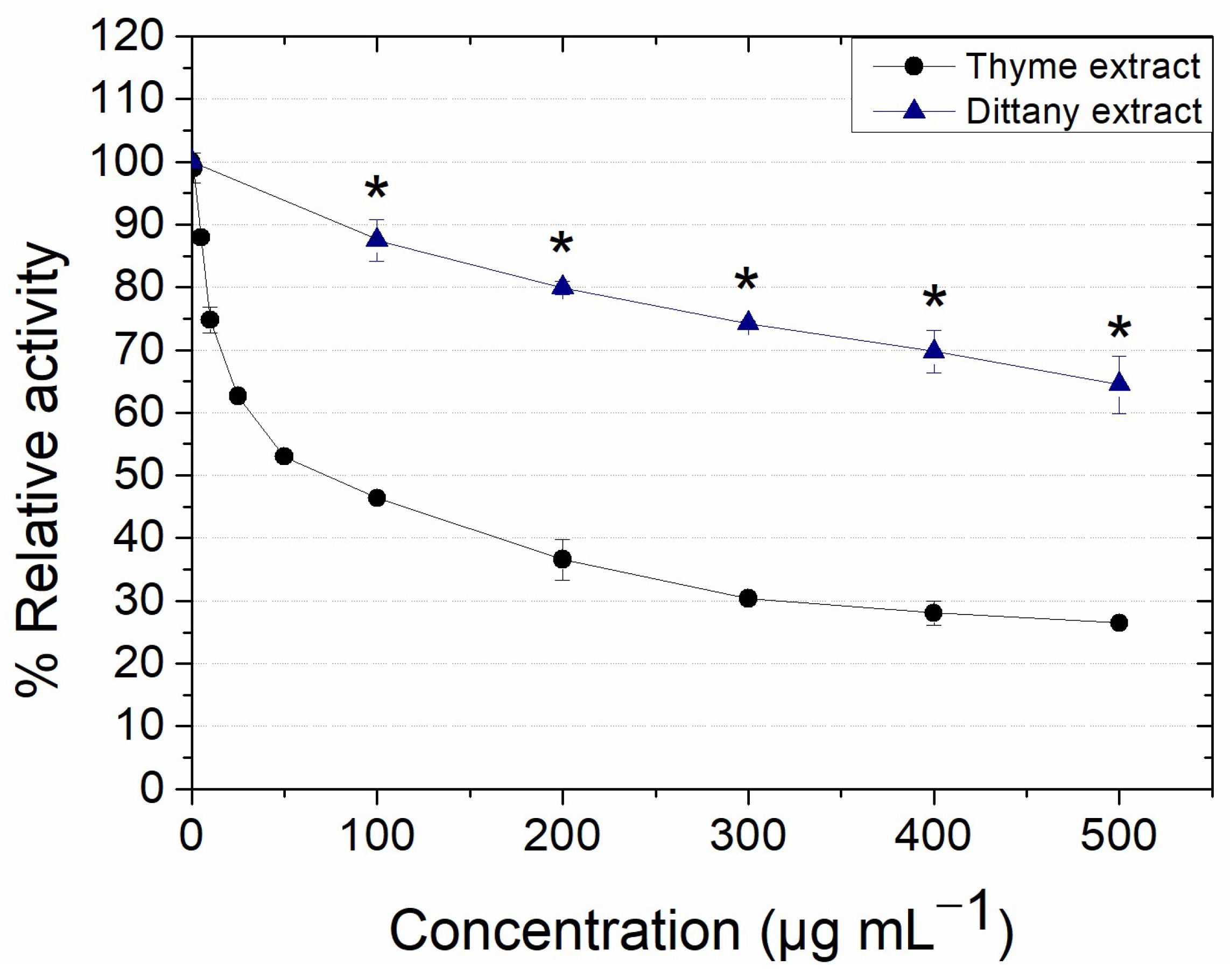
3.2.1. Effect on the Activity of Lipase from Candida rugosa

Candida rugosa lipase (Crl) inhibitors are gaining more and more attention for antifungal treatments, especially since the resistance of Candida strains in therapy is increasing [50,51]. In the present work, the prepared extracts were assessed for their potent inhibitory activity against Crl, and the results are presented in Figure 1. As demonstrated, the thyme extract presented strong inhibitory activity against Crl, with its IC50 value being 63.9 ± 23.8 μg mL−1 in contrast with the dittany extract, whose IC50 was proven to be more than 500 μg mL−1. Additionally, the IC50 of the positive control orlistat was found to be 86.9 ± 22.1 μg mL−1, in accordance with other studies [52,53]. It is noteworthy that the thyme extract had a much stronger inhibitory impact on Crl’s relative activity, comparable to that of orlistat, in comparison with the dittany one. The different compositions of the extracts is inseparably connected to their biological properties, as anticipated, also taking into account that the included components may act in a synergistic or antagonistic way. Thus, the inhibitory effect observed in both extracts could be attributed to the presence of phenolic compounds [54], as previous studies have shown that their two main phenolic compounds, rosmarinic acid and carvacrol for thyme and dittany extracts, respectively, possess an inhibitory impact on Crl’s relative activity [55,56]. It is significant to note that the components of the extracts can interact with each other in a manner that cannot be predicted, owing to the complexity of the extracts, as various components are present, and their concentrations differ.
Furthermore, extracts derived from Thymus species have been recorded as inhibitors of Candida rugosa lipase activity, using organic solvent/water mixtures as extraction solvents [57]. In addition, in the study of Slanc et al. [58], water/methanol extracts were prepared from different medicinal plants, such as Origanum vulgare and Thymus pulegoides, which were screened for their inhibitory activity against porcine pancreas lipase. According to the results, both extracts presented inhibitory activity up to 70%, depending on the substrate used. Also, in the study of Jaradat et al. [59], a hexanoic extract from Origanum dayi proved to be a stronger inhibitor against porcine pancreas lipase than orlistat, with an IC50 at 18.62 μg mL−1. All these studies confirm the potential of Origanum sp. and Thymus sp. organic extracts to inhibit the activity of lipases of different origins.

3.2.2. Effect on the Activity of Tyrosinase from Mushrooms

Tyrosinase is the key enzyme implicated in the metabolic pathway of melanin synthesis. Thus, its regulation is crucial regarding skin-related hypo- or hyper-pigmentation diseases [60]. In the present work, the prepared extracts were examined for their tyrosinase activity-regulating potential. As presented in Figure 2, the thyme extract presented inhibitory activity up to 40%, with no statistically significant differences being observed among the concentrations 100–400 μg mL−1 (p > 0.05), and its IC50 was estimated at >500 μg mL−1. On the other hand, the dittany extract did not affect the enzyme activity (p > 0.5), except for the maximum concentration used, where a possible activation effect occurred (p < 0.5). Additionally, the IC50 of the positive control kojic acid was found to be 3.2 ± 0.2 μg mL−1, in accordance with other studies [61,62], indicating that the inhibitory effect of the thyme extract against tyrosinase is rather slight when compared to the standard inhibitor. The observed inhibitory activity may be ascribed to the phenolic compounds that are present in the thyme extract, as the main phenolic compounds of the extracts (carvacrol, thymol and rosmarinic acid) have also been reported as tyrosinase inhibitors [63,64]. Concerning rosmarinic acid, it should be noted that it can simultaneously act both as substrate and inhibitor of tyrosinase [64,65]. Hence, it seems that the biological effects of the extracts on the enzyme’s activity strongly depend on the composition of each extract, including possible antagonistic or synergistic effects.
Organic extracts or essential oils from Origanum and Thymus genus have been previously presented as inhibitors of tyrosinase activity [66,67,68,69,70]. For example, in the study of Duletić-Laušević et al. [67], ethanolic extracts of Origanum majorana deriving from different regions were prepared and screened for their tyrosinase inhibitory potential. According to their results, the inhibitory activity of the prepared extracts did not exceed 25% inhibition at the maximum tested concentration of 100 μg mL−1, and, interestingly, a reduced inhibitory activity was observed with the increase in the tested extract concentrations. Additionally, in the study of Küçükaydın et al. [70], rosmarinic acid-rich methanolic extracts from Thymus cariensis and Thymus cilicicus were also used for the evaluation of their inhibitory activity against tyrosinase from mushrooms, and the estimated IC50 values were in the range of 58–161 μg mL−1, contrary to the observed results for the thyme extract in the present study.

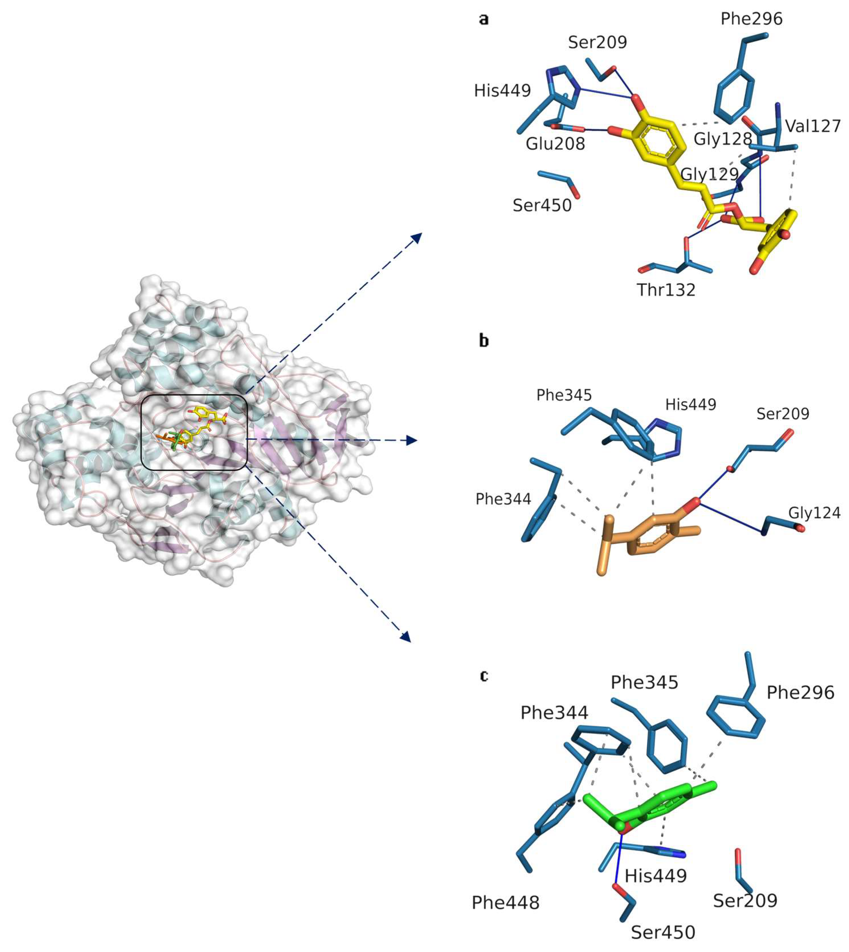
3.2.3. Docking Studies on Candida rugosa Lipase

Molecular docking is a widely used methodology to study the binding affinity of small molecules (ligands) to receptors (such as enzymes). They can provide rational explanations for the observed regioselectivity of biocatalysts and can contribute to the development of new drugs since enzymes are important inhibitor targets [71,72]. In this work, molecular docking was used as a tool to predict the binding orientation of rosmarinic acid, carvacrol and thymol in order to gain a deeper understanding of the underlying interactions that might influence their inhibitory activity. Tables S3 and S4 show data for rosmarinic acid, carvacrol and thymol from the molecular docking method onto the active site of Crl. According to the results, all compounds interacted favorably within the active site of the protein, with rosmarinic acid demonstrating the highest binding affinity (Table S3). More specifically, rosmarinic acid interacts with many residues hydrophobically, while also participating in many hydrogen bonds either as a donor or as an acceptor, due to its numerous hydroxyl groups (Figure 3a). Two key interactions that might influence its inhibitory activity are the hydrogen bonds between the catalytic residues, Ser209 and His449, with one of the hydroxyl groups of its caffeic acid ester moiety. Indeed, serine hydrolases require the Ne atom of the imidazole ring to be non-protonated to activate the catalytic serine. Thus, the binding conformation of rosmarinic acid in such a conformation is believed to hinder the ability to activate Ser209.
Carvacrol and thymol are isomeric compounds, and their difference lies in the position of the hydroxyl group (Table S3). From the molecular docking studies, their conformations appear to be different (Figure 3b,c). More specifically, carvacrol’s optimum conformation facilitates the formation of a hydrogen bond between the hydroxyl group and catalytic Ser209. By contrast, no such interaction is formed with thymol. One possible explanation for this differentiation might be the position of the hydroxyl group on the aromatic ring, which influences the final binding conformation of the molecule. Despite this difference, they share some similar interactions between the benzyl groups of Phe344 and Phe345 and their aromatic ring. Additionally, both compounds interact hydrophobically with the catalytic His449, although no hydrogen bond is formed between them. Regarding the binding affinity, thymol displays a higher affinity to Crl than carvacrol (−5.33 and −5.24 kcal mol−1 respectively). However, one of the shortcomings of molecular docking is the scoring functions [73]. A biocatalytic system is complex, and the oversimplification of interactions of such systems through scoring functions could possibly lead to inaccurate results, thus highlighting the importance of a thorough analysis of the putative complex interactions and not relying solely on scoring functions before drawing any conclusion [74]. Similar work has been conducted for the inhibition of lipase from Aspergillus niger with carvacrol and thymol [75]. It was shown that both thymol and carvacrol inhibited the lipase activity, with carvacrol showing a higher affinity due to the position of the hydroxyl group. Although a different lipase was studied, the consensus is that carvacrol, when compared to thymol, displays higher inhibitory activity, possibly because the hydroxyl group interacts with the catalytic serine. These observations seem to be in accordance with the experimental results, since, between the two extracts, the thyme extract displayed higher inhibitory activity towards Crl. Rosmarinic acid, which is present in higher concentrations in the thyme extract, is believed to greatly influence the enzymatic activity of Crl.

3.2.4. Docking Studies on Tyrosinase from Mushroom

Numerous studies have been conducted on the front of tyrosinase inhibitors that were also complemented by the implementation of computational tools (such as molecular docking and molecular dynamics simulations) [76,77]. From these studies, it is suggested that phenolic compounds that can chelate the copper ions of tyrosinase can be classified as effective inhibitors [78]. Previous studies have shown that rosmarinic acid can act as an inhibitor and a substrate of tyrosinase simultaneously, due to its two o-dihydroxyphenyl systems in its structure [64,79]. According to Lin et al. [64], rosmarinic acid displayed a non-competitive inhibitory action on tyrosinase. For this reason, we decided to exclude rosmarinic acid from the docking analysis. Carvacrol and thymol are classified as phenol monoterpenoids that could act as inhibitors of tyrosinase. Previous studies have proposed thymol’s inhibitory mechanism, demonstrating its ability to inhibit the oxidation of L-DOPA to dopaquinone [80]. In contrast, no previous work exists in the literature regarding the inhibition of mushroom tyrosinase with carvacrol. Therefore, in this work, molecular docking was employed with carvacrol and thymol to elucidate the binding mechanism that might influence their inhibitory activities on tyrosinase. From the docking results, carvacrol and thymol occupy the hydrophobic binding pocket that surrounds the active site of tyrosinase near the binuclear copper atoms (Figure 4a,b). Both molecules form a hydrogen bond with Asn260 (Table S5), which seems to play a critical role regarding the various tyrosinase inhibitors that were previously studied with computational tools [81]. Despite their similar interactions, the conformations of carvacrol and thymol differ. More specifically, thymol’s hydroxyl group is in closer proximity to the copper atoms, while carvacrol is oriented with the methyl group of the ring towards the copper atoms, leaving the hydroxyl group further away. This could indicate that thymol might have a greater influence on inhibiting tyrosinase, in addition to the lower binding energy when compared to carvacrol (Table S3). The observations from this study suggest that thymol could negatively affect the tyrosinase activity, which is in accordance with the experimental results, since the thyme extract (in which thymol is one of the major phenolic compounds) displayed inhibitory activity at the tested concentrations. On the other hand, the dittany extract did not affect the tyrosinase activity at the tested concentrations except for the higher one (500 μg mL−1), where an activation effect may occur. Carvacrol, which is a major phenolic compound in the dittany extract, binds in a favorable way and interacts with the catalytic residues, which possibly suggests its affinity within the active site of the enzyme. The discrepancy between the modeling and experimental results concerning the non-inhibitory activity of the dittany extract on tyrosinase could be ascribed to various factors, including the fact that models do not accurately reflect the conformation of this compound into the active site, while further possible interactions among the phenolic compounds or among other non-phenolic extract compounds with the enzyme may occur that cannot be predicted, leading to the obtained experimental results.

3.2.5. Evaluation of the Antioxidant Activity of the Extracts

DPPH and FRAP assays are well-established methods for evaluating the antioxidant activity of extracts [82,83]. In the present work, the prepared extracts were tested for their ability to scavenge the DPPH radical and reduce the Fe+3-TPTZ complex. According to the results, the antioxidant activity of the thyme extract was stronger than that of the dittany extract, with their SC50 values being 33.9 ± 0.7 and 47.2 ± 0.1 μg mL−1 (p < 0.05), correspondingly, and their Trolox equivalents being 351 ± 7 and 275 ± 2 mg g−1 dry extract, respectively (p < 0.05). These results are not only a function of the total phenolic and flavonoid content (higher in both cases for the thyme extract), but mainly of the composition of the phenolic profile, the concentrations of the individual compounds and the potential synergistic or antagonistic way of action [84]. Thymol, carvacrol and rosmarinic acid are strong antioxidants [85,86], and extracts/oils derived from Thymus or Origanum plants containing these compounds have been reported to exert significant antioxidant potential [7,16,17,87,88,89], conforming to the present results. For example, in the study of Gedikoğlu et al. [87], an 80% v/v thymol-rich methanolic extract from Thymus vulgaris presented an IC50 value of 29.22 μg mL−1 using the DPPH assay, whereas, in the study of Shan et al. [88], a rosmarinic acid/thymol-rich extract was considered a strong antioxidant using the ABTS assay. Accordingly, in the study of Mitropoulou et al. [7], a carvacrol-rich essential oil from Origanum dictamnus was prepared, and its antioxidant activity was evaluated using the DPPH assay, resulting in an IC50 value of 0.045% v/v.

3.2.6. Evaluation of the Antibacterial Activity of the Extracts

Phenolic compounds are well-known antibacterial agents [90]. Thus, the prepared phenolic compound-rich extracts were assessed using the broth microdilution assay, up to 10 mg mL−1, for their inhibitory effect against Bacillus subtilis and Escherichia coli, and the results are presented in Table 2 and Table 3. It is interesting to note that the extracts had a lower inhibitory impact on the Escherichia coli strain, with the MIC values being 2.5 and 8 mg mL−1 for the dittany and thyme extracts, correspondingly, contrary to the Bacillus subtilis extract, where the MIC values were found to be 0.5 and 2 mg mL−1 for the dittany and thyme extracts, respectively. It is known that the differentiated composition of bacterial cell walls between Gram-positive and Gram-negative strains affects their permeability [91]. Accordingly, many studies have tested dittany and thyme extracts or oils against different human pathogens, such as Staphylococcus sp., Enterobacter sp. and Salmonella sp., and a stronger antibacterial effect has been reported against Gram-positive bacteria, as Gram-negative ones are known to be more resistant [17,67,87,92,93], which is in accordance with our results.
More specifically, in the work of Gedikoğlu et al. [87], thymol-rich oils and rosmarinic acid-rich methanolic extracts from thyme were tested for their antimicrobial activity against different bacterial pathogens using the disk diffusion agar assay. According to the results, while thyme oils were strong antimicrobial agents, the extracts showed no antibacterial effect. On the contrary, in another work, a methanolic extract from Thymus vulgaris proved to have a significant antibacterial effect, presenting an MIC value of 31 μg mL−1 against the B.cereus strain and 250 μg mL−1 against E. coli [93]. In another study [67], where rosmarinic acid-rich Origanum majorana ethanolic extracts were employed, it was also found that they had a stronger antibacterial effect against the B.subtilis strain (MIC values up to 2.5 mg mL−1) than the E. coli strain (MIC values over 5 mg mL−1), as evaluated using the broth microdilution assay.
The prepared extracts were further tested for their bactericidal effect against these strains. In the case of the B. subtilis strain, the MBC value was found to be over 10 mg mL−1 for both plant extracts, whereas, in the case of E. coli, the MBC was found to be 8 mg mL−1 for both extracts. It is known that, if the ratio MBC/MIC is lower or equal to 4, the effect of the tested sample is bactericidal; otherwise, the effect is defined as bacteriostatic [94]. The results obtained from the MBC test and the estimated MBC/MIC ratios lead to the conclusion that these extracts have a bacteriostatic profile against Bacillus subtilis, whereas, in the case of Escherichia coli, the effect of the extracts was clearly bactericidal.

3.3. Preparation and Biological Assessment of Gelatin Hydrogels Containing Extracts

3.3.1. Preparation of Gelatin Hydrogels Containing Extracts

Gelatin hydrogels that contained methanolic extracts either from thyme or dittany were successfully prepared due to physical crosslinking (Figure 5). In physical crosslinking, gelatin chains interact with each other mainly though hydrogen bonds, leading to the formation of an extended network that can be preserved at temperatures below 35 °C [95]. Also, the DES Bet:PG (1:4) was used as a green solvent for the dissolution of the phenolic extracts, which, when compared to the conventional organic solvents (i.e., methanol), remains in the formed hydrogel, thus offering additional physicochemical properties, such as enhanced flexibility and moisture [96,97].

3.3.2. Evaluation of the Antioxidant Activity of Gelatin Hydrogels Containing Extracts

The antioxidant activity of dried hydrogel membranes (Figures S4 and S5) was estimated in terms of DPPH radical scavenging, and the results are presented in Figure 6. As shown, the implementation of thyme and dittany extracts in the preparation of hydrogels has enhanced the antioxidant activity of gelatin by up to 85% in relation to the concentration of the extracts. In particular, the HG_T membranes demonstrated higher antioxidant activity when compared to HG_D membranes (p < 0.05), which is in correlation with the above-mentioned results for the extracts (Section 3.3). Gelatin possesses slight antioxidant activity owing to the presence of specific amino acids, such as proline and the glycine and peptide fractions contained in the biopolymer, which are involved in the electron transfer [43,98,99,100], whereas the incorporation of phenols in the formation of gelatin hydrogels has been broadly implemented for the improvement of antioxidant activity [101].
Herein, the presented antioxidant activity may be ascribed both to a gradual release of the extract’s compounds from the membrane to the DPPH solution and to the antioxidant capacity of the entrapped ones. According to some preliminary results concerning the release kinetics of phenolic compounds from hydrogels containing 8 mg mL−1 extracts in ddH2O, ~15% release for HG_D and ~20% for HG_T after 24 h was observed, meaning that part of the observed antioxidant activity could be attributed to small amounts of released phenolic compounds. These results agree with other studies, where carvacrol [26], gallic acid [31] or other polyphenols [27] were used in the preparation of gelatin hydrogels with improved antioxidant activity. Furthermore, green tea [30] and oregano and rosemary aqueous extracts have also been introduced in other gelatin films, leading to enhanced antioxidant activity [29].

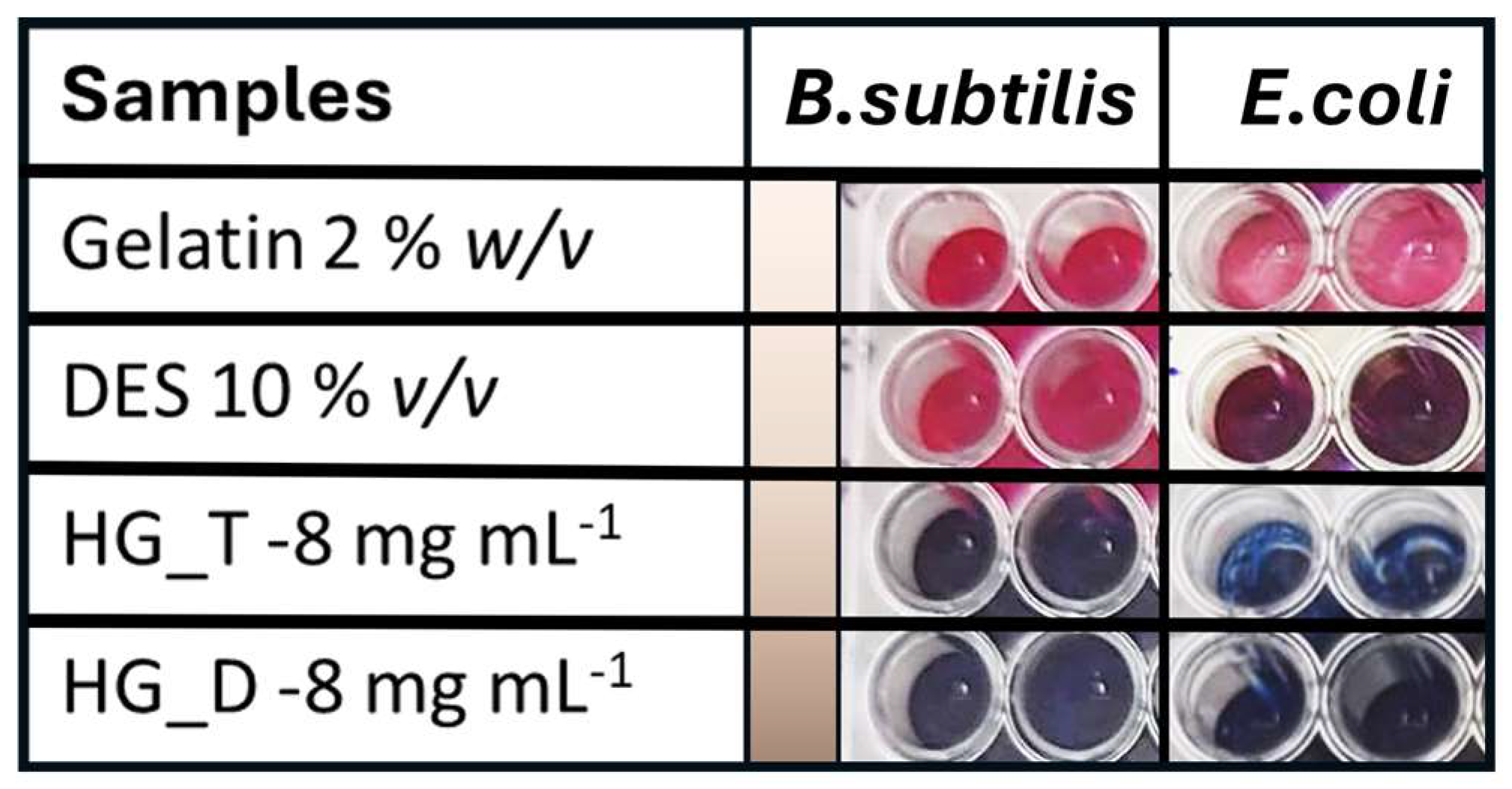
3.3.3. Evaluation of the Antibacterial Activity of Gelatin Hydrogels Containing Extracts

The liquid hydrogel mixtures that contained gelatin, DES and the extracts at the concentration of 8 mg mL−1 showed antibacterial activity against Bacillus subtilis and Escherichia coli. The growth inhibitory activity was detected through the color change in the bacterial growth indicator resazurin from blue to pink [102]. More specifically, the prepared hydrogel mixtures at the selected concentration showed antibacterial activity that is ascribed to the presence of the extracts, as gelatin and the DES Bet:PG (1:4) at concentrations of 2% w/v and 10% v/v, respectively, do not inhibit the bacterial growth (Figure 7).
It has been reported that single gelatin does not demonstrate antibacterial activity against Gram-positive and Gram-negative bacteria [27], and that Bet:PG DES’s antibacterial activity decreases when the water content increases [103], which agrees with the results of the current study. The incorporation of polyphenol (curcumin, gallic acid and quercetin)-loaded nanoemulsions [27] and carvacrol [26], as well as the introduction of Gingo biloba extract in gelatin films, offered great antibacterial activity, as these phenolic compounds cause structural damage on the bacterial cell walls and cell membranes, thus leading to other processes that hinder bacterial growth [28].

4. Conclusions

In this work, methanolic extracts from thyme and dittany plants, rich in known biologically active components such as rosmarinic acid, carvacrol and thymol, were prepared in order to evaluate their biological effectiveness and their incorporation in gelatin-based hydrogels. The main phenolic compounds determined in the thyme extract were rosmarinic acid and thymol, whereas carvacrol was the main component in the dittany extract. Also, the phenolic and flavonoid content of the thyme extract was found to be higher than that of the dittany extract. In general, the thyme extract showed stronger antioxidant (SC50 = 33.9 μg mL−1) and lipase inhibitory properties (IC50 = 63.7 μg mL−1), while the dittany extract exhibited greater antibacterial activity (MIC = 0.5 mg mL−1 against B. subtillis and 2 mg mL−1 against E. coli). Molecular docking studies confirmed the superiority of the thyme extract against inhibition assays, as thymol and rosmarinic acid proved to have the most interaction with tyrosinase and lipase, correspondingly. Overall, the difference in the extracts’ phenolic contents seems to strongly affect their antibacterial, antioxidant and enzyme inhibitory activity, and it must be noted that possible interactions among the phenolic compounds may occur in a manner that cannot be easily predicted, highlighting the need for further research.
The prepared extracts were successfully incorporated into gelatin-based hydrogels, with the intention of producing preparations with biologically active ingredients based on the biological assessment of the extracts. The prepared hydrogels, containing the extracts with concentrations in the range of 2–8 mg/mL, presented high antioxidant activity (up to 85% for HG_T and 58% for HG_D) and antimicrobial activity, which was in correlation with the activity of the extracts. These findings can pave the way for the application of such extracts to hydrogel preparations that could be applied in pharmaceutical and cosmeceutical industries for multiple uses due to their bioactivity. Further studies regarding the structural and physicochemical characterization and optimization of the hydrogel preparations could be performed in order to produce fully characterized formulations.

Supplementary Materials

The following supporting information can be downloaded at https://www.mdpi.com/article/10.3390/biochem4030009/s1, Figure S1. Prepared methanolic extracts of thyme (left) and dittany (right); Figure S2. Total ion chromatogram (up) and UV chromatograms at 280, 254 and 334 nm of dittany extract (Peak indicated with one asterisk was carvacrol, whose identification was confirmed using commercial standard, and peak indicated with two asterisks probably corresponds to thymoquinone [49]; Table S1. Peak assignments of dittany extract [33]; Figure S3. Total ion chromatogram (up) and UV chromatograms at 280, 254 and 334 nm of thyme extract. (Peak indicated with one asterisk with Rt = 24.3 min was thymol, whose identification was confirmed using commercial standard); Table S2. Peak assignments of thyme extract; Table S3. Binding energy from the docked structures inside the active site of Crl and tyrosinase; Table S4. Binding interactions of the studied compounds with residues of Crl; Table S5. Binding interactions of the studied compounds with residues of tyrosinase; Figure S4. Form of dried hydrogel, after incubation at 25 °C for 2 days; Figure S5. Hydrogel membranes strips at the size of 3 × 5 mm [104,105,106,107,108,109,110,111].

Author Contributions

Conceptualization, S.S., M.G.B., A.V.C. and H.S.; methodology, S.S., M.G.B., A.P., K.N., V.G.K., A.V.C. and H.S.; software, A.P.; validation, V.G.K., A.V.C. and H.S.; formal analysis, S.S., M.G.B., A.P., K.N. and V.G.K.; investigation, S.S., M.G.B., A.P., K.N., V.G.K. and A.V.C.; resources, H.S.; data curation, S.S., M.G.B., A.P., K.N. and V.G.K.; writing—original draft preparation, S.S., M.G.B., A.P. and V.G.K.; writing—review and editing, V.G.K., A.V.C. and H.S.; visualization, S.S., M.G.B. and A.P.; supervision, H.S.; project administration, H.S.; funding acquisition, H.S. All authors have read and agreed to the published version of the manuscript.

Funding

This research has been co-financed by the European Regional Development Fund of the European Union and Greek national funds through the Operational Program Competitiveness, Entrepreneurship, and Innovation, under the call RESEARCH-CREATE-INNOVATE (project code: T2EDK-01410).

Data Availability Statement

Data are contained within the article and Supplementary Materials.

Conflicts of Interest

The authors declare no conflicts of interest.

References

  1. Petrovska, B. Historical Review of Medicinal Plants’ Usage. Pharmacogn. Rev. 2012, 6, 1–5. [Google Scholar] [CrossRef] [PubMed]
  2. Venkateshappa, S.M.; Sreenath, K.P. Potential Medicinal Plants of Lamiaceae. Am. Int. J. Res. Form. Appl. Nat. Sci. 2013, 3, 82–87. [Google Scholar]
  3. Reddy, P.; Kandisa, R.V.; Varsha, P.V.; Satyam, S. Review on Thymus Vulgaris Traditional Uses and Pharmacological Properties. Med. Aromat. Plants 2014, 3, 167. [Google Scholar] [CrossRef]
  4. Dauqan, E.M.A.; Abdullah, A. Medicinal and Functional Values of Thyme (Thymus vulgaris L.) Herb. J. Appl. Biol. Biotechnol. 2017, 5, 17–22. [Google Scholar] [CrossRef]
  5. Patil, S.M.; Ramu, R.; Shirahatti, P.S.; Shivamallu, C.; Amachawadi, R.G. A Systematic Review on Ethnopharmacology, Phytochemistry and Pharmacological Aspects of Thymus Vulgaris Linn. Heliyon 2021, 7, e07054. [Google Scholar] [CrossRef] [PubMed]
  6. Liolios, C.C.; Graikou, K.; Skaltsa, E.; Chinou, I. Dittany of Crete: A Botanical and Ethnopharmacological Review. J. Ethnopharmacol. 2010, 131, 229–241. [Google Scholar] [CrossRef] [PubMed]
  7. Mitropoulou, G.; Fitsiou, E.; Stavropoulou, E.; Papavassilopoulou, E.; Vamvakias, M.; Pappa, A.; Oreopoulou, A.; Kourkoutas, Y. Composition, Antimicrobial, Antioxidant, and Antiproliferative Activity of Origanum dictamnus (dittany) Essential Oil. Microb. Ecol. Health Dis. 2015, 26, 26543. [Google Scholar] [CrossRef] [PubMed]
  8. Solomou, A.D.; Fountouli, A.; Molla, A.; Petrakis, M.; Manolikaki, I.; Skoufogianni, E. Ecology, Cultivation, and Utilization of the Dittany of Crete (Origanum dictamnus L.) from Ancient Times to the Present: A Short Review. Agronomy 2024, 14, 1066. [Google Scholar] [CrossRef]
  9. Uyama, H. Artificial Polymeric Flavonoids: Synthesis and Applications. Macromol. Biosci. 2007, 7, 410–422. [Google Scholar] [CrossRef]
  10. Sauceda, A.E.Q.; Sáyago-Ayerdi, S.G.; Ayala-Zavala, J.F.; Wall-Medrano, A.; De La Rosa, L.A.; González-Aguilar, G.A.; Álvarez-Parrilla, E. Biological Actions of Phenolic Compounds. Fruit. Veg. Phytochem. 2017, 2, 125–138. [Google Scholar] [CrossRef]
  11. Nabavi, S.M.; Marchese, A.; Izadi, M.; Curti, V.; Daglia, M.; Nabavi, S.F. Plants Belonging to the Genus Thymus as Antibacterial Agents: From Farm to Pharmacy. Food Chem. 2015, 173, 339–347. [Google Scholar] [CrossRef] [PubMed]
  12. Imran, M.; Aslam, M.; Alsagaby, S.A.; Saeed, F.; Ahmad, I.; Afzaal, M.; Arshad, M.U.; Abdelgawad, M.A.; El-Ghorab, A.H.; Khames, A.; et al. Therapeutic Application of Carvacrol: A Comprehensive Review. Food Sci. Nutr. 2022, 10, 3544–3561. [Google Scholar] [CrossRef] [PubMed]
  13. Chatzopoulou, A.; Karioti, A.; Gousiadou, C.; Lax Vivancos, V.; Kyriazopoulos, P.; Golegou, S.; Skaltsa, H. Depsides and Other Polar Constituents from Origanum dictamnus L. and Their in Vitro Antimicrobial Activity in Clinical Strains. J. Agric. Food Chem. 2010, 58, 6064–6068. [Google Scholar] [CrossRef] [PubMed]
  14. Pereira, D.M.; Valentão, P.; Pereira, J.A.; Andrade, P.B. Phenolics: From Chemistry to Biology. Molecules 2009, 14, 2202–2211. [Google Scholar] [CrossRef]
  15. Kouri, G.; Tsimogiannis, D.; Bardouki, H.; Oreopoulou, V. Extraction and Analysis of Antioxidant Components from Origanum dictamnus. Innov. Food Sci. Emerg. Technol. 2007, 8, 155–162. [Google Scholar] [CrossRef]
  16. Roby, M.H.H.; Sarhan, M.A.; Selim, K.A.H.; Khalel, K.I. Evaluation of Antioxidant Activity, Total Phenols and Phenolic Compounds in Thyme (Thymus vulgaris L.), Sage (Salvia officinalis L.), and Marjoram (Origanum majorana L.) Extracts. Ind. Crops Prod. 2013, 43, 827–831. [Google Scholar] [CrossRef]
  17. Gortzi, O.; Lalas, S.; Chinou, I.; Tsaknis, J. Evaluation of the Antimicrobial and Antioxidant Activities of Origanum dictamnus Extracts before and after Encapsulation in Liposomes. Molecules 2007, 12, 932–945. [Google Scholar] [CrossRef]
  18. Habashy, N.H.; Abu Serie, M.M.; Attia, W.E.; Abdelgaleil, S.A.M. Chemical Characterization, Antioxidant and Anti-Inflammatory Properties of Greek Thymus Vulgaris Extracts and Their Possible Synergism with Egyptian Chlorella Vulgaris. J. Funct. Foods 2018, 40, 317–328. [Google Scholar] [CrossRef]
  19. Paloukopoulou, C.; Govari, S.; Soulioti, A.; Stefanis, I.; Angeli, A.; Matheeussen, A.; Capasso, C.; Cos, P.; Supuran, C.T.; Karioti, A. Phenols from Origanum dictamnus L. and Thymus vulgaris L. and Their Activity against Malassezia globosa Carbonic Anhydrase. Nat. Prod. Res. 2022, 36, 1558–1564. [Google Scholar] [CrossRef]
  20. Olszewska, M.A.; Gędas, A.; Simões, M. Antimicrobial Polyphenol-Rich Extracts: Applications and Limitations in the Food Industry. Food Res. Int. 2020, 134, 109214. [Google Scholar] [CrossRef]
  21. Guimarães, I.; Baptista-Silva, S.; Pintado, M.; Oliveira, A.L. Polyphenols: A Promising Avenue in Therapeutic Solutions for Wound Care. Appl. Sci. 2021, 11, 1230. [Google Scholar] [CrossRef]
  22. Li, Z.; Chen, Z.; Chen, H.; Chen, K.; Tao, W.; Ouyang, X.; Mei, L.; Zeng, X. Polyphenol-Based Hydrogels: Pyramid Evolution from Crosslinked Structures to Biomedical Applications and the Reverse Design. Bioact. Mater. 2022, 17, 49–70. [Google Scholar] [CrossRef]
  23. Alipal, J.; Mohd Pu’ad, N.A.S.; Lee, T.C.; Nayan, N.H.M.; Sahari, N.; Basri, H.; Idris, M.I.; Abdullah, H.Z. A Review of Gelatin: Properties, Sources, Process, Applications, and Commercialisation. Mater. Today Proc. 2021, 42, 240–250. [Google Scholar] [CrossRef]
  24. Rather, J.A.; Akhter, N.; Ashraf, Q.S.; Mir, S.A.; Makroo, H.A.; Majid, D.; Barba, F.J.; Khaneghah, A.M.; Dar, B.N. A Comprehensive Review on Gelatin: Understanding Impact of the Sources, Extraction Methods, and Modifications on Potential Packaging Applications. Food Packag. Shelf Life 2022, 34, 100945. [Google Scholar] [CrossRef]
  25. Montazerian, H.; Sampath, R.R.; Annabi, N.; Khademhosseini, A.; Weiss, P.S. Polyphenolic Gelatin-Based Bioadhesives. Acc. Mater. Res. 2023, 4, 627–640. [Google Scholar] [CrossRef]
  26. Kavoosi, G.; Dadfar, S.M.M.; Mohammadi Purfard, A.; Mehrabi, R. Antioxidant and Antibacterial Properties of Gelatin Films Incorporated with Carvacrol. J. Food Saf. 2013, 33, 423–432. [Google Scholar] [CrossRef]
  27. Khan, M.R.; Sadiq, M.B.; Mehmood, Z. Development of Edible Gelatin Composite Films Enriched with Polyphenol Loaded Nanoemulsions as Chicken Meat Packaging Material. CYTA J. Food 2020, 18, 137–146. [Google Scholar] [CrossRef]
  28. Hu, X.; Yuan, L.; Han, L.; Li, S.; Song, L. Characterization of Antioxidant and Antibacterial Gelatin Films Incorporated with: Ginkgo biloba Extract. RSC Adv. 2019, 9, 27449–27454. [Google Scholar] [CrossRef]
  29. Gómez-Estaca, J.; Bravo, L.; Gómez-Guillén, M.C.; Alemán, A.; Montero, P. Antioxidant Properties of Tuna-Skin and Bovine-Hide Gelatin Films Induced by the Addition of Oregano and Rosemary Extracts. Food Chem. 2009, 112, 18–25. [Google Scholar] [CrossRef]
  30. Wu, J.; Chen, S.; Ge, S.; Miao, J.; Li, J.; Zhang, Q. Preparation, Properties and Antioxidant Activity of an Active Film from Silver Carp (Hypophthalmichthys molitrix) Skin Gelatin Incorporated with Green Tea Extract. Food Hydrocoll. 2013, 32, 42–51. [Google Scholar] [CrossRef]
  31. Limpisophon, K.; Schleining, G. Use of Gallic Acid to Enhance the Antioxidant and Mechanical Properties of Active Fish Gelatin Film. J. Food Sci. 2017, 82, 80–89. [Google Scholar] [CrossRef] [PubMed]
  32. Kontogianni, V.G.; Kasapidou, E.; Mitlianga, P.; Mataragas, M.; Pappa, E.; Kondyli, E.; Bosnea, L. Production, Characteristics and Application of Whey Protein Films Activated with Rosemary and Sage Extract in Preserving Soft Cheese. LWT 2022, 155, 112996. [Google Scholar] [CrossRef]
  33. Kontogianni, V.G.; Tomic, G.; Nikolic, I.; Nerantzaki, A.A.; Sayyad, N.; Stosic-Grujicic, S.; Stojanovic, I.; Gerothanassis, I.P.; Tzakos, A.G. Phytochemical Profile of Rosmarinus Officinalis and Salvia Officinalis Extracts and Correlation to Their Antioxidant and Anti-Proliferative Activity. Food Chem. 2013, 136, 120–129. [Google Scholar] [CrossRef]
  34. Spyrou, S.; Chatzikonstantinou, A.; Giannakopoulou, A.; Fotiadou, R.; Priska, S.; Simos, Y.; Tsakni, A.; Peschos, D.; Houhoula, D.; Voutsas, E.; et al. Fungal Laccase-Mediated Enhancement of the Bioactivity of Green Algae Extracts. Catal. Res. 2023, 3, 1–29. [Google Scholar] [CrossRef]
  35. Di Petrillo, A.; González-Paramás, A.M.; Era, B.; Medda, R.; Pintus, F.; Santos-Buelga, C.; Fais, A. Tyrosinase Inhibition and Antioxidant Properties of Asphodelus Microcarpus Extracts. BMC Complement. Altern. Med. 2016, 16, 453. [Google Scholar] [CrossRef] [PubMed]
  36. Forli, S.; Huey, R.; Pique, M.E.; Sanner, M.F.; Goodsell, D.S.; Olson, A.J. Computational Protein–Ligand Docking and Virtual Drug Screening with the AutoDock Suite. Nat. Protoc. 2016, 11, 905–919. [Google Scholar] [CrossRef] [PubMed]
  37. Williams, C.J.; Headd, J.J.; Moriarty, N.W.; Prisant, M.G.; Videau, L.L.; Deis, L.N.; Verma, V.; Keedy, D.A.; Hintze, B.J.; Chen, V.B.; et al. MolProbity: More and Better Reference Data for Improved All-Atom Structure Validation. Protein Sci. 2018, 27, 293–315. [Google Scholar] [CrossRef] [PubMed]
  38. Hanwell, M.D.; Curtis, D.E.; Lonie, D.C.; Vandermeersch, T.; Zurek, E.; Hutchison, G.R. Avogadro: An Advanced Semantic Chemical Editor, Visualization, and Analysis Platform. J. Cheminform. 2012, 4, 17. [Google Scholar] [CrossRef]
  39. Adasme, M.F.; Linnemann, K.L.; Bolz, S.N.; Kaiser, F.; Salentin, S.; Haupt, V.J.; Schroeder, M. PLIP 2021: Expanding the Scope of the Protein–Ligand Interaction Profiler to DNA and RNA. Nucleic Acids Res. 2021, 49, W530–W534. [Google Scholar] [CrossRef]
  40. Balouiri, M.; Sadiki, M.; Ibnsouda, S.K. Methods for in Vitro Evaluating Antimicrobial Activity: A Review. J. Pharm. Anal. 2016, 6, 71–79. [Google Scholar] [CrossRef]
  41. Parvekar, P.; Palaskar, J.; Metgud, S.; Maria, R.; Dutta, S. The Minimum Inhibitory Concentration (MIC) and Minimum Bactericidal Concentration (MBC) of Silver Nanoparticles against Staphylococcus aureus. Biomater. Investig. Dent. 2020, 7, 105–109. [Google Scholar] [CrossRef] [PubMed]
  42. Chatzikonstantinou, A.V.; Bellou, M.G.; Spyrou, S.; Papanikolaou, A.; Simos, Y.V.; Peschos, D.; Stamatis, H. Enhancement of the Biological Activity of Hydroxytyrosol through Its Oxidation by Laccase from Trametes Versicolor. J. Biotechnol. 2024, 385, 30–41. [Google Scholar] [CrossRef] [PubMed]
  43. Jafari, M.; Afkhami, R.; Sedaghat, N. Preparation and Characterization of Active Cirish Fructans–Fish Gelatin Film: Physicochemical, Antioxidant, and Antimicrobial Properties. Food Sci. Nutr. 2023, 11, 709–721. [Google Scholar] [CrossRef] [PubMed]
  44. Vujicic, M.; Nikolic, I.; Kontogianni, V.G.; Saksida, T.; Charisiadis, P.; Orescanin-Dusic, Z.; Blagojevic, D.; Stosic-Grujicic, S.; Tzakos, A.G.; Stojanovic, I. Methanolic Extract of Origanum vulgare Ameliorates Type 1 Diabetes through Antioxidant, Anti-Inflammatory and Anti-Apoptotic Activity. Br. J. Nutr. 2015, 113, 770–782. [Google Scholar] [CrossRef] [PubMed]
  45. Bendif, H.; Peron, G.; Miara, M.D.; Sut, S.; Dall’Acqua, S.; Flamini, G.; Maggi, F. Total Phytochemical Analysis of Thymus munbyanus subsp. coloratus from Algeria by HS-SPME-GC-MS, NMR and HPLC-MSn Studies. J. Pharm. Biomed. Anal. 2020, 186, 113330. [Google Scholar] [CrossRef]
  46. Ahmad, S.; Biosci, I.J.; Mehmood, T.; Shafique, S.; Tabassam, Q.; Afzal, M.; Author, C. Variation in Antioxidant Attributes, Individual Phenolic Acids Composition and Biological Activities of Thymus Vulgaris: Effects of Extraction Solvents. Int. J. Biosci. 2015, 6, 73–86. [Google Scholar] [CrossRef]
  47. Lagouri, V.; Alexandri, G. Antioxidant Properties of Greek O. dictamnus and R. officinalis Methanol and Aqueous Extracts - HPLC Determination of Phenolic Acids. Int. J. Food Prop. 2013, 16, 549–562. [Google Scholar] [CrossRef]
  48. Zengin, G.; Atasagun, B.; Zakariyyah Aumeeruddy, M.; Saleem, H.; Mollica, A.; Babak Bahadori, M.; Mahomoodally, M.F. Phenolic Profiling and in Vitro Biological Properties of Two Lamiaceae Species (Salvia Modesta and Thymus Argaeus): A Comprehensive Evaluation. Ind. Crops Prod. 2019, 128, 308–314. [Google Scholar] [CrossRef]
  49. Lemonis, I.; Tsimogiannis, D.; Louli, V.; Voutsas, E.; Oreopoulou, V.; Magoulas, K. Extraction of Dittany (Origanum dictamnus) Using Supercritical CO2 and Liquid Solvent. J. Supercrit. Fluids 2013, 76, 48–53. [Google Scholar] [CrossRef]
  50. Grippa, E.; Valla, R.; Battinelli, L.; Mazzanti, G.; Saso, L.; Silvestrini, B. Inhibition of Candida Rugosa Lipase by Berberine and Structurally Related Alkaloids, Evaluated by High-Performance Liquid Chromatography. Biosci. Biotechnol. Biochem. 1999, 63, 1557–1562. [Google Scholar] [CrossRef]
  51. Ruiz, C.; Falcocchio, S.; Xoxi, E.; Villo, L.; Nicolosi, G.; Pastor, F.I.J.; Diaz, P.; Saso, L. Inhibition of Candida Rugosa Lipase by Saponins, Flavonoids and Alkaloids. J. Mol. Catal. B Enzym. 2006, 40, 138–143. [Google Scholar] [CrossRef]
  52. Benarous, K.; Bombarda, I.; Iriepa, I.; Moraleda, I.; Gaetan, H.; Linani, A.; Tahri, D.; Sebaa, M.; Yousfi, M. Harmaline and Hispidin from Peganum Harmala and Inonotus Hispidus with Binding Affinity to Candida Rugosa Lipase: In Silico and in Vitro Studies. Bioorganic Chem. 2015, 62, 1–7. [Google Scholar] [CrossRef]
  53. Santana, C.C.; Silva-Júnior, E.F.; Santos, J.C.N.; Rodrigues, E.E.D.S.; da Silva, I.M.; Araújo-Júnior, J.X.; do Nascimento, T.G.; Barbosa, L.A.O.; Dornelas, C.B.; Figueiredo, I.M.; et al. Evaluation of Guanylhydrazone Derivatives as Inhibitors of Candida Rugosa Digestive Lipase: Biological, Biophysical, Theoretical Studies and Biotechnological Application. Bioorganic Chem. 2019, 87, 169–180. [Google Scholar] [CrossRef] [PubMed]
  54. Gatto, M.T.; Falcocchio, S.; Grippa, E.; Mazzanti, G.; Battinelli, L.; Nicolosi, G.; Lambusta, D.; Saso, L. Antimicrobial and Anti-Lipase Activity of Quercetin and Its C2-C16 3-O-Acyl-Esters. Bioorganic Med. Chem. 2002, 10, 269–272. [Google Scholar] [CrossRef] [PubMed]
  55. Serseg, T.; Benarous, K.; Yousfi, M. The Inhibitory Effect of Three Essential Oils on Candida Rugosa Lipase: In Vitro and In Silico Studies. Nat. Prod. J. 2020, 10, 208–215. [Google Scholar] [CrossRef]
  56. Zhang, H.; Yang, F.Q.; Gao, J.L. A Simple and Portable Personal Glucose Meter Method Combined with Molecular Docking for Screening of Lipase Inhibitors. Evid. -Based Complement. Altern. Med. 2022, 2022, 4430050. [Google Scholar] [CrossRef]
  57. Benguechoua, M.; Nia, S.; Benarous, K.; Khacheba, I.; Yousfi, M. Inhibition of Candida Rugosa Lipase by Different Extracts of Five Algerian Plants and Their Antioxidant Activities. Curr. Enzym. Inhib. 2014, 10, 121–128. [Google Scholar] [CrossRef]
  58. Slanc, P.; Doljak, B.; Kreft, S.; Lunder, M.; Janes, D.; Ítrukelj, B. Screening of Selected Food and Medicinal Plant Extracts for Pancreatic Lipase Inhibition. Phytother. Res. 2009, 23, 874–877. [Google Scholar] [CrossRef] [PubMed]
  59. Jaradat, N.; Zaid, A.; Hussein, F.; Zaqzouq, M.; Aljammal, H.; Ayesh, O. Anti-Lipase Potential of the Organic and Aqueous Extracts of Ten Traditional Edible and Medicinal Plants in Palestine; a Comparison Study with Orlistat. Medicines 2017, 4, 89. [Google Scholar] [CrossRef]
  60. Panzella, L.; Napolitano, A. Natural and Bioinspired Phenolic Compounds as Tyrosinase Inhibitors for the Treatment of Skin Hyperpigmentation: Recent Advances. Cosmetics 2019, 6, 57. [Google Scholar] [CrossRef]
  61. Lee, Y.S.; Park, J.H.; Kim, M.H.; Seo, S.H.; Kim, H.J. Synthesis of Tyrosinase Inhibitory Kojic Acid Derivative. Arch. Pharm. 2006, 339, 111–114. [Google Scholar] [CrossRef] [PubMed]
  62. Xie, W.; Zhang, H.; He, J.; Zhang, J.; Yu, Q.; Luo, C.; Li, S. Synthesis and Biological Evaluation of Novel Hydroxybenzaldehyde-Based Kojic Acid Analogues as Inhibitors of Mushroom Tyrosinase. Bioorganic Med. Chem. Lett. 2017, 27, 530–532. [Google Scholar] [CrossRef] [PubMed]
  63. Brotzman, N.; Xu, Y.; Graybill, A.; Cocolas, A.; Ressler, A.; Seeram, N.P.; Ma, H.; Henry, G.E. Synthesis and Tyrosinase Inhibitory Activities of 4-Oxobutanoate Derivatives of Carvacrol and Thymol. Bioorganic Med. Chem. Lett. 2019, 29, 56–58. [Google Scholar] [CrossRef]
  64. Lin, L.; Dong, Y.; Zhao, H.; Wen, L.; Yang, B.; Zhao, M. Comparative Evaluation of Rosmarinic Acid, Methyl Rosmarinate and Pedalitin Isolated from Rabdosia Serra (MAXIM.) HARA as Inhibitors of Tyrosinase and α-Glucosidase. Food Chem. 2011, 129, 884–889. [Google Scholar] [CrossRef]
  65. Zuo, A.R.; Dong, H.H.; Yu, Y.Y.; Shu, Q.L.; Zheng, L.X.; Yu, X.Y.; Cao, S.W. The Antityrosinase and Antioxidant Activities of Flavonoids Dominated by the Number and Location of Phenolic Hydroxyl Groups. Chin. Med. 2018, 13, 51. [Google Scholar] [CrossRef] [PubMed]
  66. Sarikurkcu, C.; Zengin, G.; Oskay, M.; Uysal, S.; Ceylan, R.; Aktumsek, A. Composition, Antioxidant, Antimicrobial and Enzyme Inhibition Activities of Two Origanum vulgare Subspecies (subsp. vulgare and subsp. hirtum) Essential Oils. Ind. Crops Prod. 2015, 70, 178–184. [Google Scholar] [CrossRef]
  67. Duletić-Laušević, S.; Aradski, A.A.; Kolarević, S.; Vuković-Gačić, B.; Oalde, M.; Živković, J.; Šavikin, K.; Marin, P.D. Antineurodegenerative, Antioxidant and Antibacterial Activities and Phenolic Components of Origanum majorana L. (Lamiaceae) Extracts. J. Appl. Bot. Food Qual. 2018, 91, 126–134. [Google Scholar] [CrossRef]
  68. El Aanachi, S.; Gali, L.; Rammali, S.; Bensouici, C.; Aassila, H.; Dari, K. In Vitro Study of the Antioxidant, Photoprotective, Anti-Tyrosinase, and Anti-Urease Effects of Methanolic Extracts from Leaves of Six Moroccan Lamiaceae. J. Food Meas. Charact. 2021, 15, 1785–1795. [Google Scholar] [CrossRef]
  69. Güven, L.; Behbudbayli, U.; Ertürk, A.; Hancı, H.; Yılmaz, B.; Kaya, Y.; Gülçin, İ. Determination of Antioxidant, Antimicrobial, Anticholinesterase, Antityrosinase, Antidiabetic and Antiglaucoma Activities of Essential Oils from Three Different Thymus Species and Their Chemical Characterization by GC-MS Analysis. J. Essent. Oil-Bear. Plants 2023, 26, 1424–1446. [Google Scholar] [CrossRef]
  70. Küçükaydın, S.; Çayan, F.; Tel-Çayan, G.; Duru, M.E. HPLC-DAD Phytochemical Profiles of Thymus Cariensis and T. Cilicicus with Antioxidant, Cytotoxic, Anticholinesterase, Anti-Urease, Anti-Tyrosinase, and Antidiabetic Activities. S. Afr. J. Bot. 2021, 143, 155–163. [Google Scholar] [CrossRef]
  71. Sethi, A.; Joshi, K.; Sasikala, K.; Alvala, M. Molecular Docking in Modern Drug Discovery: Principles and Recent Applications. In Drug Discovery and Development—New Advances; IntechOpen: London, UK, 2020. [Google Scholar]
  72. Gagic, Z.; Ruzic, D.; Djokovic, N.; Djikic, T.; Nikolic, K. In Silico Methods for Design of Kinase Inhibitors as Anticancer Drugs. Front. Chem. 2020, 7, 873. [Google Scholar] [CrossRef] [PubMed]
  73. Pantsar, T.; Poso, A. Binding Affinity via Docking: Fact and Fiction. Molecules 2018, 23, 1899. [Google Scholar] [CrossRef] [PubMed]
  74. Ramírez, D.; Caballero, J. Is It Reliable to Take the Molecular Docking Top Scoring Position as the Best Solution without Considering Available Structural Data? Molecules 2018, 23, 1038. [Google Scholar] [CrossRef] [PubMed]
  75. Naz, F.; Khan, I.; Islam, A.; Khan, L.A. Interaction of Fungal Lipase with Potential Phytotherapeutics. PLoS ONE 2022, 17, e0264460. [Google Scholar] [CrossRef] [PubMed]
  76. Da Silva, A.P.; De Silva, N.F.; Andrade, E.H.A.; Gratieri, T.; Setzer, W.N.; Maia, J.G.S.; Da Silva, J.K.R. Tyrosinase Inhibitory Activity, Molecular Docking Studies and Antioxidant Potential of Chemotypes of Lippia Origanoides (Verbenaceae) Essential Oils. PLoS ONE 2017, 12, e0175598. [Google Scholar] [CrossRef] [PubMed]
  77. Wang, X.; Zhang, D.-Y.; Yin, S.-J.; Jiang, H.; Lu, M.; Yang, F.-Q.; Hu, Y.-J. Screening of Potential Thrombin and Factor Xa Inhibitors from the Danshen–Chuanxiong Herbal Pair through a Spectrum–Effect Relationship Analysis. Molecules 2021, 26, 7293. [Google Scholar] [CrossRef]
  78. Kim, Y.J.; Uyama, H. Tyrosinase Inhibitors from Natural and Synthetic Sources: Structure, Inhibition Mechanism and Perspective for the Future. Cell. Mol. Life Sci. 2005, 62, 1707–1723. [Google Scholar] [CrossRef] [PubMed]
  79. Ha, T.J.; Lee, M.H.; Kwon, H.S.; Lee, B.W.; Park, C.H.; Pae, S.B.; Jung, C.S.; Park, K.Y. Oxidation of Rosmarinic Acid Catalyzed by Mushroom Tyrosinase. J. Korean Soc. Appl. Biol. Chem. 2011, 54, 619–622. [Google Scholar] [CrossRef]
  80. Satooka, H.; Kubo, I. Effects of Thymol on Mushroom Tyrosinase-Catalyzed Melanin Formation. J. Agric. Food Chem. 2011, 59, 8908–8914. [Google Scholar] [CrossRef]
  81. Si, Y.-X.; Yin, S.-J.; Park, D.; Chung, H.Y.; Yan, L.; Lü, Z.-R.; Zhou, H.-M.; Yang, J.-M.; Qian, G.-Y.; Park, Y.-D. Tyrosinase Inhibition by Isophthalic Acid: Kinetics and Computational Simulation. Int. J. Biol. Macromol. 2011, 48, 700–704. [Google Scholar] [CrossRef]
  82. Wong, S.P.; Leong, L.P.; William Koh, J.H. Antioxidant Activities of Aqueous Extracts of Selected Plants. Food Chem. 2006, 99, 775–783. [Google Scholar] [CrossRef]
  83. Mensor, L.L.; Menezes, F.S.; Leitão, G.G.; Reyes, A.S.; dos Santos, T.C.; Fit, C.S.; Leitão, S.G. Screening of Brazilian Plant Extracts for Antioxidant Activity by the Use of DPPH Free Radical Method. Phytother. Res. 2001, 15, 127–130. [Google Scholar] [CrossRef] [PubMed]
  84. Chen, J.; Yang, J.; Ma, L.; Li, J.; Shahzad, N.; Kim, C.K. Structure-Antioxidant Activity Relationship of Methoxy, Phenolic Hydroxyl, and Carboxylic Acid Groups of Phenolic Acids. Sci. Rep. 2020, 10, 2611. [Google Scholar] [CrossRef] [PubMed]
  85. Topal, M.; Gulcin, İ. Evaluation of the in Vitro Antioxidant, Antidiabetic and Anticholinergic Properties of Rosmarinic Acid from Rosemary (Rosmarinus officinalis L.). Biocatal. Agric. Biotechnol. 2022, 43, 102417. [Google Scholar] [CrossRef]
  86. Gavaric, N.; Mozina, S.S.; Kladar, N.; Bozin, B. Chemical Profile, Antioxidant and Antibacterial Activity of Thyme and Oregano Essential Oils, Thymol and Carvacrol and Their Possible Synergism. J. Essent. Oil-Bear. Plants 2015, 18, 1013–1021. [Google Scholar] [CrossRef]
  87. Gedikoğlu, A.; Sökmen, M.; Çivit, A. Evaluation of Thymus vulgaris and Thymbra spicata Essential Oils and Plant Extracts for Chemical Composition, Antioxidant, and Antimicrobial Properties. Food Sci. Nutr. 2019, 7, 1704–1714. [Google Scholar] [CrossRef] [PubMed]
  88. Shan, B.; Cai, Y.Z.; Sun, M.; Corke, H. Antioxidant Capacity of 26 Spice Extracts and Characterization of Their Phenolic Constituents. J. Agric. Food Chem. 2005, 53, 7749–7759. [Google Scholar] [CrossRef] [PubMed]
  89. Pizzale, L.; Bortolomeazzi, R.; Vichi, S.; Überegger, E.; Conte, L.S. Antioxidant Activity of Sage (Salvia officinalis and S fruticosa) and Oregano (Origanum onites and O indercedens) Extracts Related to Their Phenolic Compound Content. J. Sci. Food Agric. 2002, 82, 1645–1651. [Google Scholar] [CrossRef]
  90. Daglia, M. Polyphenols as Antimicrobial Agents. Curr. Opin. Biotechnol. 2012, 23, 174–181. [Google Scholar] [CrossRef]
  91. Zgurskaya, H.I.; Rybenkov, V.V. Permeability Barriers of Gram-negative Pathogens. Ann. N. Y. Acad. Sci. 2020, 1459, 5–18. [Google Scholar] [CrossRef]
  92. Anastasaki, E.; Zoumpopoulou, G.; Astraka, K.; Kampoli, E.; Skoumpi, G.; Papadimitriou, K.; Tsakalidou, E.; Polissiou, M. Phytochemical Analysis and Evaluation of the Antioxidant and Antimicrobial Properties of Selected Herbs Cultivated in Greece. Ind. Crops Prod. 2017, 108, 616–628. [Google Scholar] [CrossRef]
  93. Eguale, T.; Tilahun, G.; Debella, A.; Feleke, A.; Makonnen, E. In Vitro and in Vivo Anthelmintic Activity of Crude Extracts of Coriandrum Sativum against Haemonchus Contortus. J. Ethnopharmacol. 2007, 110, 428–433. [Google Scholar] [CrossRef] [PubMed]
  94. Mogana, R.; Adhikari, A.; Tzar, M.N.; Ramliza, R.; Wiart, C. Antibacterial Activities of the Extracts, Fractions and Isolated Compounds from Canarium Patentinervium Miq. against Bacterial Clinical Isolates. BMC Complement. Med. Ther. 2020, 20, 55. [Google Scholar] [CrossRef] [PubMed]
  95. Xing, Q.; Yates, K.; Vogt, C.; Qian, Z.; Frost, M.C.; Zhao, F. Increasing Mechanical Strength of Gelatin Hydrogels by Divalent Metal Ion Removal. Sci. Rep. 2014, 4, 4706. [Google Scholar] [CrossRef]
  96. Qin, H.; Owyeung, R.E.; Sonkusale, S.R.; Panzer, M.J. Highly Stretchable and Nonvolatile Gelatin-Supported Deep Eutectic Solvent Gel Electrolyte-Based Ionic Skins for Strain and Pressure Sensing. J. Mater. Chem. C Mater. 2019, 7, 601–608. [Google Scholar] [CrossRef]
  97. Wan, H.; Zhu, Z.; Sun, D.W. Deep Eutectic Solvents (DESs) Films Based on Gelatin as Active Packaging for Moisture Regulation in Fruit Preservation. Food Chem. 2024, 439, 4706. [Google Scholar] [CrossRef]
  98. Bhatia, S.; Al-Harrasi, A.; Jawad, M.; Shah, Y.A.; Al-Azri, M.S.; Ullah, S.; Anwer, M.K.; Aldawsari, M.F.; Koca, E.; Aydemir, L.Y. A Comparative Study of the Properties of Gelatin (Porcine and Bovine)-Based Edible Films Loaded with Spearmint Essential Oil. Biomimetics 2023, 8, 172. [Google Scholar] [CrossRef]
  99. López, D.; Márquez, A.; Gutiérrez-Cutiño, M.; Venegas-Yazigi, D.; Bustos, R.; Matiacevich, S. Edible Film with Antioxidant Capacity Based on Salmon Gelatin and Boldine. LWT 2017, 77, 160–169. [Google Scholar] [CrossRef]
  100. Chang, O.K.; Ha, G.E.; Jeong, S.G.; Seol, K.H.; Oh, M.H.; Kim, D.W.; Jang, A.; Kim, S.H.; Park, B.Y.; Ham, J.S. Antioxidant Activity of Porcine Skin Gelatin Hydrolyzed by Pepsin and Pancreatin. Korean J. Food Sci. Anim. Resour. 2013, 33, 493–500. [Google Scholar] [CrossRef]
  101. Lu, Y.; Luo, Q.; Chu, Y.; Tao, N.; Deng, S.; Wang, L.; Li, L. Application of Gelatin in Food Packaging: A Review. Polymers 2022, 14, 436. [Google Scholar] [CrossRef]
  102. Bouhdid, S.; Abrini, J.; Zhiri, A.; Espuny, M.J.; Manresa, A. Investigation of Functional and Morphological Changes in Pseudomonas aeruginosa and Staphylococcus aureus Cells Induced by Origanum Compactum Essential Oil. J. Appl. Microbiol. 2009, 106, 1558–1568. [Google Scholar] [CrossRef] [PubMed]
  103. Rodrigues, L.A.; Cardeira, M.; Leonardo, I.C.; Gaspar, F.B.; Radojčić Redovniković, I.; Duarte, A.R.C.; Paiva, A.; Matias, A.A. Deep Eutectic Systems from Betaine and Polyols—Physicochemical and Toxicological Properties. J. Mol. Liq. 2021, 335, 116201. [Google Scholar] [CrossRef]
  104. Maietta, M.; Colombo, R.; Corana, F.; Papetti, A. Cretan Tea (Origanum dictamnus L.) as a Functional Beverage: An Investigation on Antiglycative and Carbonyl Trapping Activities. Food Funct. 2018, 9, 1545–1556. [Google Scholar] [CrossRef] [PubMed]
  105. Miron, T.L.; Herrero, M.; Ibáñez, E. Enrichment of Antioxidant Compounds from Lemon Balm (Melissa Officinalis) by Pressurized Liquid Extraction and Enzyme-Assisted Extraction. J. Chromatogr. A 2013, 1288, 1–9. [Google Scholar] [CrossRef]
  106. Hossain, M.B.; Rai, D.K.; Brunton, N.P.; Martin-Diana, A.B.; Barry-Ryan, A.C. Characterization of Phenolic Composition in Lamiaceae Spices by LC-ESI-MS/MS. J. Agric. Food Chem. 2010, 58, 10576–10581. [Google Scholar] [CrossRef] [PubMed]
  107. Elshamy, S.; Abdel Motaal, A.; Abdel-Halim, M.; Medhat, D.; Handoussa, H. Potential Neuroprotective Activity of Mentha Longifolia L. in Aluminum Chloride-Induced Rat Model of Alzheimer’s Disease. J. Food Biochem. 2021, 45, 1770. [Google Scholar] [CrossRef]
  108. Sut, S.; Poloniato, G.; Malagoli, M.; Dall’acqua, S. Fragmentation of the Main Triterpene Acids of Apple by LC- APCI-MSn. J. Mass Spectrom. 2018, 53, 882–892. [Google Scholar] [CrossRef]
  109. Martins, N.; Barros, L.; Santos-Buelga, C.; Silva, S.; Henriques, M.; Ferreira, I.C.F.R. Decoction, Infusion and Hydroalcoholic Extract of Cultivated Thyme: Antioxidant and Antibacterial Activities, and Phenolic Characterisation. Food Chem. 2015, 167, 131–137. [Google Scholar] [CrossRef] [PubMed]
  110. Parejo, I.; Jauregui, O.; Sánchez-Rabaneda, F.; Viladomat, F.; Bastida, J.; Codina, C. Separation and Characterization of Phenolic Compounds in Fennel (Foeniculum Vulgare) Using Liquid Chromatography-Negative Electrospray Ionization Tandem Mass Spectrometry. J. Agric. Food Chem. 2004, 52, 3679–3687. [Google Scholar] [CrossRef]
  111. Rainis, G.; Ternes, W. Identification and Characterization of Dimeric Oxidation Products of P-Cymene-2,3-Diol Isolated from Thymus vulgaris L. J. Agric. Food Chem. 2014, 62, 235–243. [Google Scholar] [CrossRef]
Figure 1. Candida rugosa relative activity (%) in the presence of different concentrations of thyme and dittany extracts. Significant differences (p < 0.05) between the two groups at each concentration are represented by a single asterisk, using t-test analysis.
Biochem 04 00009 g001
Figure 2. Relative activity (%) of tyrosinase from mushrooms in the presence of different concentrations of thyme extracts and dittany extracts. Significant differences (p < 0.05) between the two groups at each concentration are represented by a single asterisk, using the t-test analysis.
Biochem 04 00009 g002
Figure 3. Binding modes of (a) rosmarinic acid (yellow sticks), (b) carvacrol (orange sticks) and (c) thymol (green sticks) inside the active site of Crl. Hydrogen bonds are depicted as blue solid lines, while hydrophobic interactions are represented as gray dashed lines.
Biochem 04 00009 g003
Figure 4. Binding modes of (a) carvacrol (orange sticks) and (b) thymol (green sticks) inside the active site of tyrosinase. Hydrogen bonds are depicted as blue solid lines, while hydrophobic interactions are represented as gray dashed lines. Magenta spheres represent the copper atoms and purple dashed lines the metal complexation.
Biochem 04 00009 g004
Figure 5. Photographic evidence of the formation of hydrogels containing either thyme or dittany methanolic extracts at concentrations of 4 or 2 mg mL−1. Also, dried hydrogels containing extracts at the concentration of 8 mg mL−1 are presented.
Biochem 04 00009 g005
Figure 6. Antioxidant activity of gelatin hydrogel (HG_Gelatin) and gelatin hydrogels containing extracts with increasing extract concentration. Significant differences (p < 0.05) between the two groups at each concentration are represented by asterisks (** p ≤ 0.01; **** p ≤ 0.0001), using t-test analysis.
Biochem 04 00009 g006
Figure 7. Antibacterial activity of gelatin, DES and primary hydrogel mixtures containing the extracts in the final concentration of 8 mg mL−1, gelatin at 2% w/v and DES at 10% v/v.
Biochem 04 00009 g007
Table 1. The main polyphenolic compounds contained in dittany and thyme extracts. * For thymol and carvacrol, identification was based on standard compounds as their ionization was not possible (n = 3).
Compound[M-H] (m/z) (%)Rt (min)Dittany Extract
(mg g−1 Dry Extract)
Thyme Extract
(mg g−1 Dry Extract)
Rosmarinic acid-O- hexoside521 (100), 463 (21), 447 (21)10.8-11.6 ± 1.3
Salvianolic acid K555 (100), 445 (34), 493 (21)12.2-4.8 ± 0.2
Rosmarinic acid359 (100), 719 (82)12.68.8 ± 0.357.5 ± 2.2
Lithospermic acid537( 100), 493 (28), 359 (13)13.34.4 ± 0.18.5 ± 0.7
Salvianolic acid A493 (100), 359 (8)14.63.9 ± 0.1-
Carvacrol*23.8110.2 ± 2.1-
Thymol*24.3-37.2 ± 0.0
Table 2. Antibacterial activity of extracts (MIC, MBC values in mg mL−1) and MBC/MIC ratio against Bacillus subtilis strain (n = 3).
ExtractMICMBCMBC/MIC
Dittany0.5>10>20
Thyme2>10>5
Table 3. Antibacterial activity of extracts (MIC, MBC values in mg mL−1) and MBC/MIC ratio against Escherichia coli strain (n = 3).
ExtractMICMBCMBC/MIC
Dittany2.583.2
Thyme881
Disclaimer/Publisher’s Note: The statements, opinions and data contained in all publications are solely those of the individual author(s) and contributor(s) and not of MDPI and/or the editor(s). MDPI and/or the editor(s) disclaim responsibility for any injury to people or property resulting from any ideas, methods, instructions or products referred to in the content.

