Review

New-Onset Diabetes Mellitus after COVID-19: Combined Effects of SARS-CoV-2 Variants, Molecular Mimicry, and m6A RNA Methylation

by
Jorge Luis Batista-Roche
1,
Marian Mirabent-Casals
1,
Dahis Manzanares
2,
Gertrud Lund
3 and
Alejandra García-Gasca
1,*
1
Molecular and Cellular Biology, Centro de Investigación en Alimentación y Desarrollo, Mazatlán 82112, Sinaloa, Mexico
2
School Pathway Department, West Campus, Miami Dade College, Miami, FL 33178, USA
3
Department of Genetic Engineering, CINVESTAV Irapuato Unit, Irapuato 36821, Mexico
*
Author to whom correspondence should be addressed.
COVID 2024, 4(4), 481-494; https://doi.org/10.3390/covid4040032
Submission received: 12 March 2024 / Revised: 10 April 2024 / Accepted: 12 April 2024 / Published: 14 April 2024

Abstract

:
Post-COVID syndrome, also known as long COVID, includes a range of symptoms that persist for months or even years after initial infection such as fatigue, shortness of breath, joint pain, chest pain, muscle aches, and heart palpitations, among others. In addition, long COVID is related with new-onset diseases such as diabetes mellitus. The association between SARS-CoV-2 infections and the development of diabetes mellitus is complex and not fully understood. Therefore, the objective of this article was to summarize the state of the art in possible mechanisms involved in the development of diabetes mellitus in the post-COVID-19 era, particularly the impact of SARS-CoV-2 variants on molecular mimicry, the role of viral m6A RNA methylation, and the potential associations between these factors. A better understanding of the combinatorial effects of these mechanisms is paramount for both clinicians and researchers alike because it could help tailor more effective treatment strategies, enhance patient care, and guide future research efforts.

1. Introduction

Coronavirus disease-19 (COVID-19) is caused by Severe Acute Respiratory Syndrome-Coronavirus-2 (SARS-CoV-2) in humans. The entry of the virus into the cells is favored by the affinity of the spike protein to the angiotensin-converting enzyme 2 (ACE2) receptors in the host [1]. Most SARS-CoV-2 mutations detected worldwide are found within the viral spike protein and to a lesser degree in other structural and non-structural proteins [2]. Multiple variants of SARS-CoV-2 have emerged worldwide and have been associated with greater transmissibility and/or severity [3,4,5]. According to the Centers for Disease Control and Prevention (CDC), these variants are classified as variants of concern (VOC), variants of interest (VOI), variants of high consequence (VOHC), or variants being monitored (VBM) [6].
Post-COVID syndrome is characterized by persistent symptoms following the acute phase of COVID-19 infection [7]. While COVID-19 symptoms typically resolve within weeks [8], long COVID manifestations can persist for months or years [9], and include conditions such as cardiovascular diseases, thrombotic events, cerebrovascular diseases [10], myalgic encephalomyelitis/chronic fatigue syndrome (ME/CFS) [11], and diabetes mellitus [10]. Diabetes mellitus requires careful management as it can produce serious health consequences if not properly controlled [12]. It is a chronic disease associated with the destruction or dysfunction of beta pancreatic insulin-secreting cells. The three main classes of diabetes are Type 1 (T1DM), Type 2 (T2DM), and gestational diabetes mellitus (GDM). Worldwide, approximately 1 in 11 adults live with diabetes mellitus, with around 90% of them diagnosed with T2DM; nevertheless, the occurrence of T1DM has been rising globally, accounting for approximately 5% of all diagnosed diabetes cases [13].
Recent findings from a systematic review involving insights from 20 selected articles suggest a possible connection between SARS-CoV-2 infection and the development of new-onset diabetes after COVID-19 (NODAC). This association operates as a pathophysiological mechanism supported by epidemiological data and by the clinical and biological observations obtained from the affected individuals [14]. In another study, the rate of NODAC was reported to be 29 cases per 1000 person-years over an average monitoring period of 4.6 months [15]. Moreover, a systematic review and meta-analysis, encompassing nine investigations with nearly 40 million participants, demonstrated a significant correlation between COVID-19 infection and an increased risk of diabetes. The incidence of diabetes following COVID-19 was 15.53 per 1000 person-years, exhibiting an elevated relative risk of 1.62 (1.45–1.80). Type 1 diabetes showed a relative risk of 1.48 and type 2 diabetes a relative risk of 1.70 compared to non-COVID-19 patients, with statistically significant positive associations across all age groups and a higher risk observed in patients with severe COVID-19, particularly within the first three months post-infection [16].
Chourasia et al. [17] performed another systematic review focusing on NODAC patients, using the following diagnostic criteria: hemoglobin A1C ≥ 6.5%, fasting blood glucose ≥ 126 mg/dL, and two-hour blood glucose ≥ 200 mg/dL with the oral glucose tolerance test (OGTT). For the review, the authors included adult patients who developed diabetes mellitus at least four weeks after the initial COVID-19 infection; all studies included sample sizes >35,000, with statistical and/or clinical significance. Patients with a pre-existing diagnosis of diabetes or diabetes diagnosed earlier than four weeks after the initial COVID-19 infection were excluded. The major findings of this study included an increased risk of new-onset diabetes mellitus four weeks after acute COVID-19 infection, especially within the first six months, and this was correlated with the severity of the initial infection. Another study reported that the diagnosis of new-onset diabetes after COVID-19 requires confirmation that there is no history of diabetes, that HbA1c is normal at the time of diagnosis, and that there is persistent hyperglycemia after infection [18]. According to a new-onset diabetes mellitus post-COVID-19 study in India, individuals who developed diabetes during the pandemic exhibited elevated glycemic indices and C-peptide levels compared to those who developed diabetes before the pandemic. This underscores the impact of SARS-CoV-2 on glucose metabolism [19].
Based on the current evidence reported, individuals with documented COVID-19 exhibited a higher incidence of T2DM compared to those with acute respiratory infection (AURI), with an incidence rate ratio (IRR) of 1.28 (95% CI 1.05, 1.57) per 1000 person-years, while no significant increase in incidence rate ratio was observed for other forms of diabetes, after matching for demographic and clinical characteristics [20]. Qeadan et al. [21] reported that individuals diagnosed with COVID-19 in the United States had a 42% higher risk of developing new-onset T1DM compared to those without COVID-19, with males exhibiting a slightly higher risk than females. A study carried out by Xie et al. [22] reported that patients with COVID-19 (n = 181,280) had a 40% higher risk of new diabetes compared with the control population (n = 4,118,441) after 1 year. This is consistent with another meta-analysis, which showed a 66% higher risk of developing diabetes in people who had COVID-19 (n = 4,270,747) compared with the control group (n = 43,203,759) [23].
Several uncertainties exist due to study limitations and the dynamic nature of the pandemic [24,25]. The multifactorial etiology involves host characteristics, social determinants, and various pandemic-related factors (e.g., psychosocial stress). For example, GDM prevalence was significantly higher in 2020 than in 2019, probably due to the stress induced by the pandemic that may have led to chronic inflammation [26] and greater gestational weight gain [27]. Changes in lifestyle and dietary habits during the pandemic, such as the increased consumption of processed foods, comfort foods, and sugary beverages, may have contributed to a higher calorie intake [28]. Excess calorie consumption, especially from high sugar processed foods, could contribute to insulin resistance, obesity, and, ultimately, the development of T2DM [29]. Furthermore, the lockdowns implemented during the pandemic may have led to increased sedentary behavior among individuals; this behavior is a known risk factor for the development of T2DM and GDM, and can lead to weight gain, insulin resistance, and other metabolic disturbances [30,31]; therefore, there is a need to consider diabetes mellitus as a post-COVID-19 syndrome for proper prevention and management [32].

2. Molecular Mechanisms Involved in NODAC Development

Different molecular mechanisms have been proposed to explain NODAC. For instance, the pathogenesis of T2DM has been associated with dysregulation of non-coding RNA expression, as well as alterations in epigenetic modifications to DNA or RNA [33]. Particularly, N6-methyladenosine (m6A) RNA methylation is one of the most relevant modifications of coding and non-coding RNAs, which consists of the addition of a methyl group in position 6 of the adenine of the RNA [34].
In a study carried out by De Jesus et al. [35], m6A RNA sequencing in human T2DM islets revealed several hypomethylated transcripts involved in insulin secretion; in addition, the depletion of m6A methylation levels in beta cells induced cell cycle arrest, decreased beta cell proliferation, and impaired insulin degranulation and secretion. We previously reported that m6A levels were significantly lower in individuals infected with SARS-CoV-2 variants delta and omicron compared to other variants and uninfected individuals [36]; however, the possible alterations to m6A RNA methylation levels by SARS-CoV-2 variants in beta cells have not been reported yet, even though RNA methylation is a potential key player in understanding and treating COVID-19 [37].
RNA methylation and the expression levels of m6A regulators may be altered in autoimmune diseases, and these changes may contribute to the initiation and progression of the disease. Wang et al. [38] analyzed the expression of m6A regulators and methylation patterns in immune cells obtained from T1DM patients (14–25 years old, n = 12, 6 with T1DM and 6 controls) and found that the increase in m6A methylation levels was accompanied by upregulated gene expression, while the decrease was accompanied by reduced gene expression.
Molecular mimicry is another suggested mechanism attempting to explain viral infection-related autoimmunity. Molecular mimicry involves the activation of T cells and the production of autoantibodies, which cross-react with host antigens. This process can be initiated by the presence of dual T-cell receptors, which can react to both foreign and self-antigens [39,40]. This mechanism is particularly relevant in the context of autoimmune diseases such as insulin-dependent diabetes [41], including those cases triggered by SARS-CoV-2 [42,43,44]. Thus, this review offers a more holistic understanding of the possible mechanisms involved in the development of NODAC, focusing on m6A RNA methylation, molecular mimicry, and inflammatory processes.

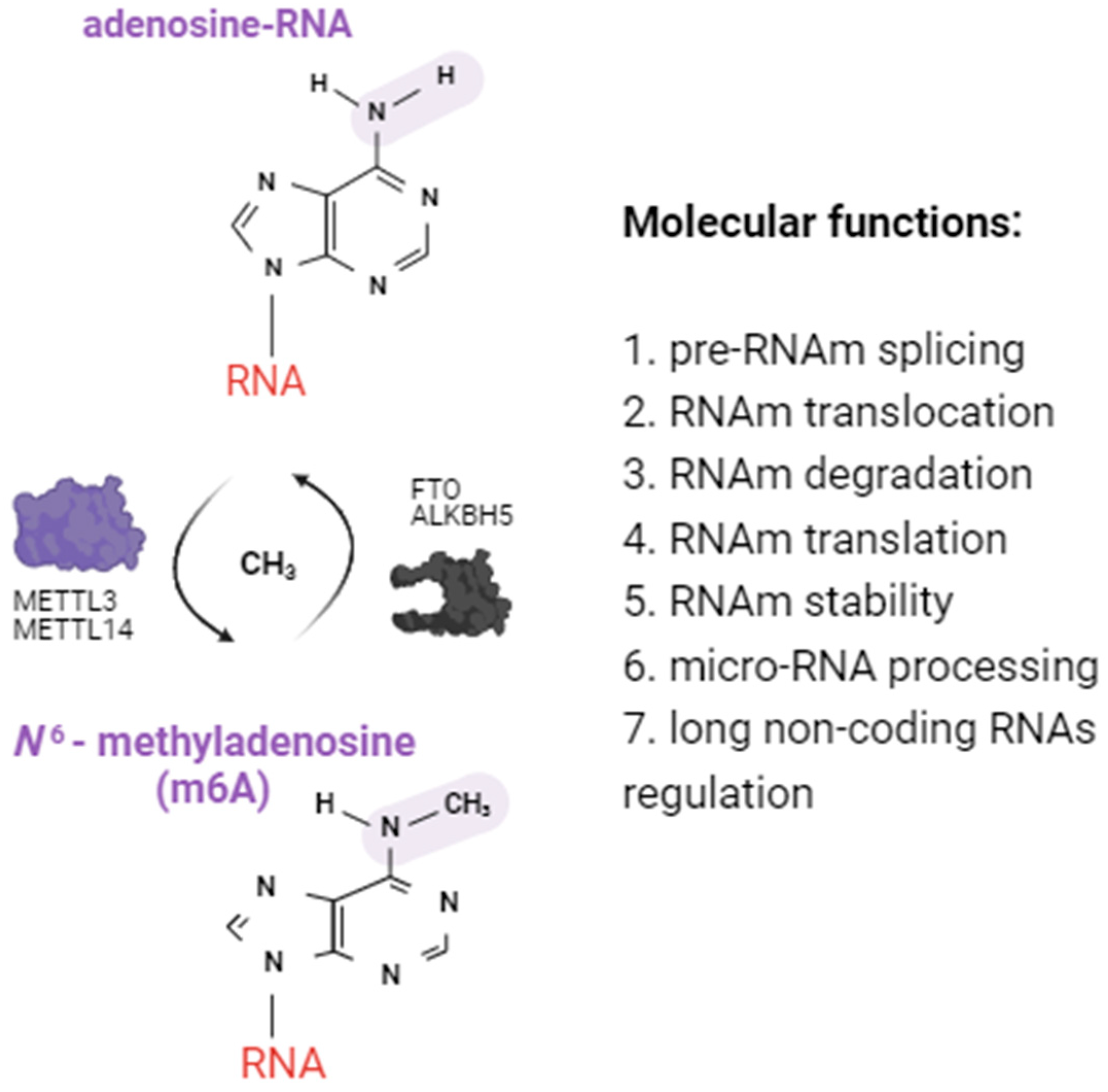
2.1. Alterations of m6A RNA Methylation Patterns

RNA methylation and demethylation are mediated by a series of enzymes, such as methyltransferases (or “writers” that transfer methyl groups to the nitrogen 6 of the adenosine of the RNA) and demethylases (or “erasers” that remove methyl groups), and have been associated with multiple functions within the cell such as RNA splicing, translocation, degradation, and stability, as well as the translation and regulation of non-coding RNAs [34] (Figure 1). Diabetic patients show alterations in the m6A methylation machinery in both immune and beta cells, which could be aggravated by SARS-CoV-2 infection, though the information available is scarce.

2.1.1. Alterations in Immune Cells from Diabetic Patients

Information regarding alterations to RNA methylation in immune cells from diabetic patients is scarce. One study by Wang et al. [38] reported that the genes involved in the regulation of m6A modifications showed notable changes in patients with T1DM (n = 6) compared to healthy individuals (n = 6). Specifically, the writer methyltransferase-like 3 (METTL3) and the reader insulin-like growth factor 2 binding protein 2 (IGF2BP2) showed decreased expression, while the readers YTH N6-methyladenosine RNA binding protein C1 (YTHDC1) and the heterogeneous nuclear ribonucleoprotein A2B1 (HNRNPA2B1) had increased expression. In addition, a microarray analysis showed that hypermethylated transcripts were enriched in the Janus kinase/signal transducers and activators of the transcription (JAK/STAT) signaling pathway, hypomethylated transcripts were enriched in the phosphatidylinositol 3-kinase/protein kinase B (PI3K/AKT) signaling pathway, and both methylation patterns were enriched in the mitogen-activated protein kinase (MAPK) pathway. Another study carried out by Yang et al. [45] reported a reduced level of m6A methylation in white blood cells from individuals with T2DM (n = 102) compared to healthy patients (n = 107). That study also revealed an increased expression of the demethylase fat mass and obesity-associated protein (FTO), and variations in expression levels of other m6A methyl-esterases. Specifically, there were elevated levels of METTL3, METTL14, and Wilms’ tumor 1-associating protein (WTAP) in T2DM patients compared to healthy individuals. Although more studies are needed, these findings suggest potential associations between m6A methylation and demethylation processes and the development of T1DM and T2DM.

2.1.2. Alterations in Beta Cells from Diabetic Patients

Research has shown that METTL3, a key component of the m6A methyltransferase complex, is crucial for maintaining beta cell function. Li and coworkers [46] demonstrated that a decrease in METTL3 under inflammatory and oxidative stress conditions leads to beta cell failure and hyperglycemia. This is likely due to decreased m6A modifications and the reduced expression of insulin secretion-related genes. Similarly, Liu et al. [47] found that the deletion of METTL14 results in glucose intolerance, decreased beta cell mass, and impaired insulin secretion. Furthermore, Cheng and coworkers [48] showed that METTL3 ameliorates the methylglyoxal-induced impairment of insulin secretion in pancreatic beta cells by regulating MAF BZIP transcription factor A (MAFA) expression. This suggests that METTL3 is a potential drug target for the treatment of beta cell failure in diabetes. Other authors have shown that FTO, a gene product associated with obesity and diabetes, plays a crucial role in maintaining beta cell function. Russell and Morgan [49] found that FTO protein enhances glucose-induced insulin secretion in pancreatic beta cells. However, Fan et al. [50] discovered that FTO overexpression inhibited insulin secretion and promoted nuclear factor kappa B (NF-κB) activation, potentially contributing to beta cell dysfunction.
In the context of T2DM, the impact of m6A methylation on the regulation of the biological function(s) of human beta cells has been recognized. m6A-sequencing of human T2DM islets revealed several hypomethylated transcripts involved in cell cycle progression and insulin secretion [35]. Moreover, beta cell-specific Mettl14 knockout mice, which displayed reduced m6A methylation levels, mimicked the islet phenotype in human T2DM with early diabetes onset and mortality due to decreased beta cell proliferation and insulin degranulation [35]. Decreased levels of METTL3, METTL14, alkB homolog 5 (ALKBH5), and YTH N6-methyladenosine RNA binding protein F1 (YTHDF1) in beta cells from individuals with T2DM (n = 7) compared to non-diabetic counterparts (n = 5) were reported before the COVID-19 pandemic [35]. Additionally, both METTL3 and METTL14 protein levels were reduced in the entire islets of T2DM patients. These findings suggest that the m6A methylation content might serve as a specific biomarker for predicting the risk of T2DM and its associated complications. However, it is crucial to note that these results warrant replication and validation in larger populations and through diverse experiments.

2.1.3. Alterations in m6A Methylation Induced by mRNA Vaccines

Some studies suggest a potential association between COVID-19 vaccination and the risk of causing elevated blood glucose or an exacerbation of pre-existing diabetes [51]. It has been reported that COVID-19 mRNA vaccination can induce T1DM in some individuals with a genetic predisposition [52], even in adults [53]. According to Alsudais et al. [52], eight studies in 12 patients diagnosed with T1DM post-vaccination were analyzed. The Pfizer-BioNTech vaccine was the most commonly administered (7/12), followed by the Moderna mRNA-1273 vaccine (2/12), CoronaVac (1/12), ChAdox1-s (1/12), and Pfizer-BioNTech/CoronaVac (1/12). The diagnostic criteria of T1DM included low C-peptide levels, and positive antibodies (e.g., anti-glutamic acid decarboxylase antibodies, anti-GAD) and/or HbA1c levels. Only five patients had reliable HbA1c data before vaccination; however, all patients showed significantly higher HbA1c levels after vaccination with an average level of 9.96%. Fifty percent of the patients (6/12) developed T1DM after the second vaccine dose; diabetic ketoacidosis occurred in 41.7% of cases, mostly within eight days post-vaccination. Genetic susceptibility was found in 41.7% of patients, with notable mutations in RNA binding motif protein 45 (RBM45)/Major Histocompatibility Complex, Class II, DR Beta 1 (DRB1) and Major Histocompatibility Complex, Class II, DQ Beta 1 (HLA-DQB1). Some limitations of that review include the lack of crucial information such as baseline body mass index (BMI) and HbA1c levels in some case reports, making it difficult to identify vaccine-induced diabetes and link vaccination to T1DM. Therefore, the authors did not rule out the possibility of diabetes onset due to COVID-19 infection.
In another study, Moon et al. [53] reported a case of a 56-year-old woman who experienced hyperglycemia following the second dose of the COVID-19 mRNA vaccine (Moderna) despite no previous diabetes history. This is an important finding of T1DM development post-vaccination with an mRNA vaccine, and notably the oldest patient under such circumstances. Nevertheless, in a much larger cohort of individuals from Hong Kong, Xiong et al. [54] found no evidence of an increased risk of diabetes following COVID-19 vaccination (CoronaVac (Sinovac) n = 5760 and BNT162b2 (Pfizer-BioNTech) n = 4411). In the BNT162b2 group, 2109 diabetes cases occurred after SARS-CoV-2 infection, and infection was associated with a higher risk of diabetes, mainly T2DM, irrespective of variants, although the risk was lower with Omicron.
We previously reported that vaccinated individuals showed significantly lower m6A methylation levels than unvaccinated individuals (n = 8 and 52, respectively), and that differences in m6A methylation levels across variants in unvaccinated individuals were significant; however, no significant correlation was observed between m6A methylation levels and viral load (nucleocapsid gene expression) or age [36]. According to the study, a complete vaccination scheme denoted the administration of one or two doses (depending on the vaccine specifications) of authorized vaccines in Mexico, including AZD1222, CoronaVac, BNT162b2, Ad5-nCOV, and mRNA-1273, developed by AstraZeneca, Sinovac, Pfizer-BioNTech, CanSinoBio, and Moderna, respectively. Although variation in m6A levels across variants could not be explained by the vaccination status alone, this first report of potential vaccination-mediated m6A methylation is intriguing and warrants further investigation, in particular regarding the possibility that COVID-19 vaccines (though generally considered safe) may induce an autoimmune response in susceptible individuals by mediating a decrease in m6A levels.

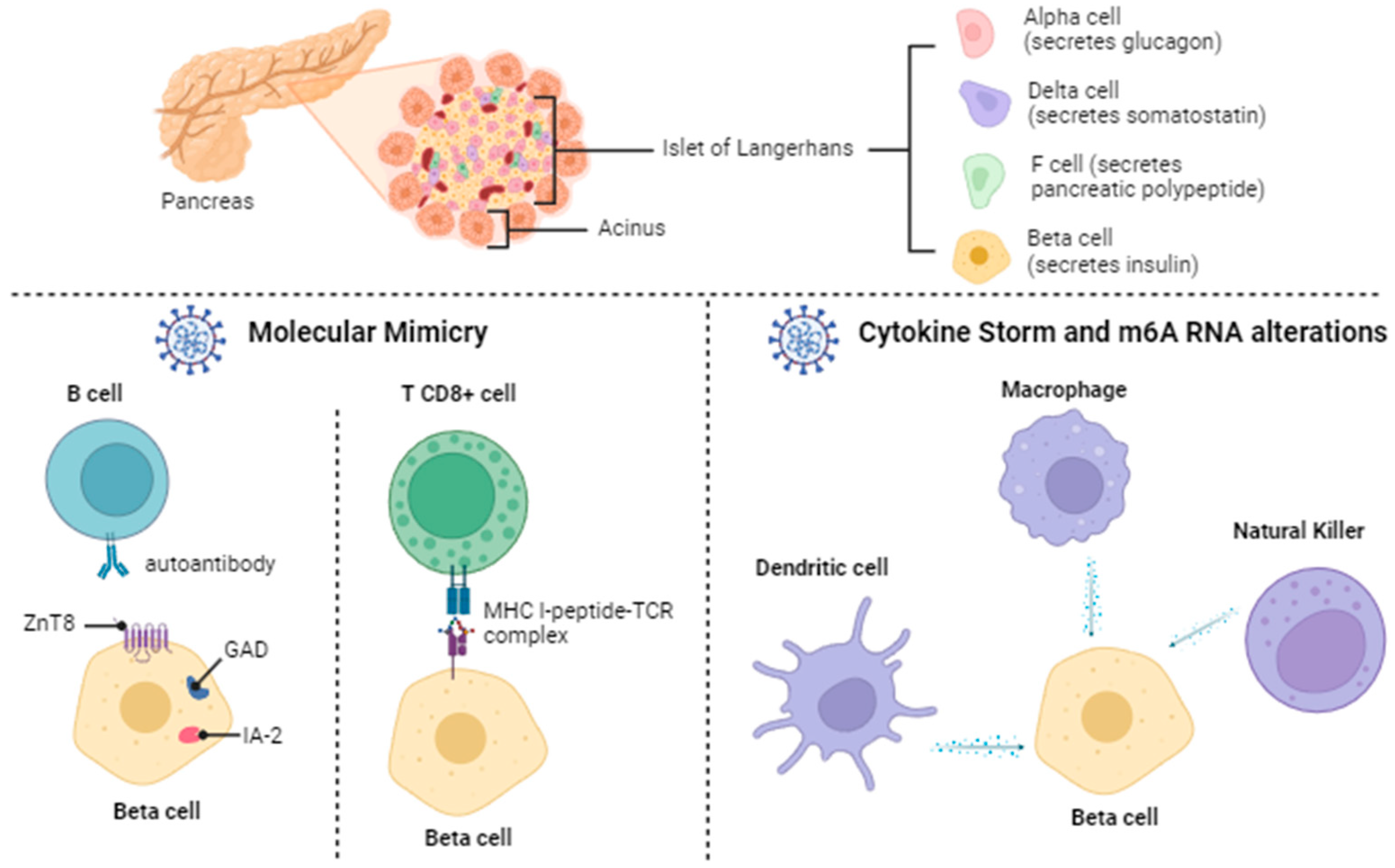
2.2. Diabetes Mellitus: Beta Pancreatic Cell Destruction and Dysfunction

T1DM is an autoimmune disease associated with the destruction of beta pancreatic cells. During the disease, several autoantibodies are produced such as those directed against insulin, tyrosine phosphatase, glutamic acid anti-decarboxylase enzymes, and the zinc transport protein [55]. The presence of these autoantibodies in an individual’s blood serum is an indication that the immune system is attacking the pancreatic beta cells that produce insulin [56,57]. The pathogenesis and progression of T1DM has been linked (among other factors) to viral infections. Viruses may lead to T1DM through a direct cytolytic activity on beta cells or by triggering autoimmune responses against beta cells.
Beta cell dysfunction and insulin resistance are key factors in the pathogenesis of T2DM [58]. The interplay between beta cell dysfunction and insulin resistance is complex, with both states influencing each other and potentially exacerbating diabetes. Insulin resistance is caused by alterations in insulin receptors in the cells and precedes T2DM [58]. Reiterer et al. [59] proposed that SARS-CoV-2 might induce dysfunction in adipose tissue, leading to insulin resistance. The authors analyzed a COVID-19 patient cohort without prior metabolic conditions (n = 4102), observing new-onset insulin resistance and hyperglycemia in 47% of the patients. In addition, SARS-CoV-2 infection was found to elevate the expression of the RE1-silencing transcription factor (REST), influencing the transcriptional regulation of key metabolic factors (such as myeloperoxidase, apelin, and myostatin) and contributing to the disruption of glucose metabolism [60]. Beta cell dysfunction impairs insulin secretion and is a critical determinant of the disease. This dysfunction is influenced by genetic and metabolic abnormalities, which affect glucose homeostasis [58].
Currently, there is no evidence supporting the notion that SARS-CoV-2 infection during pregnancy results in permanent diabetes for mothers or their offspring through autoimmunity or beta cell destruction. The heightened prevalence of GDM observed during the pandemic is likely attributed to lifestyle changes during lockdown. Nevertheless, severe COVID-19 cases may contribute to the development of GDM by exacerbating glucose tolerance [61]. GDM and T2DM share insulin-related challenges, affecting both obese and lean women. Obesity contributes to pre-existing insulin resistance, while lean women predominantly face impaired first-phase insulin secretion. Pregnancy-induced insulin resistance is influenced by factors such as placental hormones and pro-inflammatory cytokines. Ultimately, by the third trimester, insulin resistance during pregnancy reaches levels similar to T2DM [62].

2.3. Direct SARS-CoV-2 Invasion of Beta Cells

The expression of ACE2 in the pancreas (mainly in islet cells) is higher than in the lungs, and ACE2 is overexpressed in diabetic/hyperglycemic islets compared to non-diabetic/normoglycemic [63,64], so it is possible that SARS-CoV-2 could bind to this receptor and enter beta cells, producing cellular dysfunction and acute hyperglycemia [65,66,67]. Viral replication within beta cells can lead to their destruction, reducing insulin production. SARS-CoV-2 can cause direct damage to the pancreas, potentially inducing T1DM in previously non-diabetic subjects [68,69,70]. Even though this is a non-cytolytic virus, it has been reported that SARS-CoV-2 could induce cell injury mediated by different cell death mechanisms such as apoptosis, autophagy, and necrosis [71]. These mechanisms triggered by SARS-CoV-2 might facilitate viral clearance as part of the host’s antiviral immunity and also contribute to virus-induced tissue injuries and disease progression. SARS-CoV-2 may negatively affect human pancreatic islet function and survival by creating inflammatory conditions, which may in turn lead to metabolic abnormalities observed in patients with COVID-19. A direct tropism of SARS-CoV-2 for beta cells was suggested with the detection of SARS-CoV-2-specific viral RNA from pancreatic sections of newly hyperglycemic deceased patients who had COVID-19 [72]. The diabetogenic effect induced by SARS-CoV-2 infection can also be mediated by a possible direct viral cytotoxic mechanism against human pancreatic islets [72].

2.4. Immune Responses (Autoimmunity and Inflammation) Induced by SARS-CoV-2

The autoimmune responses induced by viral infections are mediated by peripheral blood mononuclear cells (PBMCs) such as T, B, natural killer (NK), dendritic cells (DCs), and monocytes. These responses are attributed to the structural similarity between viral antigens and motifs found in proteins of beta cells [73]. Boddu et al. [13] proposed that antigen exposure triggers the activation of autoreactive lymphocytes, initiating an autoimmune response. Then, an autoimmune reaction progresses to the destruction of the remaining beta cell mass, leading to the onset of insulin-dependent T1DM. This theory may not fully elucidate the immediate onset of diabetes during the acute phase of COVID-19 infection. However, it could explain the later emergence of diabetes in the weeks or months following recovery from COVID-19 [74]. Figure 2 shows the possible mechanisms of beta cell destruction induced by SARS-CoV-2 infection.

2.4.1. Molecular Mimicry

The spike protein from SARS-CoV-2 contains mimotopes that resemble human antigen epitopes, potentially leading to antigenic mimicry and the activation of immune receptors [76]. Computational methods revealed that the spike protein of SARS-CoV-2 contains clusters of molecular mimics with significant autoimmune potential. Within these clusters, multiple molecular mimics share motifs that are recurrently present in the human proteome [77]. This computational study provided insights into the autoimmune potential of SARS-CoV-2 for a therapeutic intervention or vaccine design, to avoid any autoimmune interference. During SARS-CoV-2 infections, the host immune system can activate expression of the apolipoprotein B editing catalytic polypeptide 3G and F (APOBEC3G/F) and adenosine deaminase RNA1-specific (ADAR1) genes, which leads to editing the viral RNA and host transcriptome. This response can trigger the production of autoantigens, perceived as foreign by the immune system, and therefore promotes the production of autoantibodies and potentially induces transient or chronic autoimmune diseases [78]. Thus, the viral infection could activate T cells that recognize pancreatic antigens, leading to an autoimmune attack on beta cells and the subsequent development of T1DM. Autoimmunity is also related to molecular mimicry between peptides of the SARS-CoV-2 spike protein and human antigens mainly expressed by endocrine glands such as the pituitary, adrenal [79], thyroid, and pancreas [80]. It is possible that the similarity of spike peptides with the membrane receptors in pancreatic beta cells in humans could be related to the development of T1DM in previously non-diabetic patients.
Moody et al. [75] employed in silico immuno-informatic tools to predict B-cell epitopes of eight SARS-CoV-2 variants and the original Wuhan variant. Subsequently, they studied the similarity between the predicted B-cell epitopes and human proteins. The authors reported an association between human proteins with sequences shared with SARS-CoV-2 and autoimmune diseases such as T1DM, systemic lupus erythematosus (SLE), and multiple sclerosis (MS), among others. For example, histone H3 shares an identical six amino acid sequence with the SARS-CoV-2 protein Orf8 and is in a region identified as an epitope in COVID-19 patients. Furthermore, autoantibody-targeting proteins found in autoimmune diseases, such as SLE and T1DM, among others, share similar sequences with some of the new predicted epitopes of SARS-CoV-2. Additionally, the authors identified 11 new predicted B-cell epitopes in host proteins, explaining key aspects of the extrapulmonary pathology of COVID-19. In another study, de Oliveira et al. [81] reported the potential similarity between the amino acid sequences of human insulin (4F0N) and glutamic acid decarboxylase-65 (GAD65) (2OKK) with SARS-CoV-2 proteins, such as the spike protein (6ZB5), to explain the possible trigger of T1DM. The authors found a sequence identity from 5 to 45.45%. The data suggest a possible pathogenic link between T1DM and SARS-CoV-2. Collectively, these studies (Supplementary Table S1) suggest that molecular mimicry may play a significant role in the pathogenesis of autoimmune diseases such as T1DM in the context of SARS-CoV-2.

2.4.2. Cytokine Storm

A cytokine storm can lead to inflammation and immune system dysfunction, potentially contributing to the destruction of pancreatic beta cells. During severe COVID-19, there is an increased release of counter-regulatory hormones and proinflammatory cytokines, including interleukin 6 (IL-6) and tumoral necrosis factor alpha (TNF-alpha), collectively referred to as a cytokine storm. This overwhelming surge of cytokines is recognized for its ability to induce insulin resistance, leading to elevated blood sugar levels or hyperglycemia [82]. Viral infections in diabetic animal models trigger natural killer cells and T cells to release inflammatory cytokines, destroying beta cells [83]. In COVID-19 patients, a dual immune response occurs, with T helper (Th) 1 cells activated by interferon-gamma (IFN-gamma) and monocyte chemoattractant protein-1, and Th2 cells expressing IL-4 and IL-10 to suppress inflammation. Macrophage activation syndrome in COVID-19 involves elevated levels of IL-6, IL-1-beta, TNF-alpha, INF-gamma, and ferritin [84]. Additionally, Th17 cells are activated in the cytokine storm, releasing IL-17 and granulocyte-colony stimulating factor. Th17 cells are abundant in the pancreas of T1DM patients [85]. In T2DM, elevated IL-17 levels are linked to adipose tissue inflammation, regulating pro-inflammatory cytokines, and contributing to insulin resistance [86]. The cytokine storm induced by SARS-CoV-2 in diabetic patients exacerbates the systemic immune imbalance, potentially worsening their clinical condition [87,88].

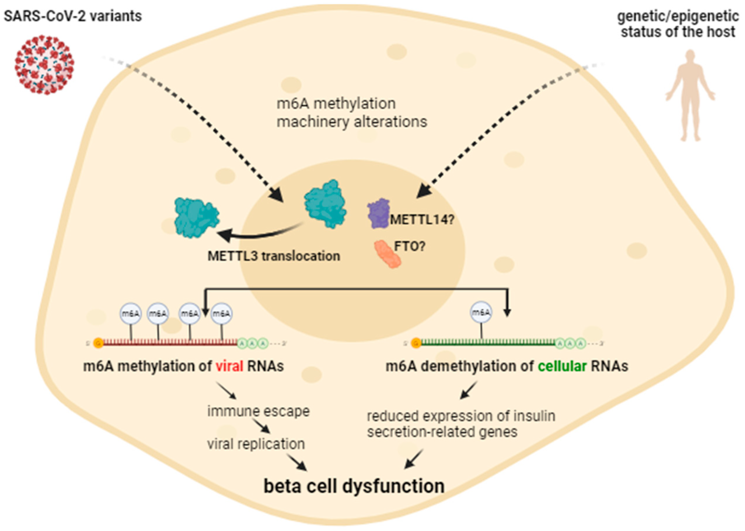
2.5. Alterations in m6A Methylation by Different SARS-CoV-2 Variants

Rangu et al. [89] suggested that new SARS-CoV-2 variants might have differential effects on the development of diabetes due to mutations associated with an alternative route of entry into beta cells, or the maintenance of higher infection loads and high levels of circulating cytokines. The authors recommended determining invasion mechanisms of SARS-CoV-2 variants in beta cells, as well as the differential effects on insulin secretion. The lack of reliable markers of in vivo beta cell death limits the studies of the potential effects induced by the different viral variants. Further research is needed to comprehensively investigate the functional consequences of changes in m6A RNA methylation patterns within beta cells caused by distinct SARS-CoV-2 variants.
We previously reported that the m6A levels of nasopharyngeal samples were significantly lower in individuals infected with SARS-CoV-2 variants delta and omicron compared to other variants and uninfected individuals [36]. Vaid et al. [90] reported that SARS-CoV-2 variants could cause a global loss of m6A methylation levels in cellular RNAs of air/liquid interface cultures of human airway epithelia, while viral RNA remains m6A-modified. METTL3 showed an unusual cytoplasmic localization post-infection. The B.1.351 variant presented a less pronounced effect on METTL3 localization and loss of m6A than the B.1 and the B.1.1.7 variants. Furthermore, transcripts with m6A modifications were preferentially downregulated post-infection [90]. Altogether, this information suggests that SARS-CoV-2-related m6A alterations could (hypothetically) lead to beta cell dysfunction (Figure 3). SARS-CoV-2 variants may differently alter the m6A methylation enzymatic machinery in pancreatic beta cells, affecting the global levels of m6A methylation of insulin-related gene transcripts. Further research is needed to confirm this hypothesis. On the other hand, the genetic background and the epigenetic status of the host pancreatic cells could also contribute to the outcome following viral infection. Epigenetic marks before viral infection may differ between individuals depending on lifestyle (e.g., physical activity or diet), previous viral infections, pharmacological treatments, and exposure to pollutants, among other factors.

3. Concluding Remarks

The onset of diabetes mellitus following COVID-19 infection appears to be influenced by a complex interplay of factors, including the emergence of SARS-CoV-2 variants, inflammation, molecular mimicry, and m6A RNA methylation. The evolving landscape of SARS-CoV-2 variants introduces new challenges in understanding their potential impact on the development of diabetes. Molecular mimicry, wherein viral components resemble host tissues, may contribute to autoimmune responses triggering diabetes in susceptible individuals. Additionally, the involvement of m6A RNA methylation, a crucial epigenetic modification, suggests a potential role in dysregulating gene expression and insulin sensitivity. As research continues, it is essential to unravel the intricate mechanisms linking COVID-19 to diabetes onset. This understanding would guide public health strategies, early detection, and targeted interventions to mitigate the long-term consequences of the pandemic on metabolic health. Furthermore, an interdisciplinary collaboration between virologists, immunologists, geneticists, and epigenetic researchers is crucial for a comprehensive grasp of the multifaceted relationship between COVID-19 and diabetes mellitus. Addressing these complexities will not only enhance our understanding of pathophysiology but will also pave the way for more effective prevention and management strategies for individuals at risk. Further research related to global, viral, and host RNA methylation is required, as well as in vivo trials assessing beta cell death, auto-antibody titers, insulin production and secretion, and the expression of insulin-related genes, pro-inflammatory cytokines, and RNA methylation-related genes.

Supplementary Materials

The following supporting information can be downloaded at https://www.mdpi.com/article/10.3390/covid4040032/s1, Table S1: Molecular mimicry of SARS-CoV-2 and the pathogenesis of autoimmune diseases.

Author Contributions

J.L.B.-R., M.M.-C. and A.G.-G.: preparation, editing, and revision of the manuscript. D.M. and G.L.: deep revision of the manuscript, according to their main areas of expertise. All authors have read and agreed to the published version of the manuscript.

Funding

This research received no external funding.

Institutional Review Board Statement

Not applicable.

Informed Consent Statement

Not applicable.

Data Availability Statement

No new data were created or analyzed in this study. Data sharing is not applicable to this article.

Acknowledgments

The authors would like to thank the National Council for Humanities, Science and Technology (CONAHCYT) for the doctoral scholarships granted to J.L.B.-R. and M.M.-C.

Conflicts of Interest

The authors declare no conflicts of interest.

References

  1. Gadanec, L.K.; McSweeney, K.R.; Qaradakhi, T.; Ali, B.; Zulli, A.; Apostolopoulos, V. Can SARS-CoV-2 Virus Use Multiple Receptors to Enter Host Cells? Int. J. Mol. Sci. 2021, 22, 992. [Google Scholar] [CrossRef]
  2. Becerra-Flores, M.; Cardozo, T. SARS-CoV-2 viral spike G614 mutation exhibits higher case fatality rate. Int. J. Clin. Pract. 2020, 74, e13525. [Google Scholar] [CrossRef] [PubMed]
  3. Davies, N.G.; Abbott, S.; Barnard, R.C.; Jarvis, C.I.; Kucharski, A.J.; Munday, J.D.; Pearson, C.A.B.; Russell, T.W.; Tully, D.C.; Washburne, A.D.; et al. Estimated transmissibility and impact of SARS-CoV-2 lineage B.1.1.7 in England. Science 2021, 372, eabg3055. [Google Scholar] [CrossRef] [PubMed]
  4. Lin, L.; Liu, Y.; Tang, X.; He, D. The Disease Severity and Clinical Outcomes of the SARS-CoV-2 Variants of Concern. Front. Public Health. 2021, 9, 775224. [Google Scholar] [CrossRef]
  5. Sanyaolu, A.; Okorie, C.; Marinkovic, A.; Haider, N.; Abbasi, A.F.; Jaferi, U.; Prakash, S.; Balendra, V. The emerging SARS-CoV-2 variants of concern. Ther. Adv. Infect. Dis. 2021, 8, 20499361211024372. [Google Scholar] [CrossRef]
  6. CDC—Centers for Disease Control and Prevention. Available online: https://www.cdc.gov/coronavirus/2019-ncov/variants/variant-classifications.html#anchor_1632237683347 (accessed on 3 January 2024).
  7. Álvarez-Santacruz, C.; Tyrkalska, S.D.; Candel, S. The Microbiota in Long COVID. Int. J. Mol. Sci. 2024, 25, 1330. [Google Scholar] [CrossRef] [PubMed]
  8. Baj, J.; Karakuła-Juchnowicz, H.; Teresiński, G.; Buszewicz, G.; Ciesielka, M.; Sitarz, R.; Forma, A.; Karakuła, K.; Flieger, W.; Portincasa, P.; et al. COVID-19: Specific and Non-Specific Clinical Manifestations and Symptoms: The Current State of Knowledge. J. Clin. Med. 2020, 9, 1753. [Google Scholar] [CrossRef]
  9. Davis, H.E.; Assaf, G.S.; McCorkell, L.; Wei, H.; Low, R.J.; Re’em, Y.; Redfield, S.; Austin, J.P.; Akrami, A. Characterizing long COVID in an international cohort: 7 months of symptoms and their impact. EClinicalMedicine 2021, 38, 101019. [Google Scholar] [CrossRef]
  10. Xie, Y.; Xu, E.; Bowe, B.; Al-Aly, Z. Long-term cardiovascular outcomes of COVID-19. Nat. Med. 2022, 28, 583–590. [Google Scholar] [CrossRef]
  11. Kedor, C.; Freitag, H.; Meyer-Arndt, L.; Wittke, K.; Hanitsch, L.G.; Zoller, T.; Steinbeis, F.; Haffke, M.; Rudolf, G.; Heidecker, B.; et al. A prospective observational study of post-COVID-19 chronic fatigue syndrome following the first pandemic wave in Germany and biomarkers associated with symptom severity. Nat. Commun. 2022, 13, 101019. [Google Scholar] [CrossRef]
  12. Antar, S.A.; Ashour, N.A.; Sharaky, M.; Khattab, M.; Ashour, N.A.; Zaid, R.T.; Roh, E.J.; Elkamhawy, A.; Al-Karmalawy, A.A. Diabetes mellitus: Classification, mediators, and complications; A gate to identify potential targets for the development of new effective treatments. Biomed. Pharmacother. 2023, 168, 115734. [Google Scholar] [CrossRef] [PubMed]
  13. Boddu, S.K.; Aurangabadkar, G.; Kuchay, M.S. New onset diabetes, type 1 diabetes and COVID-19. Diabetes Metab. Syndr. 2020, 14, 2211–2217. [Google Scholar] [CrossRef] [PubMed]
  14. Pantea-Stoian, A.; Bica, I.C.; Salmen, T.; Al Mahmeed, W.; Al-Rasadi, K.; Al-Alawi, K.; Banach, M.; Banerjee, Y.; Ceriello, A.; Cesur, M.; et al. New-Onset Diabetes Mellitus in COVID-19: A Scoping Review. Diabetes Ther. 2024, 15, 33–60. [Google Scholar] [CrossRef] [PubMed]
  15. Ayoubkhani, D.; Khunti, K.; Nafilyan, V.; Maddox, T.; Humberstone, B.; Diamond, I.; Banerjee, A. Post-covid syndrome in individuals admitted to hospital with COVID-19: Retrospective cohort study. BMJ 2021, 372, n693. [Google Scholar] [CrossRef] [PubMed]
  16. Zhang, W.; Zhang, S.; Dong, C.; Guo, S.; Jia, W.; Jiang, Y.; Wang, C.; Zhou, M.; Gong, Y. A bibliometric analysis of RNA methylation in diabetes mellitus and its complications from 2002 to 2022. Front. Endocrinol. 2022, 13, 997034. [Google Scholar] [CrossRef] [PubMed]
  17. Chourasia, P.; Goyal, L.; Kansal, D.; Roy, S.; Singh, R.; Mahata, I.; Sheikh, A.B.; Shekhar, R. Risk of New-Onset Diabetes Mellitus as a Post-COVID-19 Condition and Possible Mechanisms: A Scoping Review. J. Clin. Med. 2023, 12, 1159. [Google Scholar] [CrossRef]
  18. Raveendran, A.V. COVID-19 and diabetes mellitus: Proposed diagnostic criteria for new-onset diabetes after COVID. Int. J. Diabetes Technol. 2022, 3, 101–103. [Google Scholar] [CrossRef]
  19. Ghosh, A.; Anjana, R.M.; Shanthi-Rani, C.S.; Jeba-Rani, S.; Gupta, R.; Jha, A.; Gupta, V.; Kuchay, M.S.; Luthra, A.; Durrani, S.; et al. Glycemic parameters in patients with new-onset diabetes during COVID-19 pandemic are more severe than in patients with new-onset diabetes before the pandemic: NOD COVID India Study. Diabetes Metab. Syndr. 2021, 15, 215–220. [Google Scholar] [CrossRef]
  20. Rathmann, W.; Kuss, O.; Kostev, K. Incidence of newly diagnosed diabetes after COVID-19. Diabetologia 2022, 65, 949–954. [Google Scholar] [CrossRef]
  21. Qeadan, F.; Tingey, B.; Egbert, J.; Pezzolesi, M.G.; Burge, M.R.; Peterson, K.A.; Honda, T. The associations between COVID-19 diagnosis, type 1 diabetes, and the risk of diabetic ketoacidosis: A nationwide cohort from the US using the Cerner Real-World Data. PLoS ONE 2022, 17, e0266809. [Google Scholar] [CrossRef]
  22. Xie, Y.; Al-Aly, Z. Risks and burdens of incident diabetes in long COVID: A cohort study. Lancet Diabetes Endocrinol. 2022, 10, 311–321. [Google Scholar] [CrossRef] [PubMed]
  23. Ssentongo, P.; Zhang, Y.; Witmer, L.; Chinchilli, V.M.; Ba, D.M. Association of COVID-19 with diabetes: A systematic review and meta-analysis. Sci. Rep. 2022, 12, 1–8. [Google Scholar] [CrossRef]
  24. Lai, H.; Yang, M.; Sun, M.; Pan, B.; Wang, Q.; Wang, J.; Tian, J.; Ding, G.; Yang, K.; Song, X.; et al. Risk of incident diabetes after COVID-19 infection: A systematic review and meta-analysis. Metabolism 2022, 137, 155330. [Google Scholar] [CrossRef] [PubMed]
  25. Li, J.; Li, Y.; Wang, Z.; Liu, N.; He, L.; Zhang, H. Increased risk of new-onset diabetes in patients with COVID-19: A systematic review and meta-analysis. Front. Public Health 2023, 11, 1170156. [Google Scholar] [CrossRef] [PubMed]
  26. Zanardo, V.; Tortora, D.; Sandri, A.; Severino, L.; Mesirca, P.; Straface, G. COVID-19 pandemic: Impact on gestational diabetes mellitus prevalence. Diabetes Res. Clin. Pract. 2022, 183, 109149. [Google Scholar] [CrossRef] [PubMed]
  27. Rhou, Y.J.J.; Elhindi, J.; Melov, S.J.; Cheung, N.W.; Pasupathy, D. Western Sydney COVID-19 Pregnancy Study Group. Indirect effects of the COVID-19 pandemic on risk of gestational diabetes and factors contributing to increased risk in a multiethnic population: A retrospective cohort study. BMC Pregnancy Childbirth 2023, 23, 341. [Google Scholar] [CrossRef] [PubMed]
  28. Maharat, M.; Sajjadi, S.F.; Moosavian, S.P. Changes in dietary habits and weight status during the COVID-19 pandemic and its association with socioeconomic status among Iranians adults. Front. Public Health 2023, 10, 1080589. [Google Scholar] [CrossRef] [PubMed]
  29. Chen, Z.; Khandpur, N.; Desjardins, C.; Wang, L.; Monteiro, C.A.; Rossato, S.L.; Fung, T.T.; Manson, J.E.; Willett, W.C.; Rimm, E.B.; et al. Ultra-Processed Food Consumption and Risk of Type 2 Diabetes: Three Large Prospective U.S. Cohort Studies. Diabetes Care 1 2023, 46, 1335–1344. [Google Scholar] [CrossRef] [PubMed]
  30. Ghosal, S.; Arora, B.; Dutta, K.; Ghosh, A.; Sinha, B.; Misra, A. Increase in the risk of type 2 diabetes during lockdown for the COVID19 pandemic in India: A cohort analysis. Diabetes Metab. Syndr. 2020, 14, 949–952. [Google Scholar] [CrossRef]
  31. Silva, F.M.; Duarte-Mendes, P.; Carvalho, E.; Soares, C.M.; Farinha, C.; Serrano, J.; Paulo, R.; Massart, A.; Rodrigues, R.N.; Teixeira, A.M.; et al. Effects of combined training during the COVID-19 pandemic on metabolic health and quality of life in sedentary workers: A randomized controlled study. Front. Public Health 2022, 10, 1040714. [Google Scholar] [CrossRef]
  32. Kim, S.H.; Arora, I.; Hsia, D.S.; Knowler, W.C.; LeBlanc, E.; Mylonakis, E.; Pratley, R.; Pittas, A.G. New-Onset Diabetes After COVID-19. J. Clin. Endocrinol. Metab. 2023, 108, e1164–e1174. [Google Scholar] [CrossRef] [PubMed]
  33. Guo, Y.C.; Cao, H.D.; Lian, X.F.; Wu, P.X.; Zhang, F.; Zhang, H.; Lu, D.H. Molecular mechanisms of noncoding RNA and epigenetic regulation in obesity with consequent diabetes mellitus development. World J. Diabetes 2023, 14, 1621–1631. [Google Scholar] [CrossRef] [PubMed]
  34. Pan, T. N6-methyl-adenosine modification in messenger and long non-coding RNA. Trends Biochem. Sci. 2013, 38, 204–209. [Google Scholar] [CrossRef] [PubMed]
  35. De Jesus, D.F.; Zhang, Z.; Kahraman, S.; Brown, N.K.; Chen, M.; Hu, J.; Gupta, M.K.; He, C.; Kulkarni, R.N. m6A mRNA Methylation Regulates Human β-Cell Biology in Physiological States and in Type 2 Diabetes. Nat. Metab. 2019, 1, 765–774. [Google Scholar] [CrossRef] [PubMed]
  36. Batista-Roche, J.L.; Gómez-Gil, B.; Lund, G.; Berlanga-Robles, C.A.; García-Gasca, A. Global m6A RNA Methylation in SARS-CoV-2 Positive Nasopharyngeal Samples in a Mexican Population: A First Approximation Study. Epigenomes 2022, 6, 16. [Google Scholar] [CrossRef] [PubMed]
  37. Qian, W.; Zhou, J.; Duan, L.; Wang, H.; Xu, S.; Cao, Y. m6A methylation: A potential key player in understanding and treating COVID-2019 infection. Cell Death Discov. 2023, 9, 300. [Google Scholar] [CrossRef] [PubMed]
  38. Wang, Y.; Xu, L.; Luo, S.; Sun, X.; Li, J.; Pang, H.; Zhou, J.; Zhou, Y.; Shi, X.; Li, X.; et al. The m6A methylation profiles of immune cells in type 1 diabetes mellitus. Front. Immunol. 2022, 13, 1030728. [Google Scholar] [CrossRef]
  39. Oldstone, M.B.A.; Schwimmbeck, P.; Dyrberg, T.; Fujinami, R. Mimicry by Virus of Host Molecules: Implications for Autoimmune Disease. In Progress in Immunology VI; Cinader, B., Miller, R.G., Eds.; Academic Press Inc.: Orlando, FL, USA, 1986; pp. 787–795. [Google Scholar] [CrossRef]
  40. Cusick, M.F.; Libbey, J.E.; Fujinami, R.S. Molecular mimicry as a mechanism of autoimmune disease. Clin. Rev. Allergy Immunol. 2012, 42, 102–111. [Google Scholar] [CrossRef] [PubMed]
  41. Coppieters, K.T.; Wiberg, A.; von Herrath, M.G. Viral infections and molecular mimicry in type 1 diabetes. APMIS 2012, 120, 941–949. [Google Scholar] [CrossRef]
  42. Kovačić, D.; Jotanović, J.; Laković, J. The possible role of molecular mimicry in SARS-CoV-2-mediated autoimmunity: An immunobiochemical basis. J. Med. Sci. 2021, 90, 137–156. [Google Scholar] [CrossRef]
  43. Kanduc, D.; Shoenfeld, Y. Molecular mimicry between SARS-CoV-2 spike glycoprotein and mammalian proteomes: Implications for the vaccine. Immunol. Res. 2020, 68, 310–313. [Google Scholar] [CrossRef] [PubMed]
  44. Cappello, F.; Gammazza, A.M.; Dieli, F.; de Macario, E.C.; Macario, A.J. Does SARS-CoV-2 Trigger Stress-InducedAutoimmunity by Molecular Mimicry? A Hypothesis. J. Clin. Med. 2020, 9, 2038. [Google Scholar] [CrossRef] [PubMed]
  45. Yang, Y.; Shen, F.; Huang, W.; Qin, S.; Huang, J.T.; Sergi, C.; Yuan, B.F.; Liu, S.M. Glucose is involved in the dynamic regulation of M6A in patients with type 2 diabetes. J. Clin. Endocrinol. Metab. 2019, 104, 665–673. [Google Scholar] [CrossRef] [PubMed]
  46. Li, X.; Jiang, Y.; Sun, X.; Wu, Y.; Chen, Z. METTL3 is required for maintaining β-cell function. Metabolism 2021, 116, 154702. [Google Scholar] [CrossRef] [PubMed]
  47. Liu, J.; Luo, G.; Sun, J.; Men, L.; Ye, H.; He, C.; Ren, D. METTL14 is essential for β-cell survival and insulin secretion. Biochim. Biophys. Acta Mol. Basis Dis. 2019, 1865, 2138–2148. [Google Scholar] [CrossRef]
  48. Cheng, Y.; Yao, X.M.; Zhou, S.M.; Sun, Y.; Meng, X.J.; Wang, Y.; Xing, Y.J.; Wan, S.J.; Hua, Q. The m6A Methyltransferase METTL3 Ameliorates Methylglyoxal-Induced Impairment of Insulin Secretion in Pancreatic β Cells by Regulating MafA Expression. Front. Endocrinol. 2022, 13, 1664–2392. [Google Scholar] [CrossRef] [PubMed]
  49. Russell, M.A.; Morgan, N.G. Conditional expression of the FTO gene product in rat INS-1 cells reveals its rapid turnover and a role in the profile of glucose-induced insulin secretion. Clin. Sci. 2011, 120, 403–413. [Google Scholar] [CrossRef]
  50. Fan, H.Q.; He, W.; Xu, K.F.; Wang, Z.X.; Xu, X.Y.; Chen, H. FTO Inhibits Insulin Secretion and Promotes NF-κB Activation through Positively Regulating ROS Production in Pancreatic β cells. PLoS ONE 2015, 10, e0127705. [Google Scholar] [CrossRef]
  51. He, Y.F.; Ouyang, J.; Hu, X.D.; Wu, N.; Jiang, Z.G.; Bian, N.; Wang, J. Correlation between COVID-19 vaccination and diabetes mellitus: A systematic review. World J. Diabetes. 2023, 14, 892–918. [Google Scholar] [CrossRef]
  52. Alsudais, A.S.; Alkanani, R.S.; Fathi, A.B.; Almuntashiri, S.S.; Jamjoom, J.N.; Alzhrani, M.A.; Althubaiti, A.; Radi, S. Autoimmune diabetes mellitus after COVID-19 vaccination in adult population: A systematic review of case reports. BMC Endocr. Disord. 2023, 23, 164. [Google Scholar] [CrossRef]
  53. Moon, H.; Suh, S.; Park, M.K. Adult-Onset Type 1 Diabetes Development Following COVID-19 mRNA Vaccination. J. Korean Med. Sci. 2023, 38, e12. [Google Scholar] [CrossRef] [PubMed]
  54. Xiong, X.; Lui, D.T.W.; Chung, M.S.H.; Au, I.C.H.; Lai, F.T.T.; Wan, E.Y.F.; Chui, C.S.L.; Li, X.; Cheng, F.W.T.; Cheung, C.L.; et al. Incidence of diabetes following COVID-19 vaccination and SARS-CoV-2 infection in Hong Kong: A population-based cohort study. PLoS Med. 2023, 20, e1004274. [Google Scholar] [CrossRef] [PubMed]
  55. Vera, F.; Jofré, P.; Rodríguez, D.; Lagos, R.; Quiroga, T.; Grassi, B.; Pollak, F. Marcadores inmunológicos de diabetes tipo 1: Análisis de 4.164 perfiles en un centro de salud de Chile. Rev. Méd. Chile 2021, 149, 330–338. [Google Scholar] [CrossRef] [PubMed]
  56. Hayes-Dorado, J.P. Diabetes mellitus tipo 1. Rev. Bol. Ped. 2008, 47, 90–96. Available online: http://www.scielo.org.bo/scielo.php?script=sci_arttext&pid=S1024-06752008000200006 (accessed on 15 February 2024).
  57. Kokuina, E.; Rozada Galardy, L.; Vilar MD, L.N.; Tamargo Barbeito, T.O.; Infante Amoros, A.; Dalas Giber, M. Autoanticuerpos anti-insulina en la diabetes tipo 1. Rev. Cuba. Med. 2022, 61, e2637. Available online: https://pesquisa.bvsalud.org/portal/resource/e/biblio-1408993 (accessed on 15 February 2024).
  58. Cerf, M.E. Beta cell dysfunction and insulin resistance. Front. Endocrinol. 2013, 4, 37. [Google Scholar] [CrossRef] [PubMed]
  59. Reiterer, M.; Rajan, M.; Gómez-Banoy, N.; Lau, J.D.; Gomez-Escobar, L.G.; Ma, L.; Gilani, A.; Alvarez-Mulett, S.; Sholle, E.T.; Chandar, V.; et al. Hyperglycemia in acute COVID-19 is characterized by insulin resistance and adipose tissue infectivity by SARS-CoV-2. Cell Metab. 2021, 33, 2174–2188.e5. [Google Scholar] [CrossRef] [PubMed]
  60. He, X.; Liu, C.; Peng, J.; Li, Z.; Li, F.; Wang, J.; Hu, A.; Peng, M.; Huang, K.; Fan, D.; et al. COVID-19 induces new-onset insulin resistance and lipid metabolic dysregulation via regulation of secreted metabolic factors. Signal Transduct. Target. Ther. 2021, 6, 1–12. [Google Scholar] [CrossRef]
  61. Kleinwechter, H.J.; Weber, K.S.; Liedtke, T.P.; Schäfer-Graf, U.; Groten, T.; Rüdiger, M.; Pecks, U. COVID-19, Pregnancy, and Diabetes Mellitus. Z. Geburtshilfe Neonatol. 2024, 228, 17–31. [Google Scholar] [CrossRef]
  62. Modzelewski, R.; Stefanowicz-Rutkowska, M.M.; Matuszewski, W.; Bandurska-Stankiewicz, E.M. Gestational Diabetes Mellitus-Recent Literature Review. J. Clin. Med. 2022, 11, 5736. [Google Scholar] [CrossRef]
  63. Taneera, J.; El-Huneidi, W.; Hamad, M.; Mohammed, A.K.; Elaraby, E.; Hachim, M.Y. Expression Profile of SARS-CoV-2 Host Receptors in Human Pancreatic Islets Revealed Upregulation of ACE2 in Diabetic Donors. Biology 2020, 9, 215. [Google Scholar] [CrossRef] [PubMed]
  64. Fignani, D.; Licata, G.; Brusco, N.; Nigi, L.; Grieco, G.E.; Marselli, L.; Overbergh, L.; Gysemans, C.; Colli, M.L.; Marchetti, P.; et al. SARS-CoV-2 Receptor Angiotensin I-Converting Enzyme Type 2 (ACE2) Is Expressed in Human Pancreatic β-Cells and in the Human Pancreas Microvasculature. Front. Endocrinol. 2020, 11, 596898. [Google Scholar] [CrossRef] [PubMed]
  65. Liu, F.; Long, X.; Zhang, B.; Zhang, W.; Chen, X.; Zhang, Z. ACE2 expression in pancreas may cause pancreatic damage after SARS-CoV-2 infection. Clin. Gastroenterol. Hepatol. 2020, 18, 2128–2130. [Google Scholar] [CrossRef] [PubMed]
  66. Wu, C.T.; Lidsky, P.V.; Xiao, Y.; Lee, I.T.; Cheng, R.; Nakayama, T.; Jiang, S.; Demeter, J.; Bevacqua, R.J.; Chang, C.A.; et al. SARS-CoV-2 infects human pancreatic β cells and elicits β cell impairment. Cell Metab. 2021, 33, 1565–1576.e5. [Google Scholar] [CrossRef] [PubMed]
  67. Müller, J.A.; Groß, R.; Conzelmann, C.; Krüger, J.; Merle, U.; Steinhart, J.; Weil, T.; Koepke, L.; Bozzo, C.P.; Read, C.; et al. SARS-CoV-2 infects and replicates in cells of the human endocrine and exocrine pancreas. Nat. Metab. 2021, 3, 149–165. [Google Scholar] [CrossRef] [PubMed]
  68. Rubino, F.; Amiel, S.A.; Zimmet, P.; Alberti, G.; Bornstein, S.; Eckel, R.H. New-onset diabetes in COVID-19. N. Engl. J. Med. 2020, 383, 789–790. [Google Scholar] [CrossRef]
  69. Lima-Martínez, M.M.; Carrera Boada, C.; Madera-Silva, M.D.; Marín, W.; Contreras, M. COVID-19 and diabetes: A bidirectional relationship. COVID-19 y diabetes mellitus: Una relación bidireccional. Clin. Investig. Arterioscler. 2021, 33, 151–157. [Google Scholar] [CrossRef]
  70. Mine, K.; Nagafuchi, S.; Mori, H.; Takahashi, H.; Anzai, K. SARS-CoV-2 Infection and Pancreatic β Cell Failure. Biology 2022, 11, 22. [Google Scholar] [CrossRef] [PubMed]
  71. Morais da Silva, M.; Lira de Lucena, A.S.; Paiva Júnior, S.S.L.; Florêncio De Carvalho, V.M.; Santana de Oliveira, P.S.; da Rosa, M.M.; Barreto de Melo Rego, M.J.; Pitta, M.G.D.R.; Pereira, M.C. Cell death mechanisms involved in cell injury caused by SARS-CoV-2. Rev. Med. Virol. 2022, 32, e2292. [Google Scholar] [CrossRef]
  72. Ben Nasr, M.; D‘Addio, F.; Montefusco, L.; Usuelli, V.; Loretelli, C.; Rossi, A.; Pastore, I.; Abdelsalam, A.; Maestroni, A.; Dell’Acqua, M.; et al. Indirect and Direct Effects of SARS-CoV-2 on Human Pancreatic Islets. Diabetes 2022, 71, 1579–1590. [Google Scholar] [CrossRef]
  73. Saberzadeh-Ardestani, B.; Karamzadeh, R.; Basiri, M.; Hajizadeh-Saffar, E.; Farhadi, A.; Shapiro, A.M.J.; Tahamtani, Y.; Baharvand, H. Type 1 Diabetes Mellitus: Cellular and Molecular Pathophysiology at A Glance. Cell J. 2018, 20, 294–301. [Google Scholar] [CrossRef] [PubMed]
  74. Gavkare, A.M.; Nanaware, N.; Rayate, A.S.; Mumbre, S.; Nagoba, B.S. COVID-19 associated diabetes mellitus: A review. World J. Diabetes 2022, 13, 729–737. [Google Scholar] [CrossRef] [PubMed]
  75. Moody, R.; Wilson, K.L.; Boer, J.C.; Holien, J.K.; Flanagan, K.L.; Jaworowski, A.; Plebanski, M. Predicted B Cell Epitopes Highlight the Potential for COVID-19 to Drive Self- Reactive Immunity. Front. Bioinform. 2021, 1, 709533. [Google Scholar] [CrossRef] [PubMed]
  76. Rojas, M.; Herrán, M.; Ramírez-Santana, C.; Leung, P.S.C.; Anaya, J.M.; Ridgway, W.M.; Gershwin, M.E. Molecular mimicry and autoimmunity in the time of COVID-19. J. Autoimmun. 2023, 139, 103070. [Google Scholar] [CrossRef] [PubMed]
  77. Nunez-Castilla, J.; Stebliankin, V.; Baral, P.; Balbin, C.A.; Sobhan, M.; Cickovski, T.; Mondal, A.M.; Narasimhan, G.; Chapagain, P.; Mathee, K.; et al. Potential Autoimmunity Resulting from Molecular Mimicry between SARS-CoV-2 Spike and Human Proteins. Viruses 2022, 14, 1415. [Google Scholar] [CrossRef]
  78. Laxminarayana, D. Molecular insights into onset of autoimmunity in SARS-CoV-2 infected patients. Rheumatol. Autoimmun. 2022, 2, 198–202. [Google Scholar] [CrossRef] [PubMed]
  79. Churilov, L.P.; Kanduc, D.; Ryabkova, V.A. COVID-19: Adrenal response and molecular mimicry. Isr. Med. Assoc. J. 2021, 23, 618–619. Available online: https://www.ima.org.il/MedicineIMAJ/viewarticle.aspx?year=2021&month=10&page=618 (accessed on 15 February 2024).
  80. Churilov, L.P.; Normatov, M.G.; Utekhin, V.J. Molecular Mimicry between SARS-CoV-2 and Human Endocrinocytes: A Prerequisite of Post-COVID-19 Endocrine Autoimmunity? Pathophysiology 2022, 29, 486–494. [Google Scholar] [CrossRef] [PubMed]
  81. de Oliveira, G.L.V.; Oliveira, C.N.S.; Pinzan, C.F.; de Salis, L.V.V.; Cardoso, C.R.B. Microbiota Modulation of the Gut-Lung Axis in COVID-19. Front. Immunol. 2021, 12, 635471. [Google Scholar] [CrossRef]
  82. Papachristou, S.; Stamatiou, I.; Stoian, A.P.; Papanas, N. New-Onset Diabetes in COVID-19: Time to Frame Its Fearful Symmetry. Diabetes Ther. 2021, 12, 461–464. [Google Scholar] [CrossRef]
  83. Galleri, L.; Sebastiani, G.; Vendrame, F.; Grieco, F.A.; Spagnuolo, I.; Dotta, F. Viral Infections and Diabetes. Adv. Exp. Med. Biol. 2013, 771, 252–271. [Google Scholar] [CrossRef]
  84. Srivastava, A.; Rockman-Greenberg, C.; Sareen, N.; Lionetti, V.; Dhingra, S. An insight into the mechanisms of COVID-19, SARS-CoV-2 infection severity concerning β-cell survival and cardiovascular conditions in diabetic patients. Mol. Cell Biochem. 2022, 477, 1681–1695. [Google Scholar] [CrossRef] [PubMed]
  85. Li, Y.; Liu, Y.; Chu, C.Q. Th17 Cells in Type 1 Diabetes: Role in the Pathogenesis and Regulation by Gut Microbiome. Mediators Inflamm. 2015, 2015, 638470. [Google Scholar] [CrossRef] [PubMed]
  86. Abdel-Moneim, A.; Bakery, H.H.; Allam, G. The potential pathogenic role of IL-17/Th17 cells in both type 1 and type 2 diabetes mellitus. Biomed. Pharmacother. 2018, 101, 287–292. [Google Scholar] [CrossRef] [PubMed]
  87. Sen, S.; Chakraborty, R.; Kalita, P.; Pathak, M.P. Diabetes mellitus and COVID-19: Understanding the association in light of current evidence. World J. Clin. Cases 2021, 9, 8327–8339. [Google Scholar] [CrossRef]
  88. Mahrooz, A.; Muscogiuri, G.; Buzzetti, R.; Maddaloni, E. The complex combination of COVID-19 and diabetes: Pleiotropic changes in glucose metabolism. Endocrine 2021, 72, 317–325. [Google Scholar] [CrossRef] [PubMed]
  89. Rangu, R.; Wander, P.L.; Zraika, S. Does diabetes risk after SARS-CoV-2 infection depend on the viral variant? Diabetes Res. Clin. Pract. 2022, 191, 110054. [Google Scholar] [CrossRef]
  90. Vaid, R.; Mendez, A.; Thombare, K.; Burgos-Panadero, R.; Robinot, R.; Fonseca, B.F.; Gandasi, N.R.; Ringlander, J.; Hassan Baig, M.; Dong, J.J.; et al. Global loss of cellular m6A RNA methylation following infection with different SARS-CoV-2 variants. Genome Res. 2023, 33, 299–313. [Google Scholar] [CrossRef]
Figure 1. Functions related to m6A RNA modifications and participating enzymes. These modifications are dynamically regulated by methyltransferase enzymes (“writers” such as methyltransferase-like 3 and 14 (METTL3 and METTL14) that transfer methyl groups to the nitrogen 6 of the adenosine of the RNA) and demethylases (“erasers” such as fat mass and obesity-associated protein (FTO) and alkB homolog 5 (ALKBH5) that remove methyl groups).
Covid 04 00032 g001
Figure 2. Mechanisms of cytotoxicity involved in pancreatic beta cell destruction after SARS-CoV-2 infection. Beta pancreatic cells can be destroyed by autoreactive T and B lymphocytes due to molecular mimicry. The immune cross-reactivity between a SARS-CoV-2 protein and one or various human proteins occurs when an immune cell recognizes both the antigen and the auto-antigen due to their sequence similarity [75]. Human antibodies against SARS-CoV-2 could have cross-reactivity with the Zinc Transporter 8 (ZnT8), Glutamic Acid Decarboxylase (GAD), Insulinoma Antigen-2 (IA-2), or other highly expressed pancreatic proteins. T Lymphocytes (CD8+) recognize, through the T-cell receptor (TCR), the peptides derived from SARS-CoV-2 proteins that are presented by the Major Histocompatibility Complex I (MHC-I) of infected beta cells. Beta cells can also be destroyed by innate immune cells, such as dendritic cells, macrophages, and natural killer cells, through different mechanisms that involve pro-inflammatory cytokines.
Covid 04 00032 g002
Figure 3. A simplified model illustrating hypothetic functional consequences of changes in RNA (m6A) methylation patterns within beta cells caused by SARS-CoV-2 variants. SARS-CoV-2 variants could impact the m6A methylation process in pancreatic beta cells of susceptible individuals with certain genetic and epigenetic backgrounds. These variants could reduce the global levels of m6A methylation of cellular RNAs, particularly those associated with insulin secretion-related genes, which could result in reduced expression and further beta cell dysfunction. Moreover, the m6A methylation of viral RNAs could facilitate immune escape, enhance viral replication, and ultimately contribute to beta cell dysfunction. Hence, the interplay between viral RNA methylation, cellular RNA demethylation, and genetic and epigenetic factors in pancreatic cells could collectively influence the dysfunction of beta cells following viral infection.
Covid 04 00032 g003
Disclaimer/Publisher’s Note: The statements, opinions and data contained in all publications are solely those of the individual author(s) and contributor(s) and not of MDPI and/or the editor(s). MDPI and/or the editor(s) disclaim responsibility for any injury to people or property resulting from any ideas, methods, instructions or products referred to in the content.

