Article

Biological Reference Intervals for 17α-Hydroxyprogesterone Immunoreactive Trypsinogen, and Biotinidase in Indian Newborns

Department of Clinical Chemistry, Apollo Diagnostics Global Reference Laboratory, Hyderabad 500037, India
*
Author to whom correspondence should be addressed.
BioMed 2024, 4(3), 268-276; https://doi.org/10.3390/biomed4030021
Submission received: 30 July 2024 / Revised: 15 August 2024 / Accepted: 19 August 2024 / Published: 24 August 2024

Abstract

:
Neonatal deaths, which usually occur in the first week after delivery, account for nearly 75 percent of all deaths of children under 5 years of age. Prematurity, birth difficulties, infections, and birth defects are responsible for about 40 percent of these deaths. Although mortality rates have declined since 2000, access to quality healthcare remains a major problem for mothers and infants worldwide. In perspective, the present study aimed to establish clear biological reference intervals for 17α-hydroxyprogesterone (17-OHP), immunoreactive trypsinogen (IRT), and biotinidase in Indian neonates. The statistical analysis of data from up to 3200 dried blood spot (DBS) samples of Indian newborns provided valuable information for the new cut-off values in newborn screening (NBS) programs. We applied correlation analysis to fix the relationship for NBS parameters such as 17-OHP, IRT, and biotinidase. This study provided important information about the distribution and comparison of key cut-offs for biomarkers considering body weights and gestational age in the Indian newborn population for the first time, which can help healthcare experts make easier treatment decisions.

1. Introduction

Biomarkers are tools used to detect or confirm the occurrence of a disease in an individual for diagnosis [1]. Biochemical and experimental validations define a correlation between biomarkers and specific diseases/disorders. Disease biomarkers are not linked with all disease mechanisms; however, they correlate with disease phenotypes and their connection to the disease’s onset/progression or relapse [2,3]. They can assist as prognostic, diagnostic, or disease classification biomarkers.
Birth complications such as premature birth, neonatal infections, and congenital anomalies primarily cause neonatal mortality [4]. Newborn screening (NBS) is a public health initiative that enables screening of newborns for correctable but not yet clinically apparent disorders [5]. The objectives of NBS are to detect newborns at risk for infant disorders early, confirm diagnosis, and start treatment. Dietetic changes can cure phenylketonuria (PKU), an aminoacidopathy, but immediate intervention is required for ideal results [6]. PKU infants have long-term intellectual disabilities due to the failure of phenylalanine metabolism [7]. Gestational age (GA) is measured in weeks from the last menstrual period of the mother until delivery, and it is key for defining if a newborn is born prematurely, at full term (FT), or after the estimated due date [8]. Early or preterm (PT) infants may have health risks due to immature body parts and systems. Newborns born between 37 and 42 weeks are considered healthy and fully developed [9,10]. Timely detection of diseases, most of which are hereditary, has improved outcomes for phenylketonuria. Neonatal screenings for congenital adrenal hyperplasia (CAH) identify the second most prevalent endocrinopathy in the Indian population. The feasibility of newborn screening for CAH was established in the late 1970s by measuring 17α-hydroxyprogesterone (17-OHP) levels in heel stick capillary blood samples [11].
In peripheral circulation, elevated levels of 17-OHP in newborns is associated with a deficiency of 21-hydroxylase activity that affects the production of cortisol and increases adrenocorticotropic hormone (ACTH) [12]. 21-hydroxylase deficiency often first manifests in infancy. Steroidal deficiencies can influence the glucose and electrolyte imbalances in male neonates [12]. The non-classical CAH exhibits androgen excess symptoms, with cortisol and aldosterone deficiency effects often going unnoticed [13,14]. Premature puberty, hirsutism, acne, and subfertility are common symptoms of non-classical CAH, and the most common non-classical CAH prevalence range is 0.6–9% in Indians, Middle East, Mediterranean, and Ashkenazi Jews with a high clinical suspicion in females [13]. Manly virilization emerges from hyperandrogenism, whereas salt loss is often the result of decreased aldosterone synthesis (hypoandrogenism). 17-OHP is converted into dehydroepiandrosterone by the 17,20-lyase activity of CYP17A1, which is primarily responsible for the androgen excess in CAH [15].
Additionally, the elevated IRT in newborns is critical for detecting cystic fibrosis (CF). The pancreas typically produces IRT, an enzyme that breaks down proteins into trypsin peptides, and directs it to the small intestine [16]. Higher concentrations of IRT in the bloodstream can lead to trypsinogen blockage in the small intestine [17]. Mucus particles in CF block the pancreatic duct, inhibiting the passage of trypsinogen to the small intestine, thereby diminishing the breakdown of ingested protein [18].
Next, biotinidase deficiency is an uncommon inherited autosomal recessive genetic condition characterized by impaired recycling of the vitamin biotin due to inadequate biotin metabolism [19]. The biotinidase enzyme breaks down the biocytin found in food into biotin and lysine [20]. It is critical to remove biotin from the enzyme. The biotin-dependent carboxylase enzymes include pyruvate carboxylase, propionyl-CoA carboxylase, 3-methylcrotonyl-CoA carboxylase, and acetyl-CoA carboxylase [21].
Examining 17-OHP, immunoreactive trypsinogen (IRT), and biotinidase levels is essential for identifying CAH, CF, and biotinidase deficiency, respectively [9,22,23]. Diagnosing newborns with CAH within the first week of birth is chiefly important, as infants with the salt-losing form may experience a health emergency if left undiagnosed and untreated [11,24,25,26]. We used correlation analysis to assess the NBS parameters such as 17-OHP, IRT, and biotinidase in this study. The statistical analysis provided valuable cut-off values for insights into 7-OHP, IRT, and biotinidase in the comparison of key biomarkers in the Indian newborn population.

2. Materials and Methods

This study collected data from up to 3200 newborn DBS samples from various parts of India between November 2022 and April 2023 at Apollo Diagnostics Global Reference Lab, Hyderabad, India. The total number of DBS samples used in this study ranges from 2724 to 3200 for the NBS parameter mentioned in the respective illustrations.

2.1. Collection and Analysis of 17-OHP, IRT, and Biotinidase Samples

The NBS practice implicates collecting DBS two days after birth by wrapping the heel in warm water, positioning it below the heart, disinfecting it, and pricking it with a lancet. Blood droplets are then saturated on filter paper (RevvityTM 226 sample collection device NBS Cards, RevvityTM Health Sciences, Boston, MA, USA); the specimen is dried naturally and must be sent to the laboratory within 72 h.
To find out how much 17α-OHP, IRT, and biotinidase was in the DBS sample, we used the AutoDELFIA® immunoassay system and Victor2 D (Revvity Inc., Turku, Finland). The AutoDELFIA® Neonatal 17α-OHP assay is a solid-phase, time-resolved fluoroimmunoassay based on the competitive reaction between europium-labeled 17-OHP and sample 17-OHP to compete for binding sites on specific polyclonal rabbit IgG antibodies. Danazol eases the 17-OHP release, while the enhancement solution disassociates europium ions from the labeled antigen, determining fluorescence inversely proportionately to 17-OHP quantity in the sample [27,28]. Next, the AutoDELFIA® Neonatal IRT assay is a solid-phase, two-site fluoroimmunometric assay that follows a direct sandwich technique and uses two monoclonal antibodies (derived from mice) directed against two antigenic determinants on the IRT molecule. After the incubation step, the fluorescence in each well is proportional to the concentration of IRT in the sample [28,29,30].
The semi-quantitative fluorometric biotinidase assay uses biotin 6-aminoquinoline (BAQ) as a substrate which is found in blood samples and cleaves BAQ to produce a fluorescent product (6AQ). The enzyme activity is measured in enzyme units (1U = 1 nmol/min/dL) and the fluorescent product is measured using a plate fluorometer (Victor2 D). The test is verified with controls and the fluorescence is measured using the excitation (at 355 nm) and emission (at 460 nm) central wavelengths [20,31,32]. Consequently, an infant was classified as NBS-positive if their levels of 17-OHP, IRT, and biotinidase deviated from the required threshold for each parameter.

2.2. Statistical Analysis

Data are analyzed using the statistical software R version 4.1.1, IBM SPSS statistics (version 23.0; SPSS Inc., Chicago, UL, USA), and Microsoft Excel. Continuous variables were represented by mean ± standard deviation (SD) or the median (range) and categorical variables were represented by frequency and percentage. To compare the concentrations of NBS parameters Mann–Whitney test is used. p-values less than or equal to 0.05 indicate statistical significance.

3. Results

The analyzed data set contained up to 3200 samples. The number of samples was sufficient for calculating markers such as 17-OHP, IRT, and biotinidase. Of these samples’ gestational age (n = 2774), 24.3% were <37 weeks and 75.7% were ≥37 weeks (Figure 1A). This provides a comprehensive overview of the distribution of gestational age in the collected data set. Figure 1B shows the overall weights from 3200 DBS samples’ data: 71.1% of the samples had a weight > 2500 g, 24.2% weighed 1500–2500 g, and 4.7% had a weight < 1500 g. It is important to consider the gestational age and birth weight when analyzing the data. When examining statistics concerning neonates or infants, both gestational age and birth weight are significant factors.

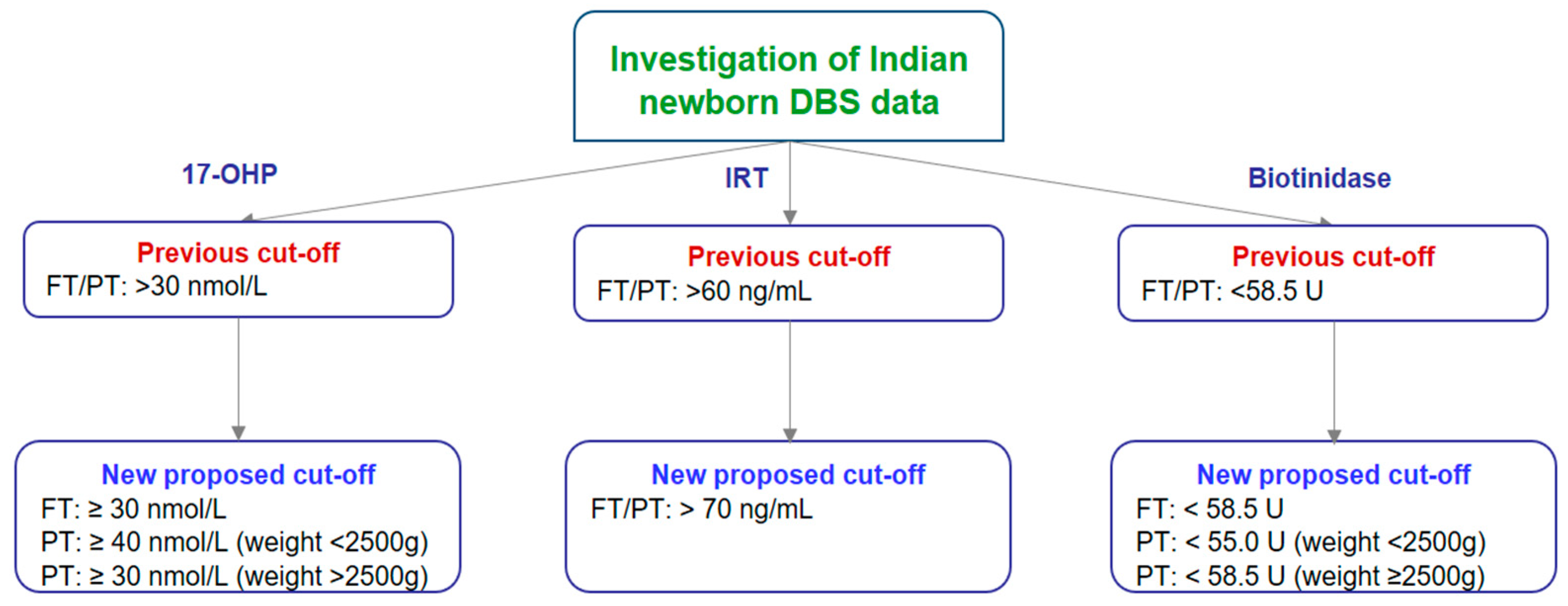
3.1. Cut-Off Establishment for 17-OHP, IRT, and Biotinidase in Indian Newborns

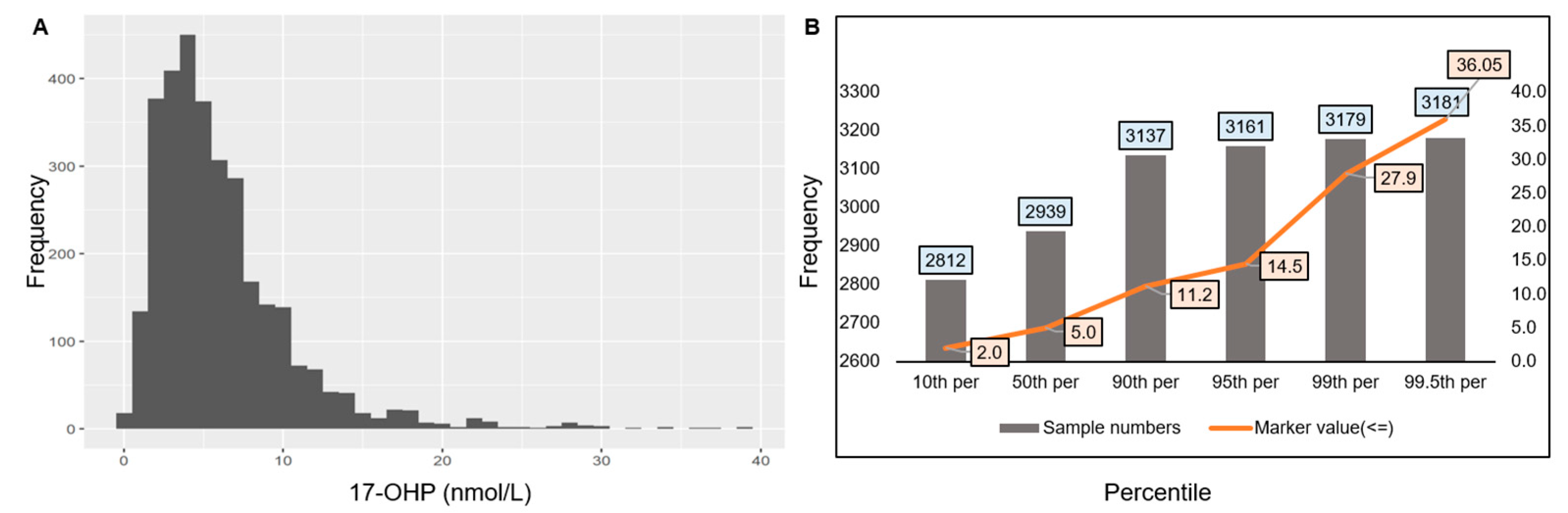
3.1.1. Cut-Off Establishment for 17-OHP

The screen-positive rate for 17-OHP with the current cut-off >30 ng/ml is 0.75% (n = 2758). The current cut-off value distribution is between the 99.2nd and 99.3rd percentile (Figure 2A). 17-OHP concentrations tend to be higher in individuals with the salt-wasting form [33]. It is crucial to exercise caution when using the correct terminology or early-normal values for comparison. It was found that 17-OHP levels increased in the DBS samples of newborns with CAH. The levels were set to >30 nmol/L for FT, ≥40 nmol/L for preterm babies weighing less than 2500 g, and ≥30 nmol/L for PT babies weighing more than 2500 g based on the percentile cut-off (Figure 2B). The amounts of hormones in newborns with salt-wasting and simple-virilizing CAH are greater than the quantities in infants with the moderate variant [33,34]. The ACTH stimulation test is useful in excluding nonclassic CAH in neonates with slightly high 17-OHP values (4–10 ng/mL). For newborns without symptoms, it is important to regularly monitor electrolyte levels during the neonatal period, especially if the levels remain within the normal range.

3.1.2. Cut-Off Establishment for IRT

With the current cut-off of >61 ng/mL, the screen-positive rate for IRT is 1.46% (n = 2733). The current cut-off value is between the 98.5th and 99.6th percentiles (Figure 3A,B). This indicates that the current cut-off value effectively identifies infants at risk for CF. A positive newborn screening result does not guarantee that a child has CF; rather, it indicates the need for additional testing for further clinical correlation. It was found that IRT levels went up in the DBS samples of newborns with CF. The established FT/PT levels are set at >70 ng/mL from the previous cut-off FT/PT: >60 ng/mL based on the 98.5th and 99.6th percentiles (Figure 3B). Early CF care recipients have superior nutrition and overall health compared to individuals who receive a diagnosis later in life. All infants should undergo NBS as cystic fibrosis can affect people of all races and ethnicities [35]. Early detection and intervention can improve growth, help maintain healthy lungs, and increase life expectancy by years.

3.1.3. Cut-Off Establishment for Biotinidase

The cut-off of biotinidase is placed at <58.5 U, where the NBS-positive rate is at 0.45% (n = 2724). Hence, the cut-offs are effective in identifying newborns with biotinidase deficiency (Figure 4A,B). Newborns who test below the current cut-off value are likely to have biotinidase deficiency, while those above are unlikely to have the normal condition. The present proposed cut-off values are <58.5 U for FT, <55.0 U for PT babies weighing <2500 g, and <58.5 U for PT babies weighing >2500 g. This suggests that we have appropriately set the current cut-off value for accurate screening for biotinidase in Indian newborns. The treatment involves providing biotin supplements to help the body break down food and ensure healthy growth and development in biotinidase deficiency.
Overall, the results provided a statistical summary and graphical representation of the results that propose updated cut-offs for the NBS parameters such as 17-OHP, IRT, and biotinidase in Indian newborns (Figure 5).

4. Discussion

Essential newborn diagnostic tests such as 17-OHP, IRT, and biotinidase at birth are used to identify metabolic disorders that potentially influence development and health. Babies born after 42 weeks, i.e., post-term infants, may have a risk for problems such as meconium aspiration, placental hypofunction, and any inborn errors [36].
Evaluating the health of a newborn just after delivery depends much on its birth weight. Post-term children delivered after 42 weeks of gestational age might have placental insufficiency or meconium aspiration [37]. Several factors influence newborn health issues such as premature birth, inadequate maternal nutrition, and other medical problems linked to low birth weight (<2500 g). As per the gestational age, the birth weight of newborns at full term (FT) is usually between 2500 and 4000 g [38]. Macrosomia refers to a high birth weight and might be a sign of maternal diabetes or other contributing issues [39]. Neonatal intensive care units (NICUs) offer specialized health attention for newborns who are born prematurely or have a low birth weight [40]. Preterm birth or maternal undernutrition may affect birth weight and influence fetal growth and nutritional status [41]. Low birth weight (<2500 g) can indicate a mother’s health issues. Low birth weight and gestational diabetes mellitus (GDM) are associated in pregnant women, producing more low-birth-weight kids [42]. Maternal glucose tolerance and insulin resistance affect infant weight. Other chronic health ailments like high blood pressure and heart, lung, renal, and drug troubles can also cause low birth weight [43,44]. Growth charts and percentiles are tools used to screen and evaluate a newborn’s growth pattern, taking into account various clinical and laboratory aspects and findings.
Every child is unique, and may not encounter the same challenges at birth. It is important to monitor the baby’s health [8]. However, infants with CAH, an inherited disorder affecting hormone production, might display elevated levels of the adrenal gland hormone 17-OHP [45]. The elevated 17-OHP-positivity prevalence rate in newborns with salt-wasting circumstances is 0.75%. Newborns with CAH show higher levels of hormones, in which the ACTH stimulation test can exclude nonclassic CAH with slightly high 17-OHP. Tippabathani et al. (2023) found that out of 220,000 tests conducted for 17-OHP, only 97 were positive using Sanger’s method. This indicates a 0.044% 17-OHP-positivity prevalence rate in India. Among these, 54 reflex gene tests accurately detected positive findings. Their investigation found that 21 hydroxylase deficits occur in 1 in 4074 patients. Our investigation revealed significant connections between aberrant 17-OHP levels and clinical manifestations [22]. Delayed diagnosis of 17-OHP and treatment of CAH can lead to salt wasting, dehydration, or other severe complications [11,33,46]. Systematic monitoring of electrolyte levels is crucial for newborns without symptoms, especially if levels remain within the normal range.
In addition, the screening for CF, a hereditary condition that affects the digestive system and lungs, is performed by an IRT test. IRT is an enzyme produced by the pancreas [47]. The present cut-off rate for CF screening for IRT is 1.46%, indicating that infants are at risk. For infants with elevated IRT levels, additional diagnostic tests may be necessary to confirm the diagnosis of CF, which may indicate that they may be at risk for the disease and treatment needs to be initiated; however, elevated IRT does not always indicate CF. Repeating the IRT-positivity test after two weeks is advisable, and if the elevated levels persist, the infant should undergo sweat chloride testing and/or cystic fibrosis transmembrane conductance regulator (CFTR) mutation analysis [48]. Moreover, it is important to consider the clinical phenotype before establishing a final confirmatory diagnosis of CF. Newborns who receive early CF care have better nutrition and overall health.
Biotinidase deficiency causes late-onset biotin-responsive multiple carboxylase deficiency (MCD), which leads to lactic acidosis or acidosis, hypoglycemia, and abnormal catabolism [49]. The symptoms may appear after a few weeks of birth or in late childhood, and some may be asymptomatic. Moreover, a deficiency in biotinidase may affect neurological functions such as convulsions, hypotonia, ataxia, psychomotor retardation, optic atrophy, sensorineural deafness, and skin problems [50]. Presently, the abnormal value of biotinidase is less than 58.5 U, and the screening positive rate in 2724 Indian newborns with DBS is 0.45%, indicating its effectiveness in identifying biotinidase deficiency. Based on the biotinidase level present in the serum, we classify biotinidase deficiency into two types: profound (<30% of enzyme activity) and partial (10–30% of enzyme activity) [51]. Partial cases can have little or no symptoms. The average amount of biotinidase in human serum ranges from 4.4 to 10 nmol/min/mL, with a mean activity of 7.1 nmol/min/mL [49,51]. Individuals with profound biotinidase deficiency cannot recycle their endogenous biotin by cleaving it from biocytin, which causes MCD. However, profound cases can lead to coma or death if treatment is not initiated rapidly. In the present study, all abnormal biotinidase levels were crosschecked by serum biotinidase and found to be correlated with clinical findings. Failure to receive treatment for biotinidase deficiency can result in significant neurological complications. The deficiency treatment includes providing biotin supplements to help the body break down food and promote healthy growth and development [51]. All infants should undergo newborn screening, as the disorders may affect people of all races and ethnicities.
Overall, the results presented valuable information on the prevalence of CAH, CF, and biotinidase abnormalities in Indian newborns. The previous 17-OHP FT and PT cut-off values were ≥30 nmol/L. For Indian newborns, the values are based on term and weight: FT ≥ 30 nmol/L, PT ≥ 40 nmol/L (weight < 2500 g), and PT ≥ 30 nmol/L (weight > 2500 g). Similarly, we established the IRT newborn reference intervals (for both FT and PT) above the 98th percentile cutoff, i.e., from >61 ng/mL to >70 ng/mL. The biotinidase cut-offs placed at 58.5 U are approximately 0.45%, i.e., FT: <58.5 U; PT: <55 U (weight < 2500 g); and PT: <58.5 U (weight ≥ 2500 g) (Figure 5).

5. Conclusions

In conclusion, these findings linked increased 17-OHP and IRT, or decreased biotinidase, in neonates to CAH, CF, and biotinidase deficiency, respectively, and suggested more accurate reference ranges taking into account gestational age and weight than previously used reference ranges. It is important to note that there can be significant variations across individuals. NBS is critical for ensuring the health and well-being of newborns, as well as improving detection, intervention strategies, and genetic guidance for subsequent pregnancies. Future research concentrating on the intricate mechanisms of these conditions for newborns is essential for early diagnosis and intervention for the disorder in its early stages.

Author Contributions

Conceptualization, E.M.P. and R.B.; methodology, E.M.P.; investigation, E.M.P. and R.B.; resources, E.M.P.; data curation, R.K. and R.B.; writing—original draft review and editing, E.M.P.; supervision, E.M.P., R.K. and R.B. All authors have read and agreed to the published version of the manuscript.

Funding

This research received no external funding.

Institutional Review Board Statement

Not applicable.

Informed Consent Statement

Not applicable.

Data Availability Statement

No new data were created or analyzed in this study. Data sharing is not applicable to this article.

Acknowledgments

The authors are grateful to Pavani Kiran, Shalini Singh, K. Anusha, Rajesh Battina, M. Sujana Reddy, and N. Sachin, Department of Clinical Chemistry, Apollo Diagnostics Global Reference Lab, Hyderabad 500037, India, for their assistance during the study, as well as Sandeep and Sujin, RevvityTM Healthcare, India, for the statistical analysis support.

Conflicts of Interest

The authors declare no conflicts of interest.

References

  1. FDA-NIH Biomarker Working Group. Diagnostic Biomarker. In BEST (Biomarkers, Endpoints, and Other Tools) Resource; Food and Drug Administration: Silver Spring, MD, USA, 2020. [Google Scholar]
  2. Bodaghi, A.; Fattahi, N.; Ramazani, A. Biomarkers: Promising and Valuable Tools towards Diagnosis, Prognosis and Treatment of COVID-19 and Other Diseases. Heliyon 2023, 9, e13323. [Google Scholar] [CrossRef]
  3. Ahmad, A.; Imran, M.; Ahsan, H. Biomarkers as Biomedical Bioindicators: Approaches and Techniques for the Detection, Analysis, and Validation of Novel Biomarkers of Diseases. Pharmaceutics 2023, 15, 1630. [Google Scholar] [CrossRef]
  4. Rosa-Mangeret, F.; Benski, A.-C.; Golaz, A.; Zala, P.Z.; Kyokan, M.; Wagner, N.; Muhe, L.M.; Pfister, R.E. 2.5 Million Annual Deaths—Are Neonates in Low- and Middle-Income Countries Too Small to Be Seen? A Bottom-Up Overview on Neonatal Morbi-Mortality. Trop. Med. Infect. Dis. 2022, 7, 64. [Google Scholar] [CrossRef]
  5. Marsden, D.; Bedrosian, C.L.; Vockley, J. Impact of Newborn Screening on the Reported Incidence and Clinical Outcomes Associated with Medium- and Long-Chain Fatty Acid Oxidation Disorders. Genet. Med. 2021, 23, 816–829. [Google Scholar] [CrossRef]
  6. Rocha, J.C.; MacDonald, A. Dietary Intervention in the Management of Phenylketonuria: Current Perspectives. Pediatr. Health Med. Ther. 2016, 7, 155–163. [Google Scholar] [CrossRef]
  7. Widaman, K.F. Phenylketonuria in Children and Mothers: Genes, Environments, Behavior. Curr. Dir. Psychol. Sci. 2009, 18, 48–52. [Google Scholar] [CrossRef]
  8. Behrman, R.E.; Butler, A.S.; Healthy Outcomes (Eds.) Mortality and Acute Complications in Preterm Infants. In Preterm Birth: Causes, Consequences, and Prevention; National Academies Press: Washington, DC, USA, 2007. [Google Scholar]
  9. Pitt, J.J. Newborn Screening. Clin. Biochem. Rev. 2010, 31, 57. [Google Scholar]
  10. Crump, C. Preterm Birth and Mortality in Adulthood: A Systematic Review. J. Perinatol. 2020, 40, 833–843. [Google Scholar] [CrossRef]
  11. Levy-Shraga, Y.; Pinhas-Hamiel, O. High 17-Hydroxyprogesterone Level in Newborn Screening Test for Congenital Adrenal Hyperplasia. BMJ Case Rep. 2016, bcr2015213939. [Google Scholar] [CrossRef]
  12. Burdea, L.; Mendez, M.D. 21-Hydroxylase Deficiency. In StatPearls; StatPearls Publishing: Treasure Island, FL, USA, 2024. [Google Scholar]
  13. Jha, S.; Turcu, A.F. Non-Classic Congenital Adrenal Hyperplasia: What Do Endocrinologists Need to Know? Endocrinol. Metab. Clin. N. Am. 2021, 50, 151–165. [Google Scholar] [CrossRef]
  14. Momodu, I.I.; Lee, B.; Singh, G. Congenital Adrenal Hyperplasia. In StatPearls; StatPearls Publishing: Treasure Island, FL, USA, 2024. [Google Scholar]
  15. Wróbel, T.M.; Jørgensen, F.S.; Pandey, A.V.; Grudzińska, A.; Sharma, K.; Yakubu, J.; Björkling, F. Non-Steroidal CYP17A1 Inhibitors: Discovery and Assessment. J. Med. Chem. 2023, 66, 6542–6566. [Google Scholar] [CrossRef]
  16. Newborn Screening for Cystic Fibrosis: Do We Need a Second IRT?—PMC. Available online: https://www.ncbi.nlm.nih.gov/pmc/articles/PMC2065957/ (accessed on 14 August 2024).
  17. Ooi, C.Y.; Sutherland, R.; Castellani, C.; Keenan, K.; Boland, M.; Reisman, J.; Bjornson, C.; Chilvers, M.A.; van Wylick, R.; Kent, S.; et al. Immunoreactive Trypsinogen Levels in Newborn Screened Infants with an Inconclusive Diagnosis of Cystic Fibrosis. BMC Pediatr. 2019, 19, 369. [Google Scholar] [CrossRef]
  18. Morrison, C.B.; Markovetz, M.R.; Ehre, C. Mucus, Mucins and Cystic Fibrosis. Pediatr. Pulmonol. 2019, 54, S84–S96. [Google Scholar] [CrossRef]
  19. Canda, E.; Kalkan Uçar, S.; Çoker, M. Biotinidase Deficiency: Prevalence, Impact And Management Strategies. Pediatr. Health Med. Ther. 2020, 11, 127–133. [Google Scholar] [CrossRef]
  20. Hymes, J.; Wolf, B. Biotinidase and Its Roles in Biotin Metabolism. Clin. Chim. Acta 1996, 255, 1–11. [Google Scholar] [CrossRef]
  21. Tong, L. Structure and Function of Biotin-Dependent Carboxylases. Cell. Mol. Life Sci. 2012, 70, 863–891. [Google Scholar] [CrossRef]
  22. Tippabathani, J.; Seenappa, V.; Murugan, A.; Phani, N.M.; Hampe, M.H.; Appaswamy, G.; Sadashiv Gambhir, P. Neonatal Screening for Congenital Adrenal Hyperplasia in Indian Newborns with Reflex Genetic Analysis of 21-Hydroxylase Deficiency. Int. J. Neonatal Screen 2023, 9, 9. [Google Scholar] [CrossRef]
  23. Chen, H.-F.; Rose, A.M.; Waisbren, S.; Ahmad, A.; Prosser, L.A. Newborn Screening and Treatment of Phenylketonuria: Projected Health Outcomes and Cost-Effectiveness. Children 2021, 8, 381. [Google Scholar] [CrossRef]
  24. Held, P.K.; Bird, I.M.; Heather, N.L. Newborn Screening for Congenital Adrenal Hyperplasia: Review of Factors Affecting Screening Accuracy. Int. J. Neonatal Screen 2020, 6, 67. [Google Scholar] [CrossRef]
  25. Bereket, A. Editorial: Neonatal Screening for Congenital Adrenal Hyperplasia in Turkey. J. Clin. Res. Pediatr. Endocrinol. 2019, 11, 1–3. [Google Scholar] [CrossRef]
  26. Kaye, C.I.; The Committee on Genetics. Newborn Screening Fact Sheets. Pediatrics 2006, 118, e934–e963. [Google Scholar] [CrossRef]
  27. Torresani, T.; Grüters, A.; Scherz, R.; Burckhardt, J.J.; Harras, A.; Zachmann, M. Improving the Efficacy of Newborn Screening for Congenital Adrenal Hyperplasia by Adjusting the Cut-off Level of 17α-Hydroxyprogesterone to Gestational Age. Screening 1994, 3, 77–84. [Google Scholar] [CrossRef]
  28. Hemmilä, I.; Dakubu, S.; Mukkala, V.-M.; Siitari, H.; Lövgren, T. Europium as a Label in Time-Resolved Immunofluorometric Assays. Anal. Biochem. 1984, 137, 335–343. [Google Scholar] [CrossRef]
  29. Rock, M.J.; Mischler, E.H.; Farrell, P.M.; Bruns, W.T.; Hassemer, D.J.; Laessig, R.H. Immunoreactive Trypsinogen Screening for Cystic Fibrosis: Characterization of Infants with a False-Positive Screening Test. Pediatr. Pulmonol. 1989, 6, 42–48. [Google Scholar] [CrossRef]
  30. Kirby, L.T.; Applegarth, D.A.; Davidson, A.G.; Wong, L.T.; Hardwick, D.F. Use of a Dried Blood Spot in Immunoreactive-Trypsin Assay for Detection of Cystic Fibrosis in Infants. Clin. Chem. 1981, 27, 678–680. Available online: https://academic.oup.com/clinchem/article-abstract/27/5/678/5666531 (accessed on 14 August 2024).
  31. Hart, P.S.; Hymes, J.; Wolf, B. Biochemical and Immunologic Characterization of Serum Biotinidase in Partial Biotinidase. Pediatr. Res. 1992, 31, 261–265. [Google Scholar] [CrossRef]
  32. Wolf, B.; Heard, G.S.; Jefferson, L.G.; Proud, V.K.; Nance, W.E.; Weissbecker, K.A. Clinical Findings in Four Children with Biotinidase Deficiency Detected through a Statewide Neonatal Screening Program. N. Engl. J. Med. 1985, 313, 16–19. [Google Scholar] [CrossRef]
  33. Yau, M.; Khattab, A.; Yuen, T.; New, M. Congenital Adrenal Hyperplasia. In Endotext; Feingold, K.R., Anawalt, B., Blackman, M.R., Boyce, A., Chrousos, G., Corpas, E., de Herder, W.W., Dhatariya, K., Dungan, K., Hofland, J., et al., Eds.; MDText.com, Inc.: South Dartmouth, MA, USA, 2000. [Google Scholar]
  34. Nimkarn, S.; Gangishetti, P.K.; Yau, M.; New, M.I. 21-Hydroxylase-Deficient Congenital Adrenal Hyperplasia. In GeneReviews®; Adam, M.P., Feldman, J., Mirzaa, G.M., Pagon, R.A., Wallace, S.E., Bean, L.J., Gripp, K.W., Amemiya, A., Eds.; University of Washington, Seattle: Seattle, WA, USA, 1993. [Google Scholar]
  35. Southern, K.W.; Mérelle, M.M.E.; Dankert-Roelse, J.E.; Nagelkerke, A. Newborn Screening for Cystic Fibrosis. Cochrane Database Syst. Rev. 2009, CD001402. [Google Scholar] [CrossRef]
  36. Patadia, C.; Desai, D. A Child Born Late: Neonatal and Paediatric Compilations Related to Post-Dated Pregnancy. preprint 2023. [Google Scholar] [CrossRef]
  37. Latif, S.; Aiken, C. Prolonged Pregnancy: Balancing Risks and Interventions for Post-Term Gestations. Obstet. Gynaecol. Reprod. Med. 2024, 34, 127–133. [Google Scholar] [CrossRef]
  38. Neggers, Y.H. Gestational Age and Pregnancy Outcomes. In Pregnancy and Birth Outcomes; Intech: London, UK, 2018; ISBN 978-1-78923-243-1. [Google Scholar] [CrossRef]
  39. Akanmode, A.M.; Mahdy, H. Macrosomia. In StatPearls; StatPearls Publishing: Treasure Island, FL, USA, 2024. [Google Scholar]
  40. Fallah, S.; Chen, X.-K.; Lefebvre, D.; Kurji, J.; Hader, J.; Leeb, K. Babies Admitted to NICU/ICU: Province of Birth and Mode of Delivery Matter. Healthc. Q. 2011, 14, 16–20. [Google Scholar] [CrossRef]
  41. Woldeamanuel, G.G.; Geta, T.G.; Mohammed, T.P.; Shuba, M.B.; Bafa, T.A. Effect of Nutritional Status of Pregnant Women on Birth Weight of Newborns at Butajira Referral Hospital, Butajira, Ethiopia. SAGE Open Med. 2019, 7, 2050312119827096. [Google Scholar] [CrossRef]
  42. Seghieri, G.; Anichini, R.; De Bellis, A.; Alviggi, L.; Franconi, F.; Breschi, M.C. Relationship between Gestational Diabetes Mellitus and Low Maternal Birth Weight. Diabetes Care 2002, 25, 1761–1765. [Google Scholar] [CrossRef]
  43. Lewandowska, M. Maternal Obesity and Risk of Low Birth Weight, Fetal Growth Restriction, and Macrosomia: Multiple Analyses. Nutrients 2021, 13, 1213. [Google Scholar] [CrossRef]
  44. Ornoy, A.; Becker, M.; Weinstein-Fudim, L.; Ergaz, Z. Diabetes during Pregnancy: A Maternal Disease Complicating the Course of Pregnancy with Long-Term Deleterious Effects on the Offspring. A Clinical Review. Int. J. Mol. Sci. 2021, 22, 2965. [Google Scholar] [CrossRef]
  45. Honour, J.W. 17-Hydroxyprogesterone in Children, Adolescents and Adults. Ann. Clin. Biochem. 2014, 51, 424–440. [Google Scholar] [CrossRef]
  46. Reddy, N.A.; Sharma, S.; Das, M.; Kapoor, A.; Maskey, U. Devastating Salt-Wasting Crisis in a Four-Month-Old Male Child with Congenital Adrenal Hyperplasia, Highlighting the Essence of Neonatal Screening. Clin. Case Rep. 2022, 10, e6010. [Google Scholar] [CrossRef]
  47. Waugh, N.; Royle, P.; Craigie, I.; Ho, V.; Pandit, L.; Ewings, P.; Adler, A.; Helms, P.; Sheldon, C. Screening for Cystic Fibrosis-Related Diabetes: A Systematic Review. Health Technol. Assess. 2012, 16, 1–179. [Google Scholar] [CrossRef]
  48. Farrell, P.M.; Rosenstein, B.J.; White, T.B.; Accurso, F.J.; Castellani, C.; Cutting, G.R.; Durie, P.R.; Legrys, V.A.; Massie, J.; Parad, R.B.; et al. Guidelines for Diagnosis of Cystic Fibrosis in Newborns through Older Adults: Cystic Fibrosis Foundation Consensus Report. J. Pediatr. 2008, 153, S4–S14. [Google Scholar] [CrossRef]
  49. Cowan, T.M.; Blitzer, M.G.; Wolf, B. Technical Standards and Guidelines for the Diagnosis of Biotinidase Deficiency. Genet. Med. 2010, 12, 464–470. [Google Scholar] [CrossRef]
  50. Saleem, H.; Simpson, B. Biotinidase Deficiency. In StatPearls; StatPearls Publishing: Treasure Island, FL, USA, 2024. [Google Scholar]
  51. Kannan, B.; Navamani, H.K.; Jayaseelan, V.P.; Arumugam, P. A Rare Biotinidase Deficiency in the Pediatrics Population: Genotype–Phenotype Analysis. J. Pediatr. Genet. 2022, 12, 1–15. [Google Scholar] [CrossRef] [PubMed]
Figure 1. (A) Distribution of gestational age in Indian newborns. (B) Distribution of weight in Indian newborns.
Biomed 04 00021 g001
Figure 2. (A) Distribution frequency of 17-OHP in Indian newborns (x-axis: concentration of 17-OHP in nmol/L; y-axis: distribution frequency). (B) Percentile cut-off values for 17-OHP levels based on weight group in Indian newborns.
Biomed 04 00021 g002
Figure 3. (A) Distribution frequency of IRT in Indian newborns (x-axis: concentration of IRT in ng/mL; y-axis: distribution frequency). (B) Percentile cut-off establishment for IRT by weight group in Indian newborns.
Biomed 04 00021 g003
Figure 4. (A) Distribution frequency of biotinidase in Indian newborns (x-axis: concentration of biotinidase in U; y-axis: distribution frequency). (B) Cut-off establishment for biotinidase by weight group in Indian newborns.
Biomed 04 00021 g004
Figure 5. The studied and new cut-off values for Indian newborn parameters, including 17-OHP, IRT, and biotinidase in India, for both FT and PT babies. Note: the cut-offs for 17-OHP and IRT are above the 98th percentile, while that for biotinidase is below the 2nd percentile.
Biomed 04 00021 g005
Disclaimer/Publisher’s Note: The statements, opinions and data contained in all publications are solely those of the individual author(s) and contributor(s) and not of MDPI and/or the editor(s). MDPI and/or the editor(s) disclaim responsibility for any injury to people or property resulting from any ideas, methods, instructions or products referred to in the content.

