Review

Cosmeceutical Applications of Phlorotannins from Brown Seaweeds

by
D. M. N. M. Gunasekara
1,
Lei Wang
2,3,
K. H. I. N. M. Herath
4,* and
K. K. A. Sanjeewa
1,*
1
Department of Biosystems Technology, Faculty of Technology, University of Sri Jayewardenepura, Homagama 10200, Sri Lanka
2
State Key Laboratory of Marine Food Processing & Safety Control, College of Food Science and Engineering, Ocean University of China, Qingdao 266003, China
3
Sanya Oceanographic Institution, Ocean University of China, Sanya 572024, China
4
Department of Biosystems Engineering, Faculty of Agriculture and Plantation Management, Wayamba University of Sri Lanka, Makandura 60170, Sri Lanka
*
Authors to whom correspondence should be addressed.
Phycology 2025, 5(2), 15; https://doi.org/10.3390/phycology5020015
Submission received: 19 March 2025 / Revised: 19 April 2025 / Accepted: 24 April 2025 / Published: 27 April 2025

Abstract

:
Due to the adverse effects associated with synthetic cosmetic ingredients, global demand is increasingly shifting toward natural formulations that offer diverse benefits for enhancing skin health and overall beauty. Researchers around the world are extensively exploring a variety of unique natural secondary metabolites for cosmeceutical applications. Among the potential candidates, phlorotannins derived from brown seaweeds have shown significant potential as an active ingredient in cosmeceutical applications. The notable properties associated with phlorotannins include antioxidant, anti-aging, whitening, anti-wrinkling, anti-inflammatory, and hair health and growth-promoting effects, making them valuable in cosmeceutical formulations. However, to date, only a limited number of studies have critically reviewed the cosmeceutical applications of phlorotannins, and most are outdated. Thus, in the present review, primary attention is given to the collected scientific data published after 2020 about the bioactive properties of brown seaweed phlorotannins related to cosmeceutical applications.

Graphical Abstract

1. Introduction

Conventional cosmetics, primarily used for esthetic purposes a few decades ago, have recently shown a remarkable increase in addressing all aspects of personal care, such as skin, hair, nails, teeth, and lips [1]. The preference for cosmetic products is rapidly growing among both men and women, changing cultural perceptions traditionally associated with women [2]. Consumer demand for cosmetic products has extensively shifted to natural and organic alternatives, leading industries to develop products derived from natural sources. According to multiple studies, the excessive use of synthetic ingredients in cosmetic formulations can lead to several drawbacks including skin bleaching, dryness, burning sensations, photosensitivity, hair loss, and other toxicities affecting overall human health, even minor to severe levels [3,4,5]. For instance, hydroxybenzoic acid esters (parabens), which are commonly used in cosmetic formulations, have been associated with negative effects on the skin and an increased risk of malignant melanoma and breast cancer by resembling the female hormone estrogen [6].
Phthalates, a principal type of plasticizer, are one of the common ingredients utilized in cosmeceutical formulations, such as in lotions, nail polish, and hair care products, and have been shown to cause DNA modification and damage to human sperm cells [7]. The term “cosmeceuticals” refers to cosmetic formulations that contain biologically active substances that offer therapeutic advantages, bridging the gap between cosmetics and pharmaceuticals [8,9]. Exploring safer or minimal-risk alternatives from natural sources has been highly expanded through the marine environment due to its abundant diversity, suggesting novel substances that represent good candidates for various industries. Seaweeds have stood out as a great natural source, rich in a broad spectrum of nutrients and bioactive compounds applicable across functional foods, nutraceuticals, pharmaceuticals, agricultural, and cosmeceuticals [10,11].
Seaweeds (marine macroalgae) are commonly classified into three main taxonomy groups: Chlorophyceae (green algae), Rhodophyceae (red algae), and Phaeophyceae (brown algae), according to their pigment profile and morphological, anatomical, and reproductive structures [10,12]. Brown seaweed is reported to be the most consumed in the world, accounting for more than 66.5% of the total production of seaweeds [13]. Usually, brown seaweeds are brownish–green to golden brown. The primary pigment fucoxanthin, overlapping with chlorophyll a, chlorophyll c (photosynthetic pigment), and β-carotene (carotenoid pigments), is responsible for the entire coloration of brown seaweeds [14]. Their fast growth rate and unique bioactive composition, including phlorotannins, fucoxanthin, alginates, laminarin, alginic acid, and fucoidan, contribute to numerous efforts focused on commercializing seaweeds [15,16]. Among them, phlorotannins are the exclusive polyphenolic compounds similar to tannins in terrestrial environments [17]. A number of studies have highlighted the specific and interesting functional properties of phlorotannins, such as anti-aging, anti-wrinkling, anti-acne, anti-inflammatory, antioxidant, anti-melanogenic, whitening, moisturizing, and photo-protective properties, as well as the ability to exhibit low cytotoxicity and low allergenic ingredients [18,19,20]. These biological products are recognized to be safe and effective alternatives to synthetic counterparts, which are closely associated with cosmeceutical preparations [10,21].

Phlorotannins

Phlorotannins are a group of polyphenolic compounds predominantly found in a range of brown seaweeds, most commonly in Laminaria japonica, Fucus vesiculosus, Undaria pinnatifida, Pelvetia siliquosa, Ecklonia kurome, Sargassum thunbergii, Sargassum horneri, Ecklonia cava, Scytosiphon lomentarius, and many others [18,22,23,24]. Phlorotannins are like tannins, which are secondary metabolites produced by terrestrial plants and other organisms as a part of their defense mechanism. Chemically, phlorotannins are composed mainly of the polymeric structure of the monomer phloroglucinol (1,3,5-hydroxybenzene), which is formed in the acetate–malonate pathway (polyketide) [25]. The natural variability of structural linkages between phloroglucinol units (PGUs) and the number of hydroxyl groups in phlorotannins has contributed to the broad spectrum of their potential biological activities [25,26,27].
The molecular weight of phlorotannins varies from low, to intermediate and high, ranging from 126 Da to 650 kDa [28]. Phlorotannins can be sub-classified into four main classes: phlorethols, fucols, fucophlorethols, and eckols, based on the type and number of bonds between the monomers and the presence of an additional hydroxyl group [25]. Their accurate identification in an extract can be difficult due to the presence of highly complex polymeric mixtures of structural and conformational isomers of phlorotannins [29]. Various attempts have been reported, and the optimal approach is to separate, quantify, and characterize phlorotannin compounds to identify specific phlorotannin bioactivities. Spectrophotometric assays are a cost-effective and efficient method for screening phlorotannin content. Moreover, advanced techniques like MS (mass spectrometry) coupled with HPLC (high-performance liquid chromatography) and UHPLC (ultra-high-performance liquid chromatography)/HRMS (high-resolution mass spectrometry) are essential for detailed separation and characterization. NMR (nuclear magnetic resonance) spectroscopy provides detailed structural information, but is less accessible due to cost. Creating standard compound libraries and linking NMR, HPLC retention times, and UV(ultraviolet) spectral data may improve the identification of common phlorotannin compounds [28]. Different phlorotannin structures are summarized in Figure 1 and Figure 2.

2. Cosmeceutical Applications of Phlorotannins Isolated from Brown Seaweeds

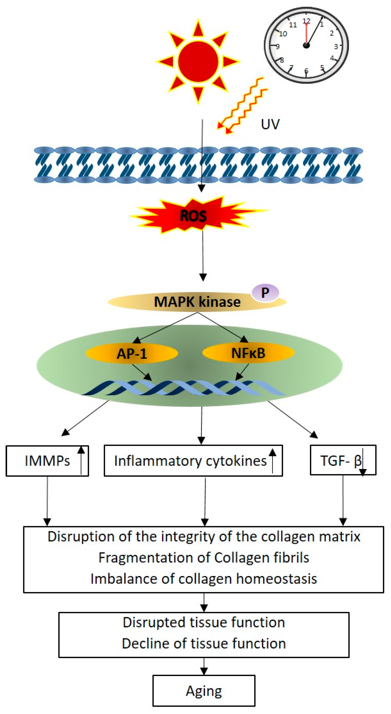
2.1. Photo-Protective and Anti-Aging Properties of Phlorotannins

Human skin comprises three main layers: the epidermis, dermis, and hypodermis (subcutaneous fat) [30,31]. Obvious changes in the dermis layer and overall degenerative changes in each layer involve the aging process [30]. Young skin is determined by proper quantitative and structural changes being made in collagen fibers, which are major components of the extracellular matrix (ECM) in the dermis and combined with other components such as fibroblasts, elastic fibers, glycosaminoglycans (GAGs), and proteoglycans (PGs) [32]. Along with these, hyaluronic acid is involved in water retention, resulting in effective skin moisturizing and making skin appear younger and healthier [33]. The reduction in and degradation of collagen leads to premature aging with skin wrinkles, thinning, and a loss of elasticity in key areas such as the forehead, corners of the eyes, and cheeks, which, along with the neck, hands, and arms, are often the most noticeable signs of aging Figure 3.
Ultraviolet radiation (UVR) is one of the main extrinsic factors that can directly contribute to dermal photodamage through oxidative stress, DNA (deoxyribosnucleic acid) damage, inflammation, and apoptosis, leading to wrinkles, skin relaxation, and hyperpigmentation [34]. Long-term and overexposure to UVR, specifically UVB (290–320 nm), causes increased skin damage that enhances the generation of reactive oxygen species (ROS), which stimulates the natural aging process by causing the loss of ECM, the degradation of matrix-supporting molecules, and other components, ultimately forming the clinical changes in the skin [35]. The aging process is driven by a number of complex and important pathways, and reactive oxygen species (ROS)-associated pathways can be illustrated as in Figure 4.
Reactive oxygen species (ROS) generated during the aging process activate mitogen-activated protein kinases (MAPKs) and trigger the activation of transcription factors such as AP-1 (activator protein 1) and NF-κB (nuclear factor-κB). This activation subsequently upregulates the expression of MMPs (matrix metalloproteinases) while downregulating TGF-β (transforming growth factor-β) signaling, resulting in the degradation of collagen and a reduction in collagen synthesis, ultimately accelerating skin aging [32]. The inhibition of the enzyme activity of intracellular collagenase, elastase, and hyaluronidase and the reduction in the expression of MMPs, pro-inflammatory cytokines, and other compounds have the potential to protect against UVB-induced skin aging [36]. These events occur through UVB-irradiated HDF (Human Dermal Fibroblast) cells exhibiting the modulation of NF-κB, AP-1, and MAPKs signaling pathways [37].
Innovating cosmeceutical products that have anti-aging properties with natural ingredients is in demand all over the world, to achieve young- and healthy-looking skin. The marine environment has proven to be a good source of compounds with recognized anti-aging properties with multifunctional potential [33,38]. A considerable amount of research has been conducted on experiments with phlorotannins in brown seaweeds. The study by [39] attempted to evaluate the potential of cosmeceutical applications of seven different brown seaweeds harvested in Brittany in France [39]. The research revealed that observation of the photoprotective and anti-aging (elastase inhibitory) effects of phlorotannin extracts from Ascophyllum nodosum shows up to 70% of elastase inhibition activity at a concentration of 0.1 mg·mL−1. While under the same concentration, Fucus serratus shows up to an 80% performance compared with the positive control EGCG (epigallocatechin gallate). In particular, the research investigation proposed by [40] has proven the photoprotective effect of (-)-loliode isolated from Sargassum horneri, demonstrating its role as an ideal ingredient in the cosmeceutical industry. According to the findings, (-)-loliode effectively protected human skin cells in vitro (HaCaT and HDF) and zebrafish in vivo against UVB-induced photodamage. Further results have shown that (-)-loliode can improve collagen synthesis, inhibit MMPs, and attenuate oxidative damage [40]. Among the different brown seaweeds that exhibit anti-aging activities (Table 1), recently, the impact of the combination of E. cava-derived extracellular vesicles (EV-EC) and phlorotannins (PT) on skin rejuvenation has been investigated by [41] using an in vitro keratinocyte senescence model and an in vivo aged mouse model. Treatment with the EV-EC and PT combination has shown an increase in the expression of heat shock protein 70 (HSP70), which leads to the decreased expression of TNF-α, MAPK, NF-κB, activator protein-1 (AP-1), and MMPs. Ultimately, this treatment was able to increase collagen fiber accumulation and improve elasticity in aged skin in the in vitro analysis, concluding that PT/EV-EC holds promise in promoting skin rejuvenation by increasing HSP70 expression, decreasing the expression of MMPs, and reducing oxidative stress in aged skin [41].

2.2. Whitening Properties of Phlorotannins

People with dark skin can be found in a variety of ethnic groups and regions throughout the world. According to the Fitzpatrick scale, type III (light brown) to type VI, (very dark brown/black) are commonly associated with commercially available whitening skin products [53]. Commonly, whitening products consist of hydroquinone, ascorbic acid, retinoic acid, and kojic acid, which are synthetic compounds that inhibit tyrosinase [54,55]. The enzyme tyrosinase is responsible for melanin synthesis, which is an essential and primary pigment determinant of skin, hair, and eye color [56]. The inhibition of tyrosinase is one of the major strategies for inhibiting the production of melanin in the body [57]. Several researchers have concluded that phlorotannins derived from brown seaweed play a considerable role as a natural ingredient suitable for whitening cosmeceutical products.
An innovative study conducted in the Philippines explores the biological properties of Turbinaria ornata, evaluating its potential as an alternative source of skin-lightening active ingredients for cosmetic applications. This study evaluated the tyrosinase inhibition properties of T. ornate, finding it to exhibit excellent inhibitory activity with EC50 of 67.50 μg GAE/mL. This level of activity was found to be more effective than that of the commercially available skin-lightening ingredient, kojic acid, which had EC50 of 109.8 μg/mL [58]. In a different study, it has been suggested that Padina boryana (PBE), a brown seaweed from the Maldives, can reduce the production of melanin in response to α-MSH (α-melanocyte-stimulating hormone), likely through a mechanism involving the activation of the ERK (extracellular signal-regulated kinase) pathway in B16F10 cells. This could potentially be of interest for applications in skin lightening or in treating conditions involving excessive pigmentation [59]. Furthermore, a related study by [60] using Eisena bicyclis showed the positive effects of phlorofucofuroeckol-A and fucofuroeckol-A on α-MSH-induced melanogenesis in murine B16 melanoma cells. This suggests that phlorotannins could offer a promising approach for the development of depigmenting agents [60].
In addition, [61] evaluated whitening effects using five different phlorotannins, dieckol, eckol, eckstolonol, phloroglucinol, and phlorofucofuroeckol A, along with a novel phlorotannin 974-A isolated from Ecklonia stolonifera okamura. The results indicated that these compounds act as potent competitive inhibitors of mushroom tyrosinase activity towards l-tyrosine and l-DOPA. Additionally, phlorofucofuroeckol-A, eckol, and 974-A have shown downregulatory effects on the expression of TRP-1 and TRP-2 (tyrosinase-related proteins 1 and 2), which are involved in melanin production in B16F10 melanoma cells. [62] reported that Sargassum fusiforme extracted using macroporous resin (SFRP) has been shown to reduce melanin levels and inhibit tyrosinase activity in B16F10 cells. It works by downregulating the expression of key proteins involved in pigmentation, such as MITF (microphthalmia-associated transcription factor), tyrosinase, and tyrosinase-related proteins 1 and 2, through the PI3K/Akt and MAPK/ERK signaling pathways. Currently, S. fusiforme extract (INCI name: Hizikia fusiforme Extract) is internationally recognized as a safe and effective alternative to synthetic ingredients in various cosmetic products, highlighting its potential application as a novel anti-melanogenic agent [62]. This was further proven by Kim, Huh et al. (2021) in that octaphlorethol A (OPA), a compound derived from the brown seaweed Ishige foliaceae, inhibits melanin synthesis and tyrosinase activity through the ERK pathway-mediated suppression of MITF, tyrosinase, and tyrosinase-related proteins 1 and 2 in α-MSH-stimulated B16F10 cells [63]. Further examples of phlorotannins isolated from brown seaweeds with whitening properties are summarized in Table 2.

2.3. Antioxidant Activity

Oxidative stress can be defined as an increase in the production of ROS and other oxidants that exceeds the antioxidant capacity. ROS such as superoxide (O2•−), hydrogen peroxide (H2O2), hydroxyl radical (OH), and singlet oxygen (1O2) can definitely exacerbate the context of skin health, especially in skin aging and inflammation [76]. The skin on the body is significantly exposed to external elements, including UV radiation, environmental pollutants, and various other factors. UVB causes oxidative damage to the skin, including apoptosis and cell death, by increasing the generation of intracellular ROS [77]. The human body has complex systems that allow it to manipulate stress-induced signaling pathways under unfavorable conditions [78]. The potential endogenous antioxidant ability will be reduced for various reasons, allowing for oxidative stress [79]. Other than endogenous antioxidants, vitamins, carotenoids, flavonoids, and phenolic compounds derived from various natural resources provide significant antioxidant activity to balance the oxidative stress of the cells [80,81]. Among algae, brown seaweeds and their derived phlorotannin compounds demonstrated strong antioxidant effects and their potential utilization as an additional exogenous supplement [82]. Furthermore, it has been reported that the photoprotection promoted by phlorotannins is strongly correlated with their radical scavenging activity. The hydroxyl (–OH) groups attached to the aromatic rings of phlorotannins act as electron donors, transferring electrons to free radicals or other reactive species, effectively neutralizing them and preventing cellular damage [83]. Compared to synthetic antioxidant compounds that are widely used in cosmetics, such as butylated hydroxytoluene (BHT), butylated hydroxyanisole (BHA), tert-butylhydroquinone (TBHQ), and propyl gallate, the natural compounds are safe, more effective, and easily absorbed by the body [84]. Phlorotannins are a natural agent, and there is strong evidence demonstrating the contribution of these compounds to overall skin health.
Ref. [85] investigated phlorotannins encapsulated by polyvinylpyrrolidone (PVP) nanoparticles (PPNPS) for their potential protective effects against oxidative stress. The findings showed that PPNPs effectively protect cells from hydrogen peroxide (H2O2)-induced oxidative damage, as seen by measuring ROS production in HaCaT keratinocytes. Particularly, PPNP treatments at concentrations of 6.25 and 12.5 μg/mL resulted in significant reductions in ROS levels of 12% and 18%, respectively. Moreover, the treatment enhanced cell viability, which increased to 60.02 ± 2.50% and 75.71 ± 1.51% (p < 0.05), compared to a mere 47.90 ± 1.49% viability observed with 1 mM H2O2 treatment alone [85]. Another experiment documented the formation of ROS on HaCaT cells following exposure to H2O2 and UVB radiation, utilizing various phenolic fractions isolated from the brown seaweed Fucus spiralis. The results showed that phlorotannins provide a substantial protective effect against the ROS formation induced by H2O2 and UVB radiation compared to the positive control (ascorbic acid). Thus, these findings indicated that phlorotannins are a prospective cosmeceutical lead for sun-protective lotions and creams, highlighting their potential as safe dermo-photo protectants [83]. In addition, [86] evaluated one of the phlorotannin compounds (dieckol) isolated from the brown seaweed E. cava, indicating that dieckol has a strong photoprotective effect against photoaging. This investigation focused on the effect of dieckol on UVB-induced skin damage in HDF cells. Surprisingly, the results showed that it had the ability to scavenge intracellular ROS activity by enhancing the extracellular matrix and reducing the enzymatic degradation of matrix-supporting molecules, making dieckol a powerful antioxidant component for cosmeceutical applications [86].

2.4. Anti-Acne Potential of Phlorotannins

Acne is a common skin condition that can affect individuals of all ages, regardless of gender, heredity, or lifestyle factors such as hormonal imbalance, drug use, and the use of oil-based cosmetics [87]. Propionibacterium acnes (P. acnes), one of the most commonly isolated skin bacteria, plays a key role in acne development. This bacterium contributes to inflammation by clogging hair follicles with a mixture of dead skin cells and sebum. This leads to the formation of blackheads and whiteheads (comedones), as well as papules, pustules, nodules, and potentially pitted or hypertrophic scars that can be mild, moderate, or severe. Acne typically affects areas with a high concentration of sebaceous glands, such as the face, neck, shoulders back, and upper chest of the body [88]. P. acnes is the main target for the prevention and medical treatment of acne [52]. Possible treatment options include anti-inflammatory medications and comedolytic medicines [89]. Additionally, antibiotics are typically used to treat acne vulgaris to either kill the bacteria or reduce inflammation [90]. The most often used medications for antibiotic therapy include triclosan, benzoyl peroxide, azelaic acid, retinoid, tetracycline, erythromycin, macrolide, and clindamycin [91,92]. The use, overuse, and misuse of antibiotics have led to an increase in pathogenic microorganisms gaining resistance to traditional therapies, which has led to an increased need for alternative therapeutic strategies. Therefore, phlorotannins with especially anti-acne properties may have greater potential to be incorporated as an ingredient in cosmeceutical products.
The acne-causing bacterium (P. acnes) activates various immune pathways that cause inflammation in the skin after activating immune cells, leading to the production of pro-inflammatory substances such as cytokines and chemokines. Eckol, derived from E. cava, examined its effect on TNF-α (tumor necrosis factor alpha)/IFN-γ (interferon gamma)-induced inflammatory responses in HaCaT cells. According to the results, eckol reduced the production of pro-inflammatory cytokines IL (Interleukin) -1β, IL-4, IL-5, IL-6, TNF-α, and IFN-γ and chemokines caused by inflammatory signals (TNF-α/IFN-γ), indicating its anti-inflammatory potential on acne formation [93]. Recently, [94] suggested that encapsulating phlorotannin extract in nanoliposomes (NLs), combined with chitosan (CH) and alginate (AL), has higher antibacterial activity against acne-related bacteria (P. acnes) than the phlorotannin extract alone. In this study, S. tenerimum-derived different phlorotannin extracts, including eckol, bieckol, dieckol, phloroglucinol, and phlorofucofuroeckol A, were examined using the Minimum Inhibitory Concentration (MIC) and Minimum Bactericidal Concentration (MBC) of the NLs, AL-CH-NLs, and CH-NLs against selected bacteria at four different periods. According to the results, NLs showed higher MIC and MBC values compared to the coated nanoliposomes. This suggests that the coating process with chitosan and alginate enhanced the effectiveness of the phenolic compounds at the minimum required dosage, ultimately reducing the potential side effects associated with higher concentrations of the extract [94].
In particular, the methanol and ethyl acetate fractions of Ecklonia sp., including E. bicyclis, E. Okamura, and E. cava, are rich in phlorotannins that possess antibacterial and anti-inflammatory properties. The compounds fucofuroeckol-A, eckol, and dieckol, which demonstrate potent antibacterial activity against P. acnes, are highlighted. Furthermore, the methanol extracts from E. cava and E. kurome can effectively suppress nitric oxide (NO) generation, which causes oxidative stress [95]. Rather than Ecklonia sp. of E. cava, Ishige sinicola (I. sinicola) has shown strong growth inhibitory activity not only against P. acnes, but also against Staphylococcus aureus and Staphylococcus epidermidis, which are bacteria frequently associated with acne [96]. The results of the bacterial growth inhibition zones were 6.3 ± 0.8 mm for I. sinicola, and 5.3 ± 0.3 mm for E. cava. In comparison, the inhibition zone around the erythromycin disk, used as a positive control, was 13 mm. Importantly, these extracts were not toxic to human immune cells (RAW 264.7 cell line), and also showed anti-inflammatory effects.

2.5. Hair Growth Activities of Phlorotannins

Awareness of the need for hair care is rising rapidly, as healthy and beautiful hair shafts enhance individuals’ physical appearance and self-confidence. The hair follicle is the critical component of the hair structure, which follows a particular growth cycle to maintain tissue homeostasis. The involvement of dermal papilla cells (DPCs) and hair follicle stem cells (hfSCs) is essential to the hair follicle, as they primarily regulate the hair cycle through complex signaling interactions [97]. Disruption to the normal hair growth cycle leads to unhealthy hair growth, resulting in temporary or permanent hair loss, while conditions such as hormonal changes, immune responses, and external factors (emotional stress, nutritional deficiencies, environmental exposure, etc.) gradually increase the rate of hair loss [98]. Alopecia, commonly known as hair loss or baldness, is the most prevalent condition associated with the majority of the aging population. Treatment drugs for hair loss and stimulating hair growth are minoxidil, finasteride, 5α-reductase inhibitors, diclofenac, and anti-inflammatory drugs [99,100]. The limitations associated with these synthetic drugs are the need to develop new therapies that effectively encourage hair regrowth and are also biocompatible. In a certain study, the hair growth capabilities of phlorotannins were examined as follows.
One study investigated the impact of E. cava on the promotion of hair growth. Treatment with E. cava enzymatic extract and its isolated compounds, including eckol, dieckol, phloroglucinol, and triphlorethol-A, stimulated the proliferation of dermal papilla cells (DPCs) in a dose-dependent manner. Further, eckol enhances the proliferation of NIH3T3 fibroblasts, which can promote hair development via the KATP (ATP-sensitive potassium) channel opening, associated with mitogenesis. The results suggest that dieckol and eckol have the potential for treatment of androgenetic alopecia (AGA), the most common type of alopecia, by inhibiting 5α-reductase activities in rat vibrissa. Dieckol was also shown to promote hair growth in C57BL/6 mice cells. These findings suggest that compounds from E. cava may promote hair growth by enhancing cell proliferation and blocking hair-loss mechanisms [101]. However, the effects of ECPs on human hair growth have not been thoroughly investigated.
Another study evaluated the effect of 7-phloroeckol from E. cava on hair growth in vitro by observing the proliferation of dermal papilla cells (DPCs) and outer root sheath (ORS) cells, as well as hair shaft elongation in cultured human scalp hair follicles. The study found that 7-phloroeckol induced IGF-1 mRNA expression and increased IGF-1 protein concentration in both DPCs and the conditioned media. These results suggest that 7-phloroeckol promotes hair growth by stimulating both DPCs and ORS cells [4]. It is confirmed that dieckol promotes the proliferation of DPCs in rat vibrissa, and these effects have been found in humans as well. Different associated peptides associated with hair growth, IGF-1, VEGF, TGF-β1, and β-catenin, are significantly influenced by purified polyphenols from E. cava. Androgens, a major cause of androgenic alopecia, increase reactive oxygen species (ROS) in human dermal papilla cells (hDPCs) with overexpressed androgen receptors, leading to enhanced TGF-β1 secretion. Thus, reducing ROS with antioxidants like E. cava phlorotannin may support hair growth [33].

2.6. Anti-Inflammatory and Anti-Allergenic Activities of Phlorotannins

Sensitive skin is a widespread concern that impacts a significant percentage of the global population. It refers specifically to a highly responsive state of the skin that occurs under physiological (UV exposure, pollution, toxic cosmetics) or pathological (fungal, bacterial, viral infections) conditions, primarily affecting the face but potentially involving other areas of the body [102]. A recent meta-analysis revealed that more than 20% of reports have contact sensitivity, and four types of allergens (fragrances, preservatives, nickel, and parabens) are described as being due to cosmetic products [103]. Inflammation is a primary response in many pathophysiological and toxic conditions. It occurs as a reaction to tissue damage and as a way for the body to protect itself against infections, and macrophages play a crucial role in this process [104]. The IκB (Inhibitor of κB)/NF-κB, MAPK, phosphoinositide 3-kinase, and JAK (Janus Kinases)/STAT (Transducers and Activators of Transcription) are the four main categories of signaling pathways involved in inflammation that trigger the release of inflammatory mediators [105]. Several researchers have increasingly focused on screening hypoallergenic, anti-inflammatory cosmeceutical products derived from marine organisms to identify promising compounds that can ensure human health by using cosmetics.
The anti-allergy activity of the phlorotannin extract from Eisenia arborea has been reported by [106] All of the isolated phlorotannin compounds were tested for β-hexosaminidase release from the rat basophilic leukemia 2H3 cells against epigallocatechin gallate (EGCg) as an inhibitor. Phlorofucofuroeckol-B showed the strongest inhibitory activity at IC/50 = 7.8 μM, greater than EGCg (IC/50 = 22.0 μM), compared with the suppression values of eckol, 6,6′-bieckol, 6,8′-bieckol, 8,8′-bieckol, and phlorofucofuroeckol-A [106]. The anti-inflammatory activity of eckol isolated from E. cava has been suggested as a potential therapeutic agent for the treatment of inflammatory skin diseases stimulated by TNF-α/IFN-γ [107]. After treating with different eckol concentrations (25, 50, and 100 µg/mL), it effectively reduced pro-inflammatory chemokines and cytokines by inhibiting the phosphorylation of MAPKs and NF-κB in HaCaT cells. Additionally, eckol has shown the ability to inhibit NF-κB p65 by reaching the cell nucleus, which is essential in activating inflammatory genes. An experiment led by Bong et al. (2022) investigated the anti-inflammatory activity of the phlorotannin trifuhalol-A derived from Agarum cribrosum using LPS (Lipopolysaccharide)-stimulated RAW264.7 cells through the NF-κB and MAPK main signaling pathways [108]. The results indicated the anti-inflammatory effects of trifuhalol-A by reducing the production of inflammatory markers like NO, iNOS (inducible Nitric Oxide Synthase), IL-1β, IL-6, TNF-α, and COX-2. It works by lowering the mRNA expression of these markers through the NF-κB and MAPK pathways, helping to reduce the immune response. According to [109] the authors investigated Undaria pinnatifida as a sustainable approach to create valuable health-promoting products, particularly in wound healing and anti-inflammatory applications. The results indicated that the ethanolic wash fraction (wE100) of phlorotannin extract significantly inhibited nitric oxide (NO) production by ~47% in LPS-stimulated RAW 264.7 cells, even at a very low, non-cytotoxic concentration (0.01 µg·mL−1) [109]. Another study showed evidence of the anti-inflammatory activities of Fucus vesiculosus phlorotannin with biological experiments using LPS-stimulated Raw 264.7 cells, which showed strong radical scavenging activity against NO production in macrophages. Furthermore, it inhibited key inflammatory markers like inducible NO synthase, interleukin 1β, and cyclooxygenase 2. It also blocked the NF-κB activation pathway, preventing inflammation at the transcriptional level and endorsing its use as a possible natural source of anti-inflammatory compounds [110]. Not only phlorofucofuroeckol-A (PFF-A), but also PFF-B, dieckol, and 6,6′-bieckol are considered to be major anti-inflammatory components of phlorotannins [111].

3. Conclusions

Consumers are increasingly turning to natural cosmeceuticals, with the exception of synthetic products, which might have adverse effects on an individual’s health and appearance. Phlorotannins, derived from brown seaweed, are emerging as highly attractive, renewable, and versatile natural compounds with no direct terrestrial equivalent. These compounds can function by targeting key biological pathways, providing promising benefits including anti-aging, antioxidant, anti-melanogenic, anti-acne, and anti-inflammatory effects, as well as enhancing hair care and overall skin health. As of today, phlorotannin-based cosmeceutical products are still in the early stages of development, and while there has been increasing research into their potential, they are not yet widely available on the market. However, certain brands, particularly within the skincare and hair care industries, are initiating the incorporation of phlorotannins as marine-derived ingredients into their formulations. Consequently, it is essential to carefully assess the authenticity and safety of these products through comprehensive in vivo and in vitro evaluations to establish phlorotannins as a functional ingredient in cosmeceuticals. Given the substantial and increasing consumer demand for natural, sustainable, and efficacious skincare solutions, it is possible that the widespread availability of phlorotannin-based cosmeceuticals may be realized in the near future.

Author Contributions

K.K.A.S.: conceptualization, writing—review and editing, supervision, and software. K.H.I.N.M.H.: writing—review and editing and supervision. L.W.: writing—review and editing. D.M.N.M.G.: data curation and writing—original draft preparation. All authors have read and agreed to the published version of the manuscript.

Funding

This research was supported by a grant received from the Research Council, University of Sri Jayewardenepura, Sri Lanka. Grant number RC/URG/FOT/2024/59.

Data Availability Statement

Not applicable.

Conflicts of Interest

The authors declare no conflicts of interest.

Abbreviations

The following abbreviations are used in this manuscript:
MSMass spectrometry
HPLCHigh-performance liquid chromatography
PGUPhloroglucinol units
ECMExtracellular matrix
GAGsGlycosaminoglycans
PGsProteoglycans
UHPLCUltra-high-performance liquid chromatography
UVUltraviolet
UVRUltraviolet radiation
DNADeoxyribosnucleic acid
NMRNuclear magnetic resonance
MAPKsMitogen-activated protein kinases
NF-κB Nuclear factor-κb
MMPsMatrix metalloproteinases
HDFHuman dermal fibroblast
EGCG Epigallocatechin gallate
PTPhlorotannins
ECE. Cava
PBEPadina boryana
EVExtracellular vesicle
HSP70Heat shock protein 70
α-MSH A-melanocyte-stimulating hormone
EC50Effective concentration 50
AP-1 Activator protein 1
TRPTyrosinase-related protein
MITFMicrophthalmia-associated transcription factor
INCIInternational Nomenclature of Cosmetic Ingredients
BHTButylated hydroxytoluene
BHAButylated hydroxyanisole
TBHQTert-butylhydroquinone
TNF-α Tumor necrosis factor alpha
IFN-γ Interferon gamma
ILInterleukin
DPCsDermal papilla cells
hfSCsHair follicle stem cells
AGAAndrogenetic alopecia
mRNAMessenger RNA
JAK Janus Kinases
STATTransducers and Activators of Transcription

References

  1. Dini, I.; Laneri, S. The New Challenge of Green Cosmetics: Natural Food Ingredients for Cosmetic Formulations. Molecules 2021, 26, 3921. [Google Scholar] [CrossRef]
  2. Dimitrov, D.; Kroumpouzos, G. Beauty perception: A historical and contemporary review. Clin. Dermatol. 2023, 41, 33–40. [Google Scholar] [CrossRef] [PubMed]
  3. Alnuqaydan, A.M. The dark side of beauty: An in-depth analysis of the health hazards and toxicological impact of synthetic cosmetics and personal care products. Front. Public Health 2024, 12, 1439027. [Google Scholar] [CrossRef] [PubMed]
  4. Kalasariya, H.S.; Pereira, L. Dermo-Cosmetic Benefits of Marine Macroalgae-Derived Phenolic Compounds. Appl. Sci. 2022, 12, 11954. [Google Scholar] [CrossRef]
  5. Lourenço-Lopes, C.; Fraga-Corral, M.; Jimenez-Lopez, C.; Pereira, A.G.; Garcia-Oliveira, P.; Carpena, M.; Prieto, M.A.; Simal-Gandara, J. Metabolites from Macroalgae and Its Applications in the Cosmetic Industry: A Circular Economy Approach. Resources 2020, 9, 101. [Google Scholar] [CrossRef]
  6. Kalasariya, H.S.; Maya-Ramírez, C.E.; Cotas, J.; Pereira, L. Cosmeceutical Significance of Seaweed: A Focus on Carbohydrates and Peptides in Skin Applications. Phycology 2024, 4, 276–313. [Google Scholar] [CrossRef]
  7. Kim, M.K.; Kim, K.B.; Yoon, S.; Kim, H.S.; Lee, B.-M. Risk assessment of unintentional phthalates contaminants in cosmetics. Regul. Toxicol. Pharmacol. 2020, 115, 104687. [Google Scholar] [CrossRef]
  8. Thiyagarasaiyar, K.; Goh, B.-H.; Jeon, Y.-J.; Yow, Y.-Y. Algae Metabolites in Cosmeceutical: An Overview of Current Applications and Challenges. Mar. Drugs 2020, 18, 323. [Google Scholar] [CrossRef]
  9. Alves, P.L.M.; Nieri, V.; Moreli, F.d.C.; Constantino, E.; de Souza, J.; Oshima-Franco, Y.; Grotto, D. Unveiling New Horizons: Advancing Technologies in Cosmeceuticals for Anti-Aging Solutions. Molecules 2024, 29, 4890. [Google Scholar] [CrossRef]
  10. Dissanayake, D.S.; Sanjeewa, K.K.A.; Jayawardena, T.U. What is Seaweed? General Facts about Seaweeds. In The Role of Seaweeds in Blue Bioeconomy; Bentham Science Publishers: Sharjah, United Arab Emirates, 2024. [Google Scholar]
  11. Chukwuma, O.C.; Tan, S.P.; Hughes, H.; McLoughlin, P.; O’Toole, N.; McCarthy, N. The potential of seaweeds as a rich natural source for novel bioherbicide formulation/development. Weed Sci. 2024, 72, 216–224. [Google Scholar] [CrossRef]
  12. Abdel-Kareem, M.S.M.; ElSaied, A.A.F.; Abdel-Kareem, M.S.M.; ElSaied, A.A.F. Global seaweeds diversity. In Handbook of Algal Biofuels; El-Sheekh, M., Abomohra, A.E.-F., Eds.; Elsevier: Amsterdam, The Netherlands, 2022; pp. 39–55. [Google Scholar]
  13. Anjana, K.; Arunkumar, K. Brown algae biomass for fucoxanthin, fucoidan and alginate; update review on structure, biosynthesis, biological activities and extraction valorisation. Int. J. Biol. Macromol. 2024, 280 Pt 2, 135632. [Google Scholar] [CrossRef] [PubMed]
  14. Cikoš, A.-M.; Šubarić, D.; Roje, M.; Babić, J.; Jerković, I.; Jokić, S. Recent advances on macroalgal pigments and their biological activities (2016–2021). Algal Res. 2022, 65, 102748. [Google Scholar] [CrossRef]
  15. Remya, R.R.; Samrot, A.V.; Kumar, S.S.; Mohanavel, V.; Karthick, A.; Chinnaiyan, V.K.; Umapathy, D.; Muhibbullah, M.; Nassar, A. Bioactive Potential of Brown Algae. Adsorpt. Sci. Technol. 2022, 9104835. [Google Scholar] [CrossRef]
  16. Gan, A.; Baroutian, S. Current status and trends in extraction of bioactives from brown macroalgae using supercritical CO2 and subcritical water. J. Chem. Technol. Biotechnol. 2022, 97, 1929–1940. [Google Scholar] [CrossRef]
  17. Thomas, N.V.; Diyya AS, M.; Ghafour, D.D.; Kim, S.-K. Marine Algal Phlorotannins and their Biological Importance. In Encyclopedia of Marine Biotechnology; John Wiley & Sons, Inc.: Hoboken, NJ, USA, 2020; pp. 1535–1558. [Google Scholar]
  18. Zheng, H.; Zhao, Y.; Guo, L. A Bioactive Substance Derived from Brown Seaweeds: Phlorotannins. Mar. Drugs 2022, 20, 742. [Google Scholar] [CrossRef]
  19. Kumar, L.R.G.; Paul, P.T.; Anas, K.K.; Tejpal, C.S.; Chatterjee, N.S.; Anupama, T.K.; Mathew, S.; Ravishankar, C.N. Phlorotannins-bioactivity and extraction perspectives. J. Appl. Phycol. 2022, 34, 2173–2185. [Google Scholar] [CrossRef] [PubMed]
  20. Sanjeewa, K.K.A.; Kim, E.A.; Son, K.T.; Jeon, Y.J. Bioactive properties and potentials cosmeceutical applications of phlorotannins isolated from brown seaweeds: A review. J. Photochem. Photobiol. B 2016, 162, 100–105. [Google Scholar] [CrossRef] [PubMed]
  21. Thiviya, P.; Gamage, A.; Gama-Arachchige, N.S.; Merah, O.; Madhujith, T. Seaweeds as a Source of Functional Proteins. Phycology 2022, 2, 216–243. [Google Scholar] [CrossRef]
  22. Gómez, I.; Huovinen, P. Brown Algal Phlorotannins: An Overview of Their Functional Roles. In Antarctic Seaweeds; Gómez, I., Huovinen, P., Eds.; Springer International Publishing: Cham, Switzerland, 2020; pp. 365–388. [Google Scholar]
  23. Pradhan, B.; Nayak, R.; Bhuyan, P.P.; Patra, S.; Behera, C.; Sahoo, S.; Ki, J.-S.; Quarta, A.; Ragusa, A.; Jena, M. Algal Phlorotannins as Novel Antibacterial Agents with Reference to the Antioxidant Modulation: Current Advances and Future Directions. Mar. Drugs 2022, 20, 403. [Google Scholar] [CrossRef]
  24. Duan, X.; Agar, O.T.; Barrow, C.J.; Dunshea, F.R.; Suleria, H.A.R. Improving potential strategies for biological activities of phlorotannins derived from seaweeds. Crit. Rev. Food Sci. Nutr. 2023, 65, 833–855. [Google Scholar] [CrossRef]
  25. Meng, W.; Mu, T.; Sun, H.; Garcia-Vaquero, M. Phlorotannins: A review of extraction methods, structural characteristics, bioactivities, bioavailability, and future trends. Algal Res. 2021, 60, 102484. [Google Scholar] [CrossRef]
  26. Ford, L.; Theodoridou, K.; Sheldrake, G.N.; Walsh, P.J. A critical review of analytical methods used for the chemical characterisation and quantification of phlorotannin compounds in brown seaweeds. Phytochem. Anal. 2019, 30, 587–599. [Google Scholar] [CrossRef] [PubMed]
  27. Sardari, R.R.R.; Prothmann, J.; Gregersen, O.; Turner, C.; Karlsson, E.N. Identification of Phlorotannins in the Brown Algae, Saccharina latissima and Ascophyllum nodosum by Ultra-High-Performance Liquid Chromatography Coupled to High-Resolution Tandem Mass Spectrometry. Molecules 2020, 26, 43. [Google Scholar] [CrossRef]
  28. Catarino, M.D.; Pires, S.M.G.; Silva, S.; Costa, F.; Braga, S.S.; Pinto, D.C.G.A.; Silva, A.M.S.; Cardoso, S.M. Overview of Phlorotannins’ Constituents in Fucales. Mar. Drugs 2022, 20, 754. [Google Scholar] [CrossRef] [PubMed]
  29. Shrestha, S.; Zhang, W.; Smid, S.D. Phlorotannins: A review on biosynthesis, chemistry and bioactivity. Food Biosci. 2021, 39, 100832. [Google Scholar] [CrossRef]
  30. Mohamed, S.A.; Hargest, R. Surgical anatomy of the skin. Surgery 2022, 40, 1–7. [Google Scholar] [CrossRef]
  31. Workman, V.L.; Giblin, A.V.; Green, N.H.; MacNeil, S.; Hearnden, V. Development of a tissue-engineered skin model with epidermal, dermal and hypodermal components. Vitr. Models 2023, 2, 297–306. [Google Scholar] [CrossRef]
  32. Liu, H.; Dong, J.; Du, R.; Gao, Y.; Zhao, P. Collagen study advances for photoaging skin. Photodermatol. Photoimmunol. Photomed. 2024, 40, e12931. [Google Scholar] [CrossRef]
  33. Jayawardhana, H.H.A.C.K.; Jayawardena, T.U.; Sanjeewa, K.K.A.; Liyanage, N.M.; Nagahawatta, D.P.; Lee, H.-G.; Kim, J.-I.; Jeon, Y.-J. Marine Algal Polyphenols as Skin Protective Agents: Current Status and Future Prospectives. Mar. Drugs 2023, 21, 285. [Google Scholar] [CrossRef]
  34. Wei, M.; He, X.; Liu, N.; Deng, H. Role of reactive oxygen species in ultraviolet-induced photodamage of the skin. Cell Div. 2024, 19, 1. [Google Scholar] [CrossRef]
  35. Gromkowska-Kępka, K.J.; Puścion-Jakubik, A.; Markiewicz-Żukowska, R.; Socha, K. The impact of ultraviolet radiation on skin photoaging—Review of in vitro studies. J. Cosmet. Dermatol. 2021, 20, 3427–3431. [Google Scholar] [CrossRef] [PubMed]
  36. Feng, C.; Chen, X.; Yin, X.; Jiang, Y.; Zhao, C. Matrix Metalloproteinases on Skin Photoaging. J. Cosmet. Dermatol. 2024, 23, 3847–3862. [Google Scholar] [CrossRef]
  37. Jin, Y.-J.; Ji, Y.; Jang, Y.-P.; Choung, S.-Y. Acer tataricum subsp. ginnala Inhibits Skin Photoaging via Regulating MAPK/AP-1, NF-κB, and TGFβ/Smad Signaling in UVB-Irradiated Human Dermal Fibroblasts. Molecules 2021, 26, 662. [Google Scholar] [CrossRef]
  38. Yang, H.; Zhang, Q.; Zhang, B.; Zhao, Y.; Wang, N. Potential Active Marine Peptides as Anti-Aging Drugs or Drug Candidates. Mar. Drugs 2023, 21, 144. [Google Scholar] [CrossRef]
  39. Gager, L.; Connan, S.; Molla, M.; Couteau, C.; Arbona, J.F.; Coiffard, L.; Cerantola, S.; Pouvreau, V.S. Active phlorotannins from seven brown seaweeds commercially harvested in Brittany (France) detected by 1H NMR and in vitro assays: Temporal variation and potential valorization in cosmetic applications. J. Appl. Phycol. 2020, 32, 2375–2386. [Google Scholar] [CrossRef]
  40. Wang, L.; Kim, H.-S.; Je, J.-G.; Fu, X.; Huang, C.; Ahn, G.; Oh, J.-Y.; Sanjeewa, K.K.A.; Xu, J.; Gao, X.; et al. In Vitro and In Vivo Photoprotective Effects of (-)-Loliode Isolated from the Brown Seaweed, Sargassum horneri. Molecules 2021, 26, 6898. [Google Scholar] [CrossRef] [PubMed]
  41. Batsukh, S.; Oh, S.; Lee, J.M.; Joo, J.H.; Son, K.H.; Byun, K. Extracellular Vesicles from Ecklonia cava and Phlorotannin Promote Rejuvenation in Aged Skin. Mar. Drugs 2024, 22, 223. [Google Scholar] [CrossRef]
  42. Sharma, G.; John, J. Identification of polyphenols using UPLC-QTOF MS/MS, in-vitro photoprotective and antiaging activities of brown macroalga Padina tetrastromatica. Algal Res. 2023, 75, 103255. [Google Scholar] [CrossRef]
  43. Kalasariya, H.S.; Pereira, L.; Patel, N.B. Pioneering Role of Marine Macroalgae in Cosmeceuticals. Phycology 2022, 2, 172–203. [Google Scholar] [CrossRef]
  44. Landa-Cansigno, C.; Serviere-Zaragoza, E.; Morales-Martínez, T.K.; Ascacio-Valdes, J.A.; Morreeuw, Z.P.; Gauyat, C.; Stiger-Pouvreau, V.; Reyes, A.G. The antioxidant and anti-elastase activity of the brown seaweed Sargassum horridum (Fucales, Phaeophyceae) and their early phenolics and saponins profiling for green cosmetic applications. Algal Res. 2023, 75, 103271. [Google Scholar] [CrossRef]
  45. Santos, T.C.D.; Levi, P.M.; Vidal, P.S.A.L.; Carlos, C.M.R.; Negrão, C.D.; Guilherme, W.B.; Letícia, M.S.A.; Marcela, C.O.J. Dermocosmetic properties of bioproducts from Sargassum macroalgae: Chemical aspects, challenges, and opportunities. Front. Mar. Sci. 2024, 11, 1500778. (In English) [Google Scholar]
  46. Guerrero-Higareda, S.; Carrillo-Nieves, D. Green extraction cascade of UV-absorbing compounds, alginate, and fucoidan from Sargassum using ethanol and natural deep eutectic solvents. Heliyon 2025, 11, e41810. [Google Scholar] [CrossRef] [PubMed]
  47. Arunkumar, K.; Raj, R.; Raja, R.; Carvalho, I.S. Brown seaweeds as a source of anti-hyaluronidase compounds. S. Afr. J. Bot. 2021, 139, 470–477. [Google Scholar] [CrossRef]
  48. Shah, B.J.L.S.L. Innovation in cosmetic products with Sargassam sp. Life Sci. Leafl. 2024, 166, 8–17. [Google Scholar]
  49. Cao, L.; Lee, S.G.; Lim, K.T.; Kim, H.-R. Potential Anti-Aging Substances Derived from Seaweeds. Mar. Drugs 2020, 18, 564. [Google Scholar] [CrossRef]
  50. Mirata, S.; Asnaghi, V.; Chiantore, M.; Salis, A.; Benvenuti, M.; Damonte, G.; Scarfì, S. Photoprotective and Anti-Aging Properties of the Apical Frond Extracts from the Mediterranean Seaweed Ericaria amentacea. Mar. Drugs 2023, 21, 306. [Google Scholar] [CrossRef]
  51. Utami, D.T.; Setyowati, E.P.; Murti, Y.B.; Meiyanto, E. Marine resources with melanogenic regulatory properties: Seagrass, seaweed, and marine sponges as anti-melanogenic agents. J. Appl. Pharm. Sci. 2024, 14, 045–058. [Google Scholar]
  52. Alves, A.; Sousa, E.; Kijjoa, A.; Pinto, M. Marine-Derived Compounds with Potential Use as Cosmeceuticals and Nutricosmetics. Molecules 2020, 25, 2536. [Google Scholar] [CrossRef]
  53. Liu, J.-K. Natural products in cosmetics. Nat. Prod. Bioprospect. 2022, 12, 40. [Google Scholar] [CrossRef]
  54. Zhao, W.; Yang, A.; Wang, J.; Huang, D.; Deng, Y.; Zhang, X.; Qu, Q.; Ma, W.; Xiong, R.; Zhu, M.; et al. Potential application of natural bioactive compounds as skin-whitening agents: A review. J. Cosmet. Dermatol. 2022, 21, 6669–6687. [Google Scholar] [CrossRef]
  55. Gam, D.-H.; Park, J.-H.; Hong, J.-W.; Jeon, S.-J.; Kim, J.-H.; Kim, J.-W. Effects of Sargassum thunbergii Extract on Skin Whitening and Anti-Wrinkling through Inhibition of TRP-1 and MMPs. Molecules 2021, 26, 7381. [Google Scholar] [CrossRef] [PubMed]
  56. Sharma, S.; Bhatt, K.; Shrivastava, R.; Nadda, A.K. Chapter 14—Tyrosinase and Oxygenases: Fundamentals and Applications. In Biotechnology of Microbial Enzymes, 2nd ed.; Brahmachari, G., Ed.; Academic Press: Cambridge, MA, USA, 2023; pp. 323–340. [Google Scholar] [CrossRef]
  57. Carradori, S.; Melfi, F.; Rešetar, J.; Şimşek, R. Tyrosinase enzyme and its inhibitors: An update of the literature. In Metalloenzymes; Supuran, C.T., Donald, W.A., Eds.; Academic Press: Cambridge, MA, USA, 2024; pp. 533–546. [Google Scholar] [CrossRef]
  58. Arguelles, E.; Sapin, A. Bioprospecting of Turbinaria ornata (Fucales, phaeophyceae) for cosmetic application: Antioxidant, tyrosinase inhibition and antibacterial activities. J. Int. Soc. Southeast Asian Agric. Sci. 2020, 26, 30–41. [Google Scholar]
  59. Jayawardena, T.U.; Sanjeewa, K.K.A.; Kim, H.S.; Lee, H.G.; Wang, L.; Lee, D.S.; Jeon, Y.J. Padina boryana, a brown alga from the Maldives: Inhibition of α-MSH-stimulated melanogenesis via the activation of ERK in B16F10 cells. Fish. Aquat. Sci. 2020, 23, 8. [Google Scholar] [CrossRef]
  60. Ohno, Y.; Kondo, S.; Tajima, K.; Shibata, T.; Itoh, T. Effect of Phlorotannins Isolated from Eisenia bicyclis on Melanogenesis in Mouse B16 Melanoma Cells. Nat. Prod. Commun. 2021, 16, 1934578X211019264. [Google Scholar] [CrossRef]
  61. Men, X.; Han, X.; Lee, S.-J.; Oh, G.; Jin, H.; Oh, H.-J.; Kim, E.; Kim, J.; Lee, B.-Y.; Choi, S.-I.; et al. In-Depth Understanding of Ecklonia stolonifera Okamura: A Review of Its Bioactivities and Bioactive Compounds. Mar. Drugs 2022, 20, 607. [Google Scholar] [CrossRef]
  62. Chen, B.; Chen, H.; Qiao, K.; Xu, M.; Wu, J.; Su, Y.; Shi, Y.; Ke, L.; Liu, Z.; Wang, Q. Anti-Melanogenic Activities of Sargassum fusiforme Polyphenol-Rich Extract on α-MSH-Stimulated B16F10 Cells via PI3K/Akt and MAPK/ERK Pathways. Foods 2024, 13, 3556. [Google Scholar] [CrossRef]
  63. Kim, K.; Huh, Y.; Lim, K.-M. Anti-Pigmentary Natural Compounds and Their Mode of Action. Int. J. Mol. Sci. 2021, 22, 6206. [Google Scholar] [CrossRef]
  64. Byun, K.-A.; Park, Y.; Oh, S.; Batsukh, S.; Son, K.H.; Byun, K. Co-Treatment with Phlorotannin and Extracellular Vesicles from Ecklonia cava Inhibits UV-Induced Melanogenesis. Antioxidants 2024, 13, 408. [Google Scholar] [CrossRef]
  65. Kirindage, K.G.I.S.; Jayasinghe, A.M.K.; Ko, C.L.; Ahn, T.S.; Heo, S.J.; Oh, J.Y.; Kim, E.A.; Cha, S.H.; Ahn, G. Unveiling the potential of ultrasonic-assisted ethanol extract from Sargassum horneri in inhibiting tyrosinase activity and melanin production in B16F10 murine melanocytes. Front. Biosci. 2024, 29, 194. [Google Scholar] [CrossRef]
  66. Fernando, I.P.S.; Dias, M.K.H.M.; Madusanka, D.M.D.; Han, E.J.; Kim, M.J.; Jeon, Y.-J.; Ahn, G. Step gradient alcohol precipitation for the purification of low molecular weight fucoidan from Sargassum siliquastrum and its UVB protective effects. Int. J. Biol. Macromol. 2020, 163, 26–35. [Google Scholar] [CrossRef]
  67. Lee, M.-K.; Ryu, H.; Lee, J.Y.; Jeong, H.H.; Baek, J.; Van, J.Y.; Kim, M.-J.; Jung, W.-K.; Lee, B. Potential Beneficial Effects of Sargassum spp. in Skin Aging. Mar. Drugs 2022, 20, 540. [Google Scholar] [CrossRef]
  68. Lee, H.-H.; Kim, J.-S.; Jeong, J.-H.; Lee, S.-Y.; Kim, C.-S. Comparative analysis of biological activities and phenolic content between fresh and steamed Sargassum fusiforme in different extraction solvents. Appl. Sci. 2022, 12, 12161. [Google Scholar] [CrossRef]
  69. Jang, B.; Chung, H.; Jung, H.; Song, H.-K.; Park, E.; Choi, H.S.; Jung, K.; Choe, H.; Yang, S.; Oh, E.-S. Extracellular vesicles from Korean Codium fragile and Sargassum fusiforme negatively regulate melanin synthesis. Mol. Cells 2021, 44, 736–745. [Google Scholar] [CrossRef]
  70. Ryu, J.W.; Yim, M.J.; Kim, J.Y.; Lee, J.M.; Lee, M.S.; Lee, D.S.; Hwang, J.Y.; Kim, K.T.; Kim, Y.M.; Eom, S.H. Tyrosinase inhibition effects of Korean edible brown, green, and red seaweed extracts. Fish. Aquat. Sci. 2024, 27, 468–473. [Google Scholar] [CrossRef]
  71. Thomas, N.V.; Diyya, A.S.M.; Ghafour, D.D.; Kim, S.K.J. Marine Algal Phlorotannins and their biological importance. Encycl. Mar. Biotechnol. 2020, 3, 1535–1558. [Google Scholar]
  72. Fernando, I.P.S.; Lee, W.; Ahn, G. Marine algal flavonoids and phlorotannins; an intriguing frontier of biofunctional secondary metabolites. Crit. Rev. Biotechnol. 2022, 42, 23–45. [Google Scholar] [CrossRef]
  73. Cotas, J.; Leandro, A.; Monteiro, P.; Pacheco, D.; Figueirinha, A.; Gonçalves, A.M.M.; da Silva, G.J.; Pereira, L. Seaweed Phenolics: From Extraction to Applications. Mar. Drugs 2020, 18, 384. [Google Scholar] [CrossRef]
  74. Arguelles, E. Bioactive properties of Halymenia durvillei Bory 1828 for pharmaceutical application: Antioxidant, antidiabetic, antiwrinkling and skin-whitening activities. Yuz. Yıl Univ. J. Agric. Sci. 2022, 32, 57–68. [Google Scholar] [CrossRef]
  75. Subbiah, V.; Xie, C.; Dunshea, F.R.; Barrow, C.J.; Suleria, H.A.R. The Quest for Phenolic Compounds from Seaweed: Nutrition, Biological Activities and Applications. Food Rev. Int. 2022, 39, 5786–5813. [Google Scholar] [CrossRef]
  76. Nakai, K.; Tsuruta, D. What Are Reactive Oxygen Species, Free Radicals, and Oxidative Stress in Skin Diseases? Int. J. Mol. Sci. 2021, 22, 10799. [Google Scholar] [CrossRef]
  77. Wang, L.; Jayawardena, T.U.; Wang, K.; Ahn, G.; Cha, S.-H.; Jeon, Y.-J. Protective effect of the brown seaweed Padina boryana against UVB-induced photoaging in vitro in skin cells and in vivo in zebrafish. Algal Res. 2023, 76, 103316. [Google Scholar] [CrossRef]
  78. Turcov, D.; Zbranca-Toporas, A.; Suteu, D. Bioactive Compounds for Combating Oxidative Stress in Dermatology. Int. J. Mol. Sci. 2023, 24, 17517. [Google Scholar] [CrossRef] [PubMed]
  79. Hoang, H.T.; Moon, J.-Y.; Lee, Y.-C. Natural Antioxidants from Plant Extracts in Skincare Cosmetics: Recent Applications, Challenges and Perspectives. Cosmetics 2021, 8, 106. [Google Scholar] [CrossRef]
  80. de Lima Cherubim, D.J.; Martins, C.V.B.; Farina, L.O.; da Silva de Lucca, R.A. Polyphenols as natural antioxidants in cosmetics applications. J. Cosmet. Dermatol. 2020, 19, 33–37. [Google Scholar] [CrossRef] [PubMed]
  81. Chen, J.; Liu, Y.; Zhao, Z.; Qiu, J. Oxidative stress in the skin: Impact and related protection. Int. J. Cosmet. Sci. 2021, 43, 495–509. [Google Scholar] [CrossRef] [PubMed]
  82. Begum, R.; Howlader, S.; Mamun-Or-Rashid, A.N.M.; Rafiquzzaman, S.M.; Ashraf, G.M.; Albadrani, G.M.; Sayed, A.A.; Peluso, I.; Abdel-Daim, M.M.; Uddin, M.S. Antioxidant and Signal-Modulating Effects of Brown Seaweed-Derived Compounds against Oxidative Stress-Associated Pathology. Oxid. Med. Cell Longev. 2021, 2021, 9974890. [Google Scholar] [CrossRef]
  83. Freitas, R.; Martins, A.; Silva, J.; Alves, C.; Pinteus, S.; Alves, J.; Teodoro, F.; Ribeiro, H.M.; Gonçalves, L.; Petrovski, Ž.; et al. Highlighting the Biological Potential of the Brown Seaweed Fucus spiralis for Skin Applications. Antioxidants 2020, 9, 611. [Google Scholar] [CrossRef]
  84. Wang, W.; Xiong, P.; Zhang, H.; Zhu, Q.; Liao, C.; Jiang, G. Analysis, occurrence, toxicity and environmental health risks of synthetic phenolic antioxidants: A review. Environ. Res. 2021, 201, 111531. [Google Scholar] [CrossRef]
  85. Bai, Y.; Sun, Y.; Gu, Y.; Zheng, J.; Yu, C.; Qi, H. Preparation, Characterization and Antioxidant Activities of Kelp Phlorotannin Nanoparticles. Molecules 2020, 25, 4550. [Google Scholar] [CrossRef]
  86. Wang, L.; Je, J.-G.; Yang, H.-W.; Jeon, Y.-J.; Lee, S. Dieckol, an Algae-Derived Phenolic Compound, Suppresses UVB-Induced Skin Damage in Human Dermal Fibroblasts and Its Underlying Mechanisms. Antioxidants 2021, 10, 352. [Google Scholar] [CrossRef]
  87. Maddheshiya, S.; Ahmad, A.; Ahmad, W.; Zakir, F.; Aggarwal, G. Essential oils for the treatment of skin anomalies: Scope and potential. S. Afr. J. Bot. 2022, 151, 187–197. [Google Scholar] [CrossRef]
  88. Abozeid, D.; Fawzy, G.; Issa, M.; Abdeltawab, N.; Soliman, F.J.B.R.A.C. Medicinal Plants and their Constituents in the Treatment of Acne vulgaris. Biointerface Res. Appl. Chem. 2022, 13, 189. [Google Scholar]
  89. Leung, A.K.; Barankin, B.; Lam, J.M.; Leong, K.F.; Hon, K.L. Dermatology: How to manage acne vulgaris. Drugs Context 2021, 10, 2021-8-6. (In English) [Google Scholar] [CrossRef]
  90. Dréno, B.; Dagnelie, M.A.; Khammari, A.; Corvec, S. The Skin Microbiome: A New Actor in Inflammatory Acne. Am. J. Clin. Dermatol. 2020, 21, 18–24. [Google Scholar] [CrossRef]
  91. Dallo, M.; Patel, K.; Hebert, A.A. Topical Antibiotic Treatment in Dermatology. Antibiotics 2023, 12, 188. [Google Scholar] [CrossRef] [PubMed]
  92. Ghasemiyeh, P.; Mohammadi-Samani, S.; Noorizadeh, K.; Zadmehr, O.; Rasekh, S.; Mohammadi-Samani, S.; Dehghan, D. Novel topical drug delivery systems in acne management: Molecular mechanisms and role of targeted delivery systems for better therapeutic outcomes. J. Drug Deliv. Sci. Technol. 2022, 74, 103595. [Google Scholar] [CrossRef]
  93. Cho, S.H.; Kim, H.S.; Lee, W.; Han, E.J.; Kim, S.Y.; Fernando, I.P.S.; Ahn, G.; Kim, K.N. Eckol from Ecklonia cava ameliorates TNF-alpha/IFN-gamma-induced inflammatory responses via regulating MAPKs and NF-kappaB signaling pathway in HaCaT cells. Int. Immunopharmacol. 2020, 82, 106146. [Google Scholar] [CrossRef] [PubMed]
  94. Mohammadi, E.; Shabanpour, B.; Pourashouri, P.; Payamnoor, V.; Sharifian, S.; Bita, S. Nanoliposome-encapsulated and biopolymer-coated phlorotannin extract of Sargassum tenerrimum: A promising approach for enhanced antibacterial activity against acne-related bacteria. Iran. J. Fish. Sci. 2024, 23, 31–49. (In English) [Google Scholar]
  95. Ramsis, T.; Refat, M.S.H.M.; Elseedy, H.; Fayed, E.A. The role of current synthetic and possible plant and marine phytochemical compounds in the treatment of acne. RSC Adv. 2024, 14, 24287–24321. [Google Scholar] [CrossRef]
  96. Besednova, N.N.; Andryukov, B.G.; Zaporozhets, T.S.; Kryzhanovsky, S.P.; Kuznetsova, T.A.; Fedyanina, L.N.; Makarenkova, I.D.; Zvyagintseva, T.N. Algae Polyphenolic Compounds and Modern Antibacterial Strategies: Current Achievements and Immediate Prospects. Biomedicines 2020, 8, 342. [Google Scholar] [CrossRef]
  97. Park, S.; Lim, Y.J.; Kim, H.S.; Shin, H.J.; Kim, J.S.; Lee, J.N.; Lee, J.H.; Bae, S. Phloroglucinol Enhances Anagen Signaling and Alleviates H2O2-Induced Oxidative Stress in Human Dermal Papilla Cells. J. Microbiol. Biotechnol. 2024, 34, 812–827. [Google Scholar] [CrossRef] [PubMed]
  98. Sadgrove, N.; Batra, S.; Barreto, D.; Rapaport, J. An Updated Etiology of Hair Loss and the New Cosmeceutical Paradigm in Therapy: Clearing ‘the Big Eight Strikes’. Cosmetics 2023, 10, 106. [Google Scholar] [CrossRef]
  99. Wisuitiprot, V.; Ingkaninan, K.; Chakkavittumrong, P.; Wisuitiprot, W.; Neungchamnong, N.; Chantakul, R.; Waranuch, N. Effects of Acanthus ebracteatus Vahl. extract and verbascoside on human dermal papilla and murine macrophage. Sci. Rep. 2022, 12, 1491. [Google Scholar] [CrossRef] [PubMed]
  100. Mishra, P.; Handa, M.; Ujjwal, R.R.; Singh, V.; Kesharwani, P.; Shukla, R. Potential of nanoparticulate based delivery systems for effective management of alopecia. Colloids Surf. B Biointerfaces 2021, 208, 112050. [Google Scholar] [CrossRef] [PubMed]
  101. Manandhar, B.; Paudel, P.; Seong, S.H.; Jung, H.A.; Choi, J.S. Characterizing Eckol as a Therapeutic Aid: A Systematic Review. Mar. Drugs 2019, 17, 361. [Google Scholar] [CrossRef]
  102. Guo, M.-m.; Xue, W.-t.; Liao, L.-y.; Ling, X.; Yu, D.; Lan, X.-l.; Zhou, W.-q.; Li, L. Anti-allergic activity of natural plant products for the treatment of sensitive skin: A review. Pharmacol. Res. Mod. Chin. Med. 2022, 3, 100117. [Google Scholar] [CrossRef]
  103. Jovanović, M.J.M.P. Contact sensitization and allergens in the composition of cosmetic products-current knowledge. Med. Rev. 2021, 74, 159–166. [Google Scholar] [CrossRef]
  104. Sun, Q.; Hu, S.; Lou, Z.; Gao, J. The macrophage polarization in inflammatory dermatosis and its potential drug candidates. Biomed. Pharmacother. 2023, 161, 114469. [Google Scholar] [CrossRef]
  105. Lee, J.; Hyun, C.-G. Natural Products for Cosmetic Applications. Molecules 2023, 28, 534. [Google Scholar] [CrossRef]
  106. Okeke, E.S.; Nweze, E.J.; Chibuogwu, C.C.; Anaduaka, E.G.; Chukwudozie, K.I.; Ezeorba, T.P.C. Aquatic Phlorotannins and Human Health: Bioavailability, Toxicity, and Future Prospects. Nat. Prod. Commun. 2021, 16, 1934578X211056144. [Google Scholar] [CrossRef]
  107. Kirindage, K.G.I.S.; Jayasinghe, A.M.K.; Han, E.-J.; Han, H.-J.; Kim, K.-N.; Wang, L.; Heo, S.-J.; Jung, K.-S.; Ahn, G. Phlorofucofuroeckol-A refined by edible brown algae Ecklonia cava indicates anti-inflammatory effects on TNF-α/IFN-γ-stimulated HaCaT keratinocytes and 12-O-tetradecanoylphorbol 13-acetate-induced ear edema in BALB/c mice. J. Funct. Foods 2023, 109, 105786. [Google Scholar] [CrossRef]
  108. Bong, S.-K.; Park, N.-J.; Lee, S.H.; Lee, J.W.; Kim, A.T.; Liu, X.; Kim, S.M.; Yang, M.H.; Kim, Y.K.; Kim, S.-N. Trifuhalol A Suppresses Allergic Inflammation through Dual Inhibition of TAK1 and MK2 Mediated by IgE and IL-33. Int. J. Mol. Sci. 2022, 23, 10163. [Google Scholar] [CrossRef] [PubMed]
  109. Ferreira, C.A.M.; Félix, R.; Félix, C.; Januário, A.P.; Alves, N.; Novais, S.C.; Dias, J.R.; Lemos, M.F.L. A Biorefinery Approach to the Biomass of the Seaweed Undaria pinnatifida (Harvey Suringar, 1873): Obtaining Phlorotannins-Enriched Extracts for Wound Healing. Biomolecules 2021, 11, 461. [Google Scholar] [CrossRef] [PubMed]
  110. Catarino, M.D.; Silva, A.; Cruz, M.T.; Mateus, N.; Silva, A.M.S.; Cardoso, S.M. Phlorotannins from Fucus vesiculosus: Modulation of Inflammatory Response by Blocking NF-κB Signaling Pathway. Int. J. Mol. Sci. 2020, 21, 6897. [Google Scholar] [CrossRef]
  111. Phasanasophon, K.; Kim, S.M. Anti-Inflammatory Activity of the Phlorotannin Trifuhalol A Using LPS-Stimulated RAW264.7 Cells Through NF-κB and MAPK Main Signaling Pathways. Nat. Prod. Commun. 2019, 14, 1934578X19849798. [Google Scholar] [CrossRef]
Figure 1. Chemical structures of phlorotannins Chemical structures of phlorotannins (Part I).
Phycology 05 00015 g001
Figure 2. Chemical structures of phlorotannins (Part II).
Phycology 05 00015 g002
Figure 3. Comparison of young skin versus aged skin, highlighting major differences in texture, elasticity, and appearance.
Phycology 05 00015 g003
Figure 4. The schematic diagram of ROS-mediated skin aging.
Phycology 05 00015 g004
Table 1. The different bioactive properties of phlorotannins isolated from brown seaweeds with reference to anti-aging properties.
Name of the
Seaweed
Activity References
Padina tetrastromaticaAnti-tyrosinase
Anti-elastase
Anti-collagenase
[42,43]
Sargassum horridumAnti-elastase[44,45,46]
Sargassum tenerrimumAnti-hyaluronidase [47,48]
Ecklonia cavaDownregulating
matrix metalloproteinases
[8,33,39,41,43,49]
Ericaria amentaceaAnti-tyrosinase
Anti-hydrolytic
[50,51]
Laminaria japonica
(Kombu)
Anti-hyaluronidase
Anti-tyrosinase
Anti-UVB-induced
[8,52]
Table 2. Skin-whitening properties of phlorotannins isolated from different brown seaweeds.
Name of Brown SeaweedActivitiesReferences
Sargassum horridumAntioxidant[44,63]
Ecklonia stolonifera OkamuraTyrosinase inhibition [18,63]
Ecklonia cavaAnti-melanogenesis[8,64]
Sargassum horneriAntioxidant[40,45,65]
Sargassum siliquastrumPhotoprotective effect [45,66,67]
Sargassum fusiformeAnti-melanogenesis,
downregulated the expression of tyrosinase-1 (TRP-1)
[45,62,68,69]
Ishige okamuraeTyrosinase inhibition[70,71,72]
Padina tetrastromaticaPhotoprotective
effect
[42,73,74]
Ecklonia bicyclisTyrosinase inhibition[18,60,73,75]
Disclaimer/Publisher’s Note: The statements, opinions and data contained in all publications are solely those of the individual author(s) and contributor(s) and not of MDPI and/or the editor(s). MDPI and/or the editor(s) disclaim responsibility for any injury to people or property resulting from any ideas, methods, instructions or products referred to in the content.

