Opinion

Leveraging Functional Genomics and Engineering Approaches to Uncover the Molecular Mechanisms of Cnidarian–Dinoflagellate Symbiosis and Broaden Biotechnological Applications

Department of Bioengineering, University of California, Riverside, CA 92521, USA
*
Author to whom correspondence should be addressed.
These authors contributed equally to this work.
Phycology 2025, 5(2), 14; https://doi.org/10.3390/phycology5020014 (registering DOI)
Submission received: 31 March 2025 / Revised: 17 April 2025 / Accepted: 24 April 2025 / Published: 26 April 2025

Abstract

:
Functional genomics is a powerful approach for uncovering molecular mechanisms underlying complex biological processes by linking genetic changes to observable phenotypes. In the context of algal symbiosis, this framework offers significant potential for advancing our understanding of the molecular interactions between marine dinoflagellates and their cnidarian hosts, such as corals—organisms that are foundational to marine ecosystems and biodiversity. As coral bleaching and reef degradation intensify due to environmental stressors, novel strategies are urgently needed to enhance the resilience of these symbiotic partnerships. This opinion piece explores emerging directions in functional genomics as applied to coral–algal symbiosis, with a focus on uncovering the molecular pathways that govern photosynthesis and stress tolerance. We discuss the challenges and opportunities in applying functional genomics to support coral health, improve ecosystem resilience, and inform biotechnological applications in agriculture and medicine. Together, these insights posit the potential for engineered symbioses as a needed focus in mitigating biodiversity loss and supporting sustainable ecosystem management in the face of accelerating environmental change.

1. Introduction

Endosymbiotic dinoflagellates in the family Symbiodiniaceae play a foundational role in the health and resilience of coral reef ecosystems [1] (Figure 1). In symbiosis with cnidarian hosts—including corals, sea anemones, and jellyfish—these algae fuel reef productivity by supplying photosynthates (e.g., sugars, amino acids, lipids) that support host metabolism and calcification, in exchange for protection and inorganic nutrients [2,3]. This metabolic exchange is central to the stability of cnidarian–Symbiodiniaceae symbiosis and supports the productivity and ecological function of coral reef ecosystems, which are among the most biologically diverse habitats in the ocean [4,5,6,7]. However, this symbiosis is increasingly threatened by environmental stressors such as ocean warming and acidification, leading to coral bleaching, energetic deficits, and increased susceptibility to disease [8,9,10,11,12,13]. Beyond their ecological role, endosymbiotic dinoflagellates are garnering interest in biotechnology for their ability to produce diverse bioactive metabolites with potential applications in pharmaceuticals, nutraceuticals, and other industries [14,15].

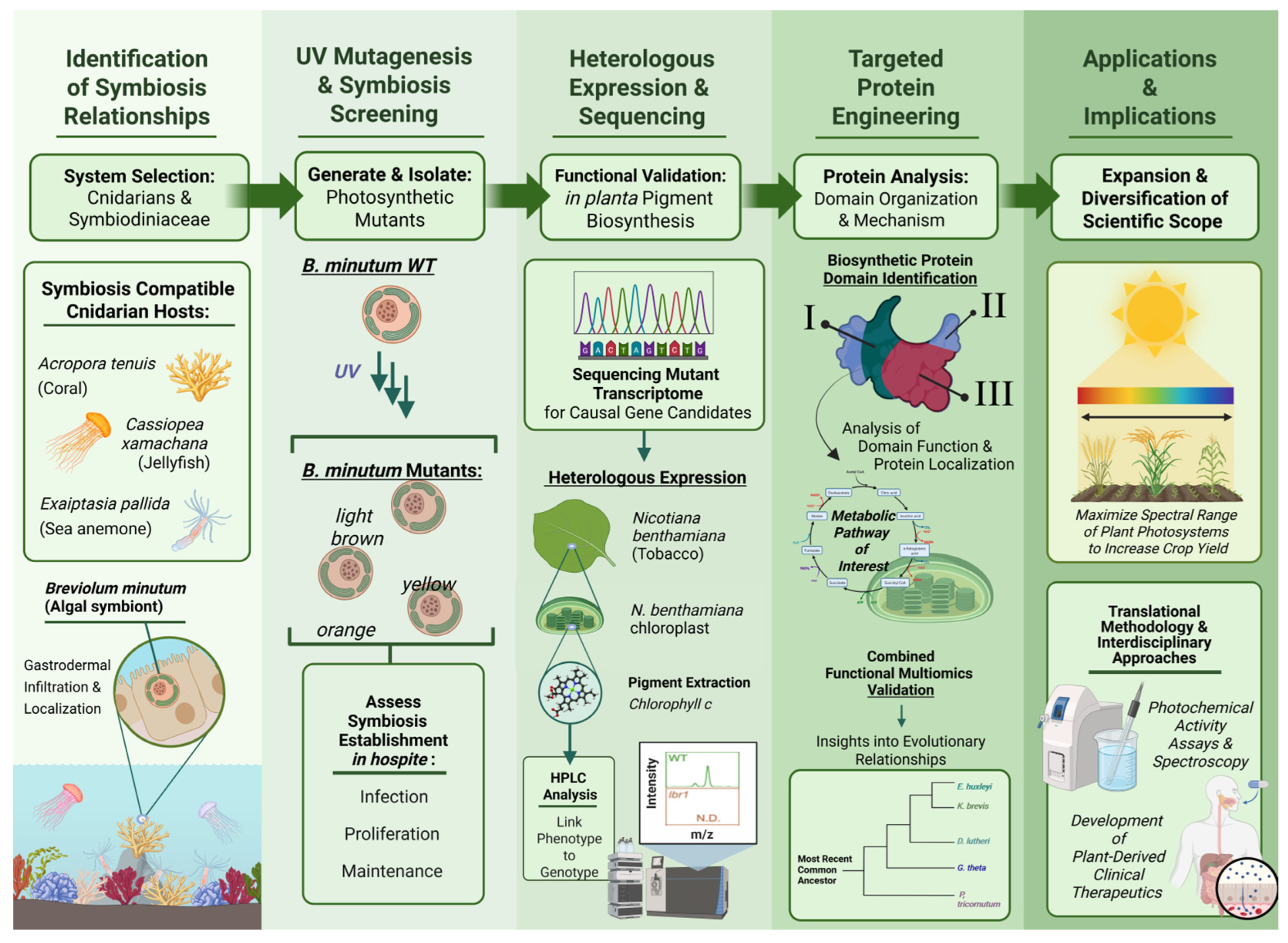
Functional Genomics to Decipher and Engineer Marine Symbioses

In this context, functional genomics offers a powerful framework for understanding biological processes and linking genotype to phenotype, thereby advancing insights into gene function [16]. For this purpose, functional genomics often involves disrupting or modifying an organism’s genome to observe the resulting phenotypic changes [17,18]. Techniques such as CRISPR (Clustered Regularly Interspaced Short Palindromic Repeats)-based genome editing, UV mutagenesis, and RNA interference (RNAi) have been instrumental in elucidating pathways underlying processes as diverse as osteoarthritis progression and chlorophyll production in marine algae [19,20,21,22,23]. In marine systems, functional genomics provides unique opportunities to unravel novel and unknown biosynthetic pathways and investigate mutualistic relationships, such as those between algae and cnidarians like corals [24,25]. This opinion piece highlights the potential of functional genomics to guide the engineering of symbiotic partners to enhance theirresilience against coral bleaching and mitigate biodiversity loss. These advances can also be applied to agricultural improvements in terms of photosynthetic efficiency and environmental stress tolerance [26,27].

2. Photosynthesis in Marine Algal Symbiosis

The study of photosynthesis within cnidarian–algal symbiosis is critical in understanding how environmental changes affect marine ecosystems [28]. Functional genomics has been instrumental in advancing this understanding, as demonstrated by the application of UV mutagenesis to Symbiodiniaceae algae, which provides a promising platform to investigate symbiont gene function and its role in the partnership [29]. Recent experiments using photosynthetically impaired algal mutants revealed that active photosynthesis is not required for the establishment of symbiosis, although it appears to be critical for supporting a healthy symbiont population [29,30,31]. These findings underscore the complexity of host–symbiont interactions and highlight the value of functional genomics in uncovering molecular mechanisms underlying symbiotic resilience.
Cnidarian–algal symbiosis establishment is characterized by three stages: infection, proliferation, and maintenance. Infection marks the initiation of cnidarian–algal symbiosis where cnidarian hosts acquire algal cells from the environment into their gastrodermis [2,29,32]. During infection, symbiont selection is thought to involve a winnowing process, potentially mediated by algal properties and host recognition events [33]. Upon successful infection, symbiont algae (and to a degree, host cells) undergo a period of rapid proliferation in hospite [34]. Maintenance occurs after infection and proliferation, when a steady-state algal population within the host is achieved under given environmental conditions [2].

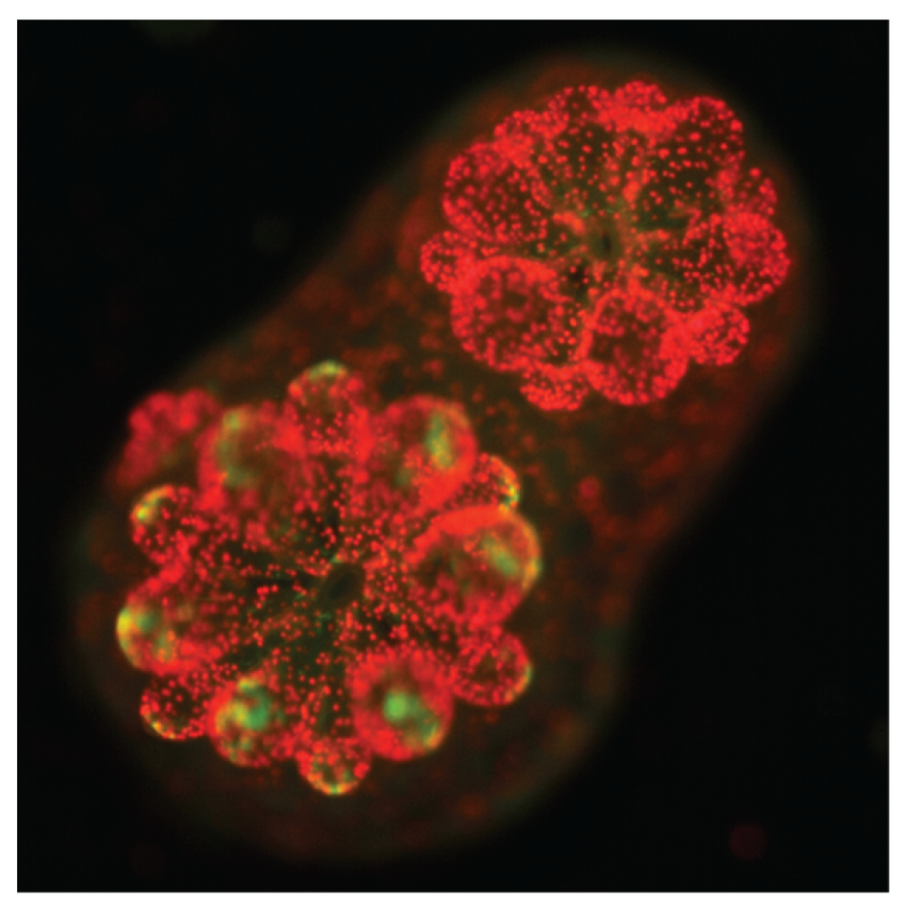
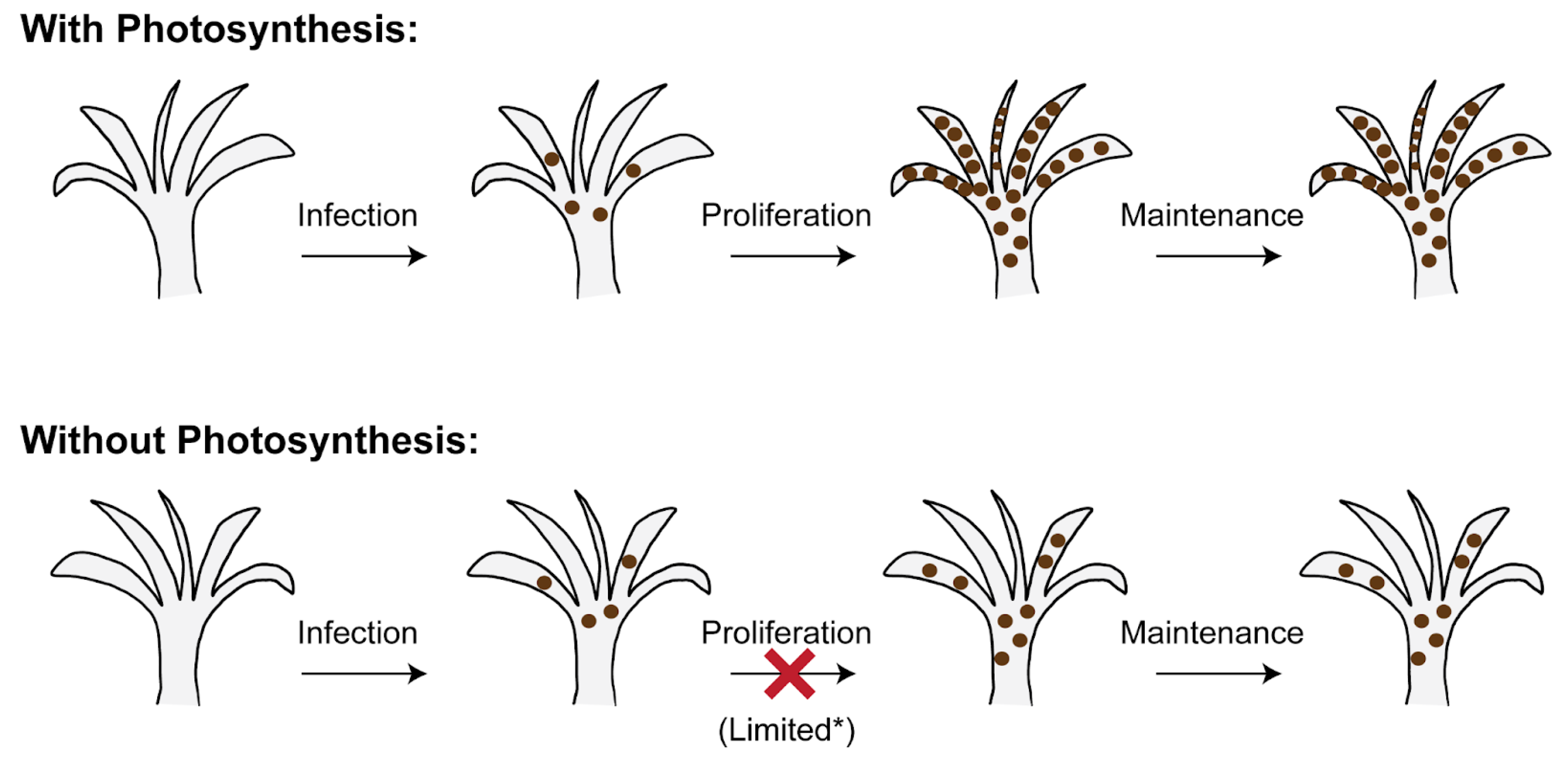
2.1. The Establishment of Cnidarian–Symbiodiniaceae Symbiosis Is Independent of Algal Photosynthesis

One characteristic trait of Symbiodiniaceae algae—photosynthesis—is thought to be a factor in symbiosis establishment. Studies with the Breviolum minutum strain SSB01 show that culturing algae in glucose-containing media under continuous light reduces their photosynthetic function and ability to infect sea anemone hosts [35]. However, these conditions also alter other physiological traits, such as cell surface complexity, complicating efforts to establish causality. To address this knowledge gap, Jinkerson et al., 2022 further investigated the role of photosynthesis during infection, proliferation, and maintenance stages of cnidarian–Symbiodiniaceae symbiosis by using darkness, a chemical inhibitor of photosynthesis (DCMU), and photosynthetic B. minutum mutants [29]. Cnidarian model systems, including with Exaiptasia pallida (Aiptasia), coral Acropora tenuis, and jellyfish Cassiopea xamachana, were used as hosts with B. minutum SSB01 and four additional symbiont species—Symbiodinium linucheae, Cladocopium goreaui, Durusdinium trenchii, and Symbiodinium necro appetens—to evaluate symbiotic establishment and account for species-specific host–symbiont interactions. Symbiont infection, proliferation, and maintenance of algal cells were evaluated and quantified using fluorescent imaging, revealing that symbiosis establishment occurs in continuous darkness and in the presence of DCMU. Following these initial experiments, the study employs functional genomics to generate B. minutum mutants and evaluate whether symbiosis establishment between cnidarian hosts and algal symbionts can occur independently of photosynthesis. Due to the lack of available transformation methods for Symbiodiniaceae, the study employs UV mutagenesis to introduce genetic disruptions. Mutants, initially screened for altered coloration, are followed by secondary screening to identify photoautotrophic growth deficiencies [36]. This approach yields six symbiont mutants, including LIGHT BROWN 1 (lbr1), ORANGE 1 (ora1), and YELLOW 7-9,12 (yel7, yel8, yel9, yel12). These mutants are then tested for their ability to establish, proliferate, and maintain symbiosis across three host systems [29].
The findings of the study by Jinkerson et al., 2022 reveals that the establishment of this symbiosis is specific to the cnidarian host and can occur independently of the photosynthetic ability of symbiont algae [29]. Specifically, their assessment across 31 host–symbiont pairs shows that photosynthesis is not necessary for Symbiodiniaceae to infect Aiptasia, coral, or Cassiopea, nor is it required for symbiont maintenance within cnidarian hosts. In particular, B. minutum was observed to be present in Aiptasia tissue for 180 days post infection in continuous darkness. However, the extent of symbiont proliferation in the absence of photosynthesis varied by host. While Acropora polyps supported algal proliferation without photosynthesis, both Aiptasia and Cassiopea showed limited symbiont growth under similar conditions [29] (Figure 2). This plasticity points to alternative metabolic interactions that may support symbiosis during stress or developmental transitions, such as in early life stages. For instance, juvenile Acropora polyps were able to support algal proliferation even in the absence of photosynthesis, hinting at life-stage-specific nutrient provisioning strategies.

2.2. Other Molecular and Environmental Modulators of Symbiosis Establishment

These observations raise important questions about the alternative molecular and cellular mechanisms that facilitate symbiosis in the absence of photosynthesis. For example, the ability of cnidarian–algal associations to initiate independently of photosynthesis highlights the possible involvement of host immune modulation and recognition pathways [32,37]. In the HydraChlorella model, for instance, microbe-associated molecular pattern (MAMP) recognition has been shown to mediate immune tolerance and help maintain symbiosis, suggesting that immune signaling may be central to initiating stable host–symbiont interactions [38]. Complementing these molecular insights, environmental conditions—including light, temperature, and pH—further modulate the physiological dynamics of the symbiotic relationship [39,40,41]. Light intensity, in particular, has a profound effect on dinoflagellate physiology, regulating growth, pigment composition, and stress responses. Transcriptomic analyses reveal that changes in environmental light conditions significantly alter gene expression in B. minutum, including genes involved in light harvesting, cell adhesion, and nutrient transport [42]. In Durusdinium glynnii, high light exposure induces the accumulation of peridinin, a photoprotective carotenoid that shields chlorophyll from photo-oxidation, suggesting an adaptive response to elevated irradiance [43]. Symbionts in the tropical hydroid Myrionema amboinense further demonstrate the ability to acclimate to low-light environments [44]. Beyond light, environmental stressors such as ocean acidification and pollutants can lower pH, often acting synergistically with warming and irradiance to trigger bleaching responses [45,46]. These findings underscore the multifactorial nature of symbiotic regulation and point to immune signaling, environmental sensing, and metabolic plasticity as key targets for engineering more resilient host–symbiont partnerships.

3. Research Beyond the Role of Photosynthesis Within Cnidarian–Algal Symbiosis

While photosynthesis is not essential for some steps in cnidarian–algal symbiosis, it remains a critical energetic pathway that ultimately supports nearly all life on Earth [4,47]. Beyond its ecological importance, photosynthesis also supplies and sustains human agriculture, yet its energy conversion efficiency in crop plants is estimated to be as low as 1% [48,49]. Therefore, improving photosynthetic efficiency is vital to meet growing agricultural demands; strategies include widening the usable light spectrum, enhancing light capture, and optimizing carbon fixation [50,51].

3.1. Identification of Chlorophyll c Biosynthesis Pathway in Dinoflagellates

In particular, researchers have explored strategies to enhance photosynthetic efficiency in crops by incorporating foreign chlorophyll pigments. One such pigment, chlorophylls c (Chls c), is predominantly found in marine algae such as diatoms, brown algae, dinoflagellates, and many other lineages of eukaryotic algae, where it captures blue-green wavelengths (447–452 nm) critical for photosynthesis in aquatic environments [52,53,54]. The absence of Chls c in terrestrial plants implies distinct ecological and evolutionary adaptations in marine algal species and represents the importance of understanding its biosynthetic pathways [55,56]. Elucidating the biological production pathway of critical chemicals, like chlorophyll c, is urgently needed for leveraging these pathways to optimize agricultural yields [57].
As such, the biosynthesis of Chls c in dinoflagellate Symbiodiniaceae has been investigated through the analysis of B. minutum photosynthesis mutants [20]. Among them, the mutant lbr1 exhibits less light absorbance from its typical blue–green spectrum. Upon further investigation with ultra-high-performance liquid chromatography–high-resolution mass spectrometry (UHPLC-HRMS), they discover that the lbr1 mutant, in particular, was unable to produce Chls c. Sequencing of its transcriptome further reveals multiple mutations, including a frameshift mutation in gene s6_3623. The study identifies the s6_3623 gene as the causal gene for Chls c production by heterologous expression of s6_3623 in Nicotiana benthamiana. Determining that this gene codes for an enzyme, identified and named “BmCHLCS (CHLOROPHYLL C SYNTHASE),” the study confirms its vital role in algal Chls c biosynthesis through the heterologous production of Chls c in N. benthamiana [20].
To address the initially limited production of Chls c in N. benthamiana extracts, the researchers use a dual-intervention approach consisting of the usage of an δ-Aminolevulinic acid (ALA) substrate followed by dark incubation. The resulting increase in Chls c production in N. benthamiana enables mass spectrometry detection of peaks corresponding to Chls c1 and c2., confirming the light independence of BmCHLCS. In order to determine the catalytic domain responsible for Chls c production, the study evaluates BmCHCLS function through assessment of the chlorophyll a/b binding and 2-oxoglutarate-Fe(II) dioxygenase (2OGD) domains. Subsequent assessment of BmCHCLS function by selective deletion of either of the two domains reveals that 2OGD is the catalytic domain as only 2OGD deletion affects the production of Chls c, resulting in no detectable levels of chlorophyll c1 and c2 in the leaves of N. benthamiana [20]. Enzymes with 2OGD dioxygenase domains are known to biosynthesize a wide range of metabolites in plants, including hormones, alkaloids, and other natural products, with most characterized pathways localized to the cytosol or mitochondria [58,59,60]. This study identifies a 2OGD-dependant pathway in the chloroplast, expanding the known sites for 2OGD-associated biosynthetic reactions and illuminating new opportunities for chloroplast engineering. Further investigation into BmCHLCS reveals that a bipartite signal and transit peptide sequence directs the enzyme to the chloroplast, enabling its function in Chls c biosynthesis. Interestingly, the chlorophyll a/b binding domain, while not essential for Chls c biosynthesis, appears to facilitate localization of BmCHLC in the chloroplast, highlighting a unique mechanism with potential implications for the enzyme’s structure and function [20].
A comprehensive analysis of CHLCS distribution reveals its presence in peridinin-containing dinoflagellates, while all chlorophyll c-dependent algal lineages encode 2OGD-containing proteins (CHLCs), forming a strongly supported monophyletic group in phylogenetic analyses. However, a subset of Ochrophyta lacks CHLCS or chlorophyll c homologs despite their reliance on chlorophyll c, suggesting the presence of alternative enzyme(s) responsible for chlorophyll c biosynthesis [20]. Investigating these pathways offers an exciting opportunity to uncover novel enzymes, shedding light on the evolution of pigment biosynthesis in ochrophytes and their adaptation to diverse ecological niches [61,62,63].

3.2. From Dinoflagellates to Plants for Enhanced Light Capture and Artificial Photosynthesis

Overall, the identification of the gene responsible for chlorophyll c production represents a significant milestone, given the inherent challenges of functional genomics in dinoflagellates. This study marks the first successful heterologous production of chlorophyll c in plants, opening the door to engineering terrestrial plants with expanded pigment diversity and enhanced light-harvesting capacity. As efforts intensify to develop crops with improved resilience to climate stress and more efficient solar energy capture, biomimetic strategies inspired by photosynthetic systems are gaining momentum [64]. Recent studies demonstrate how bioinspired designs can enhance photovoltaic performance while reducing heat output [65]. Parallel advances in artificial photosynthesis have demonstrated efficient CO2 fixation and solar-to-food energy conversion, enabling the production of concentrated acetate streams that can support heterotrophic growth of various organisms—including photosynthetic algae—without reliance on natural photosynthesis [66]. Together, these innovations point to new frontiers in sustainable agriculture and controlled-environment food production, with the potential to surpass the efficiency of traditional photosynthetic systems.

4. Marine Pharmacology: A Sustainable Frontier for Therapeutic Discovery

Beyond just the use of functional genomics to uncover biosynthetic pathways in plant photosynthetic pigments, these strategies offer immense potential in marine systems, where they can illuminate the molecular mechanisms underlying the production of bioactive compounds [67]. Marine pharmacology represents a sustainable and innovative frontier for therapeutic discovery, bridging ecological research with clinical advancement. By exploring the vast chemical diversity of marine organisms—such as algae, cyanobacteria, sponges, and corals—this field seeks to identify novel bioactive compounds with applications in treating cancer, infection, inflammation, and neurological disorders [68,69,70]. These compounds, often shaped by evolutionary pressures in complex symbiotic or competitive marine ecosystems, offer unique structural scaffolds and mechanisms of action that are rarely found in terrestrial organisms [71,72]. To date, over 13,000 molecules derived from marine species have been identified, with approximately 3000 exhibiting significant pharmacological activity [73,74]. These compounds, primarily secondary metabolites, are not directly involved in essential biological processes such as growth or reproduction but possess immense therapeutic potential. They could offer solutions to pressing medical challenges, including cancer; bacterial, viral, parasitic, and fungal infections; and applications in bioregenerative medicine [75,76,77].

Bridging Gaps in Biosynthetic Mechanisms Is Critical for Novel Therapeutic Development

While the therapeutic potential of marine-derived compounds spans numerous fields, gastrointestinal research presents particularly promising and readily feasible translational opportunities [78,79,80]. Advances in gut microbiome modulation—such as microbial fecal implants and supplements—could mitigate current challenges, including poorly characterized disease mechanisms, reliance on complex animal models with limited clinical relevance, immune rejection of treatments, and uncertainties surrounding long-term outcomes [81,82,83]. Chronic and acute inflammatory gastrointestinal disorders, such as ulcerative colitis, diverticulitis, pancreatitis, and irritable bowel syndrome, are often linked to microbial imbalances or disruptions in microbe–host homeostasis [84,85]. Insights into these conditions could be gained by applying functional systems biology approaches that are informed by analogies to algal symbiosis and biosynthesis, facilitating improved diagnosis, prognosis, and therapeutic outcomes [14,86].
Despite its vast potential, marine pharmacology faces significant challenges. Comprehensive reviews by Malve et al., 2016 and Mayer et al., 2019 highlight the therapeutic promise of marine-derived pharmaceuticals and their capacity to expand the repertoire of available therapeutic agents [73,74]. However, only a small fraction of marine natural products and their derivatives have been approved or are undergoing clinical testing by the U.S. Food and Drug Administration (USFDA) [87,88,89,90]. This slow progress stems from gaps in understanding the biosynthetic pathways of these compounds, limiting the efficiency and sustainability of their extraction and formulation [15]. Addressing these obstacles through functional genomics and other advanced techniques will be critical for unlocking the full potential of marine systems in drug discovery and development.

5. Advancing Genomic Research in Marine Symbiosis and Beyond

Understanding marine symbiosis, particularly in cnidarian–algal interactions, is pivotal for ecological and biotechnological progress. This symbiosis establishment is impacted by a complex interplay of environmental factors such as light, temperature, salinity, and nutrient availability. Investigating the molecular and cellular mechanisms underlying algal infection into host gastrodermal cells, including processes like cell-surface recognition and symbiosome formation, remains an essential frontier. Unraveling these processes will provide crucial insights into the specificity and stability of symbiotic relationships, with broad implications for ecosystem resilience and health (Figure 3).

5.1. Limitations in Genetic Engineering Tools for Studying Symbiodiniaceae Algae

Genomic and genetic research in Symbiodiniaceae species also represents a critical avenue for future work. However, challenges posed by their unique genomic organization, including permanently condensed chromosomes and lack of traditional transcriptional regulation, limit the application of conventional genetic tools like CRISPR-Cas9 [91,92,93]. Expanding these efforts to sequence a broader range of Symbiodiniaceae genomes across diverse species is essential for capturing the genetic diversity and evolutionary adaptations within this ecologically important family [94,95,96]. Such diversity will provide valuable insights into species-specific traits, host compatibility, and environmental resilience [97]. Despite the importance of functional genomic studies, progress is limited by the complexity of genetic manipulation, which contrasts with the simpler methods used in bacterial, viral, or algal systems [98,99,100,101,102]. Promising advancements in cell-wall deconstruction have shown potential for improving transformation efficiency, offering a path forward for developing robust genetic manipulation methods [103]. Further inquiry into novel genomic approaches is vital for overcoming these barriers. Ultimately, integrating functional genomics with metabolomics and proteomics will help elucidate the molecular mechanisms of symbiosis and provide a framework for addressing broader ecological and biotechnological challenges [104,105].

5.2. Conclusions and Outlook

These insights hold broad applications beyond symbiosis research. Marine pharmacology, for example, highlights the therapeutic potential of marine bioactive compounds, which could address critical medical challenges. Specifically, understanding the biosynthetic pathways of these compounds through functional genomics could enhance sustainable drug development. Similarly, advancements in photosynthetic biology, such as research on chlorophyll c synthase in marine algae, offer pathways to improve photosynthetic efficiency and resilience in terrestrial crops. Integrating genomics with metabolomics and proteomics enables the discovery of the molecular mechanisms underlying symbiosis and photosynthesis, fostering innovations in sustainable agriculture, climate adaptation, and biomedicine. Robust genomic resources and methodologies tailored to Symbiodiniaceae are essential not only for unraveling the complexities of marine symbiosis, but also for leveraging marine systems to address global challenges in health, agriculture, and environmental sustainability.

Author Contributions

Conceptualization, T.X.; writing—original draft preparation, G.M., J.P.H., A.T. and T.X.; writing—review and editing, G.M., A.T., J.P.H. and T.X.; figure contributions, G.M. and T.X.; funding acquisition, T.X. All authors have read and agreed to the published version of the manuscript.

Funding

This work was supported by funds provided by NSF-IOS EDGE Award to T.X. (2308644).

Acknowledgments

We would like to acknowledge Michelle Taepakdee for her help with editing this paper. Additionally, this work was conducted as part of the Cellular and Molecular Engineering course. We thank our classmates for their valuable feedback and support throughout the project. This class provided the foundational knowledge and collaborative environment essential to the development of this study.

Conflicts of Interest

The authors declare no competing interests.

Abbreviations

The following abbreviations are used in this manuscript:
ALAδ-Aminolevulinic acide
BmCHCLSBreviolum minutum Chlorophyll c Synthase
Chls cChlorophyll c
CRISPRClustered Regularly Interspaced Short Palindromic Repeats
DCMU3-(3,4-dichlorophenyl)-1,1-dimethylurea
MAMPMicrobe Associated Molecular Patterns
RNAiRibonucleic Acid Interference
UHPLC-HRMSUltra-High-Performance Liquid Chromatography–High-Resolution Mass Spectrometry
USFDAUnited States Food and Drug Administration
UVUltraviolet
2OGD2-oxyglutarate-Fe(II) dioxygenase
B. minutumBreviolum minutum
N. benthamianaNicotiana benthamiana
B. minutum Mutants
lbr1Light Brown 1
ora1Orange 1
yel7-12Yellow 7-12

References

  1. Weis, V.M.; Allemand, D. Physiology. What Determines Coral Health? Science 2009, 324, 1153–1155. [Google Scholar] [CrossRef]
  2. Davy, S.K.; Allemand, D.; Weis, V.M. Cell Biology of Cnidarian-Dinoflagellate Symbiosis. Microbiol. Mol. Biol. Rev. 2012, 76, 229–261. [Google Scholar] [CrossRef]
  3. Gordon, B.R.; Leggat, W. Symbiodinium-Invertebrate Symbioses and the Role of Metabolomics. Mar. Drugs 2010, 8, 2546–2568. [Google Scholar] [CrossRef]
  4. Baker, A. Flexibility and Specificity in Coral-Algal Symbiosis: Diversity, Ecology, and Biogeography of Symbiodinium. Annu. Rev. Ecol. Evol. Syst. 2003, 34, 661–689. [Google Scholar] [CrossRef]
  5. Yellowlees, D.; Rees, T.A.V.; Leggat, W. Metabolic Interactions between Algal Symbionts and Invertebrate Hosts. Plant Cell Environ. 2008, 31, 679–694. [Google Scholar] [CrossRef]
  6. Fisher, R.; O’Leary, R.A.; Low-Choy, S.; Mengersen, K.; Knowlton, N.; Brainard, R.E.; Caley, M.J. Species Richness on Coral Reefs and the Pursuit of Convergent Global Estimates. Curr. Biol. 2015, 25, 500–505. [Google Scholar] [CrossRef]
  7. Hoegh-Guldberg, O.; Poloczanska, E.S.; Skirving, W.; Dove, S. Coral Reef Ecosystems under Climate Change and Ocean Acidification. Front. Mar. Sci. 2017, 4, 158. [Google Scholar] [CrossRef]
  8. Ainsworth, T.D.; Brown, B.E. Coral Bleaching. Curr. Biol. 2021, 31, R5–R6. [Google Scholar] [CrossRef]
  9. Douglas, A.E. Coral Bleaching—How and Why? Mar. Pollut. Bull. 2003, 46, 385–392. [Google Scholar] [CrossRef]
  10. Hoegh-Guldberg, O. Climate Change, Coral Bleaching and the Future of the World’s Coral Reefs. Mar. Freshw. Res. 1999, 50, 839. [Google Scholar] [CrossRef]
  11. Helgoe, J.; Davy, S.K.; Weis, V.M.; Rodriguez-Lanetty, M. Triggers, Cascades, and Endpoints: Connecting the Dots of Coral Bleaching Mechanisms. Biol. Rev. Camb. Philos. Soc. 2024, 99, 715–752. [Google Scholar] [CrossRef] [PubMed]
  12. Allen-Waller, L.; Barott, K.L. Symbiotic Dinoflagellates Divert Energy Away from Mutualism during Coral Bleaching Recovery. Symbiosis 2023, 89, 173–186. [Google Scholar] [CrossRef]
  13. Fitt, W.; Brown, B.; Warner, M.; Dunne, R. Coral Bleaching: Interpretation of Thermal Tolerance Limits and Thermal Thresholds in Tropical Corals. Coral Reefs 2001, 20, 51–65. [Google Scholar] [CrossRef]
  14. Dai, N.; Wang, Q.; Xu, B.; Chen, H. Remarkable Natural Biological Resource of Algae for Medical Applications. Front. Mar. Sci. 2022, 9, 912924. [Google Scholar] [CrossRef]
  15. Oliveira, C.Y.B.; Oliveira, C.D.L.; Müller, M.N.; Santos, E.P.; Dantas, D.M.M.; Gálvez, A.O. A Scientometric Overview of Global Dinoflagellate Research. Publications 2020, 8, 50. [Google Scholar] [CrossRef]
  16. Przybyla, L.; Gilbert, L.A. A New Era in Functional Genomics Screens. Nat. Rev. Genet. 2022, 23, 89–103. [Google Scholar] [CrossRef] [PubMed]
  17. Fields, S.; Kohara, Y.; Lockhart, D.J. Functional Genomics. Proc. Natl. Acad. Sci. USA 1999, 96, 8825–8826. [Google Scholar] [CrossRef]
  18. Hieter, P.; Boguski, M. Functional Genomics: It’s All How You Read It. Science 1997, 278, 601–602. [Google Scholar] [CrossRef]
  19. Kim, H.S.; Kweon, J.; Kim, Y. Recent Advances in CRISPR-Based Functional Genomics for the Study of Disease-Associated Genetic Variants. Exp. Mol. Med. 2024, 56, 861–869. [Google Scholar] [CrossRef]
  20. Jinkerson, R.E.; Poveda-Huertes, D.; Cooney, E.C.; Cho, A.; Ochoa-Fernandez, R.; Keeling, P.J.; Xiang, T.; Andersen-Ranberg, J. Biosynthesis of Chlorophyll c in a Dinoflagellate and Heterologous Production in Planta. Curr. Biol. 2024, 34, 594–605.e4. [Google Scholar] [CrossRef]
  21. Razzaq, M.K.; Aleem, M.; Mansoor, S.; Khan, M.A.; Rauf, S.; Iqbal, S.; Siddique, K.H.M. Omics and CRISPR-Cas9 Approaches for Molecular Insight, Functional Gene Analysis, and Stress Tolerance Development in Crops. Int. J. Mol. Sci. 2021, 22, 1292. [Google Scholar] [CrossRef]
  22. Todor, H.; Silvis, M.R.; Osadnik, H.; Gross, C.A. Bacterial CRISPR Screens for Gene Function. Curr. Opin. Microbiol. 2021, 59, 102–109. [Google Scholar] [CrossRef] [PubMed]
  23. Shi, H.; Doench, J.G.; Chi, H. CRISPR Screens for Functional Interrogation of Immunity. Nat. Rev. Immunol. 2023, 23, 363–380. [Google Scholar] [CrossRef]
  24. Martín-Zamora, F.M.; Liang, Y.; Guynes, K.; Carrillo-Baltodano, A.M.; Davies, B.E.; Donnellan, R.D.; Tan, Y.; Moggioli, G.; Seudre, O.; Tran, M.; et al. Annelid Functional Genomics Reveal the Origins of Bilaterian Life Cycles. Nature 2023, 615, 105–110. [Google Scholar] [CrossRef]
  25. Hirooka, S.; Itabashi, T.; Ichinose, T.M.; Onuma, R.; Fujiwara, T.; Yamashita, S.; Jong, L.W.; Tomita, R.; Iwane, A.H.; Miyagishima, S.-Y. Life Cycle and Functional Genomics of the Unicellular Red Alga Galdieria for Elucidating Algal and Plant Evolution and Industrial Use. Proc. Natl. Acad. Sci. USA 2022, 119, e2210665119. [Google Scholar] [CrossRef] [PubMed]
  26. Falk, D.A.; Watts, A.C.; Thode, A.E. Scaling Ecological Resilience. Front. Ecol. Evol. 2019, 7, 275. [Google Scholar] [CrossRef]
  27. Wang, Y.; Cai, Y.; Xie, Y.; Zhang, P.; Chen, L. A Quantitative Framework to Evaluate Urban Ecological Resilience: Broadening Understanding through Multi-Attribute Perspectives. Front. Ecol. Evol. 2023, 11, 1144244. [Google Scholar] [CrossRef]
  28. Venn, A.A.; Loram, J.E.; Douglas, A.E. Photosynthetic Symbioses in Animals. J. Exp. Bot. 2008, 59, 1069–1080. [Google Scholar] [CrossRef]
  29. Jinkerson, R.E.; Russo, J.A.; Newkirk, C.R.; Kirk, A.L.; Chi, R.J.; Martindale, M.Q.; Grossman, A.R.; Hatta, M.; Xiang, T. Cnidarian-Symbiodiniaceae Symbiosis Establishment Is Independent of Photosynthesis. Curr. Biol. 2022, 32, 2402–2415.e4. [Google Scholar] [CrossRef]
  30. Jones, R.J. Testing the ‘photoinhibition’ Model of Coral Bleaching Using Chemical Inhibitors. Mar. Ecol. Prog. Ser. 2004, 284, 133–145. [Google Scholar] [CrossRef]
  31. Zaquin, T.; Zaslansky, P.; Pinkas, I.; Mass, T. Simulating Bleaching: Long-Term Adaptation to the Dark Reveals Phenotypic Plasticity of the Mediterranean Sea Coral Oculina Patagonica. Front. Mar. Sci. 2019, 6, 662. [Google Scholar] [CrossRef]
  32. Jacobovitz, M.R.; Hambleton, E.A.; Guse, A. Unlocking the Complex Cell Biology of Coral-Dinoflagellate Symbiosis: A Model Systems Approach. Annu. Rev. Genet. 2023, 57, 411–434. [Google Scholar] [CrossRef] [PubMed]
  33. Nyholm, S.V.; McFall-Ngai, M.J. The Winnowing: Establishing the Squid-Vibrio Symbiosis. Nat. Rev. Microbiol. 2004, 2, 632–642. [Google Scholar] [CrossRef]
  34. Tivey, T.R.; Parkinson, J.E.; Weis, V.M. Host and Symbiont Cell Cycle Coordination Is Mediated by Symbiotic State, Nutrition, and Partner Identity in a Model Cnidarian-Dinoflagellate Symbiosis. MBio 2020, 11, e02626-19. [Google Scholar] [CrossRef]
  35. Xiang, T.; Jinkerson, R.E.; Clowez, S.; Tran, C.; Krediet, C.J.; Onishi, M.; Cleves, P.A.; Pringle, J.R.; Grossman, A.R. Glucose-Induced Trophic Shift in an Endosymbiont Dinoflagellate with Physiological and Molecular Consequences. Plant Physiol. 2018, 176, 1793–1807. [Google Scholar] [CrossRef] [PubMed]
  36. Russo, J.A.; Xiang, T.; Jinkerson, R.E. Protocol for the Generation of Symbiodiniaceae Mutants Using UV Mutagenesis. STAR Protoc. 2023, 4, 102627. [Google Scholar] [CrossRef]
  37. Rosset, S.L.; Oakley, C.A.; Ferrier-Pagès, C.; Suggett, D.J.; Weis, V.M.; Davy, S.K. The Molecular Language of the Cnidarian-Dinoflagellate Symbiosis. Trends Microbiol. 2021, 29, 320–333. [Google Scholar] [CrossRef]
  38. Douglas, A.; Smith, D.C. The Green Hydra Symbiosis. VIII. Mechanisms in Symbiont Regulation. Proc. R. Soc. Lond. 1984, 221, 291–319. [Google Scholar] [CrossRef]
  39. Aihara, Y.; Maruyama, S.; Baird, A.H.; Iguchi, A.; Takahashi, S.; Minagawa, J. Green Fluorescence from Cnidarian Hosts Attracts Symbiotic Algae. Proc. Natl. Acad. Sci. USA 2019, 116, 2118–2123. [Google Scholar] [CrossRef]
  40. Roth, M.S. The Engine of the Reef: Photobiology of the Coral–Algal Symbiosis. Front. Microbiol. 2014, 5, 422. [Google Scholar] [CrossRef]
  41. Rehman, A.U.; Szabó, M.; Deák, Z.; Sass, L.; Larkum, A.; Ralph, P.; Vass, I. Symbiodinium Sp. Cells Produce Light-Induced Intra- and Extracellular Singlet Oxygen, Which Mediates Photodamage of the Photosynthetic Apparatus and Has the Potential to Interact with the Animal Host in Coral Symbiosis. New Phytol. 2016, 212, 472–484. [Google Scholar] [CrossRef] [PubMed]
  42. Xiang, T.; Nelson, W.; Rodriguez, J.; Tolleter, D.; Grossman, A.R. Symbiodinium Transcriptome and Global Responses of Cells to Immediate Changes in Light Intensity When Grown under Autotrophic or Mixotrophic Conditions. Plant J. 2015, 82, 67–80. [Google Scholar] [CrossRef]
  43. Oliveira, C.Y.B.; Abreu, J.L.; Santos, E.P.; Matos, Â.P.; Tribuzi, G.; Oliveira, C.D.L.; Veras, B.O.; Bezerra, R.S.; Müller, M.N.; Gálvez, A.O. Light Induces Peridinin and Docosahexaenoic Acid Accumulation in the Dinoflagellate Durusdinium Glynnii. Appl. Microbiol. Biotechnol. 2022, 106, 6263–6276. [Google Scholar] [CrossRef] [PubMed]
  44. Fitt, W.K.; Cook, C.B. Photoacclimation and the Effect of the Symbiotic Environment on the Photosynthetic Response of Symbiotic Dinoflagellates in the Tropical Marine Hydroid Myrionema Amboinense. J. Exp. Mar. Bio. Ecol. 2001, 256, 15–31. [Google Scholar] [CrossRef]
  45. Sun, Y.; Jiang, L.; Gong, S.; Guo, M.; Yuan, X.; Zhou, G.; Lei, X.; Zhang, Y.; Yuan, T.; Lian, J.; et al. Impact of Ocean Warming and Acidification on Symbiosis Establishment and Gene Expression Profiles in Recruits of Reef Coral Acropora Intermedia. Front. Microbiol. 2020, 11, 532447. [Google Scholar] [CrossRef]
  46. Grottoli, A.G.; Dalcin Martins, P.; Wilkins, M.J.; Johnston, M.D.; Warner, M.E.; Cai, W.-J.; Melman, T.F.; Hoadley, K.D.; Pettay, D.T.; Levas, S.; et al. Coral Physiology and Microbiome Dynamics under Combined Warming and Ocean Acidification. PLoS ONE 2018, 13, e0191156. [Google Scholar] [CrossRef] [PubMed]
  47. Galston, A.W. Photosynthesis as a Basis for Life Support on Earth and in Space. Bioscience 1992, 42, 490–493. [Google Scholar] [CrossRef]
  48. Grayson, K.J.; Faries, K.M.; Huang, X.; Qian, P.; Dilbeck, P.; Martin, E.C.; Hitchcock, A.; Vasilev, C.; Yuen, J.M.; Niedzwiedzki, D.M.; et al. Augmenting Light Coverage for Photosynthesis through YFP-Enhanced Charge Separation at the Rhodobacter Sphaeroides Reaction Centre. Nat. Commun. 2017, 8, 13972. [Google Scholar] [CrossRef]
  49. Hitchcock, A.; Hunter, C.N.; Sobotka, R.; Komenda, J.; Dann, M.; Leister, D. Redesigning the Photosynthetic Light Reactions to Enhance Photosynthesis—The PhotoRedesign Consortium. Plant J. 2022, 109, 23–34. [Google Scholar] [CrossRef]
  50. Vullev, V.I. From Biomimesis to Bioinspiration: What’s the Benefit for Solar Energy Conversion Applications? J. Phys. Chem. Lett. 2011, 2, 503–508. [Google Scholar] [CrossRef]
  51. Su, J.; Vayssieres, L. A Place in the Sun for Artificial Photosynthesis? ACS Energy Lett. 2016, 1, 121–135. [Google Scholar] [CrossRef]
  52. Myśliwa-Kurdziel, B.; Latowski, D.; Strzałka, K. Chlorophylls c—Occurrence, Synthesis, Properties, Photosynthetic and Evolutionary Significance. In Advances in Botanical Research; Elsevier: Amsterdam, The Netherlands, 2019; pp. 91–119. ISBN 9780081027523. [Google Scholar] [CrossRef]
  53. Reinbothe, S.; Reinbothe, C. The Regulation of Enzymes Involved in Chlorophyll Biosynthesis. Eur. J. Biochem. 1996, 237, 323–343. [Google Scholar] [CrossRef]
  54. Averina, S.G.; Velichko, N.V.; Pinevich, A.A.; Senatskaya, E.V.; Pinevich, A.V. Non-a Chlorophylls in Cyanobacteria. Photosynthetica 2019, 57, 1109–1118. [Google Scholar] [CrossRef]
  55. Wang, P.; Grimm, B. Connecting Chlorophyll Metabolism with Accumulation of the Photosynthetic Apparatus. Trends Plant Sci. 2021, 26, 484–495. [Google Scholar] [CrossRef]
  56. Peterhansel, C.; Niessen, M.; Kebeish, R.M. Metabolic Engineering towards the Enhancement of Photosynthesis. Photochem. Photobiol. 2008, 84, 1317–1323. [Google Scholar] [CrossRef]
  57. Liu, Y.; Gui, Z.; Liu, J. Research Progress of Light Wavelength Conversion Materials and Their Applications in Functional Agricultural Films. Polymers 2022, 14, 851. [Google Scholar] [CrossRef] [PubMed]
  58. Eckhardt, U.; Grimm, B.; Hörtensteiner, S. Recent Advances in Chlorophyll Biosynthesis and Breakdown in Higher Plants. Plant Mol. Biol. 2004, 56, 1–14. [Google Scholar] [CrossRef]
  59. Rebeiz, C.A.; Benning, C.; Bohnert, H.J.; Daniell, H.; Hoober, J.K.; Lichtenthaler, H.K.; Portis, A.R.; Tripathy, B.C. The Chloroplast: Basics and Applications; Springer: Dordrecht, The Netherlands, 2010; p. 426. [Google Scholar]
  60. Wei, S.; Zhang, W.; Fu, R.; Zhang, Y. Genome-Wide Characterization of 2-Oxoglutarate and Fe(II)-Dependent Dioxygenase Family Genes in Tomato during Growth Cycle and Their Roles in Metabolism. BMC Genomics 2021, 22, 126. [Google Scholar] [CrossRef]
  61. Bolte, K.; Bullmann, L.; Hempel, F.; Bozarth, A.; Zauner, S. Maier Protein Targeting into Secondary Plastids 1. J. Eukaryot. Microbiol. 2009, 56, 9–15. [Google Scholar] [CrossRef]
  62. Cavalier-Smith, T. Principles of Protein and Lipid Targeting in Secondary Symbiogenesis: Euglenoid, Dinoflagellate, and Sporozoan Plastid Origins and the Eukaryote Family Tree. J. Eukaryot. Microbiol. 1999, 46, 347–366. [Google Scholar] [CrossRef]
  63. Raven, J.A. Cells inside Cells: Symbiosis and Continuing Phagotrophy. Curr. Biol. 2013, 23, R530–R531. [Google Scholar] [CrossRef]
  64. Liu, G.; Gao, F.; Gao, C.; Xiong, Y. Bioinspiration toward Efficient Photosynthetic Systems: From Biohybrids to Biomimetics. Chem Catal. 2021, 1, 1367–1377. [Google Scholar] [CrossRef]
  65. Huang, G.; Xu, J.; Markides, C.N. High-Efficiency Bio-Inspired Hybrid Multi-Generation Photovoltaic Leaf. Nat. Commun. 2023, 14, 3344. [Google Scholar] [CrossRef]
  66. Hann, E.C.; Overa, S.; Harland-Dunaway, M.; Narvaez, A.F.; Le, D.N.; Orozco-Cárdenas, M.L.; Jiao, F.; Jinkerson, R.E. A Hybrid Inorganic-Biological Artificial Photosynthesis System for Energy-Efficient Food Production. Nat. Food 2022, 3, 461–471. [Google Scholar] [CrossRef] [PubMed]
  67. Liang, X.; Luo, D.; Luesch, H. Advances in Exploring the Therapeutic Potential of Marine Natural Products. Pharmacol. Res. 2019, 147, 104373. [Google Scholar] [CrossRef] [PubMed]
  68. Karthikeyan, A.; Joseph, A.; Nair, B.G. Promising Bioactive Compounds from the Marine Environment and Their Potential Effects on Various Diseases. J. Genet. Eng. Biotechnol. 2022, 20, 14. [Google Scholar] [CrossRef]
  69. Blunt, J.W.; Carroll, A.R.; Copp, B.R.; Davis, R.A.; Keyzers, R.A.; Prinsep, M.R. Marine Natural Products. Nat. Prod. Rep. 2018, 35, 8–53. [Google Scholar] [CrossRef]
  70. Yang, J.; Tan, S.; Ge, S.; Yang, M.; Liu, H.; Liu, W.; Zhang, K.; Zhang, Z.; Wang, Z.-H.; Shi, J.; et al. Cyanobacteria-Probiotics Symbionts for Modulation of Intestinal Inflammation and Microbiome Dysregulation in Colitis. Proc. Natl. Acad. Sci. USA 2024, 121, e2403417121. [Google Scholar] [CrossRef]
  71. Datta, D.; Nath Talapatra, S.; Swarnakar, S. Bioactive Compounds from Marine Invertebrates for Potential Medicines—An Overview. Int. Lett. Nat. Sci. 2015, 34, 42–61. [Google Scholar] [CrossRef]
  72. Stonik, V.A. Marine Natural Products: A Way to New Drugs. Acta Naturae 2009, 1, 15–25. [Google Scholar] [CrossRef]
  73. Mayer, A.M.S.; Guerrero, A.J.; Rodríguez, A.D.; Taglialatela-Scafati, O.; Nakamura, F.; Fusetani, N. Marine Pharmacology in 2014–2015: Marine Compounds with Antibacterial, Antidiabetic, Antifungal, Anti-Inflammatory, Antiprotozoal, Antituberculosis, Antiviral, and Anthelmintic Activities; Affecting the Immune and Nervous Systems, and Other Miscellaneous Mechanisms of Action. Mar. Drugs 2019, 18, 5. [Google Scholar] [CrossRef] [PubMed]
  74. Malve, H. Exploring the Ocean for New Drug Developments: Marine Pharmacology. J. Pharm. Bioallied Sci. 2016, 8, 83–91. [Google Scholar] [CrossRef] [PubMed]
  75. Hanif, N.; Murni, A.; Tanaka, C.; Tanaka, J. Marine Natural Products from Indonesian Waters. Mar. Drugs 2019, 17, 364. [Google Scholar] [CrossRef] [PubMed]
  76. Zheng, L.-H.; Wang, Y.-J.; Sheng, J.; Wang, F.; Zheng, Y.; Lin, X.-K.; Sun, M. Antitumor Peptides from Marine Organisms. Mar. Drugs 2011, 9, 1840–1859. [Google Scholar] [CrossRef]
  77. Romano, G.; Almeida, M.; Varela Coelho, A.; Cutignano, A.; Gonçalves, L.G.; Hansen, E.; Khnykin, D.; Mass, T.; Ramšak, A.; Rocha, M.S.; et al. Biomaterials and Bioactive Natural Products from Marine Invertebrates: From Basic Research to Innovative Applications. Mar. Drugs 2022, 20, 219. [Google Scholar] [CrossRef]
  78. Rudi, K.; Zhao, L. Grand Challenges in Understanding Gut Microbes. Front. Microbiol. 2021, 12, 752829. [Google Scholar] [CrossRef]
  79. Diaz, J.; Reese, A.T. Possibilities and Limits for Using the Gut Microbiome to Improve Captive Animal Health. Anim. Microbiome 2021, 3, 89. [Google Scholar] [CrossRef]
  80. Kim, D.; Hofstaedter, C.E.; Zhao, C.; Mattei, L.; Tanes, C.; Clarke, E.; Lauder, A.; Sherrill-Mix, S.; Chehoud, C.; Kelsen, J.; et al. Optimizing Methods and Dodging Pitfalls in Microbiome Research. Microbiome 2017, 5, 52. [Google Scholar] [CrossRef]
  81. Walter, J.; Armet, A.M.; Finlay, B.B.; Shanahan, F. Establishing or Exaggerating Causality for the Gut Microbiome: Lessons from Human Microbiota-Associated Rodents. Cell 2020, 180, 221–232. [Google Scholar] [CrossRef]
  82. Wang, G.-J.; Gao, C.-F.; Wei, D.; Wang, C.; Ding, S.-Q. Acute Pancreatitis: Etiology and Common Pathogenesis. World J. Gastroenterol. 2009, 15, 1427–1430. [Google Scholar] [CrossRef]
  83. Chang, L.; Chey, W.D.; Imdad, A.; Almario, C.V.; Bharucha, A.E.; Diem, S.; Greer, K.B.; Hanson, B.; Harris, L.A.; Ko, C.; et al. American Gastroenterological Association-American College of Gastroenterology Clinical Practice Guideline: Pharmacological Management of Chronic Idiopathic Constipation. Gastroenterology 2023, 164, 1086–1106. [Google Scholar] [CrossRef]
  84. Saha, L. Irritable Bowel Syndrome: Pathogenesis, Diagnosis, Treatment, and Evidence-Based Medicine. World J. Gastroenterol. 2014, 20, 6759–6773. [Google Scholar] [CrossRef]
  85. Guo, M.; Wang, X. Pathological Mechanism and Targeted Drugs of Ulcerative Colitis: A Review. Medicine 2023, 102, e35020. [Google Scholar] [CrossRef] [PubMed]
  86. Chávez, M.N.; Moellhoff, N.; Schenck, T.L.; Egaña, J.T.; Nickelsen, J. Photosymbiosis for Biomedical Applications. Front. Bioeng. Biotechnol. 2020, 8, 577204. [Google Scholar] [CrossRef] [PubMed]
  87. Radjasa, O.K.; Vaske, Y.M.; Navarro, G.; Vervoort, H.C.; Tenney, K.; Linington, R.G.; Crews, P. Highlights of Marine Invertebrate-Derived Biosynthetic Products: Their Biomedical Potential and Possible Production by Microbial Associants. Bioorg. Med. Chem. 2011, 19, 6658–6674. [Google Scholar] [CrossRef] [PubMed]
  88. Gerwick, W.H.; Moore, B.S. Lessons from the Past and Charting the Future of Marine Natural Products Drug Discovery and Chemical Biology. Chem. Biol. 2012, 19, 85–98. [Google Scholar] [CrossRef]
  89. Proksch, P.; Edrada-Ebel, R.; Ebel, R. Drugs from the Sea—Opportunities and Obstacles. Mar. Drugs 2003, 1, 5–17. [Google Scholar] [CrossRef]
  90. Molinski, T.F.; Dalisay, D.S.; Lievens, S.L.; Saludes, J.P. Drug Development from Marine Natural Products. Nat. Rev. Drug Discov. 2009, 8, 69–85. [Google Scholar] [CrossRef]
  91. Costas, E.; Goyanes, V. Architecture and Evolution of Dinoflagellate Chromosomes: An Enigmatic Origin. Cytogenet. Genome Res. 2005, 109, 268–275. [Google Scholar] [CrossRef]
  92. Gornik, S.G.; Hu, I.; Lassadi, I.; Waller, R.F. The Biochemistry and Evolution of the Dinoflagellate Nucleus. Microorganisms 2019, 7, 245. [Google Scholar] [CrossRef]
  93. Lin, S. A Decade of Dinoflagellate Genomics Illuminating an Enigmatic Eukaryote Cell. BMC Genomics 2024, 25, 932. [Google Scholar] [CrossRef]
  94. De Saeger, J.; Coulembier Vandelannoote, E.; Lee, H.; Park, J.; Blomme, J. Genome Editing in Macroalgae: Advances and Challenges. Front. Genome Ed. 2024, 6, 1380682. [Google Scholar] [CrossRef]
  95. Marinov, G.K.; Chen, X.; Swaffer, M.P.; Xiang, T.; Grossman, A.R.; Greenleaf, W.J. Genome-Wide Distribution of 5-Hydroxymethyluracil and Chromatin Accessibility in the Breviolum Minutum Genome. Genome Biol. 2024, 25, 115. [Google Scholar] [CrossRef]
  96. Marinov, G.K.; Trevino, A.E.; Xiang, T.; Kundaje, A.; Grossman, A.R.; Greenleaf, W.J. Transcription-Dependent Domain-Scale Three-Dimensional Genome Organization in the Dinoflagellate Breviolum Minutum. Nat. Genet. 2021, 53, 613–617. [Google Scholar] [CrossRef] [PubMed]
  97. LaJeunesse, T.C.; Parkinson, J.E.; Gabrielson, P.W.; Jeong, H.J.; Reimer, J.D.; Voolstra, C.R.; Santos, S.R. Systematic Revision of Symbiodiniaceae Highlights the Antiquity and Diversity of Coral Endosymbionts. Curr. Biol. 2018, 28, 2570–2580.e6. [Google Scholar] [CrossRef] [PubMed]
  98. Gornik, S.G.; Maegele, I.; Hambleton, E.A.; Voss, P.A.; Waller, R.F.; Guse, A. Nuclear Transformation of a Dinoflagellate Symbiont of Corals. Front. Mar. Sci. 2022, 9, 1035413. [Google Scholar] [CrossRef]
  99. Ciche, T.A.; Goffredi, S.K. General Methods to Investigate Microbial Symbioses. In Methods for General and Molecular Microbiology; ASM Press: Washington, DC, USA, 2014; pp. 394–419. ISBN 9781683671619. [Google Scholar] [CrossRef]
  100. Gutiérrez, S.; Lauersen, K.J. Gene Delivery Technologies with Applications in Microalgal Genetic Engineering. Biology 2021, 10, 265. [Google Scholar] [CrossRef]
  101. Neupert, J.; Shao, N.; Lu, Y.; Bock, R. Genetic Transformation of the Model Green Alga Chlamydomonas Reinhardtii. Methods Mol. Biol. 2012, 847, 35–47. [Google Scholar] [CrossRef]
  102. Zaslavskaia, L.A.; Lippmeier, J.C.; Kroth, P.G.; Grossman, A.R.; Apt, K.E. Transformation of the Diatom Phaeodactylum tricornutum (Bacillariophyceae) with a Variety of Selectable Marker and Reporter Genes. J. Phycol. 2000, 36, 379–386. [Google Scholar] [CrossRef]
  103. Pairs, P.I.; Dundon, M.L.; Narváez-Vásquez, J.; Orozco-Cárdenas, M.L.; Xiang, T.; Jinkerson, R.E.; Rao, M.P. Cell Wall Digestion of the Dinoflagellate Breviolum Minutum. J. Appl. Phycol. 2024, 36, 181–189. [Google Scholar] [CrossRef]
  104. Gaubert, J.; Greff, S.; Thomas, O.P.; Payri, C.E. Metabolomic Variability of Four Macroalgal Species of the Genus Lobophora Using Diverse Approaches. Phytochemistry 2019, 162, 165–172. [Google Scholar] [CrossRef] [PubMed]
  105. Stevens, D.R.; Purton, S. Genetic Engineering of Eukarygtic Algae: Progress and Prospects. J. Phycol. 1997, 33, 713–722. [Google Scholar] [CrossRef]
Figure 1. Symbiosis Between Cnidarians and Symbiodiniaceae. Fluorescence image of juvenile polyps of the stony coral Acropora tenuis hosting the dinoflagellate algae Symbiodiniaceae (Breviolum minutum). B. minutum cells are visible through their chlorophyll autofluorescence (red) in the top-down view of the polyps.
Phycology 05 00014 g001
Figure 2. The establishment of cnidarian–Symbiodiniaceae symbiosis is independent of algal photosynthesis. Photosynthesis is not required for Symbiodiniaceae to infect Aiptasia, coral, or Cassiopea. Evidence shows that infection proceeds independent of photosynthesis in all evaluated cnidarian–Symbiodiniaceae relationships. Photosynthesis is also not required for Symbiodiniaceae maintenance within cnidarian hosts, as demonstrated by the presence of B. minutum in Aiptasia tissue for 180 days post infection. Additionally, photosynthesis is not required for symbiont proliferation in some relationships, such as in Acropora juvenile polyps, with limited proliferation observed in Aiptasia and Cassiopea. [* indicates limited proliferation occurrence].
Phycology 05 00014 g002
Figure 3. Overview and outlook on functional multiomics approach to dissecting and engineering cnidarian–algal symbiosis. Schematic overview of functional genomics and transcriptomics to investigate symbiotic systems, facilitating a wide range of applications, such as enhancing organismal resilience to environmental stressors. By delving into the dynamics of microbial interactions and employing functional validation through heterologous expression systems, this approach serves as a foundation for future research into symbiotic relationships across diverse ecological and biological contexts. Such insights have the potential to transform our understanding of symbiosis and drive innovations in biotechnology, agriculture, and environmental sustainability. Created in BioRender. Mannur, G. (2025) https://BioRender.com/h42n006.
Phycology 05 00014 g003
Disclaimer/Publisher’s Note: The statements, opinions and data contained in all publications are solely those of the individual author(s) and contributor(s) and not of MDPI and/or the editor(s). MDPI and/or the editor(s) disclaim responsibility for any injury to people or property resulting from any ideas, methods, instructions or products referred to in the content.

