Review

Hardwoods: Anatomy and Functionality of Their Elements—A Short Review

by
Luis G. Esteban
1,*,
Paloma de Palacios
1,
Peter Gasson
2,
Alberto García-Iruela
3,
Francisco García-Fernández
1 and
Lydia García-Esteban
1
1
Departamento de Sistemas y Recursos Naturales, Escuela Técnica Superior de Ingeniería de Montes, Forestal y del Medio Natural, Universidad Politécnica de Madrid, Ciudad Universitaria, 28040 Madrid, Spain
2
Royal Botanic Gardens, Kew, Richmond, Surrey TW9 3AB, UK
3
Departamento de Ingeniería y Gestión Forestal y Ambiental, Escuela Técnica Superior de Ingeniería de Montes, Forestal y del Medio Natural, Universidad Politécnica de Madrid, Ciudad Universitaria, 28040 Madrid, Spain
*
Author to whom correspondence should be addressed.
Forests 2024, 15(7), 1162; https://doi.org/10.3390/f15071162
Submission received: 6 May 2024 / Revised: 17 June 2024 / Accepted: 28 June 2024 / Published: 4 July 2024
(This article belongs to the Special Issue Reviews on Structure and Physical and Mechanical Properties of Wood)

Abstract

:
Hardwoods are complex heterogeneous and orthotropic structures that have evolved to the present day, adapting to successive climate episodes to prosper on Earth. Attributing a specific functionality to individual hardwood elements is difficult because of their interconnection in this heterogeneous three-dimensional network. However, tree physiology research is helping to enhance knowledge in this field. This work is a short review of the possible functionalities of hardwood elements, in some cases supported by experimentation and in others by comparative anatomy. It is intended for students or researchers starting out in the world of hardwood anatomy to aid their understanding of the functionality of hardwood elements.

1. Introduction

The wood of dicotyledonous angiosperms (angiosperms) is a complex orthotropic and heterogeneous tissue made up of several types of cells: vessels and tracheids capable of transporting water; fibres, primarily responsible for providing mechanical support; and parenchyma, charged with storing and transporting nutrients.
This complex tissue, the result of years of evolution and environmental adaptability, not only provides storage and structural support to the plant but also allows water to ascend under negative hydrostatic pressure due to evapotranspiration, giving rise to one of the greatest flows of water on Earth. Each element of the wood structure has one or more specific functions.
It seems clear that the conductive efficiency of angiosperms led to anatomical diversity in wood, allowing for the development of new types of cells with specific purposes [1] and resulting in diversification and the high number of species in this group.
With reference to the geological past, much has been speculated about the presence of angiosperms before the Cretaceous, but the accepted evidence is the Early Cretaceous–Hauterivian pollen fossil record from 130 million years ago [2], while the oldest angiosperm forests with vessels date from the Albian period (110–100 million years ago) [3]. These records had simple or scalariform perforation plates in vessels or even both [4,5].
Some questions about the evolution of angiosperm wood remain unanswered, such as whether the vessels of Gnetales, relatives of the angiosperms, have the same origin and a common ancestor. Nor is it clear whether the first angiosperms had vessels. Vessels are lacking in relatively basal groups, such as Amborella and Winteraceae, but their presence in angiosperms is confirmed by the oldest fossil records. However, there is a time difference between the pollen fossil record and fossil wood with vessels, as the oldest angiosperm corresponding to the period of the pollen sample has not been found.
Similarly, the distinction between early angiosperm and homoxylous wood is also unclear, particularly because the features that could differentiate them appear to be plesiomorphic, i.e., ancestral or primitive [6]. For example, some homoxylous wood has parenchyma in a scalariform arrangement between rays, which is unlike any other modern conifer but is similar to an arrangement characteristic of present-day angiosperms.
Angiosperm wood has evolved from the time of the early fossil records, adapting to the climate conditions in different geographical regions and causing marked differences in the anatomy of the group. It is sufficient to compare wood from a temperate zone and a tropical region to note that their anatomy is very different. As Baas and Wheeler [7] commented, this affirmation is as old as wood anatomy itself: in the 17th century, Leeuwenhoek noted the lack of growth rings in wood from Ceylon.
Evolutionary trends in angiosperm vessels and tracheids have been suggested [8], such as the shift from vesselless wood with very long tracheids to wood with vessels of a similar length to tracheids, with scalariform perforation plates and pits, to wood with short vessels with simple perforation plates and alternate pits. Angiosperm fibres have evolved towards the presence of small pits to function primarily as mechanical support. Other solid hypotheses on the evolution of parenchyma and rays have been put forward [9,10]. The fossil record is incomplete, but its use alongside phylogenetic DNA analysis certainly supports Bayesian trends, although other analyses of this type have revealed homoplasy in most anatomical features [7]. The presence or absence of certain features in the angiosperm wood anatomy allows for diagnosis at the family level, e.g., perforation plate types, while the presence of vestured pits allows for diagnosis at the order level (e.g., Myrtales) or family level (e.g., Fabaceae). Jansen et al. [11] demonstrated that both of these features are strongly associated with ecological conditions: scalariform perforation plates with hydraulic security at frost-prone sites and vestured pits with risk of cavitation in water-stress environments with high negative pressures (e.g., deserts, Mediterranean climates, and monsoon forests).
The overall functionality of the main angiosperm elements is not difficult to explain, but the morphology, arrangements, and variations of these elements remain under study. Their presence has sometimes been explained from an evolutionary point of view and sometimes because of adaptability and in some cases, from both perspectives. Many unanswered questions remain about the functionality of the specialised cells of angiosperm xylem. Modern woods, both softwoods and hardwoods, have unique anatomical elements that not only allow for differentiation but also provide answers to their physiology and why they have been able to adapt to a range of climate, soil, and altitude conditions and the current scenario of climate change.
The objective of this study is to use the list of the main hardwood elements from the IAWA Committee [12] to review their functionality based on the literature, even when it is speculative, as a reference for students or researchers starting out in the world of hardwood anatomy to aid their understanding of the functionality of hardwood elements.

2. Vessels

Vessels are hollow tubes and the major cells transporting water and nutrients from the roots to the leaves. Like all axial cells in the secondary xylem, they are the product of the fusiform initials in the vascular cambium. The initial cambial fusiform cell that gives rise to a vascular element is imperforate and has a primary wall and a protoplast. First, the cell enlarges transversely, sometimes up to several hundred times the initial size, with only limited elongation along the grain. When the cell reaches its maximum size, the secondary wall starts to form, leaving points or regions where the wall is not formed, giving rise to pits. In the meantime, perforation plates develop in the end walls of the vessel elements [13].
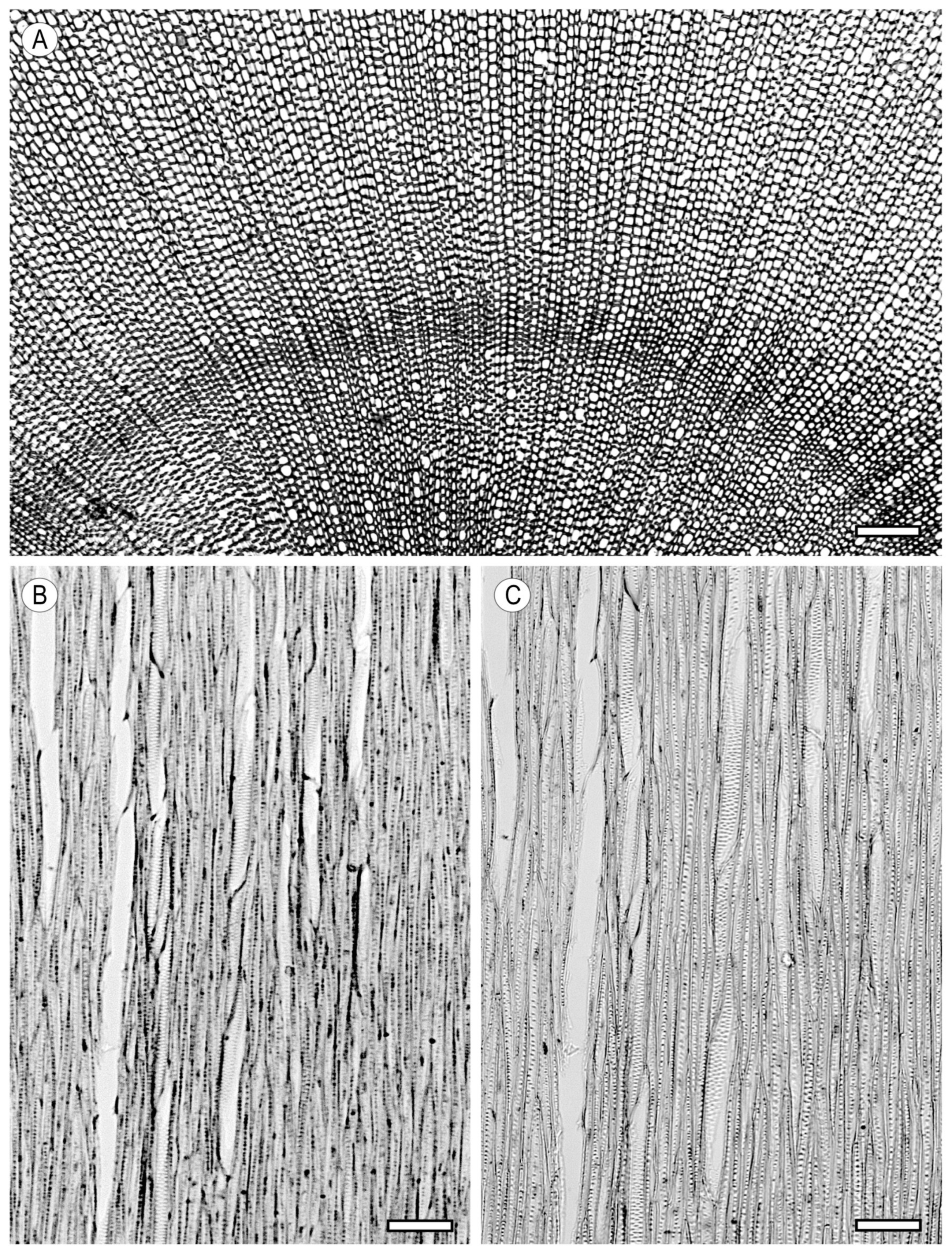
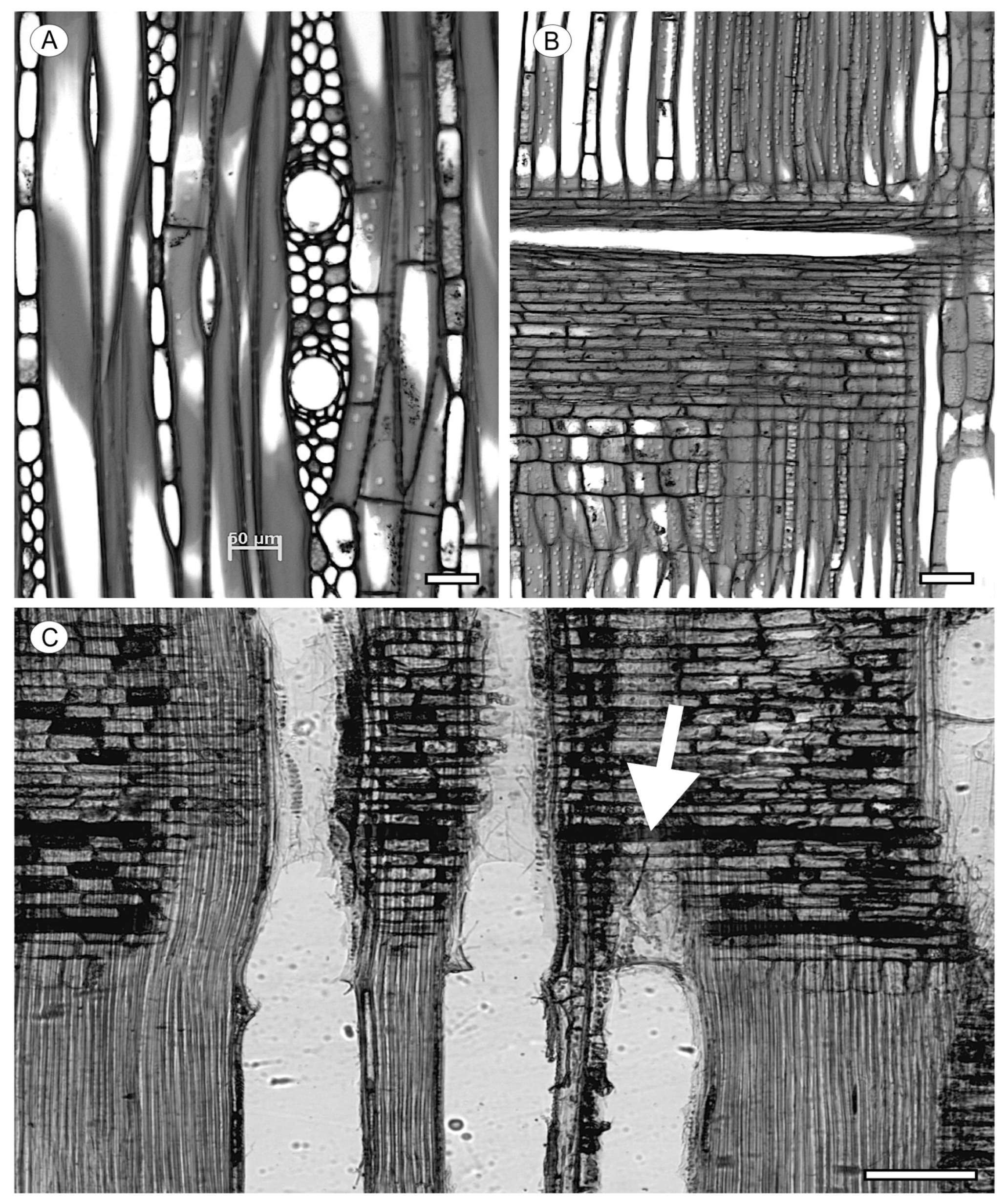
The presence of vessels in wood indicates that it is a hardwood. However, some families lack vessels, and their xylem tissue is made up entirely of imperforate longitudinal elements and parenchyma, e.g., Amborellaceae, Tetracentraceae, Trochodendraceae, and Winteraceae. Although their fibrous tissue resembles conifer wood, it is distinguished from conifer tissue by the presence of multiseriate rays (Figure 1). The vessel diameter varies considerably in hardwoods, and this depends on the age of the cambium and the position of the secondary xylem in the tree or shrub. There is a tapering effect, as described by Olson et al. [14], where vessels towards the top and extremities are smaller than those closer to the base. Vessels in roots are often wider, at a given cambial age, than those in aerial xylem (pers. obs. P.G.). For detailed papers on vessels and their functions, especially water transport, the two IAWA Journal special issues [15,16] and references therein are of particular interest.
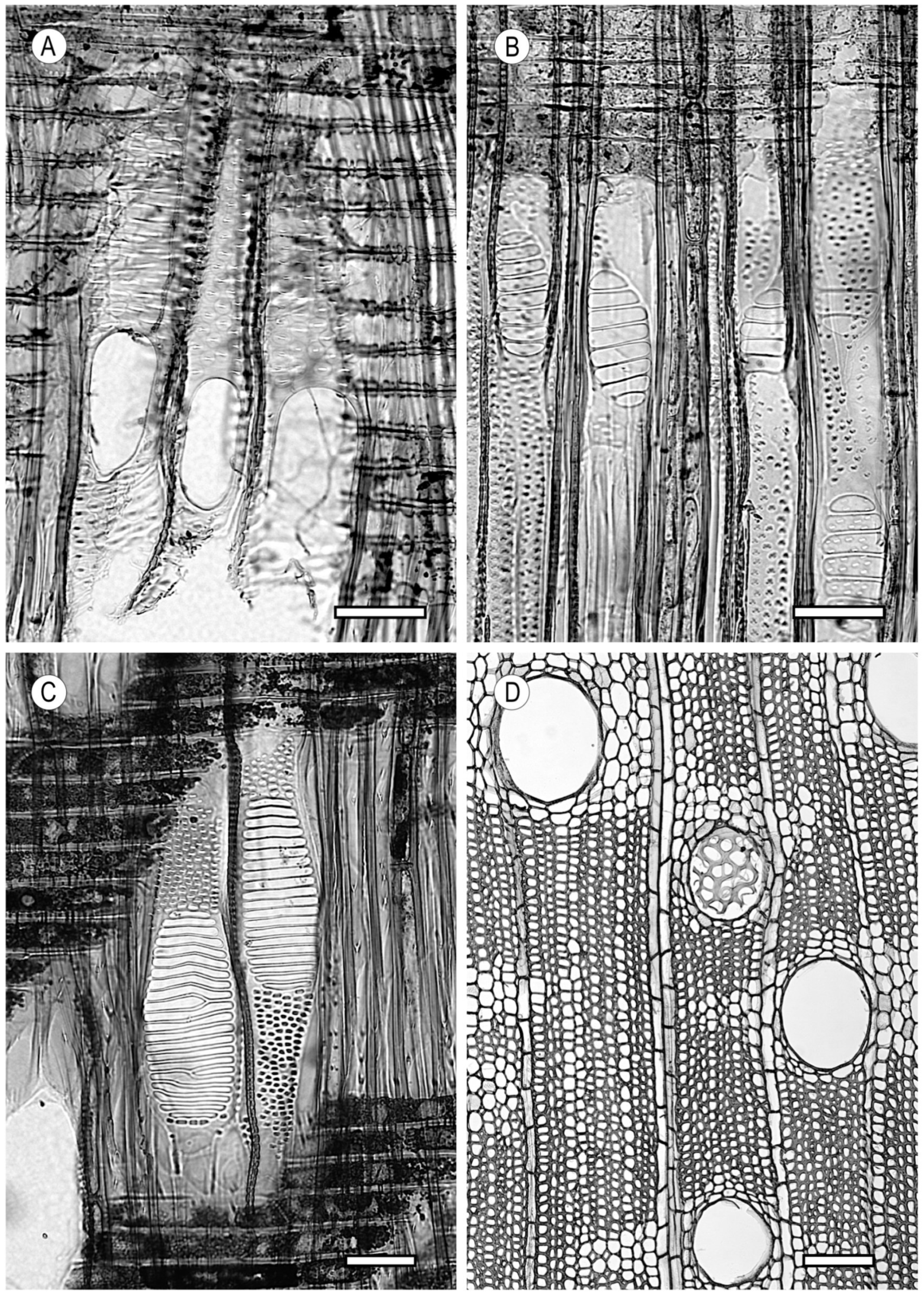
Vessels are sap-conducting “tubes” in hardwoods, extending in the axial direction of the tree and formed by a chain of cells, each of which is known as a vessel or vascular element. These elements are interconnected longitudinally through perforation plates. The three types of perforation plates are: 1—simple, 2—scalariform, and 3—reticulate, foraminate, and other types (complex and radiate) [17] (Figure 2). The considerable conductive efficiency of vessels compared to tracheids is partly because of the ease with which water flows axially through vessel element perforation plates, unlike tracheids, in which water must pass through pit membranes [18]. Vessel perforation plates are openings at the end of the vessel caused by the partial or total hydrolysis of the middle lamella and the primary wall. The plate between the vessel elements, which may be perforated to varying degrees, is made up of the remnants of the cell wall corresponding to each vessel element. Vessel elements have at least two perforated walls, one at each end, but sometimes have more than two perforated regions because they are contiguous with other vessel elements. The perforation process begins in a large borderless pit and is caused by the reabsorption of the pit membrane.
Simple perforation plates (Figure 2A) are the most abundant type and are present in more than 80% of the world’s wood [19]. The pit membrane is completely reabsorbed, leaving a gap between the vessel elements. Occasionally, a single wood can contain simple and scalariform perforation plates, e.g., Fagus sylvatica, in which scalariform perforation plates are commonly found in latewood vessels [20].
Scalariform perforation plates (Figure 2B,C) are formed when the reabsorption of the pit membrane occurs in grooves, leaving bars between them. The thresholds proposed by the IAWA Committee [12] are as follows: fewer than 10 bars (e.g., Apollonias barbujana, Arbutus menziesii, Cinnamomum camphora, Scottellia klaineana, and Virola koschnyi); from 10 to 20 bars (e.g., Alnus rubra, Betula alleghaniensis, Buxus wallichiana, and Dillenia spp.); from 20 to 40 bars (e.g., Altingia excelsa, Alnus rubra, Betula lenta, Cornus florida, Dillenia spp., and Nyssa spp.); and more than 40 bars (e.g., Ilex parviflora, Nyssa javanica, and Adinandra spp.).
Reticulate and foraminate perforation plates (Figure 2D) occur in few species and always in combination with another type (e.g., Kielmeyera spp., Fagus sylvatica, Oroxylum indicum, and Sorbus aucuparia).
Perforation plates are one of the most studied characteristics from the evolutionary point of view. The thesis that scalariform perforation plates are older than simple plates and are a relic of hydrolysed pits between tracheids [8] is supported by current phylogeny studies. However, the persistence of scalariform pits in some lineages and their reappearance in others suggests an adaptive function to some environments [4]. Although plants evolved towards simple perforation plates to facilitate transport in environments of high hydraulic demand, there is no doubt about the advantages of scalariform perforation plates in unfavourable conducting situations. Christman and Sperry [21] described scalariform perforation plates as elements that enhance flow resistance, are capable of capturing large freezing-induced air bubbles (thus avoiding the formation of large air bubbles, which are more difficult to dissolve), provide increased rates of vessel refilling, and give greater mechanical support to prevent vessel implosion. Under stress conditions, when the vessel pressure is close to the atmospheric pressure, in plants with scalariform perforation plates, the bubble is divided up between the elements of contiguous vessels and dissolves more easily, regardless of whether cavitation was induced by freezing or water stress [22]. Perforation plates have evolved to simple plates in environments requiring a high rate of hydraulic efficiency, and it is very likely that the shift from scalariform to simple perforation plates occurred many times independently during evolution [23]. The greater strength conferred by scalariform perforation plates can be an advantage at sites with low transpiration rates, such as tropical montane regions and cold/temperate environments [24]. Plants evolved and adapted, modifying their structural elements to survive in these ecosystems. The loss of vessels in Winteraceae, caused by plant adaptation to freezing-prone environments characteristic of wet temperate forests and tropical alpine forests, as reported by Feild et al. [25], may be an example of this.
In any event, it is clear that alpine and Arctic flora have a very high prevalence of scalariform perforation plates, whereas simple perforation plates predominate in warm, dry climates with a seasonal or constant demand for greater hydraulic efficiency [11].
Vessels vary enormously in length among plants and even in a single plant [26,27,28]. The widest and longest vessels occur in ring-porous earlywood. Vessels occupying the entire trunk have been reported, e.g., up to 18 m long in Fraxinus americana [29].
Vessel elements are also interconnected laterally with other vessels through intervessel pits. As water moves from roots to leaves, it has to cross intervessel pits hundreds or thousands of times, and, therefore, the membrane thickness and porosity have a very significant influence on the plant’s hydraulic resistance [30,31]. Plant physiology studies have determined that intervessel pit membranes are responsible for at least 50% of the xylem hydraulic resistance [32,33,34].
The structure of bordered intervessel pits is made up of small openings, where the secondary wall was not deposited over the primary wall, permitting water flow between adjacent vessels. However, these small holes are also weak points of vessels, through which air bubbles can form as a consequence of the transpiration flow [11]. Pit types vary in size, shape, and arrangement as follows [12]:
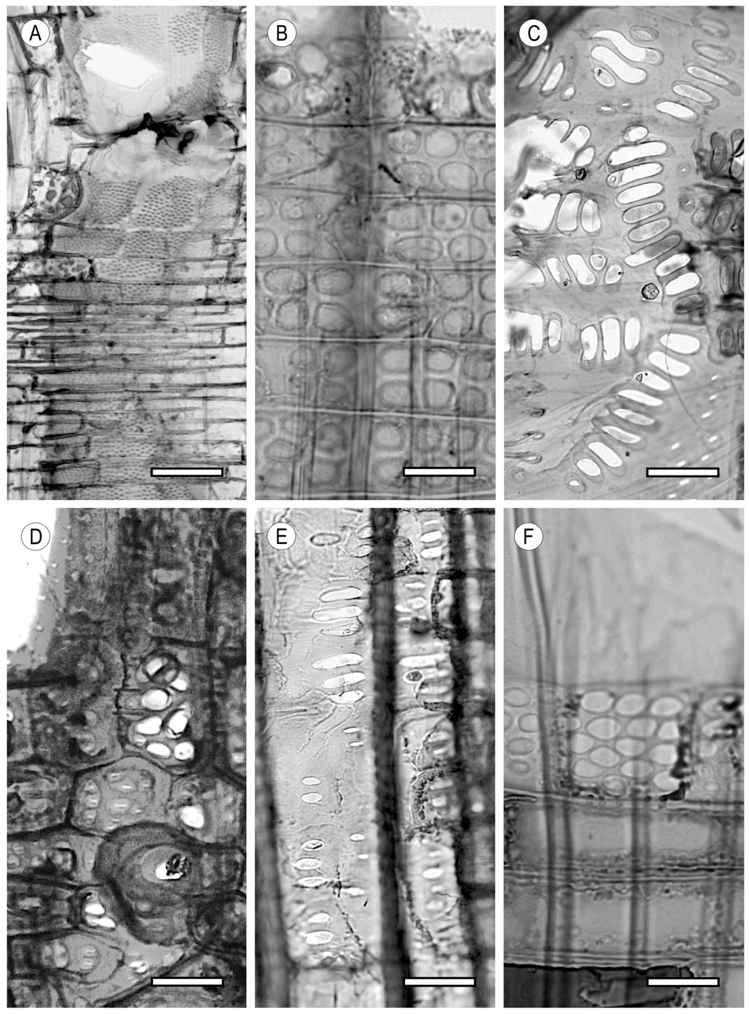
Scalariform intervessel pits (Figure 3A). Pits are linear, with their axis perpendicular to the vessel axis. This is another feature that indicates a small specialisation in wood and, therefore, the persistence of ancestral structures (e.g., Dillenia spp., Liquidambar styraciflua, Nyssa spp., Nothofagus menziesii, and Scottellia coriacea).
The pits between vessels and vascular tracheids are analogous to these pits. In contrast, pits that are interconnected with libriform fibres and fibre-tracheids are arranged in vertical alignments and are narrow and almost vertical.
Opposite intervessel pits (Figure 3B). Pits are arranged in horizontal formations transverse to the axis of the vessel element (e.g., Fagus sylvatica, Liriodendron spp., Nothofagus obliqua, Platanus spp., and Rhododendron maximum).
Alternate intervessel pits (Figure 3C). These appear in diagonal rows relative to the axis of the vessel (e.g., Aceraceae, Leguminosae, Meliaceae, and Sapindaceae) (e.g., Alstonia boonei, Astronium urundeuva, Aucoumea klaineana, Avicennia spp., Betula pendula, and Tabebuia rosea). When the pit outline is a polygon with more than four sides, the term used is shape of alternate pits polygonal (Figure 3D) (e.g., Salicaceae and Leguminosae) (e.g., Canarium schweinfurthii, Corylus avellana, Mangifera indica, and Schinopsis balansae).
Alternate intervessel pits are the most common, while opposite and scalariform intervessel pits occur in only a few species. However, combinations of pit types can occur in some species, e.g., in Buxus, they are alternate and opposite, and in Liquidambar, opposite and scalariform pitting can be observed simultaneously.
The size of intervessel pits can be used to differentiate genera within families and to distinguish between families. Many genera of Meliaceae have very small-diameter pits, whereas Anacardiaceae has large pits. The thresholds for this feature were included in the IAWA Committee list [12] (minute ≤ 4 μm, small 4–7 μm, medium 7–10 μm, and large ≥ 10 μm).
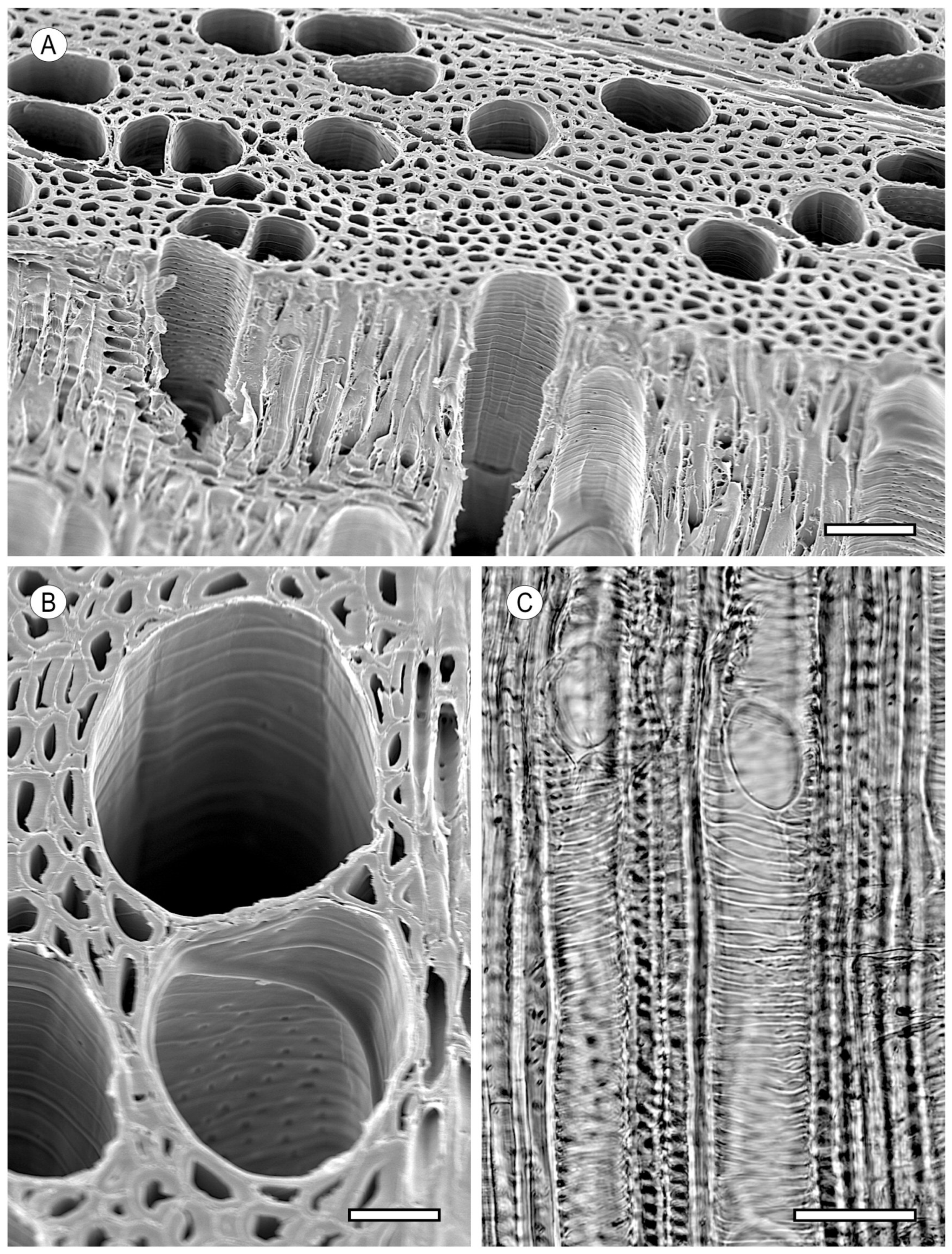
Some intervessel pits have projections from the secondary wall of the pit and/or the outer pit aperture. Known as vestured pits (Figure 3E), they also occur in vessel–ray pits, vessel–axial parenchyma pits, or interfibre pits. The first reports of this type of pitting are from the late 19th to early 20th centuries, although it was Bailey [35,36] who demonstrated that the sieve-like appearance under a light microscope was not because of perforations of the pit membrane but because of small outgrowths from the free surface of the secondary wall. Bailey introduced the terms “vestures” and “vestured pit” [37].
Vestured pits are very common in Combretaceae, Lythraceae, Myrtaceae, Rubiaceae, and most species of Leguminosae. They can be characteristic of a family or groups within a family, and their number, size and arrangement can have diagnostic value [12] (e.g., Afzelia africana, Brachystegia laurentii, Guibourtia ehie, Shorea negrosensis, and Terminalia ivorensis). They may also occur in latewood vessels but not in earlywood, as in Platanus [38].
Carlquist [39,40] acknowledged the difficulties in determining the possible functions of vestured pits but put forward three hypotheses: (1) a lower resistance to water flow, (2) a mechanism for removing air from cavitations in vessels and tracheids and restoring normal water columns, and (3) a mechanism to support higher water tensions and prevent the formation of embolisms by increasing the surface area and hydration.
According to some researchers, these pits have a protective function that prevents a strong deviation of the membrane due to pressure difference [41,42], thus enabling the porosity to increase when the pressure difference increases.
Although Zimmermann [43] found that it is not easy to establish correlations between the pit structure and habitat, Jansen et al. [11] studied 11,843 species and 6428 genera and concluded that the presence of vestured pits decreases considerably from the tropics to the tundra, and they are more numerous in desert regions and at tropical sites. The presence of vestured pits may, therefore, be associated with adaptation to stress-prone environments in plants that have them. Their function could be to increase hydraulic resistance or minimise vulnerability to cavitation. It is still unknown why some plants in water-stress environments lack vestured pits.
Zimmermann [43] reported that vestured pits, like vessels with helical thickenings or scalariform perforation plates, may have the function for trapping air bubbles when ice in vessels thaws at the end of winter. When bubbles are fragmented, they dissolve faster and cavitation is avoided.
In any event, the function of vestured pits is not yet clear. Their presence could be explained as outgrowths from the secondary wall [35,36], or their development may be associated with the adaptability of species to hostile environments. More research is needed to understand their functions.
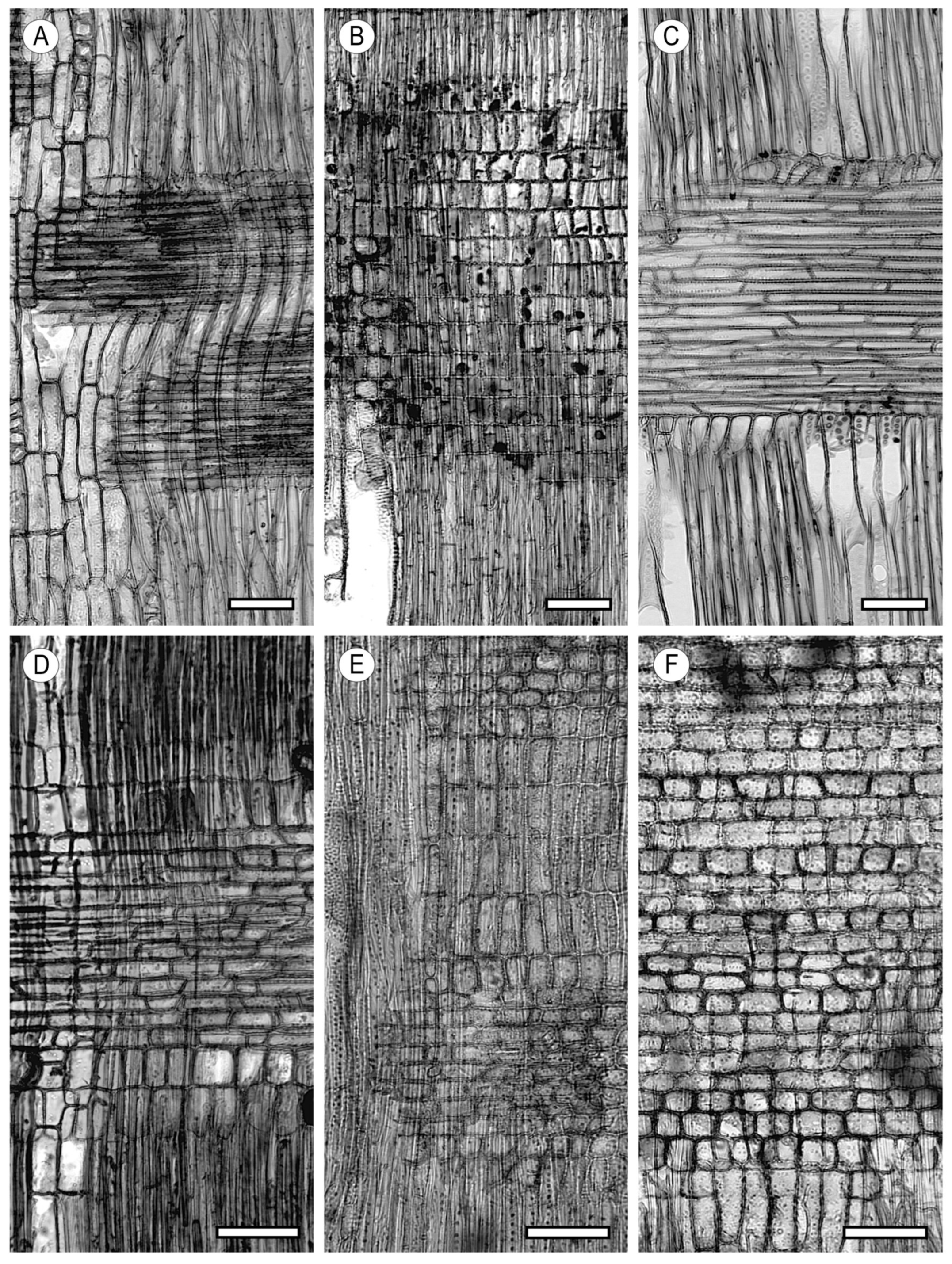
Pits between vessels and rays often have a different appearance from intervessel pits, but in species with solitary vessels, it is sometimes impossible to distinguish between the two types. Moreover, combinations of different types of vessel–ray pits can occur in a single wood (Figure 4).
The vessel wall can have helical thickenings in all or only some vessels (Figure 5), normally in only the narrower vessels of ring porous species (Robinia pseudoacacia and Ulmus spp.). The location of thickenings in vessels provides information about the wood species, as they can occur only in vessel element tails (Liquidambar styraciflua) or throughout the vessel (Acer spp., Aesculus spp., and Tilia spp.). Thickenings can also occur in vascular/vasicentric tracheids and, although rarely, in axial parenchyma cells. Costa and Wiedenhoeft [44] postulated that in addition to a mechanical role, helical thickenings in conductive cells may be involved in vessel refilling after cavitation and increased hydraulic efficiency. These two functions could be questionable in taxa where helical thickenings are found only in latewood vessels, such as Ulmus spp.
Helical thickenings appear to be more frequent in woods from subtropical and temperate regions than in tropical woods [9,10]. This is probably because of the association between helical thickenings and conductive efficiency. Carlquist [40] reported that the presence of helical thickenings may be associated with drought- or frost-induced water stress. Thickenings decrease the lumen volume inside the vessel, helping to prevent bubble formation and propagation. Zimmermann [43] indicated that their development may be associated with increased vessel wall strength. However, Ohtani et al. [45] considered it to be simply because of genetic information. The exact role of helical thickenings is unclear and, as in conifers, their ridges do not appear to be large enough to have an influence on mechanical properties, although they may influence conduction. One explanation was put forward based on biomechanical comparisons between the structure of hardwood vessels and pressure vessels with fibre-reinforced layers in helical patterns, applying the constant-strength design principle; i.e., loading all the pieces to the maximum allowable stress minimises the amount of material used, and this can be applied to explain secondary wall thickening patterns [46].

2.1. Morphology

The vessel element shape can vary from a barrel-like appearance to tapered vessels with perforated ends, known as fibriform vessels (a term coined by Woodworth [47] in Passifloraceae and maintained by Carlquist [40]); i.e., from having the same transverse and longitudinal dimensions to a linear shape considerably larger in size longitudinally than transversely. In the latter case, ligulate extensions at the ends are thought to be vestiges of the original immature cell.
However, the longitudinal growth of vessel elements is generally very limited compared to the size of the initial cambial cell. Despite this, there is a noticeable difference in length among the vessel elements of different woods.

2.2. Arrangement

Carlquist [40] explained the grouping of vessels by their greater vulnerability to embolism compared to tracheids. At sites prone to embolism and the risk of air bubbles entering through intervessel pit membranes, the presence of subsidiary tissue allowing for alternative three-dimensional pathways is essential. If the vessel is surrounded by libriform fibres or fibre-tracheids, there is no alternative, but where there are other vessels or vasicentric tracheids, recovery from embolism is possible. Carlquist [40] concluded that in wood where vasicentric tracheids are abundant, e.g., Quercus spp., vessels are solitary, whereas in wood where they are scarce, e.g., Calycanthus spp., the vessel grouping is an adaptation to xericity. Bissing [48] tested this by growing the same genetic stock in environments with different water availabilities, evaluating the more stable anatomical elements and finding that grouping was greater at dry sites.
The three types of vessel grouping in the transverse section are [12]:
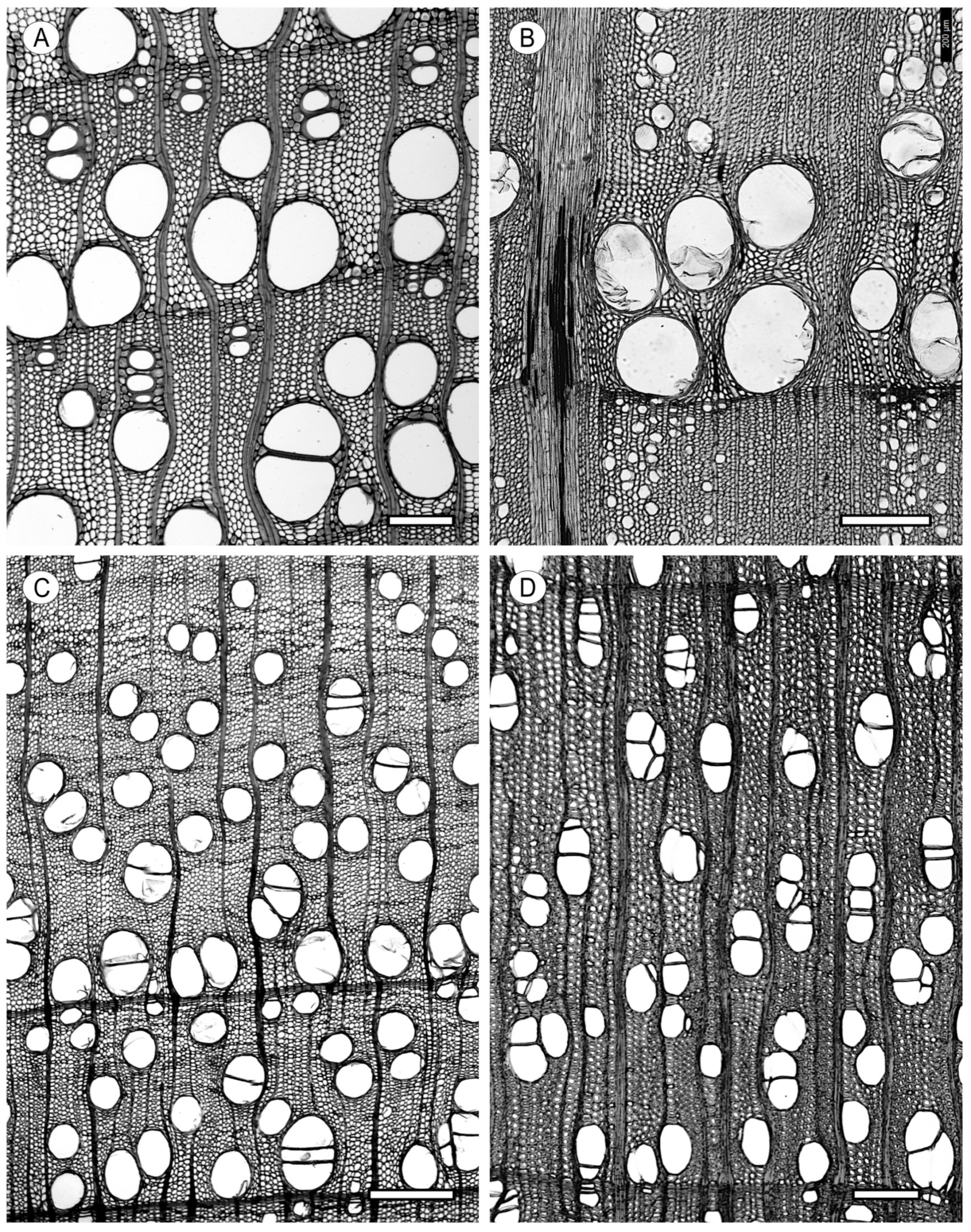
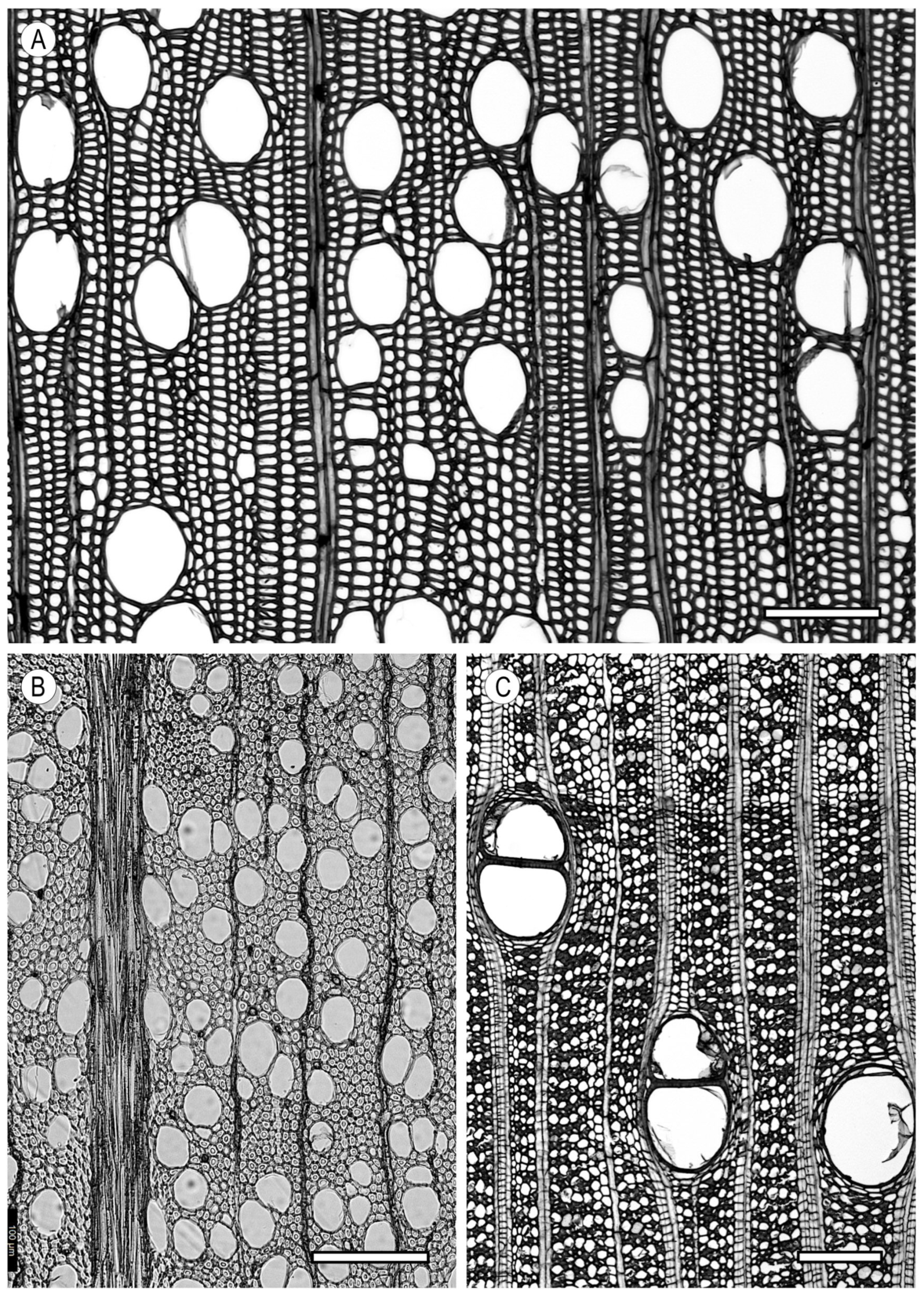
Exclusively solitary vessels (Figure 6A). Each vessel is alone and separated from the others by other tissues. These vessels are typically circular or oval in outline, with the larger axis in the radial direction, although pores sometimes have an angular outline (e.g., Aextoxicon punctatum and the latewood of some Quercus (white oaks)). At least 90% of the vessels must be solitary to apply this feature to wood (e.g., Altingia excelsa, Buxus sempervirens, Dillenia indica, and Dryobalanops rappa).
Vessels in radial multiples (Figure 6B). Vessels are in groups of four or more in the radial direction. Intermediate vessels have flattened tangential walls, and the outer ends of the group are circular (e.g., Alnus rubra, Corylus avellana, Pouteria spp., and Aquilaria malaccensis). Radial multiples from two to four with solitary vessels in a variable proportion are the most common vessel grouping.
Vessels in clusters (Figure 6C). Vessels in groups of three or more are in contact with each other both tangentially and radially (e.g., Ailanthus altissima, Pistacia vera, Celtis occidentalis, Melia spp., and Morus alba).
With regard to the vessel arrangement, the IAWA Committee [12] distinguishes the following:
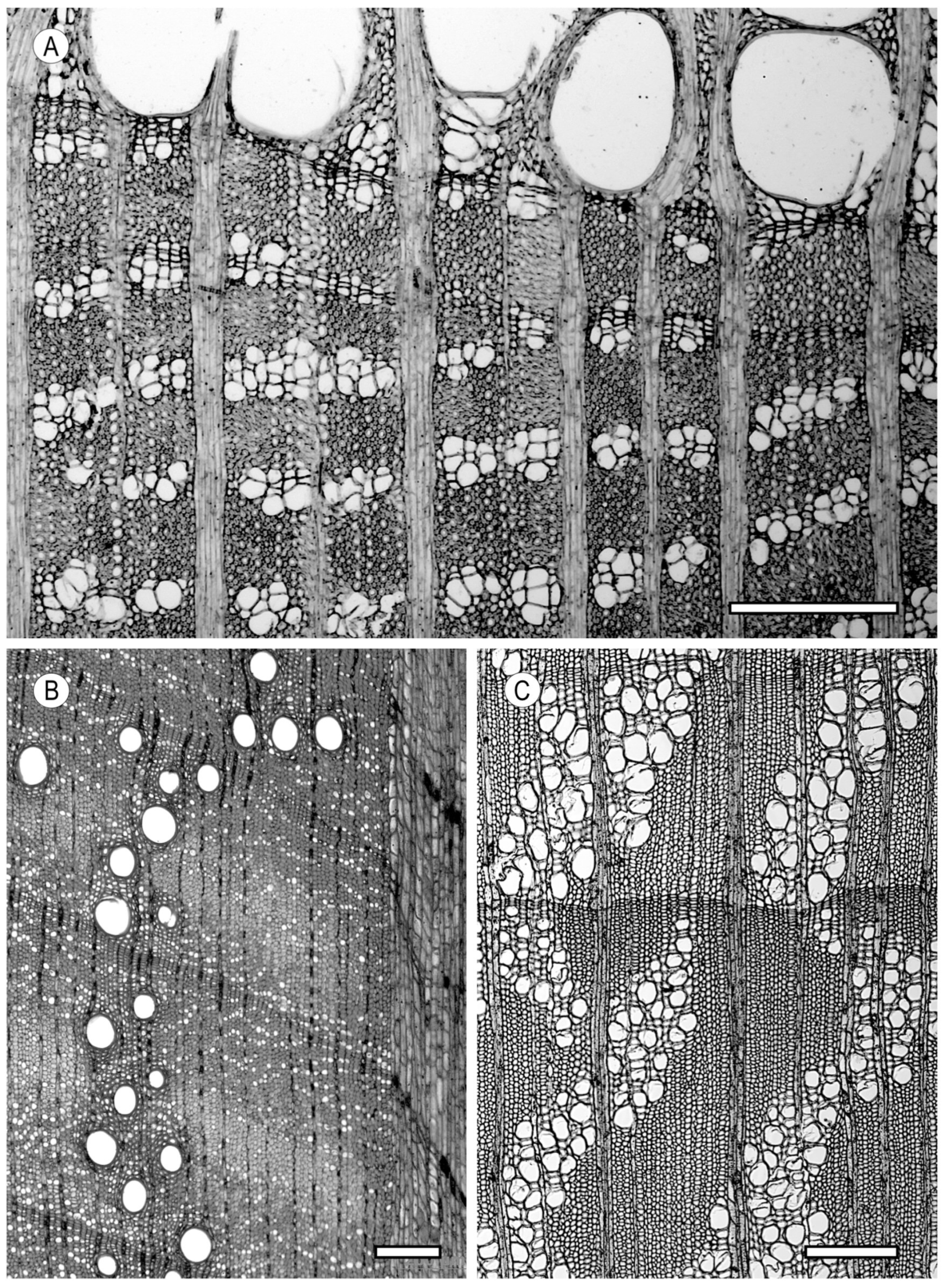
Vessels in tangential bands (Figure 7A). Bands may be short or long and straight or wavy (e.g., Celtis occidentalis, Ulmus americana, and Zelkova serrata).
Vessels in diagonal and or/radial pattern (Figure 7B). (e.g., Avicennia marina, Calophyllum brasiliense, Castanea spp., and Pistacia vera).
Vessels in dendritic pattern (Figure 7C). (e.g., Castanea dentata and Rhamnus cathartica).
Lastly, in terms of the porosity, wood can be
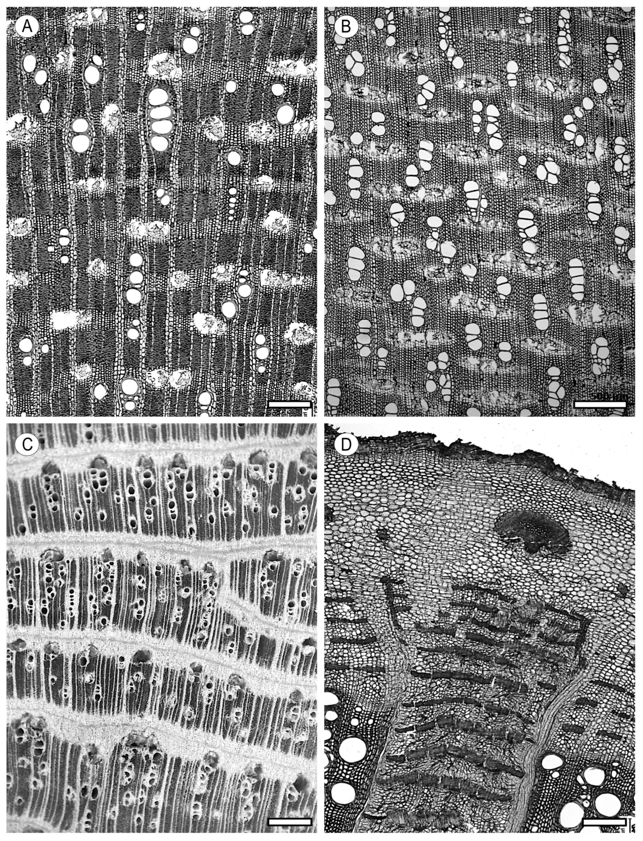
Ring porous (Figure 8A,B), when the diameter of earlywood vessels is distinctly larger than in vessels formed in the latewood (Quercus robur, Fraxinus excelsior, and Ulmus americana). The growth ring has a region that, even to the naked eye, has a porous aspect that is easily observed because of its different appearance or colouring; Semi-ring porous (Figure 8C), when the diameter of vessels gradually decreases from wood formed in earlywood to wood formed in latewood (Juglans regia, Pterocarpus indicus, and Cedrela odorata); Diffuse porous (Figure 8D), when the vessel diameter is more or less the same size throughout the growth ring (Acer spp., Populus spp., Swietenia spp., and Enterolobium spp.). Ring porosity is almost confined to temperate regions and is seen only in about 4.6% of hardwoods [49], whereas diffuse porosity is worldwide and occurs in virtually all tropical species (92.8%). Semi-ring porosity is more difficult to define, overlaps with both to some extent, and can be found in about 9.7% of hardwoods.

2.3. Tyloses and Deposits

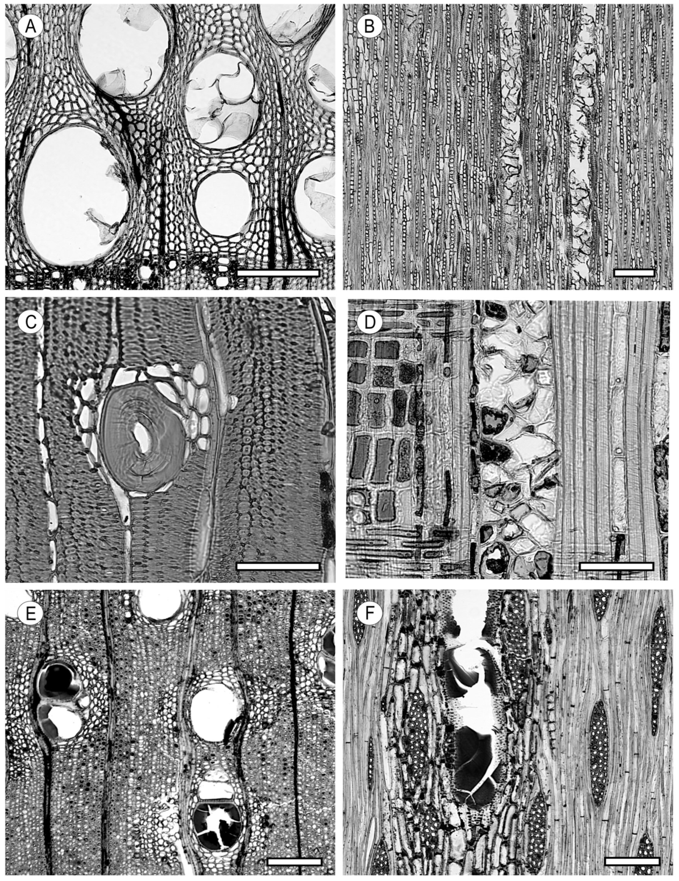
During the ageing process of a woody plant, the sapwood loses its conducting and storage functions and becomes heartwood, which mission is to provide mechanical support and resistance to deterioration. Tylosis formation and gum secretion (Figure 9) are characteristic processes of heartwood formation, although tyloses can also occur in sapwood in response to embolism [50] or wounding [51].
Tyloses play key roles in limiting the spread of pathogens and wood-decomposing organisms [52] and as compartmentalisation elements after wounding. They are formed both in ring-porous and diffuse-porous woods [50].
Malpighi [53] was the first to describe “balloon-shaped sacs” in heartwood vessels. The term “tylosis” comes from the Greek “thyllen”, meaning bag or container. It was first mentioned in an anonymous paper of 1845 attributed to Hermine, Baroness von Reichenbach of Vienna [50], in which tyloses were described as outgrowths produced by neighbouring parenchyma cells passing through the pits but never between two vessels in contact.
The first records of tyloses in fossil plants are from the early Carboniferous (Viseense), around 340 million years ago, in the French region of Vosges, from a progymnosperm, Protopitys buchiana [54].
Tyloses are formed mainly by ray parenchyma cells, and to a lesser extent by axial parenchyma cells, entering the vessel through vessel–ray pits [55]. There seems to be consensus that the prerequisite for occlusion by tyloses is cavitation, which would explain the higher frequency of tyloses in large-diameter vessels [18]. Tyloses can occupy all or part of the vessel, appear singly or in groups, and be thin-walled or sclerotic, pitted, or smooth, and with or without inclusions, such as starch, crystals, resins, gums, gels, and other storage products [50]. They also occur occasionally in softwood and hardwood tracheids and in hardwood fibres [56].
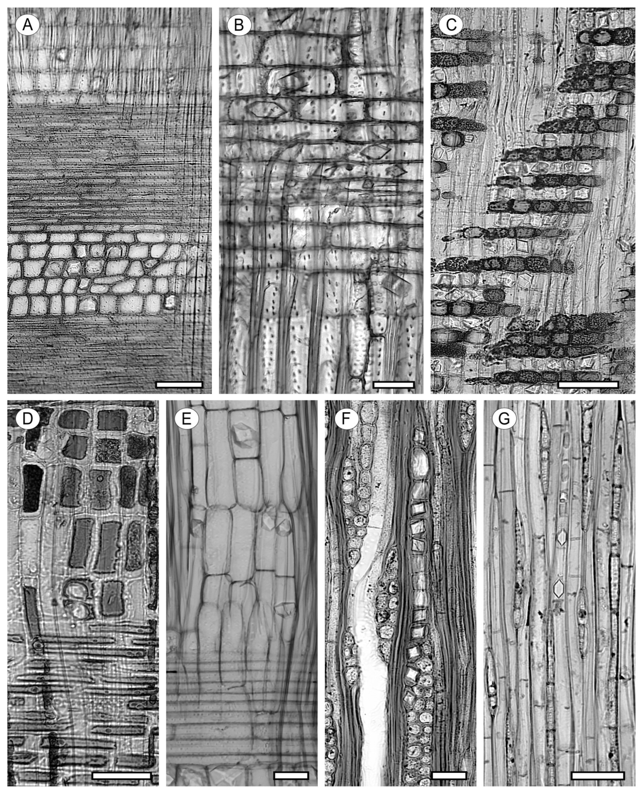
Brown et al. [13] explained tylosis formation as the pressure difference between the living parenchyma cell, which is in a state of turgor, and the dead vessel element, which has ceased, or will soon cease, its conducting functions. The pit membrane enlarges and arches into the cavity of the vessel segment, and a part of the protoplast of the parenchyma cell enters the cavity or hollow formed, constituting what is known as thyllos or tyloses (Figure 9A,B). Not only the primary wall of the pit is stretched and arched into the cavity but also new cell wall materials (cellulose, hemicellulose, pectin, suberin, and lignin) are actively deposited [57].
Tylosis formation takes place in the region of contact because of a special layer of a pectocellulosic composition overlying the pit membrane and the portions of the cell wall in contact with the vessel. Initially transparent, tyloses can enlarge until they occupy all the vessel lumen or undergo cell divisions; when expansion is complete, the formation of the secondary wall and pits begins [50].
Tyloses are common in species such as Astronium graveolens, Calophyllum lucidum, Castanea sativa, Hura crepitans, Quercus spp., Milicia excelsa, Robinia pseudoacacia, and Shorea guiso.
When tyloses are abundant, they prevent the entry of liquids, but, as a general rule, they do not increase wood durability. However, the presence of tyloses is a very important feature to consider in preservative impregnation processes, as abundant tyloses hinder impregnation.
Tyloses are generally thin-walled, although their walls are sometimes lignified, giving rise to sclerotic tyloses (Figure 9C,D) (e.g., in Brosimum guianense, Pouteria guianensis, Schinopsis lorentzii, and Staudtia stipitata).
Normal and sclerotic tyloses can occur in a single wood, and tyloses and deposits may occur simultaneously [13]. Very rarely, gums can be secreted by a previously formed tylosis [55].
Gums, like tyloses, occlude vessels and are produced by parenchyma cells in contact with vessels, mainly ray cells (Figure 9E,F). These non-water-soluble compounds have variable chemical compositions and are of chemotaxonomic interest [58].
Examples of wood with gums include Aspidosperma album, Dicorynia guianensis, Dillenia spp., Diospyros crassiflora, Guibourtia tessmannii, and Oxandra lanceolata).
De Micco et al. [50] provided a detailed review of the structure, functions, and occurrences of tyloses and gums.

3. Tracheids and Fibres

3.1. Vascular and Vasicentric Tracheids

Tracheary elements lacking perforation plates (i.e., not vessels) are usually described as vascular tracheids when not associated with vessels and as vasicentric tracheids when they are. The IAWA list [12] used a simple definition for both, but Olson [59] pointed out that for functional and physiological investigations, more sophisticated definitions are needed. Vascular tracheids are considered by some authors to be imperfect or degenerate vessel elements. Arranged in longitudinal series, they resemble small-diameter vessels in shape, size, and wall pitting but are differentiated from vessels by their imperforate ends (e.g., Celtis occidentalis, Sambucus nigra, and Sophora japonica). Their inner walls can have helical thickenings [12].
Vasicentric tracheids are imperforate cells with bordered pits in the tangential and radial walls arranged around the vessels (Castanea spp., Quercus spp., and many species of Shorea (Dipterocarpaceae) and Eucalyptus spp. (Myrtaceae)) [12]. They are normally very abundant in ring-porous wood, near the earlywood vessels, as in Quercus spp. and Castanea spp., and are often associated with longitudinal parenchyma, from which they are easily differentiated in longitudinal sections by their pitting (Figure 10).
The functions of vasicentric tracheids have rarely been studied [60,61,62]. They may constitute the final and safest conduction inside the growth ring by retaining columns of water adjacent to the cambium at the end of the growing season. Moreover, because they are imperforate elements, like true tracheids, vasicentric tracheids provide the maximum protection against embolism and are less likely to cavitate [40]. Some authors [62] consider them to be a three-dimensional network of alternative or secondary conduction to the vessels in case of failure because of water stress, thus increasing the xylem connectivity. They could even be a subsidiary system as an alternative to the vascular system when vessels have cavitated [60]. This theory implies that vasicentric tracheids are less vulnerable than vessels to cavitation, and, therefore, wood with a higher proportion of vasicentric tracheids would be less vulnerable to cavitation. For other authors, vasicentric tracheids may act as water reservoirs [63,64].
In wood with solitary vessels, tracheids could have an essential role in interconnection between vessels [61], as they increase the xylem connectivity [62,65,66]. The conductive function of tracheids depends mainly on the structure and density of the pits connecting them. As one might expect from observation, embolism tolerance appears to be greater in eudicots with both tracheids and vessels [67].
Barotto et al. [68] demonstrated a correlation between cells that accompany vessels (vasicentric tracheids, fibre-tracheids, and parenchyma) and functional variables. This suggests that the cells surrounding vessels contribute to increase the connectivity between contiguous vessels, thus improving the xylem conduction and decreasing the likelihood of embolism.
In any event, the specific function of each cell has not been determined, and further study is necessary.

3.2. Fibrous Tissue

The fibrous tissue of hardwoods is made up of libriform fibres and fibre-tracheids (Figure 11), whose main function is to provide mechanical support. Both types of cells enlarge considerably from the cambial fusiform initials but vary greatly in length, wall thickness, and diameter from one species to another and in the proportion in which they form a part of the wood structure. They expand more radially than tangentially, remaining much the same in the tangential diameter as the fusiform initials from which they are derived. Although highly variable, Brown et al. [13] indicated that fibrous tissue accounts for 50% of the total volume of the wood in some hardwood species.
The IAWA Committee [12] purposely avoided the terms libriform fibres, fibre-tracheids, and “true tracheids” as descriptors in its list because of a lack of consensus on their definitions. Fibre-tracheids are very elongated cells with tapered ends and walls, with bordered pits, and with chambers larger than 3 mm in diameter (e.g., Ilex spp., Dillenia spp., and Camellia spp.). Libriform fibres are also elongated cells with tapered ends, but they differ from the former in their simple pitting or pits with a border of less than 3 mm in diameter (e.g., Fraxinus spp., Olea europea, Populus spp., Rhizophora spp., and Ulmus spp.) [12]. The major function of both libriform fibres and fibre-tracheids is mechanical support, although the latter may have a very minor role in water transport.
Helical thickenings in fibrous tissue (Figure 11C) can be present both in libriform fibres and fibre-tracheids but are much more common in the latter (e.g., Ilex spp., Ligustrum spp., Lonicera spp., and Rosa spp.). Whereas their functions can be speculated upon most easily in vessels and tracheids, they are more obscure in fibres and fibre-tracheids.
In some species, fibres appear with thin transverse walls without pitting, formed after the secondary walls of the fibres have been deposited and, therefore, lack middle lamella. Known as septate fibres (Figure 11D), they occur, for example, in Aniba spp., Aucoumea klaineana, Gmelina arborea, and Scottellia coriacea. Their arrangement differs greatly from one wood to another. In some woods, all the fibres are septate (e.g., Canarium spp. and Tapirira spp.), while in others, septate and non-septate fibres co-occur (e.g., Phoebe porosa, Tectona grandis, and Cedrela spp.). They can also be scattered irregularly near the vessels or rays or arranged in tangential bands.
Tropical trees with axial parenchyma rare or absent have a larger quantity of septate fibres [69], probably because these fibres perform the function of the axial parenchyma [70,71,72]. Achariaceae, Araliaceae, Burseraceae, and Salicaceae are examples of families in temperate and tropical climates that have this feature [73]. It has long been known that in some species, septate fibres contain starch [74], which can hydrolyse and produce the sugars necessary for translocation to vessels. For Carlquist [40], the presence of starch in fibres suggests a pattern of alternative storage and photosynthate conduction in taxa that have constant flowering events rather than sudden flushes.
Most identification keys include entries for the cell wall thickness of the fibrous tissue (Figure 12) as an analytical feature, with three differentiated states: very thin-walled fibres (Tilia spp.), from thin- to thick-walled fibres (Ilex spp.), and very thick-walled fibres (Strombosia pustulata and Rhizophora mangle). This measurement is actually a ratio between the lumen and the cell wall thickness. Chattaway [75] proposed ranges that are very useful for standardising the values used in the biometry of hardwood anatomical elements.
With the appearance of vessels, tracheids that subsequently evolved into fibres seem to have been freed from their conductive function [76,77], although those that evolved into modern vascular and vasicentric tracheids appear to have remained in the structure of some angiosperm families as a conductive safety measure.
With regard to fibre-tracheids, Carlquist [40] indicated that they are transition elements between tracheids and libriform fibres.

4. Parenchyma

It is commonly taken for granted that all the cells in mature wood are dead. All the conductive elements (vessels and tracheids) in the functional sapwood undergo a process of cell autolysis, or programmed suicide, resulting in hollow conduits reinforced by a lignified cell wall. Autolysis is caused by the hydrolases that had remained inactive inside the vacuoles. At a signal, yet unknown, a calcium flux causes the release of hydrolases, and they degrade the entire cell contents but not the cell wall [78]. However, cells with a protoplast are also present in the secondary xylem, giving rise to wood parenchyma. Parenchyma has thinner walls than fibrous tissue and can be divided into two types by orientation or arrangement: axial parenchyma (cells arranged along the axis of the stem and the product of the fusiform cambial initials, as are vessels, tracheids, and fibres) and ray parenchyma (the product of the ray cambial initials) [79].
Unlike sclerenchyma tissue, whose main function is to provide mechanical support and, in some cases, conduction, the functions of parenchymatous tissue are primarily storage and conduction. Morris et al. [80] explored this subject in detail.
In both softwoods and hardwoods, sucrose, glucose, and fructose are the predominant soluble carbohydrates, while starch is the main non-soluble storage carbohydrate [81]. Carbohydrate storage is essential, first, so the tree can cope with adverse environmental conditions, and, second, so it can reactivate growth when conditions are favourable.
As a result of the lignification of their walls, parenchyma cells also contribute to the mechanical support of the tree while, at the same time, acting as a defence against pathogenic fungi because of the durable nature of lignin [82,83]. The lignification process is the deposition of phenolic polymers (lignin) on the extracellular polysaccharidic matrix [84]. The lignin produced acts as a defensive barrier to infection from pathogens, as it is not degradable by most microorganisms [85]. Parenchyma cells are also capable of producing tyloses to plug the vessels of damaged xylem, thus avoiding the propagation of pathogens in the tree.
In general, these cells are relatively short and maintain their vitality much longer than other xylem cells. In Rhododendron lapponicum, the presence of live parenchyma cells as old as 200 years has been confirmed [86,87]. In Fraxinus, live axial parenchyma cells were found more than 45 years after vessels stopped transporting water [86].
The amount of the total parenchyma (axial + radial) in angiosperms ranges from 20% to 40%, much higher than in gymnosperms (5%–10%), with values of 40%–60% commonly found in tropical woods [79]. Despite the scarce presence of axial parenchyma in conifers, this feature can be used for identification at the genus level. The presence of axial parenchyma permits the exclusion of the families Araucariaceae and Sciadopityaceae and the genera Taxus and Pseudotaxus in Taxaceae; Neocallitropsis, Thuja, and Xanthocyparis in Cupressaceae; and Halocarpus, Lagarostrobos, Lepidothamnus, Manoao, Microcachrys, Phyllocladus, and Sundacarpus in Podocarpaceae [88]. In contrast, the greater abundance of axial parenchyma cells in hardwoods and, in particular, their varied patterns of distribution, can frequently be used for wood identification [12]. Morris et al. [89] found a 29-fold variation in ray and axial parenchyma proportion from data from a wide range of literature sources and showed that both the temperature and growth form influenced this.
Non-structural carbohydrates (NSCs) are the most abundant reserves in wood parenchyma. The role of NSCs is essential to tree growth and functioning [90]. They provide the basic components of plant structures and the metabolic resources for their development and for the synthesis of other organic components [91]. Starch and the water-soluble sugar fraction make up NSCs. Starch is considered as the main method of long-term storage. Its large molecules cannot move freely between cells, but they can hydrolyse and produce more mobile soluble sugars capable of performing the most active physiological functions. NSCs play key roles in plant responses to drought and frost stress [92] and against fire [93]. A larger proportion of parenchyma may suggest a greater amount of NSCs, but this has not been confirmed [79].

4.1. Axial Parenchyma

Axial parenchyma is produced by the division of fusiform cambial cells through transverse divisions that result in axial columns with two or more parenchyma cells.
The presence of axial parenchyma in hardwoods has been determined as 1%–25% of the xylem [94] and is greater in tropical woods than in wood from temperate forests. This may seem strange in view of the limited variation in seasons in tropical regions and lower need for storage, but the presence of axial and radial parenchyma has many functions, including NSC storage and transport, defence against pathogens, water storage, mineral storage, and sapwood-to-heartwood transition [95]. The parenchyma is the only living component of the wood structure, although in some species of Araliaceae and Salicaceae, fibres can take the place of the axial parenchyma and act as a functional substitute [69,96]. The presence of a secondary wall in the parenchyma has been questioned because the typical layered structure is absent, resembling instead a thickened primary wall [97]. However, the presence of a primary wall and two or more concentric layers has been observed in hardwood axial parenchyma, with a structure essentially similar to that of fibres and tracheids [98]. Parenchyma is normally lignified, but some woody plants, including many lianas [99], and some tropical trees of Urticaceae, have unlignified parenchyma [100]. Alvarado and Terrazas [101] suggested a synchronisation in Cochlospermum vitifolium between parenchyma proliferation and water availability in months with highest precipitation. This synchronisation supports the idea that unlignified parenchyma aids rapid water deposition because the unlignified walls allow for efficient water storage.
Although many researchers maintain that parenchyma does not contribute to water and mineral transport in the xylem, an extensive list of publications supports the claim that it does [102,103,104,105,106,107,108]. One of the reasons for this may be that they do not envisage wood as three-dimensional, strongly interconnected tissue. Ray cells connect the axial tissue to the ray tissue and connect the ray tissue to the phloem. The phloem is vital to maintain the radial transport capacity and, therefore, the whole hydraulic system of the plant [73]. Although the participation of the axial and radial parenchyma in plant transport mechanisms should not be questioned, embolism repair mechanisms by parenchymatous tissue remain unconfirmed. Speculations include vibration mechanisms capable of activating living parenchyma cells [109], osmotic changes between parenchyma cells and vessels [110,111], and even tylosis production as a last resort to plug vessels [52,112,113]. The release of sugars in the vessel lumen is thought to be the mechanism that triggers the quick reversal of drought-induced embolism [110,114].
Mechanical studies on the percentage of the parenchyma in hardwoods have revealed a negative correlation between the presence of the axial parenchyma and the modulus of the elasticity [115], probably associated with lower wood density at higher amounts of axial parenchyma [116].
Hardwoods have more parenchyma than softwoods, although parenchyma may exceptionally be rare or lacking, e.g., in Berberidaceae, Punicaceae, and Violaceae.
The complexity of hardwoods, a result of their evolution, also means they have very diverse parenchyma types, which can be separated into three groups by their arrangement [12]: apotracheal, paratracheal, and banded.
Apotracheal (Figure 13). The parenchyma is not associated with the vessels. This class includes two types:
Axial parenchyma diffuse. Parenchyma cells are irregularly distributed in the fibrous tissue (e.g., Betula pendula, Buxus spp., and Fagus sylvatica).
Axial parenchyma diffuse-in-aggregates. Cells are solitary and in small tangential or oblique linear groupings generally formed by very few cells (e.g., Caryocar costaricense, Dalbergia baronii, Ochroma pyramidale, and Pterocarpus soyauxii).
Paratracheal (Figure 14). The parenchyma is associated with the vessels and/or vascular tracheids. This class is very frequent and occurs in 28% of hardwoods [69]. It includes the following types:
Axial parenchyma scanty paratracheal. This type appears as occasional cells alongside the vessels or forming an incomplete sheath around them (e.g., Cedrela odorata, Mansonia altissima, Tapirira spp., and Entandrophragma cylindricum).
Axial parenchyma vasicentric. The parenchyma forms a circular or oval sheath around a vessel or group of vessels (e.g., Celtis adolfi-friderici, Cordia africana, and Terminalia ivorensis).
Axial parenchyma aliform. This occurs when vasicentric parenchyma has lateral wing-shaped extensions. It includes two subtypes: lozenge-aliform, with short lateral extensions in a lozenge shape (e.g., Handroanthus serratifolius, Hymenaea courbaril, and Milicia regia), and elongated, narrow winged-aliform (e.g., Terminalia superba, Koompassia malaccensis, and Gonystylus spp.).
Axial parenchyma confluent. The parenchyma connects contiguous vessels, often forming irregular bands (e.g., Cordia africana, Caesalpinia paraguariensis, and Koompassia malaccensis). Confluent in bands has the appearance of a continuous band, but the band narrows when it is no longer influenced by the vessel.
Axial parenchyma unilateral paratracheal. The parenchyma forms hoods or caps associated with only one side of the vessel (e.g., Hopea ferrea and Testulea gabonensis), sometimes forming an aliform, confluent, or banded pattern.
Banded parenchyma (Figure 15). The parenchyma forms groups in bands that may or may not be associated with the vessels. The band has a homogeneous thickness throughout and does not narrow when it is no longer influenced by vessels. Identification keys usually differentiate ranges for the width of bands in terms of the number of cells.
Parenchyma in broad bands. When the parenchyma band is more than three cells wide (e.g., Andira inermis, Endiandra spp., and Millettia laurentii).
Parenchyma in narrow bands. When the parenchyma band is up to three cells wide (e.g., Diospyros ebenum, Hevea brasiliensis, and Juglans regia).
Parenchyma reticulate. This type of parenchyma has the appearance of a net or mesh made up of rays and parenchyma bands very similar in width, with approximately the same distance between the rays as between the parenchyma bands (e.g., Cariniana spp., Diospyros crassiflora, and Tieghemella heckelii).
Parenchyma scalariform. The parenchyma is arranged in fine lines or tangential bands between the rays, horizontally or in arcs, narrower and closer together than the rays, creating a ladder-like appearance (e.g., Alphonsea spp., Couratari spp., and Meiogyne spp.).
Parenchyma in marginal or in seemingly marginal bands. When the parenchyma is in a more or less continuous band of variable width at the growth ring margin (terminal and/or initial) or irregularly zonate (seemingly marginal) (e.g., Cedrela spp., Entandrophragma angolense, Swietenia spp., and Toona spp.).
Fusiform parenchyma is uncommon and normally occurs in wood with storied structures (Figure 16). It occurs without subdivisions and is most easily recognised in tangential longitudinal sections, e.g., in Cordia africana, Pterocarpus soyauxii, Triplochiton scleroxylon, and Guaiacum spp. The IAWA Committee [12] established the following ranges for axial parenchyma strands: 2 cells (e.g., Dalbergia spp. and Pterocarpus spp.), 3–4 cells (e.g., Ligustrum spp., Nesogordonia spp., and Terminalia spp.), 5–8 cells (e.g., Nerium oleander and Fraxinus spp.), and more than 8 cells (e.g., Lophira spp. and Minquartia spp.).
Lastly, unlignified parenchyma can appear in some species (e.g., Apeiba spp. and Heliocarpus spp.), generally in broad bands.

4.2. Ray Parenchyma

Occupying 8%–25% of the total xylem volume [117], ray parenchyma is a tissue that extends transversely to the tree axis and originates from cambial ray initials, whose shape is completely different from fusiform initials. It is a structure typical of secondary growth.
Rays provide the ideal pathway for nutrient mobilisation [118]. Soluble sugars, a product of starch mobilisation, have to move radially within the stem to supply carbon and energy to the cambium and the sapwood/heartwood transition zone. Ray parenchyma cells are interconnected by pits perforated by numerous plasmodesmata that facilitate the flow of components in what is known as symplastic transport [79]. However, these plasmodesmata that allow for molecules to pass between cells put up a certain amount of resistance, and, therefore, ray conduction could be expected to be more efficient in procumbent cells than in upright and/or square cells, simply because of the fewer connections between cells [119].
Sugars can also be discharged into the vessel lumen through the vessel–ray pits, which are different from the simple or slightly bordered pits between parenchyma cells. The membranes of these pits are compact, dense, and free of plasmodesmata. An amorphous, or protective, pectin-rich layer is deposited under the pit membrane covering the vessel/parenchyma interface. It has been speculated that this layer can extend the flow of substances [120], although other authors [121] have described it as having a buffer effect against variations in the xylem pressure.
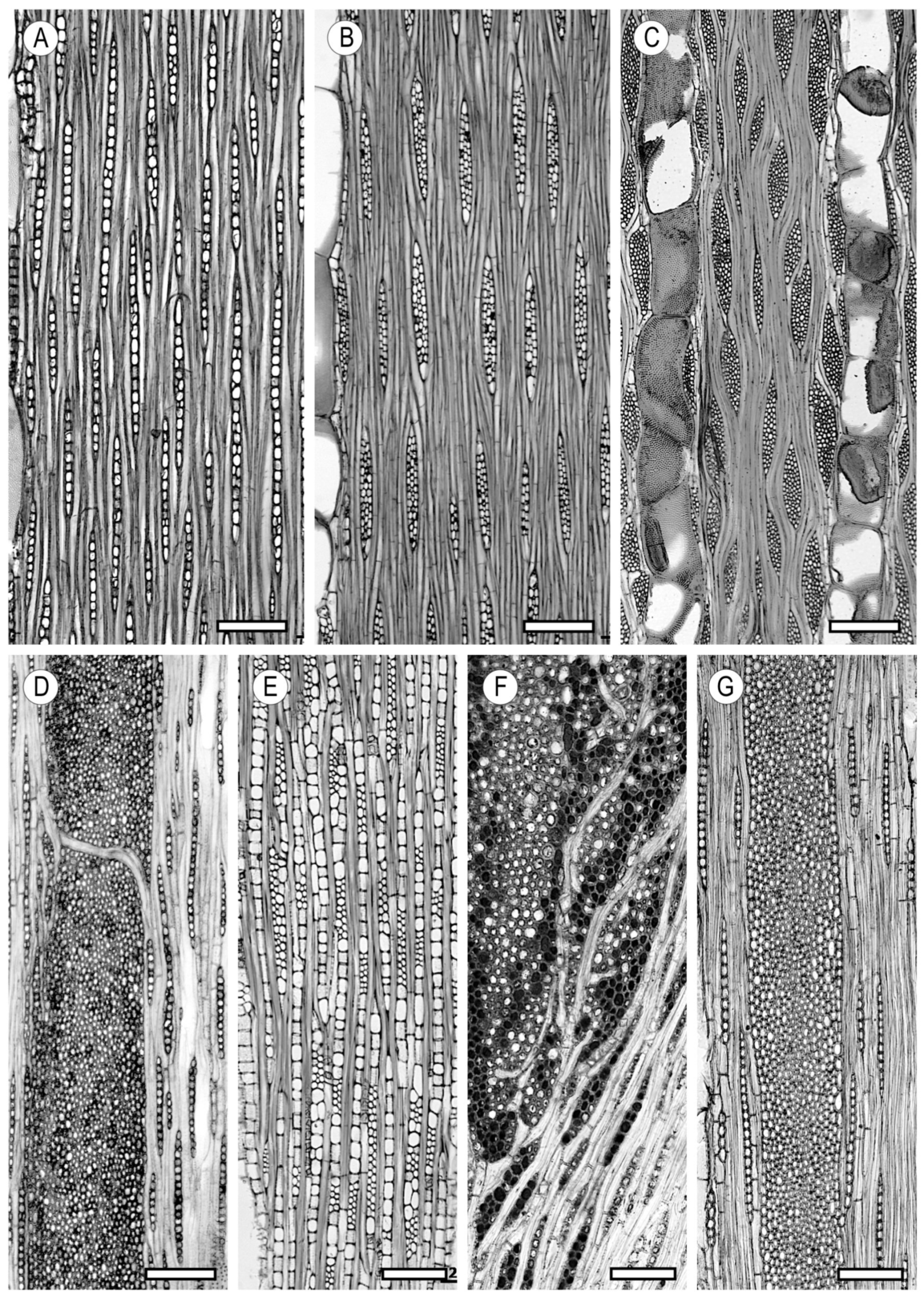
As rays develop in the radial direction, they separate. When the distance between two rays is very large, an intermediate ray appears. The IAWA Committee [12] established the following intervals by ray width in terms of the number of cells counted in the tangential section: exclusively uniseriate (Castanea sativa, Populus spp., and Terminalia superba), from one to three cells (Aucoumea klaineana, Dialium guianense, and Albizzia saman), from four to ten cells (Acer saccharum, Khaya anthotheca, and Celtis sinensis), and more than 10 cells (Quercus spp. and Platanus spp.). The multiseriate portion of the ray is occasionally the same width as the uniseriate portion and can be used as a differentiating diagnostic feature (Caryocar costaricense and Strombosia pustulata) (Figure 17A-E). Uniseriate rays and multiseriate rays more than 10 cells wide are uncommon in hardwoods, while multiseriate rays from three to ten cells wide are the most frequent.
In some species, rays are grouped to form wider rays that appear macroscopically to be a single large ray, although they are actually several rays separated by axial elements (aggregate rays) (e.g., Alnus spp. and Quercus spp.) (Figure 17F).
In some species (e.g., Quercus spp., Ilex aquifolium, and Poga oleosa), rays have two clearly distinct sizes in width, normally associated with different heights (Figure 17G). They do not have to be uniseriate in some cases and multiseriate in others, but the difference between the two groups must be clear, with no third intermediate group; or, where such a group occurs, it must be infrequent, e.g., Fagus spp. The IAWA Committee [12] stressed that aggregate rays must not, in themselves, be viewed as a separate class or group size and should be considered as having two different sizes only if they are composed of much broader rays than in nonaggregate rays, as in Quercus spp.
Viewed in the tangential section, the ray shape is tapered, with ends normally finishing in a single line of cells. Like the ray width, the ray height is highly variable, although most identification keys take this feature into account only when rays exceed the threshold of 1 mm.
Some hardwood species have no horizontal tissue, and their wood tissue is made up exclusively of axial elements (Figure 18). This unusual feature occurs in only a few species (e.g., Pisonia umbellifera and Veronica speciosa). The absence of rays has been interpreted as an adaptation to greater mechanical resistance [122].
With regard to the ray spacing or frequency, the IAWA Committee [12] established the ranges ≤ 4/mm, 4–12, and ≥12, with 4–12 cells per mm being the most common.
Hardwood rays can have various cell shapes: procumbent, arranged horizontally with their longer axis perpendicular to the longitudinal direction of the tree; upright, with their longer axis parallel to the longitudinal direction; and square, with the two axes similar in size.
The ray classification described by Kribs [123] has now been mostly superseded. The ray composition was defined by the IAWA Committee [12] as follows: all cells procumbent (Acer spp., Albizia spp., and Tabebuia spp.), all cells upright and/or square (Aucuba japonica and Hedyosmum scabrum), ray cells procumbent with one row of upright and/or square marginal cells (Aucoumea klaineana), ray cells procumbent with 24 rows of upright and/or square marginal cells (Liquidambar styraciflua), ray cells procumbent with more than four rows of upright and/or square marginal cells (Turpinia spp.), and rays with procumbent, square, and upright cells mixed throughout the ray (Heliocarpus spp.) (Figure 19).
Uniseriate and multiseriate rays are not necessarily composed of the same cell types in a single wood. The ray cell type must always be determined in mature wood because the cell composition can differ between mature and juvenile wood.
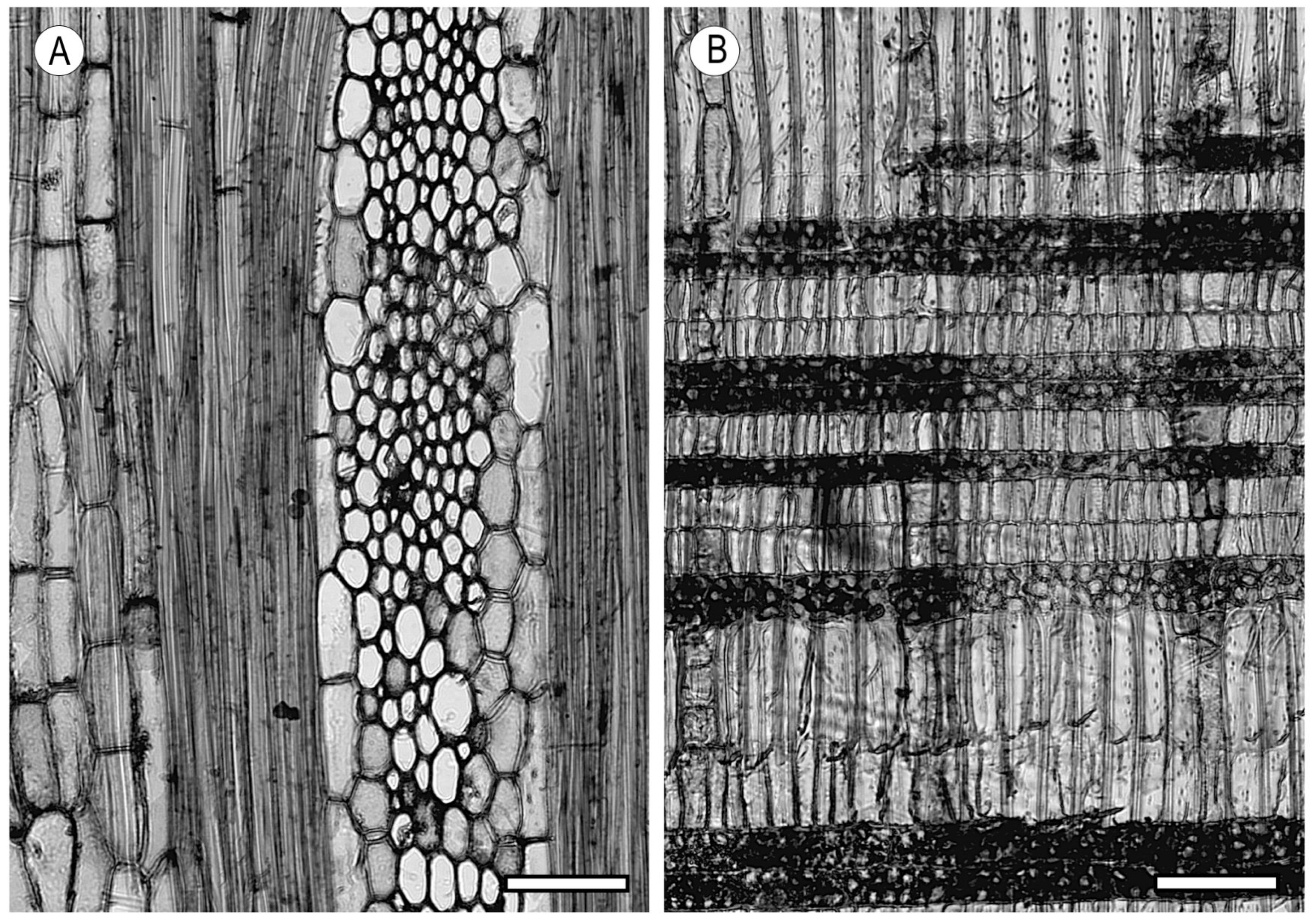
Some species have distinctive cells known as sheath cells (Figure 20A), located on each side of rays more than three-seriate, which are larger (normally taller than broad) than central ray cells (e.g., Heritiera utilis, Ochroma pyramidale, and Triplochiton scleroxylon). These cells must be observed in the tangential section. Other species also have tile cells, most easily seen in radial sections, which have the appearance of upright, very narrow cells (rarely square) arranged in horizontal series among procumbent cells (e.g., Durio spp., Luehea spp., Ochroma pyramidale, and Triplochiton scleroxylon) (Figure 20B).

5. Storied Structures

Some anatomical elements of hardwoods have a storied structure in horizontal series observed in the tangential section. The origin of this structure is unknown, although some theories associated with cambial activity have been proposed. It is probably associated with the homogeneity of the height of their axial elements and a low level of intrusion from fusiform cells. From the evolutionary point of view, storied wood is more highly specialised than nonstoried wood [18]. This structure can occur in all the rays of a wood (e.g., Dalbergia nigra, Dipteryx odorata, Millettia laurentii, and Pterocarpus soyauxii), only in the lower rays (e.g., Dialium guineense and Heritiera utilis), in axial parenchyma cells and/or vessel elements (e.g., Dalbergia baronii, Dicorynia guianensis, and Robinia pseudoacacia), and in fibres (e.g., Dalbergia nigra, Guaiacum officinale, Millettia laurentii, and Triplochiton scleroxylon) (Figure 21A–D).
An intermediate situation between storied structure and alternate arrangement occurs in some wood of Lauraceae (e.g., Aspidostemon spp.), Leguminosae (e.g., Brachystegia cynometroides, Erythrophleum suaveolens, and Platymiscium pinnatum), and Meliaceae (e.g., Entandrophragma cylindricum and Pseudocedrela kotschyi), as their elements are arranged in oblique formations that the IAWA Committee [12] has termed as rays and/or axial elements being irregularly storied because of their wavy or oblique rather than horizontal arrangement (Figure 21E).

6. Secretory Elements

All the substances produced by plants are the result of cell metabolism and can appear and disappear throughout the life of the cell. Most are storage products, some are involved in plant defence, and a few are waste products. Secretory cells generate a large variety of products, including oils, resins, mucilages, rubbers, tannins, and crystals [18].

6.1. Oil Cells and Mucilage Cells

These cells are present in the longitudinal and radial elements of hardwoods and differ only in the nature of the substances they contain, either oils or mucilages. Both types of cells are parenchymatous idioblasts, enlarged and rounded, occasionally with considerable axial extension. When they occur in rays, they usually occupy the ends of the ray, and when associated with axial parenchyma, they are differentiated from other cells by their size. They can also be associated with fibrous tissue (Figure 22).
Oil cells occur in many Lauraceae woods, including Nectandra grandis, in some species of Ocotea spp., and in Phoebe porosa. Mucilage cells associated with axial parenchyma occur in Endlicheria spp., and when associated with ray parenchyma, they occur in Persea spp. and others [12].
Many functions have been attributed to mucilage cells (carbohydrate and water storage, transpiration reduction, protection against radiation, and protection against herbivores), but experimental evidence is lacking, and since the review by Gregory and Baas [124], their functions remain mostly unexplained.

6.2. Intercellular Canals

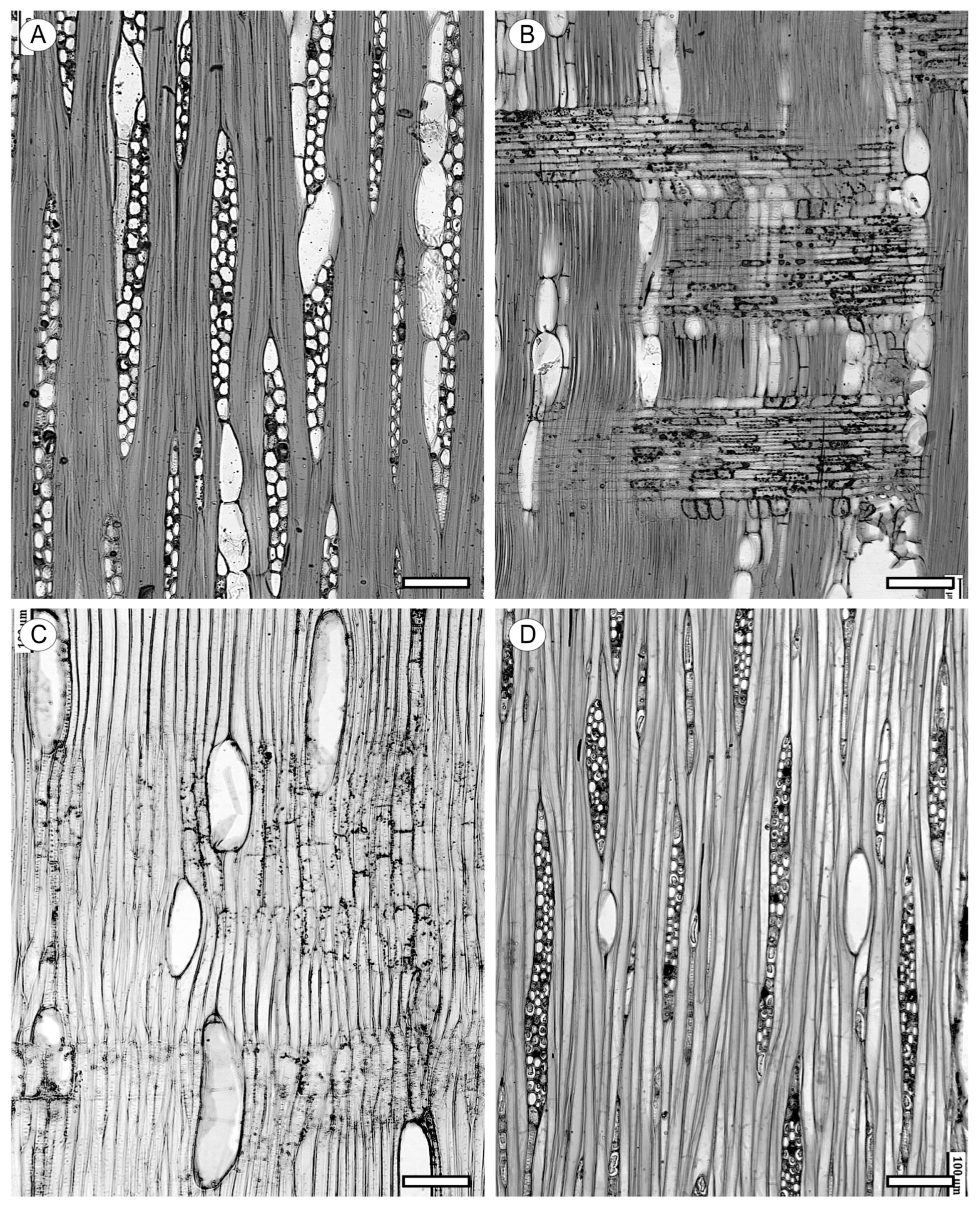
Intercellular canals in hardwoods, both longitudinal (axial) and radial, are surrounded by epithelial cells capable of secreting gums, resins, and other substances. Axial canals are in tangential lines or diffusely arranged. For tangential arrangement, the IAWA Committee [12] has made divisions between long tangential lines of more than five canals (e.g., Dryobalanops spp., Hopea spp., Shorea spp., and Sindora spp.) or in short lines from two to five canals (e.g., Dryobalanops sumatrensis, Hopea nervosa, and Shorea robusta). In diffuse arrangement, canals are isolated and randomly distributed (e.g., Dipterocarpus indicus, Upuna borneensis, and Vatica spp.) (Figure 23A–C).
Radial canals occur in the rays and create a fusiform appearance (e.g., Astronium graveolens, Melanochyla spp., Pistacia spp., Shorea section Richetia, and Tapirira spp.) (Figure 23D). The size and number of canals per ray are useful for wood identification. The colour of resins in the canals of Dipterocarpaceae can also be used for identification.
Like conifers, some hardwoods form axial traumatic resin canals in tangential rows, generally irregular in outline and closely spaced, in response to wounding (e.g., Altingia excelsa, Berlinia bracteosa, Liquidambar styraciflua, and Terminalia ivorensis) (Figure 23E).
Much has been speculated about the presence of intercellular canals in hardwoods, but they appear to be simply associated with tree metabolic processes.

6.3. Laticiferous and Tanniniferous Tubes

Tubes are series of cells of indeterminate length, arranged horizontally or vertically, containing only two types of substances: latex or tannins (Figure 24).
In laticiferous tubes, latex can be from colourless or light yellow to brown. These tubes occur in rays of the wood of Apocynaceae, Campanulaceae, Caricaceae, Euphorbiaceae, and Moraceae and, axially, among the fibrous tissue observed so far only in Moraceae. Tanniniferous tubes are reddish brown and have been observed so far only in Myristicaceae. They are very difficult to differentiate from other ray cells in the tangential section, but in the radial section, they are longer than normal ray cells. To distinguish the type of tube, chemical tests are used rather than colour [12].
It is estimated that around 12,500 plants belonging to 900 genera contain latex, from small annual herbaceous plants to large trees, such as the rubber tree, Hevea brasiliensis [18], whose bark (but not wood) contains latex. Laticiferous tubes have been studied since the 19th century [125], and because of the liquid or milky content that flows from them when the plant is cut, early botanists compared them to the circulatory system in animals. Various functions have been attributed to them, including storage, although the starch they contain is not easily mobilised [126]; scarring [127]; a response to excess metabolites [128]; and even for sequestering secondary metabolites that can repel herbivores [129].
Tannins are a heterogeneous group of astringent-tasting polyphenols. They are abundant in fruits, seeds, and galls and can occur in idioblasts and tubular cells [18]. Because of their astringency, tannins appear to have the primary function for repelling predators and invasion by parasitic organisms.

7. Included Phloem

This is a very infrequent feature and responds to cambial variants, resulting in diffuse (e.g., Aquilaria malaccensis and Erisma spp.) or concentric (e.g., Avicennia marina and Koompassia malaccensis) arrangements. It is a regular feature in the species in which it occurs and must never be termed as being anomalous. Other arrangements can occur, particularly in lianas with lobed, fissured, or furrowed stems (Figure 25).
Carlquist [40] discussed the presence of included phloem produced by a single cambium as an ideal mechanism for distributing photosynthates more quickly and efficiently to their storage sites, raising the question as to why this system has not evolved in other plants.

8. Crystal Inclusions and Silica

Crystal inclusions are much more abundant in hardwoods than in conifers. The chemical composition of the crystals is calcium oxalate, which is birefringent under polarised light. Prismatic crystals are the most frequent type, although crystals can occur in a large variety of shapes. They can be present in any of the cell types: in non-chambered axial parenchyma cells (e.g., Carya spp., Ceiba pentandra, Cordia africana, Parashorea spp., and Shorea subgr. Richetia) and chambered axial parenchyma cells (e.g., Cerbera spp., Caryocar costarricense, Hevea spp., and Parashorea spp.); in ray parenchyma in upright and/or square cells (e.g., Astronium graveolens, Mangifera spp., Tapirira spp., Khaya ivorensis, and Toona ciliata), in procumbent cells (e.g., Aspidosperma quebracho-blanco, Hopea spp., and Castanopsis spp.), in radial alignment in procumbent cells (e.g., Anogeissus spp. and Barringtonia spp.), and in chambered upright and/or square cells (e.g., Distemonanthus benthamianus, Scottellia coriacea, and Ilex spp.); in fibres (e.g., Julbernardia pellegriniana and Tetraberlinia tubmaniana); and even in tyloses (e.g., Hopea odorata and Strombosia glaucescens) (Figure 26).
The presence, type, and location of crystals are distinguishing diagnostic features, although they should be used with caution. The absence of crystals is not recommended for use as a diagnostic feature, even though prismatic crystals are absent in some genera (Betula spp., Dipterocarpus spp., Liriodendron spp., and Tilia spp.) [12].
Other types of crystals are druses, raphides, acicular crystals, styloids, elongate crystals, and even small crystals sometimes grouped in a granular mass.
Druses are star-shaped crystal inclusions that occur in ray parenchyma cells (e.g., Amygdalus communis, Celtis africana, Gleditsia triacanthos, and Prunus africana), axial parenchyma (e.g., Terminalia complanata and Toona sureni), fibres (e.g., Combretum fruticosum), and chambered cells (e.g., Celtis africana and Prunus spp.) (Figure 27A–D).
Raphides are needle-like crystals grouped in bundles (e.g., Dillenia spp., Tetramerista spp., and Vitis spp.) (Figure 27E). Acicular crystals are similar to raphides but do not occur in bundles (e.g., Gmelina arborea, Olea europea, and Tecoma stans). Styloids are elongated crystals, normally four times as long as broad, with pointed or square ends (e.g., Gonystylus macrophyllus), and elongate crystals are crystals from two to four times as long as broad, with pointed ends (e.g., Ligustrum vulgare). Smaller crystals of other shapes can also be observed, including cubic (Aporusa villosa), navicular (Litsea reticulata), and spindle-shaped (Dehaasia spp.), and even very small granular masses of crystals, known as crystal sand because of their appearance. Apart from these types in hardwoods, crystals with other morphologies can also occur, but because of their specific characteristics, they cannot be included with any of the other types. Because of this, hardwood identification keys should include a feature for the presence of crystals of shapes other than the commonly known ones. In some species, crystals are included in a particular type of cell, as in the case of idioblasts (e.g., Carpinus carolinianum, Juglans nigra, Pyrus communis, and Zelkova serrata). Two or more similar-sized crystals can occur in a single chamber or cell (e.g., Gmelina arborea and Ligustrum vulgare), and, rarely, two different-sized crystals can occur in a single chamber or cell (e.g., Gonystylus bancanus). Lastly, in some hardwoods, it is possible to observe a globular mass of crystals around an organic nucleus, attached to the cell wall by a cellulosic stalk impregnated with calcium carbonate (e.g., Sparattanthelium spp. and Trichanthera spp.). These formations are known as cystoliths [12] (Figure 28).
Accumulations slightly differ between plants growing in calcium-rich soil and plants growing in soil without calcium. Calcium oxalate crystals are thought to be metabolic waste substances. Active deposits of calcium oxalate by fungi [130] could be a defence mechanism against fungivores. Moreover, Carlquist [40] stated that although plants evidently have other defence mechanisms to avoid being eaten (toxic substances, thorns, etc.) closer to the surface of a plant rather than inside the wood, the presence of crystals can be an effective element against chewing insects and molluscs, and even raphides can be a deterrent for herbivorous vertebrates [131,132]. The hardness and insolubility of calcium oxalate crystals makes them an effective plant defence mechanism.
The presence of silicon dioxide particles (silica) is frequent in wood from tropical regions. Like crystals, their location in wood varies considerably. They can occur in ray parenchyma cells (e.g., Aucoumea klaineana, Dacryodes spp., Dipterocarpus spp., and Guarea cedrata), in axial parenchyma (e.g., Entandrophragma candollei and Qualea spp.), and even in fibres (e.g., Dacryodes spp. and Dysoxylum spectabile) (Figure 29).
At low magnifications (4–10×), silica is observed as dark nonbirefringent particles. At higher magnifications (25–40×) they can have a glassy appearance.
The abundance of silica in wood sometimes requires the maceration of the sample for observation when the wood cannot be sliced for the microscope.
The presence of silica in wood is a disadvantage during machining because a lack of suitable aspiration systems can cause rhinitis, dermatitis, dizziness, vomiting, nasal haemorrhage, etc., because of inhalation [133]. Silica accumulation in plants helps to increase the stem strength and provides a resistance mechanism to attack by fungal pathogens, xylophagous insects, and herbivores [134].

9. Conclusions

The functionality of hardwood elements remains mostly unexplained. Tools and research capable of distinguishing the presence or absence of elements as a result of plant genetics or adaptive capacity will enable significant advances. For now, it has been shown that some hardwood anatomical elements have a clear functionality in the plant structure. For other elements, however, more specific research is necessary to fully explain their functions.
The functions of the various hardwood elements (vessels, other tracheary elements, fibres, fibre-tracheids, and axial and ray parenchyma) are broadly understood individually, and there is a wide variety of patterns that they exhibit, which reflect the taxon, habit, and environment where the tree or shrub grows. Understanding how these cell types act together and combining anatomical observations with physiological experiments that support or refute hypotheses on function is much more difficult, and reconciling speculation from observations with experimental data is challenging. Such a wide range of woody plant forms and wood anatomies provide endless opportunities for research. In the current climate change scenario and with trees rapidly changing their distribution in response, elucidating in detail the relationship between wood structure and ecological/physiological functions is increasingly urgent.

Author Contributions

Conceptualisation, L.G.E., P.d.P., and P.G.; methodology, L.G.E., P.d.P., and P.G.; validation, L.G.E. and P.d.P.; investigation, L.G.E., P.d.P., P.G., A.G.-I., F.G.-F., and L.G.-E.; resources, L.G.E. and P.G.; writing—original draft preparation, L.G.E., P.d.P., P.G., A.G.-I., F.G.-F., and L.G.-E.; writing—review and editing, L.G.E., P.d.P., and P.G.; visualisation, A.G.-I., F.G.-F., and L.G.-E.; supervision, L.G.E.; project administration, L.G.E., P.d.P. and P.G. All authors have read and agreed to the published version of the manuscript.

Funding

This research received no external funding.

Data Availability Statement

Not applicable.

Acknowledgments

The authors thank Sarah Wilson for taking some of the photographs. The SEM photo in Figure 3E was taken by Nigel Warwick.

Conflicts of Interest

The authors declare no conflicts of interest.

References

  1. Hudson, P.J.; Razanatsoa, J.; Feild, T.S. Early vessel evolution and the diversification of wood function: Insights from Malagasy Canellales. Am. J. Bot. 2010, 97, 80–93. [Google Scholar] [CrossRef]
  2. Brenner, G.; Bickoff, I.S. Palynology and age of the Lower Cretaceous basal Kurnub Group from the coastal plain to the northern Negev of Israel. Palynology 1992, 16, 137–185. [Google Scholar] [CrossRef]
  3. Herendeen, P.S.; Wheeler, E.A.; Baas, P. Angiosperm wood evolution and the potential contribution of paleontological data. Bot. Rev. 1999, 65, 278–300. [Google Scholar] [CrossRef]
  4. Baas, P.; Wheeler, E.A. Parallelism and reversibility in xylem evolution—A review. IAWA J. 1996, 17, 351–364. [Google Scholar] [CrossRef]
  5. Wheeler, E.A.; Baas, P. Wood evolution: Baileyan trends and functional traits in the fossil record. IAWA J. 2019, 40, 488–529. [Google Scholar] [CrossRef]
  6. Philippe, M.; Cuny, G.; Bashforth, A. Ecpagloxylon mathiesenii gen. nov. et sp. nov., a Jurassic wood from Greenland with several primitive angiosperm features. Plant Syst. Evol. 2010, 287, 153–165. [Google Scholar] [CrossRef]
  7. Baas, P.; Wheeler, E.A. Wood anatomy and climate change. In Climate Change, Ecology and Systematics; Hodkinson, T.R., Jones, M.B., Waldren, S., Parnell, J.A.N., Eds.; Cambridge University Press: Cambridge, UK, 2011; pp. 141–155. [Google Scholar] [CrossRef]
  8. Bailey, I.W.; Tupper, W.W. Size variation in tracheary cells. I. A comparison between the secondary xylems of vascular cryptograms, gymnosperms and angiosperms. Proc. Am. Art. Sci. Soc. 1918, 54, 149–204. [Google Scholar] [CrossRef]
  9. Baas, P. Ecological patterns in xylem anatomy. In On the Economy of Plant Form and Function; Givnish, T.J., Ed.; Cambridge University Press: Cambridge, UK, 1986; pp. 327–349. [Google Scholar]
  10. Carlquist, S. Comparative Wood Anatomy: Systematic, Ecological, and Evolutionary Aspects of Dicotyledon Wood, 2nd ed.; Springer: Heidelberg, Germany, 2001; 448p. [Google Scholar] [CrossRef]
  11. Jansen, S.; Baas, P.; Gasson, P.; Lens, F.; Smets, E. Variation in xylem structure from tropics to tundra: Evidence from vestured pits. Proc. Natl. Acad. Sci. USA 2004, 101, 8833–8837. [Google Scholar] [CrossRef]
  12. IAWA Committee. IAWA List of microscopic features for hardwood identification. IAWA Bull. 1989, 10, 219–332. [Google Scholar]
  13. Brown, H.P.; Panshin, A.J.; Forsaith, C.C. Textbook of Wood Technology. Volume I. Structure, Identification, Defects and Uses of the Commercial Woods of the United States, 1st ed.; McGraw-Hill: New York, NY, USA, 1949; p. 652. [Google Scholar]
  14. Olson, M.E.; Anfodillo, T.; Gleason, S.M.; McCulloh, K.A. Tip-to-base xylem conduit widening as an adaptation: Causes, consequences, and empirical priorities. New Phytol. 2021, 229, 1877–1893. [Google Scholar] [CrossRef]
  15. Baas, P.; Beeckman, H.; Cufar, K.; De Micco, V. Functional traits in wood anatomy. IAWA J. 2016, 37, 121–368. [Google Scholar] [CrossRef]
  16. Olson, M.E.; Pace, M.R. Tributes to Sherwin Carlquist. IAWA J. 2023, 44, 273–556. [Google Scholar] [CrossRef]
  17. Esteban, L.G.; Guindeo, A.; Peraza, C.; de Palacios, P. La Madera y su Anatomía; Fundación Conde del Valle de Salazar, Ed.; Mundi-Prensa y AiTiM: Madrid, Spain, 2003; 327p. [Google Scholar]
  18. Evert, R.F. Esau’s Plant Anatomy: Meristems, Cells, and Tissues of the Plant Body: Their Structure, Function, and Development, 3rd ed.; John Wiley & Sons, Inc.: Hoboken, NJ, USA, 2006; p. 601. [Google Scholar] [CrossRef]
  19. Wheeler, E.A.; Pearson, R.G.; LaPasha, C.A.; Zack, T.; Hatley, W. Computer-Aided Wood Identification: Reference Manual; North Carolina Agricultural Research Service Bulletin 474; NCRS: Raleigh, NC, USA, 1986. [Google Scholar]
  20. Esteban, L.G.; Guindeo, A. Anatomía de las maderas de frondosas españolas; AiTiM: Madrid, Spain, 1990; p. 619. [Google Scholar]
  21. Christman, M.A.; Sperry, J.S. Single vessel flow measurements indicate scalariform perforation plates confer higher flow resistance than previously estimated. Plant Cell Environ. 2010, 33, 431–443. [Google Scholar] [CrossRef]
  22. Sperry, J.S. Evolution of water transport and xylem structure. Int. J. Plant Sci. 2003, 164, 115–127. [Google Scholar] [CrossRef]
  23. Jansen, S.; Nardini, A. From systematic to ecological wood anatomy and finally plant hydraulics: Are we making progress in understanding xylem evolution? New Phytol. 2014, 203, 12–15. [Google Scholar] [CrossRef]
  24. Baas, P. Some functional and adaptive aspects of vessel member morphology. Leiden Bot. Ser. 1976, 3, 157–181. [Google Scholar]
  25. Feild, T.S.; Brodribb, T.; Holbrook, N.M. Hardly a relict: Freezing and the evolution of vesselless wood in Winteraceae. Evolution 2002, 56, 464–478. [Google Scholar] [CrossRef]
  26. Zimmermann, M.H.; Jeje, A. Vessel-length distribution in stems of some American woody plants. Can. J. Bot. 1981, 59, 1882–1892. [Google Scholar] [CrossRef]
  27. Ewers, F.W.; Fisher, J.B.; Chiu, S.T. A survey of vessel dimensions in stems of tropical lianas and other growth forms. Oecologia 1990, 8, 544–552. [Google Scholar] [CrossRef]
  28. Sperry, J.; Hacke, G.; Feild, T.S.; Sano, Y.; Sikkema, E.H. Hydraulic consequences of vessel evolution in angiosperms. Int. J. Plant Sci. 2007, 168, 1127–1139. [Google Scholar] [CrossRef]
  29. Greenidge, K.N.H. An approach to the study of vessel length in hardwood species. Am. J. Bot. 1952, 39, 570–574. [Google Scholar] [CrossRef]
  30. Jansen, S.; Choat, B.; Pletsers, A. Morphological variation of intervessel pit membranes and implications to xylem function in angiosperms. Am. J. Bot. 2009, 96, 409–419. [Google Scholar] [CrossRef]
  31. Li, S.; Lens, F.; Espino, S.; Karimi, Z.; Klepsch, M.; Schenk, H.J.; Schmitt, M.; Schuldt, B.; Jansen, S. Intervessel pit membrane thickness as a key determinant of embolism resistance in angiosperm xylem. IAWA J. 2016, 37, 152–171. [Google Scholar] [CrossRef]
  32. Wheeler, J.K.; Sperry, J.S.; Hacke, U.G.; Hoang, N. Intervessel pitting and cavitation in woody Rosaceae and other vesselled plants: A basis for a safety versus efficiency trade-off in xylem transport. Plant Cell Environ. 2005, 28, 800–812. [Google Scholar] [CrossRef]
  33. Choat, B.; Brodie, T.W.; Cobb, A.; Zwieniecki, M.A.; Holbrook, N.M. Direct measurements of intervessel pit membrane hydraulic resistance in two angiosperm tree species. Am. J. Bot. 2006, 93, 993–1000. [Google Scholar] [CrossRef]
  34. Hacke, U.G.; Sperry, J.S.; Wheeler, J.K.; Castro, L. Scaling of angiosperm xylem structure with safety and efficiency. Tree Physiol. 2006, 26, 619–701. [Google Scholar] [CrossRef]
  35. Bailey, L.W. Preliminary notes on cribriform and vestured pits. Trop. Woods 1932, 3, 46–48. [Google Scholar]
  36. Bailey, L.W. The cambium and its derivative tissues: No. VIII. Structure, distribution, and diagnostic significance of vestured pits in dicotyledons. J. Arnold Arbor. 1933, 14, 259–273. [Google Scholar] [CrossRef]
  37. Jansen, S.; Smets, E.; Baas, P. Vestures in woody plants: A review. IAWA J. 1998, 19, 347–382. [Google Scholar] [CrossRef]
  38. Parham, R.A.; Baird, W.M. Warts in the evolution of angiosperm wood. Wood Sci. Technol. 1974, 8, 1–10. [Google Scholar] [CrossRef]
  39. Carlquist, S. Wood anatomy of Onagraceae: Further species; root anatomy; significance of vestured pits and allied structures in dicotyledons. Ann. Mo. Bot. Gard. 1982, 69, 755–769. [Google Scholar] [CrossRef]
  40. Carlquist, S. Comparative Wood Anatomy. Systematic, Ecological, and Evolutionary Aspects of Dicotyledon Wood; Springer: Berlin/Alemania, Germany, 1988; 436p. [Google Scholar]
  41. Zweypfenning, R.C.V.J. A hypothesis on the function of vestured pits. IAWA Bull. 1978, 1, 13–15. [Google Scholar]
  42. Jansen, S.; Baas, P.; Gasson, P.; Smets, E. Vestured. Pits: Do they promote safer water transport? Int. J. Plant Sci. 2003, 164, 405–413. [Google Scholar] [CrossRef]
  43. Zimmerman, M.H. Xylem Structure and the Ascent of Sap; Springer: Heidelberg, Germany, 1983; p. 146. [Google Scholar] [CrossRef]
  44. Costa, A.; Wiedenhoeft, A.C. On the possible functions of helical thickenings in conductive cells of wood. IAWA J. 2023, 44, 381–398. [Google Scholar] [CrossRef]
  45. Ohtani, J.; Meylan, B.A.; Butterfield, B.G. A note on vestures on helical thickenings. IAWA Bull. 1984, 5, 9–11. [Google Scholar] [CrossRef]
  46. Karam, G.N. Biomechanical Model of the Xylem Vessels in Vascular Plants. Ann. Bot. 2005, 95, 1179–1186. [Google Scholar] [CrossRef] [PubMed]
  47. Woodworth, R.H. Fibriform vessel members in the Passifloraceae. Trop. Woods 1935, 41, 8–16. [Google Scholar]
  48. Bissing, D.R. Variation in qualitative anatomical features of the xylem of selected dicotyledonous woods in relation to water availability. Bull. Torrey Bot. Club. 1982, 109, 371–384. [Google Scholar] [CrossRef]
  49. Wheeler, E.A.; Gasson, P.E.; Baas, P. Using the InsideWood web site: Potentials and pitfalls. IAWA J. 2020, 41, 412–462. [Google Scholar] [CrossRef]
  50. De Micco, V.; Balzano, A.; Wheeler, E.; Baas, P. Tyloses and gums: A review of structure, function and occurrence of vessel occlusions. IAWA J. 2016, 37, 186–205. [Google Scholar] [CrossRef]
  51. Murmanis, L. Formation of tyloses in felled Quercus rubra L. Wood Sci. Technol. 1975, 9, 3–14. [Google Scholar] [CrossRef]
  52. Sun, Q.; Ros, T.L.; Reid, M.S.; Matthews, M.A. Ethylene and not embolism is required for wound-induced tylose development in stems of grapevines (Vitis vinifera L.). Plant Physiol. 2007, 145, 1629–1636. [Google Scholar] [CrossRef] [PubMed]
  53. Malpighi, M. Opera omnia. In Opere Scelte di Marcello Malpighi; Belloni, L., Ed.; Unione Tipografico-Editrice Torinese: Torino, Italy, 1967; pp. 75–99. [Google Scholar]
  54. Scheckler, S.E.; Galtier, J. Tyloses and ecophysiology of the early carboniferous progymnosperm tree Protopitys buchiana. Ann. Bot. 2003, 91, 739–747. [Google Scholar] [CrossRef] [PubMed]
  55. Chattaway, M.M. The development of tyloses and secretion of gum in heartwood formation. Aust. J. Biol. Sci. 1949, 2, 227–240. [Google Scholar] [CrossRef]
  56. Esau, K. Plant Anatomy, 2nd ed.; John Wiley & Sons: New York, NY, USA, 1965; p. 767. [Google Scholar]
  57. Pearce, R.B.; Holloway, P.J. Suberin in the sapwood of oak (Quercus robur L.): Its composition from a compartmentalization barrier and its occurrence in tyloses in undecayed wood. Physiol. Plant Pathol. 1984, 24, 71–81. [Google Scholar] [CrossRef]
  58. Hillis, W.E. Heartwood and Tree Exudates; Springer: Heidelberg, Germany, 1987; p. 268. [Google Scholar] [CrossRef]
  59. Olson, M.E. Imperforate tracheary element classification for studies of xylem structure-function relations. IAWA J. 2023, 44, 439–464. [Google Scholar] [CrossRef]
  60. Carlquist, S. Vasicentric tracheids as a drought survival mechanism in the woody flora of southern California and similar regions: Review of vasicentric tracheids. Aliso 1985, 11, 37–68. [Google Scholar] [CrossRef]
  61. Carlquist, S. How wood evolves: A new synthesis. Botany 2012, 90, 901–940. [Google Scholar] [CrossRef]
  62. Sano, Y.; Morris, H.; Shimada, H.; Ronse de Craene, L.P.; Jansen, S. Anatomical features associated with water transport in imperforate tracheary elements of vessel-bearing angiosperms. Ann. Bot. 2011, 107, 953–964. [Google Scholar] [CrossRef]
  63. Scholz, F.G.; Bucci, S.J.; Goldstein, G.; Meinzer, F.C.; Franco, A.C.; Miralles-Wilhelm, F. Biophysical properties and functional significance of stem water storage tissues in Neotropical savanna trees. Plant. Cell. Environ. 2007, 30, 236–248. [Google Scholar] [CrossRef]
  64. Pfautsch, S.; Renard, J.; Tjoelker, M.G.; Salih, A. Phloem as capacitor: Radial transfer of water into xylem of tree stems occurs via symplastic transport in ray parenchyma. Plant Physiol. 2015, 167, 963–971. [Google Scholar] [CrossRef] [PubMed]
  65. Loepfe, L.; Martínez-Vilalta, J.; Piñol, J.; Mencuccini, M. The relevance of xylem network structure for plant hydraulic efficiency and safety. J. Theor. Biol. 2007, 247, 788–803. [Google Scholar] [CrossRef] [PubMed]
  66. Martínez-Vilalta, J.; Mencuccini, M.; Álvarez, X.; Camacho, J.; Loepfe, L.; Piñol, J. Spatial distribution and packing of xylem conduits. Am. J. Bot. 2012, 99, 1189–1196. [Google Scholar] [CrossRef] [PubMed]
  67. Pratt, R.B.; Castro, V.; Jacobsen, A.L. The functional significance of tracheids co-occurring with vessels in xylem of Eudicots suggests a role in embolism tolerance. IAWA J. 2023, 44, 477–494. [Google Scholar] [CrossRef]
  68. Barotto, A.J.; Fernandez, M.E.; Gyenge, J.; Meyra, A.; Martinez-Meier, A.; Monteoliva, A. First insights into the functional role of vasicentric tracheids and parenchyma in eucalyptus species with solitary vessels: Do they contribute to xylem efficiency or safety? Tree Physiol. 2016, 36, 1485–1497. [Google Scholar] [CrossRef] [PubMed]
  69. Wheeler, E.A.; Baas, P.; Rodgers, S. Variations in dicot anatomy: A global analysis based on the Inside Wood database. IAWA J. 2007, 28, 229–258. [Google Scholar] [CrossRef]
  70. Harrar, E.S. Notes on starch grains in septate fiber-tracheids. Trop. Woods 1946, 85, 1–9. [Google Scholar]
  71. Spackman, W.; Swamy, B.G.L. The nature and occurrence of septate fibers in dicotyledons. Am. J. Bot. 1949, 36, 804. [Google Scholar]
  72. Plavcova, L.; Olson, M.E.; Jandova, V.; Dolezal, J. Parenchyma is not the sole site of storage: Storage in living fibres. IAWA J. 2023, 44, 465–476. [Google Scholar] [CrossRef]
  73. Morris, H.R. The Structure and Function of Ray and Axial Parenchyma in Woody Seed Plants. Ph.D. Thesis, Universität Ulm, Ulm, Germany, 20 July 2016. [Google Scholar] [CrossRef]
  74. Frison, E. De Ia presence d’amidon dans le Iumen des fibres du bois. Bull. Agric. Congo Belg. 1948, 39, 869–874. [Google Scholar]
  75. Chattaway, M.M. Proposed standards for numerical values used in describing woods. Trop. Woods 1932, 29, 20–28. [Google Scholar]
  76. Carlquist, S. Further concepts in ecological wood anatomy, with comments on recent work in wood anatomy and evolution. Aliso 1980, 9, 499–553. [Google Scholar] [CrossRef]
  77. Carlquist, S.; Hoekman, D.A. Ecological Wood Anatomy of the Woody Southern Californian Flora. IAWA J. 1985, 6, 319–347. [Google Scholar] [CrossRef]
  78. Plomion, C.; Leprovost, G.; Stokes, A. Wood Formation in Trees. Plant Physiol. 2001, 127, 1513–1523. [Google Scholar] [CrossRef] [PubMed]
  79. Plavcová, L.; Jansen, S. The Role of Xylem Parenchyma in the Storage and Utilization of Nonstructural Carbohydrates. In Functional and Ecological Xylem Anatomy; Hacke, U., Ed.; Springer: Cham, Switzerland, 2015; pp. 209–234. [Google Scholar] [CrossRef]
  80. Morris, H.; Jansen, S.; Arber, A. Opinion paper—Secondary xylem parenchyma—from classical terminology to functional traits. IAWA J. 2016, 37, 1–15. [Google Scholar] [CrossRef]
  81. Magel, E.; Einig, W.; Hampp, R. Carbohydrates in trees. Dev. Crop Sci. 2000, 26, 317–336. [Google Scholar] [CrossRef]
  82. Eriksson, K.E.L.; Blanchette, R.A.; Ander, P. Microbial and Enzymatic Degradation of Wood and Wood Components; Springer: Heidelberg, Germany, 1990; p. 407. [Google Scholar] [CrossRef]
  83. Lundquist, R.; Brunow, G. Lignins: Natural products from oxidative coupling of 4 hydroxyphenylpropanoids. Phytochem. Rev. 2004, 3, 29–60. [Google Scholar] [CrossRef]
  84. Ros Barceló, A. Lignification in plant cell walls. Int. Rev. Cytol. 1997, 176, 87–132. [Google Scholar] [CrossRef] [PubMed]
  85. Moura, J.C.; Bonine, C.A.; de Oliveira Fernandes Viana, J.; Dornelas, M.C.; Mazzafera, P. Abiotic and biotic stresses and changes in the lignin content and composition in plants. J. Integr. Plant Biol. 2010, 52, 360–376. [Google Scholar] [CrossRef]
  86. Spicer, R.; Holbrook, N.M. Parenchyma cell respiration and survival in secondary xylem: Does metabolic activity decline with cell age? Plant Cell Environ. 2007, 30, 934–943. [Google Scholar] [CrossRef]
  87. Schweingruber, F.; Hellmann, L.; Tegel, W.; Braun, S.; Nievergelt, D.; Buntgen, U. Evaluating the wood anatomical and dendroecological potential of Arctic dwarf shrub communities. IAWA J. 2013, 34, 485–497. [Google Scholar] [CrossRef]
  88. Esteban, L.G.; de Palacios, P.; Heinz, I.; Gasson, P.; García-Iruela, A.; García-Fernández, F. Softwood Anatomy: A Review. Forests 2023, 14, 323. [Google Scholar] [CrossRef]
  89. Morris, H.; Brodersen, C.; Schwarze, F.W.M.R.; Jansen, S. The parenchyma of secondary xylem and its critical role in tree defense against fungal decay in relation to the CODIT model. Front. Plant Sci. 2016, 7, 1665. [Google Scholar] [CrossRef]
  90. Kramer, P.J.; Kozlowski, T.T. Physiology of Woody Plants, 2nd ed.; Academic Press: New York, NY, USA, 1979; p. 811. [Google Scholar]
  91. Hartmann, H.; Trumbore, S. Understanding the roles of nonstructural carbohydrates in forest trees—From what we can measure to what we want to know. New Phytol. 2016, 211, 386–403. [Google Scholar] [CrossRef]
  92. Tomasella, M.; Petrussa, E.; Petruzzellis, F.; Nardini, A.; Casolo, V. The Possible Role of Non-Structural Carbohydrates in the Regulation of Tree Hydraulics. Int J. Mol. Sci. 2020, 21, 144. [Google Scholar] [CrossRef] [PubMed]
  93. Piper, F.I.; Paula, S. The Role of Nonstructural Carbohydrates Storage in Forest Resilience under Climate Change. Curr. For. Rep. 2020, 6, 1–13. [Google Scholar] [CrossRef]
  94. Spicer, R. Symplastic networks in secondary vascular tissues: Parenchyma distribution and activity supporting long-distance transport. J. Exp. Bot. 2014, 65, 1829–1848. [Google Scholar] [CrossRef] [PubMed]
  95. Morris, H.; Plavcova, L.; Cvecko, P.; Fichtler, E.; Gillingham, M.A.F.; Martinez-Cabrera, H.I.; McGlinn, D.J.; Wheeler, E.; Zheng, J.; Zieminska, K.; et al. A global analysis of parenchyma tissue fractions in secondary xylem of seed plants. New Phytol. 2016, 209, 1553–1565. [Google Scholar] [CrossRef] [PubMed]
  96. Fahn, A.; Leshem, B. Fibras de madera con protoplastos vivos. New Phytol. 1963, 62, 91–98. [Google Scholar] [CrossRef]
  97. Panshin, A.J.; de Zeeuw, C. Textbook of Wood Technology, 4th ed.; McGraw-Hill: New York, NY, USA, 1980; p. 722. [Google Scholar]
  98. Chafe, S.C.; Chauret, G. Cell wall structure in the xylem parenchyma of trembling aspen. Protoplasma 1974, 80, 129–147. [Google Scholar] [CrossRef]
  99. Carlquist, S. Anatomy of Vine and Liana Stems: A Review and Synthesis. In The Biology of Vines; Putz, F.E., Mooney, H.A., Eds.; Cambridge University Press: Cambridge, UK, 1992; pp. 53–72. [Google Scholar] [CrossRef]
  100. Cocoletzi, E.; Angeles, G.; Sosa, V.; Patrón, A. The chloroplasts and unlignified parenchyma of two tropical pioneer forest tree species (Urticaceae). Bot. Sci. 2013, 91, 251–260. [Google Scholar] [CrossRef]
  101. Alvarado, M.; Terrazas, T. Ontogenesis and variation of wood ‘parenchymatization’ in Cochlospermum vitifolium (Bixaceae). IAWA J. 2024; published online ahead of print. [Google Scholar] [CrossRef]
  102. Smith, A.M. The cohesive strength of water and xylem transport. Ann. Bot. 1994, 74, 64–651. [Google Scholar] [CrossRef]
  103. Zimmermann, U.; Meinzer, F.; Benkert, R.; Zhu, J.J.; Schneider, H.; Goldstein, G.; Kuchenbrod, E.; Haase, A. Xylem water transport: Is the available evidence consistent with the cohesion theory? Plant Cell Environ. 1994, 17, 1169–1181. [Google Scholar] [CrossRef]
  104. Zwieniecki, M.A.; Melcher, P.J.; Holbrook, N.M. Hydrogel control of xylem hydraulic resistance in plants. Science 2001, 291, 1059–1062. [Google Scholar] [CrossRef] [PubMed]
  105. Salleo, S.; Trifilo, P.; Esposito, S.; Nardini, A.; Lo Gullo, M.A. Starch-to-sugar conversion in wood parenchyma of field-growing Laurus nobilis plants: A component of the signal pathway for embolism repair. Funct. Plant Biol. 2009, 36, 815–825. [Google Scholar] [CrossRef] [PubMed]
  106. Jansen, S.; Gortan, E.; Lens, F.; Lo Gullo, M.A.; Salleo, S.; Scholz, A.; Stein, A.; Trifilò, P.; Nardini, A. Do quantitative vessel and pit characters account for ion-mediated changes in the hydraulic conductance of angiosperm xylem? New Phytol. 2011, 189, 218–228. [Google Scholar] [CrossRef]
  107. Brodersen, C.R.; McElrone, A.J. Maintenance of xylem network transport capacity: A review of embolism repair in vascular plants. Front. Plant Sci. 2013, 4, 1–11. [Google Scholar] [CrossRef]
  108. Trifilò, P.; Barbera, P.M.; Raimondo, F.; Nardini, A.; Lo Gullo, M.A. Coping with drought induced xylem cavitation: Coordination of embolism repair and ionic effects in three Mediterranean evergreens. Tree Physiol. 2014, 34, 109–122. [Google Scholar] [CrossRef]
  109. Salleo, S.; Trifilò, P.; Lo Gullo, M.A. Vessel wall vibrations: Trigger for embolism repair? Funct. Plant Biol. 2008, 35, 289–297. [Google Scholar] [CrossRef]
  110. Secchi, F.; Zwieniecki, M.A. Sensing embolism in xylem vessels: The role of sucrose as a trigger for refilling. Plant Cell Environ. 2011, 34, 564–579. [Google Scholar] [CrossRef]
  111. Secchi, F.; Zwieniecki, M.A. Analysis of xylem sap from functional (non-embolized) and non-functional (embolized) vessels of Populus nigra: Chemistry of refilling. Plant Physiol. 2012, 160, 955–964. [Google Scholar] [CrossRef] [PubMed]
  112. Klein, G. Zur Ätiologie der Thyllen. Z. Botanik 1923, 15, 418–439. [Google Scholar]
  113. Zimmermann, M.H. Hydraulic architecture of some diffuse-porous trees. Can. J. Bot. 1978, 56, 2286–2295. [Google Scholar] [CrossRef]
  114. Brodersen, C.R.; McElrone, A.J.; Choat, B.; Matthews, M.A.; Shackel, K.A. The dynamics of embolism repair in xylem: In vivo visualizations using high-resolution computed tomography. Plant Physiol. 2010, 154, 1088–1095. [Google Scholar] [CrossRef] [PubMed]
  115. Pratt, R.B.; Jacobsen, A.L.; Ewers, F.W.; Davis, S.D. Relationships among xylem transport, biomechanics and storage in stems and roots of nine Rhamnaceae species of the California chaparral. New Phytol. 2007, 174, 787–798. [Google Scholar] [CrossRef] [PubMed]
  116. Zheng, J.; Martínez-Cabrera, H.I. Wood anatomical correlates with theoretical conductivity and wood density across China: Evolutionary evidence of the functional differentiation of axial and radial parenchyma. Ann. Bot. 2013, 112, 927–935. [Google Scholar] [CrossRef] [PubMed]
  117. Gregory, R.A. Cambial activity and ray cell abundance in Acer saccharum. Can. J. Bot. 1977, 55, 2559–2564. [Google Scholar] [CrossRef]
  118. Van Bel, A.J. Xylem-phloem exchange via the rays: The undervalued route of transport. J. Exp. Bot. 1990, 41, 631–644. [Google Scholar] [CrossRef]
  119. Carlquist, S. Wood anatomy of Onagraceae, with notes on alternative modes of photosynthate movement in dicotyledon woods. Ann. Mo. Bot. Gard. 1975, 62, 386–424. [Google Scholar] [CrossRef]
  120. Barnett, J.; Cooper, P.; Bonner, L.J. The protective layer as an extension of the apoplast. IAWA J. 1993, 14, 163–171. [Google Scholar] [CrossRef]
  121. Van Bel, A.J.; Van der Schoot, C. Primary function of the protective layer in contact cells. Buffer against oscillations in hydrostatic pressure in the vessels. IAWA Bull. 1988, 9, 285–288. [Google Scholar] [CrossRef]
  122. Carlquist, S. Types of cambial activity and wood anatomy of Stylidium (Stylidiaceae). Am. J. Bot. 1981, 68, 778–785. [Google Scholar] [CrossRef]
  123. Kribs, D.A. Commercial Foreign Woods on the American Market; Dover Publications: New York, NY, USA, 1968; p. 241. [Google Scholar]
  124. Gregory, M.; Baas, P. A survey of mucilage cells in vegetative organs of the dicotyledons. Isr. J. Bot. 1989, 38, 125–174. [Google Scholar] [CrossRef]
  125. de Bary, A. Comparative Anatomy of the Vegetative Organs of the Phanerogams and Ferns; Clarendon Press: Oxford, UK, 1884. [Google Scholar]
  126. Nissen, S.J.; Foley, M.E. No latex starch utilization in Euphorbia esula L. Plant Physiol. 1986, 81, 696–698. [Google Scholar] [CrossRef] [PubMed]
  127. Spilatro, S.R.; Mahlberg, P.G. Characterization of Starch Grains in the Nonarticulated Laticifer of Euphorbia pulcherrima (Poinsettia). Am. J. Bot. 1990, 77, 153––158. [Google Scholar] [CrossRef]
  128. Paterson-Jones, J.C.; Gilliland, M.G.; Van Staden, J. The biosynthesis of natural rubber. J. Plant Physiol. 1990, 136, 257–263. [Google Scholar] [CrossRef]
  129. Raven, P.H.; Evert, R.F.; Eichhorn, S.E. Biology of Plants, 7th ed.; Freeman: New York, NY, USA, 2005; p. 686. [Google Scholar]
  130. Homer, H.T.; Tiffany, L.H.; Cody, A.M.; Knaphu, G. Development of fungal calcium oxalate crystals associated with the basidiocarps of Geastrum minus (Lycoperdales). Scanning Electron Microscroscopy 1985, 2, 789–801. [Google Scholar]
  131. Finley, D.S. Patterns of calcium oxalate crystals in Young tropical leaves: A possible role as an anti-herbivory defense. Rev. Biol. Trop. 1999, 47, 27–31. [Google Scholar] [CrossRef]
  132. Molano-Flores, B. Herbivory and calcium concentrations affect calcium oxalate crystal formation in leaves of Sida (Malvaceae). Ann. Bot. 2001, 88, 387–391. [Google Scholar] [CrossRef]
  133. Innocenti, A.; del Monaco, S. Patologia dovuta a polveri di legno. In Contributi Scientifico Pratici per una Migliore Conoscenza del Legno CNR Instituto del Legno Firenze; Consiglio Nazionale delle Ricerche: Rome, Italy, 1980; Volume 27, pp. 1–64. [Google Scholar]
  134. Mcnaughton, S.J.; Tarrants, J.L. Grass leaf silicification: Natural selection for an inducible defense against herbivores. Proc. Natl. Acad. Sci. USA 1983, 80, 790–791. [Google Scholar] [CrossRef]
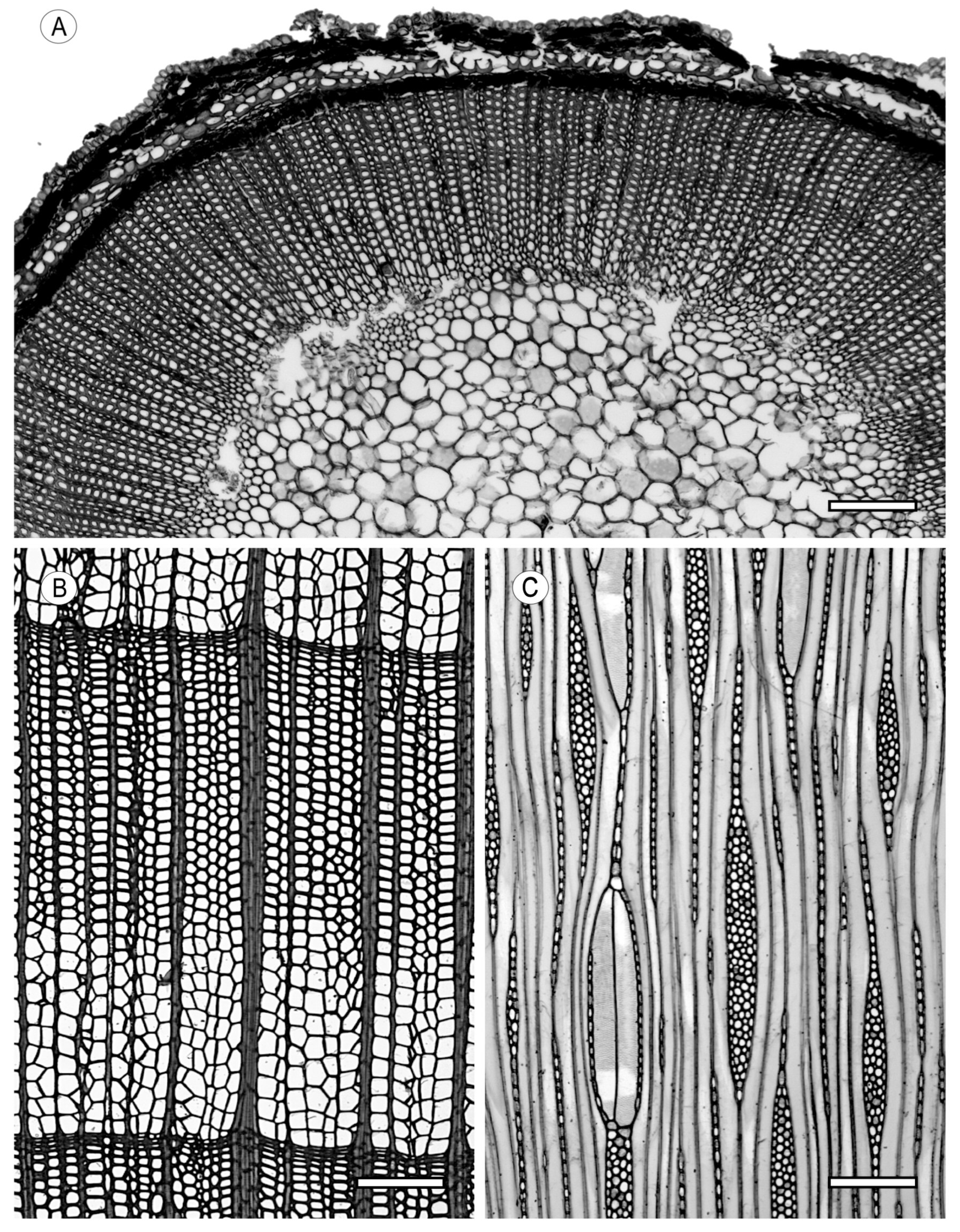
Figure 1. Wood vesseless. (A) Amborella trichopoda; (B,C) Tetracentron sinense. Scale bars: (AC) 200 μm. (A,B) Transverse section (TS); (C) tangential longitudinal section (TLS).
Forests 15 01162 g001
Figure 2. Perforation plates. (A) Simple perforation plates in Carpinus betulus; (B) scalariform perforation plates with ≤10 bars in Corylus maxima; (C) scalariform perforation plates with 10–20 bars in Alnus glutinosa; (D) foraminate perforation plate in Oroxylum indicum. Scale bars: (AC) 50 µm; (D) 100 µm. (AC) Radial longitudinal section (RLS); (D) TS.
Forests 15 01162 g002
Figure 3. Intervessel pits. (A) Scalariform intervessel pits in Magnolia liliifera var. obovata; (B) opposite intervessel pits in Liquidambar styraciflua; (C) alternate intervessel pits in Populus tremula; (D) shape of alternate pits polygonal in Salix alba; (E) vestured pits in Acacia excelsa (SEM). Scale bars: (A) 100 µm; (B) 100 µm; (C,D) 30 µm; (E) 15 µm. (A,C,D) TLS; (B,E) RLS.
Forests 15 01162 g003
Figure 4. Vessel–ray pitting. (A) Vessel–ray pits with distinct borders, similar to intervessel pits in size and shape throughout the ray cell in Swietenia macrophylla; (B) vessel–ray pits from much-reduced borders to apparently simple pits, rounded or angular, in Populus tremula; (C) vessel–ray pits from much-reduced borders to apparently simple pits from horizontal (scalariform and gash-like) to vertical (palisade) in Hirtella carbonaria; (D) vessel–ray pits of two distinct sizes or types in the same ray cell in Bumelia obovata; (E) vessel–ray pits unilaterally compound and coarse (over 10 μm) in Magnolia liliifera var. obovata; (F) vessel–ray pits restricted to marginal rows in Salix alba. Scale bars: (A) 100 µm; (BD,F) 25 µm; (E) 50 µm. (AF) RLS.
Forests 15 01162 g004
Figure 5. Helical thickenings in vessel elements. (A,B) Helical thickenings throughout body of vessel element in Prunus serotina (SEM); (C) helical thickenings throughout body of vessel element in Arbutus unedo. Scale bars: (A) 75 µm; (B) 20 µm; (C) 50 µm. (A,B) TS; (C) RLS.
Forests 15 01162 g005
Figure 6. Vessel groupings. (A) Solitary vessel with angular outline in Liquidambar styraciflua; (B) vessels in radial multiples of four or more in Alnus glutinosa; (C) vessel clusters in Kalopanax septemlobus. Scale bars: (A,B) 100 µm; (C) 200 µm. (AC) TS.
Forests 15 01162 g006
Figure 7. Vessel arrangements. (A) Vessels in tangential bands in Ulmus pumila; (B) vessels in diagonal and/or radial pattern in Quercus ilex; (C) vessels in dendritic pattern in Rhamnus cathartica. Scale bars: (AC) 200 µm. (AC) TS.
Forests 15 01162 g007
Figure 8. Porosity. (A) Ring-porous wood in Fraxinus excelsior; (B) ring-porous wood in Quercus petraea; (C) semi-ring-porous wood in Juglans mandshurica; (D) diffuse-porous wood in Betula pendula. Scale bars: (A) 100 µm; (B,D) 200 µm; (C) 500 µm. (AD) TS.
Forests 15 01162 g008
Figure 9. Tyloses and gums/deposits. (A,B) Tyloses common in Quercus petraea; (C) sclerotic tyloses in Chlorocardium rodiei; (D) sclerotic tyloses in Strombosia pustulata; (E,F) gums/deposits in heartwood vessels in Albizia lebbeck. Scale bars: (A,B) 200 µm; (C,D) 100 µm; (E,F) 250 µm. (A,C,E) TS; (B,F) TLS; (D) RLS.
Forests 15 01162 g009
Figure 10. Vascular/vasicentric tracheids (A,B) in Castanea sativa; (C) in Quercus robur. Scale bars: (AC) 100 µm. (A,C) RLS; (B) TLS.
Forests 15 01162 g010
Figure 11. Fibres. (A) Fibres with from simple to minutely bordered pits in Alphitonia ponderosa; (B) fibres with distinctly bordered pits (or fibre-tracheids) in Illicium anisatum; (C) helical thickenings in ground tissue fibres in Viburnum tinus; (D) septate fibres present in Khaya ivorensis. Scale bars: (A,B) 100 µm; (C) 50 µm; (D) 150 µm. (A,C) RLS; (B,D) TLS.
Forests 15 01162 g011
Figure 12. Fibres. (A) Very thin-walled fibres in Juglans mandschurica; (B) from thin- to thick-walled fibres in Swietenia mahagoni; (C) very thick-walled fibres in Schefflerodendron gazense. Scale bars: (A) 100 µm; (B) 150 µm; (C) 50 µm. (AC) TS.
Forests 15 01162 g012
Figure 13. Axial parenchyma. (A) Axial parenchyma absent or extremely rare in Campnosperma brevipetiolata; apotracheal axial parenchyma; (B) axial parenchyma diffuse in Fagus sylvatica; (C) axial parenchyma diffuse in aggregates in Triplochiton scleroxylon. Scale bars: (A) 200 µm; (B,C) 250 µm. (AC) TS.
Forests 15 01162 g013
Figure 14. Paratracheal axial parenchyma. (A) Axial parenchyma scanty paratracheal in Dillenia borneensis; (B) axial parenchyma vasicentric in Dalbergia spruceana; (C) axial parenchyma lozenge-aliform in Afzelia africana; (D) axial parenchyma winged-aliform in Gonystylus punctatus; (E) axial parenchyma confluent in Acacia stenophylla; (F) axial parenchyma unilateral paratracheal in Caraipa densifolia. Scale bars: (A) 150 µm; (B) 250 µm; (C,D) 200 µm; (E,F) 100 µm. (AF) TS.
Forests 15 01162 g014
Figure 15. Banded parenchyma. (A) Axial parenchyma bands more than three cells wide in Schefflerodendron gazense; (B) axial parenchyma in narrow bands or lines up to three cells wide in Platypodium elegans; (C) axial parenchyma in aggregates and scalariform in Palaquium clarkeana; (D) axial parenchyma reticulate in Cariniana estrellensis; (E) axial parenchyma in marginal or in seemingly marginal bands in Swietenia mahagoni. Scale bars: (A) 50 µm; (BE) 200 µm. (AE) TS.
Forests 15 01162 g015
Figure 16. Fusiform parenchyma/strand length. (A) Fusiform parenchyma cells in Sophora tetraptera; (B) two cells per parenchyma strand in Etaballia guianensis; (C) (3–4) cells per parenchyma strand in Fraxinus uhdei; (D) (5–8) cells per parenchyma strand in Fraxinus excelsior; (E) over eight cells per parenchyma strand in Lophira alata. Scale bars: (AD) 100 µm; (E) 200 µm. (AE) TLS.
Forests 15 01162 g016
Figure 17. Rays: ray width and aggregate rays. (A) Rays exclusively uniseriate in Gonystylus punctatus; (B) ray width from one to three cells in Swietenia macrophylla; (C) larger rays commonly from four- to ten-seriate in Guibourtia coleosperma; (D) larger rays commonly >10-seriate in Quercus robur; (E) rays with multiseriate portion(s) as wide as uniseriate portions in Caryocar costaricense; (F) aggregate rays in Quercus ilex; (G) rays of two distinct sizes in Quercus petraea (also in (D,F)). Scale bars: (AG) 200 µm. (AG) TLS.
Forests 15 01162 g017
Figure 18. Rayless wood. (A) Cross section; (B) tangential longitudinal section; (C) radial longitudinal section in Veronica speciosa. Scale bars: (AC) 100 µm.
Forests 15 01162 g018
Figure 19. Rays: cellular composition. (A) All ray cells procumbent in Albizia tomentosa; (B) all ray cells upright and/or square in Senna alata; (C) ray cells procumbent with one row of upright and/or square marginal cells in Kalopanax septemlobus; (D) ray cells procumbent with 2–4 rows of upright and/or square marginal cells in Carapa procera; (E) ray cells procumbent with more than four rows of upright and/or square marginal cells in Coffea spp.; (F) procumbent, square, and upright cells mixed throughout the ray in Butea monosperma. Scale bars: (AF) 100 µm. (AF) RLS.
Forests 15 01162 g019
Figure 20. Sheath and tile cells. (A) Sheath cells in Sterculia excelsa; (B) tile cells in Durio lowianus. Scale bars: (A,B) 100 µm. (A) TLS; (B) RLS.
Forests 15 01162 g020
Figure 21. Storied structures. (A) All rays storied in Dalbergia retusa; (B) low rays storied and high rays nonstoried in Pterospermum acerifolium; (C) axial parenchyma and/or vessel elements storied in Spartium junceum; (D) fibres storied in Brachystegia leonensis; (E) rays and/or axial elements irregularly storied in Swietenia mahagoni. Scale bars: (A,B,D) 200 µm; (C) 100 µm; (E) 150 µm. (AE) TLS.
Forests 15 01162 g021
Figure 22. Oil and mucilage cells. (A) Oil and/or mucilage cells associated with ray parenchyma in Ocotea rubra; (B) oil and/or mucilage cells associated with axial parenchyma in Ocotea rubra; (C,D) oil and/or mucilage cells present among fibres in Cryptocarya griffithiana. Scale bars: (AD) 100 µm. (A,D) TLS; (BC) RLS.
Forests 15 01162 g022
Figure 23. Intercellular canals. (A) Axial canals in long tangential lines in Sindora cochinchinensis; (B) axial canals in short tangential lines in Dipterocarpus grandiflorus; (C) axial canals diffuse in Vateria indica; (D) radial canals in Campnosperma pteripentandra; (E) intercellular canals of traumatic origin in Guibourtia demeusei. Scale bars: (A) 150 µm; (BE) 200 µm. (AC,E) TS; (D) TLS.
Forests 15 01162 g023
Figure 24. Laticifers (latex tubes) or tanniniferous tubes. (A,B) Laticifers in Dyera costulata; (C) tanniniferous tubes in Virola gardneri. Scale bars: (A) 50 µm; (B) 100 µm; (C) 200 µm. (A) TLS; (B,C) RLS.
Forests 15 01162 g024
Figure 25. Cambial variants. (A) Included phloem, diffuse in Salvadora persica; (B) included phloem, diffuse in Aquilaria agallocha; (C) included phloem, concentric in Avicennia officinalis; (D) other cambial variants in Chodanthus puberulus. Scale bars: (A,D) 200 µm; (B) 500 µm; (C) 250 µm. (AD) TS.
Forests 15 01162 g025
Figure 26. Prismatic crystals. (A) Prismatic crystals in upright and/or square ray cells in Homalium letestui; (B) prismatic crystals in procumbent ray cells in Gonystylus punctatus; (C) prismatic crystals in radial alignment in procumbent ray cells in Sonneratia apetala; (D) prismatic crystals in chambered upright and/or square ray cells in Strombosia pustulata; (E) prismatic crystals in non-chambered axial parenchyma cells in Milicia excelsa; (F) prismatic crystals in chambered axial parenchyma cells in Sorbus domestica; (G) prismatic crystals in fibres in Punica granatum. Scale bars: (A,C) 100 µm; (B,DG) 50 µm. (AE) RLS; (F,G) TLS.
Forests 15 01162 g026
Figure 27. Druses and raphides. (AC) Druses in ray parenchyma cells in Hibiscus similis; (D) druses in axial parenchyma cells in Hibiscus similis; (E) raphides in ray cells in Vitis vinifera. Scale bars: (A,B,E) 100 µm; (C,D) 50 µm. (A,E) TS; (B) TLS; (C,D) RLS.
Forests 15 01162 g027
Figure 28. Other crystal types. (A) Styloids in Tecoma stans; (B) styloids and elongate crystals in Ligustrum vulgare; (C) crystal sand in Cordia subcordata; (D) crystals in enlarged cells (idioblasts) in Acacia aneura; (E) more than one crystal of about the same size per cell or chamber and (a) two distinct sizes of crystals per cell or chamber in (b) in Milicia excelsa; (F) crystals in tyloses in Strombosia pustulata. Scale bars: (A,B,F) 100 µm; (C,D,E) 50 µm. (AF) RLS.
Forests 15 01162 g028
Figure 29. Silica. (A,B) Silica bodies in axial parenchyma cells in Distemonanthus benthamianus; (C) silica bodies in ray cells in Sclerolobium guianense; (D) silica bodies in ray cells in Shorea bracteolata. Scale bars: (AD) 100 µm. (A,C,D) RLS; (B) TLS.
Forests 15 01162 g029
Disclaimer/Publisher’s Note: The statements, opinions and data contained in all publications are solely those of the individual author(s) and contributor(s) and not of MDPI and/or the editor(s). MDPI and/or the editor(s) disclaim responsibility for any injury to people or property resulting from any ideas, methods, instructions or products referred to in the content.