applsci-logo

Journal Browser

Journal Browser

Finite Element Modeling of Joint

A special issue of Applied Sciences (ISSN 2076-3417). This special issue belongs to the section "Chemical and Molecular Sciences".

Deadline for manuscript submissions: closed (20 April 2022) | Viewed by 8036

Special Issue Editor


E-Mail Website
Guest Editor
Department of Applied Physics, University of Eastern Finland, Kuopio 70211, Finland
Interests: finite element modeling of knee; gait cycle analysis; MRI; CT/x-ray imaging; functional imaging; segmentation; tissue degeneration and adaptation; osteoarthritis; quality of life

Special Issue Information

Dear Colleagues,

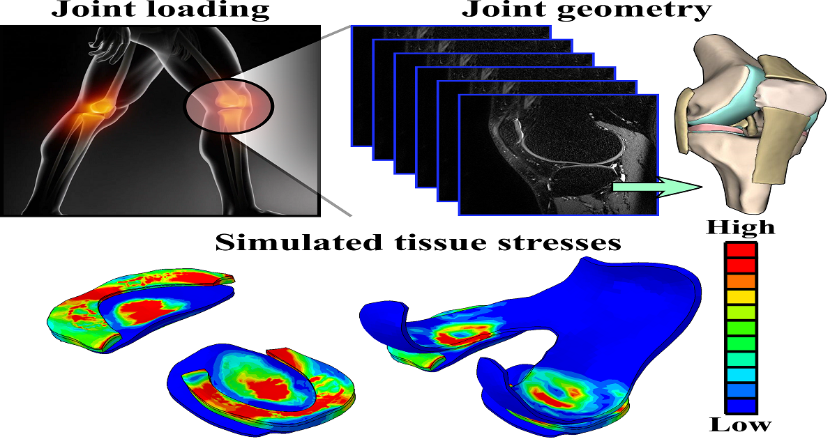
In a healthy joint, a thin cartilage layer on the bone surface enables frictionless motion between the contacting bones. During daily living activities, various loading conditions are generated in joints, which produces different mechanical responses in the soft tissues within the joints. As these loading patterns are highly subject specific, substantial differences of the simulated mechanical responses of the soft tissues and their locations are usually found between different subjects. Furthermore, it is well known that exceeded levels of soft tissue stress or deformations may lead to tissue failure or degeneration in joints. In order to avoid tissue failures or degeneration, computational approaches are needed to give subject-specific information about possible risks before tissue failure or degeneration occurs in joints. We are interested in articles that utilize finite element modeling to explore mechanical responses of the soft tissues within different joints under different loading conditions. Potential topics include, but are not limited to, the following:

- Joint injury and soft tissue responses;

- Different physical activities and mechanical responses of soft tissues in joints;

- Soft tissue degeneration in joints;

- Clinical applications and finite element modeling of joints;

- Subject characteristics and mechanical responses of soft tissues in joints.

Dr. Mika Mononen
Guest Editor

Manuscript Submission Information

Manuscripts should be submitted online at www.mdpi.com by registering and logging in to this website. Once you are registered, click here to go to the submission form. Manuscripts can be submitted until the deadline. All submissions that pass pre-check are peer-reviewed. Accepted papers will be published continuously in the journal (as soon as accepted) and will be listed together on the special issue website. Research articles, review articles as well as short communications are invited. For planned papers, a title and short abstract (about 100 words) can be sent to the Editorial Office for announcement on this website.

Submitted manuscripts should not have been published previously, nor be under consideration for publication elsewhere (except conference proceedings papers). All manuscripts are thoroughly refereed through a single-blind peer-review process. A guide for authors and other relevant information for submission of manuscripts is available on the Instructions for Authors page. Applied Sciences is an international peer-reviewed open access semimonthly journal published by MDPI.

Please visit the Instructions for Authors page before submitting a manuscript. The Article Processing Charge (APC) for publication in this open access journal is 2400 CHF (Swiss Francs). Submitted papers should be well formatted and use good English. Authors may use MDPI's English editing service prior to publication or during author revisions.

Keywords

  • finite element modeling
  • joint
  • soft tissue
  • injury
  • degeneration

Published Papers (2 papers)

Order results
Result details
Select all
Export citation of selected articles as:

Research

Jump to: Review

9 pages, 1969 KiB  
Communication
High-Resolution Phosphorescence Lifetime Imaging (PLIM) of Bones
by Hans Georg Breunig and Karsten König
Appl. Sci. 2022, 12(3), 1066; https://doi.org/10.3390/app12031066 - 20 Jan 2022
Cited by 3 | Viewed by 1581
Abstract
For the first time, the time-resolved two-photon excited autophosphorescence of non-labeled biological specimens was investigated by phosphoresce lifetime imaging with microscopic spatial resolution. A modified multiphoton tomograph was employed to record both photoluminescence contributions, autofluorescence and autophosphorescence, simultaneously, induced by two-photon excitation using [...] Read more.
For the first time, the time-resolved two-photon excited autophosphorescence of non-labeled biological specimens was investigated by phosphoresce lifetime imaging with microscopic spatial resolution. A modified multiphoton tomograph was employed to record both photoluminescence contributions, autofluorescence and autophosphorescence, simultaneously, induced by two-photon excitation using an 80 MHz near infrared femtosecond-pulse-laser scanning beam, an acousto-optic modulator, and a time-correlated single-photon counting module for lifetime measurements from the picosecond to the microsecond range. In particular, the two-photon-excited luminescence of thermally altered bones was imaged. A strong dependence of the phosphorescence intensity on exposure temperature, with a maximum emission for an exposure temperature of approximately 600 °C was observed. Furthermore, the phosphorescence lifetime data indicated a bi-exponential signal decay with both a faster few µs decay time in the range of 3–10 µs and a slower one in the range of 30–60 µs. The recording of fluorescence and phosphorescence allowed deriving the relative signal proportion as an unbiased measure of the temperature dependence. The measurements on thermally altered bones are of particular interest for application to forensic and archeological investigations. Full article
(This article belongs to the Special Issue Finite Element Modeling of Joint)
Show Figures

Figure 1

Review

Jump to: Research

24 pages, 4940 KiB  
Review
Expediting Finite Element Analyses for Subject-Specific Studies of Knee Osteoarthritis: A Literature Review
by Alexander Paz, Gustavo A. Orozco, Rami K. Korhonen, José J. García and Mika E. Mononen
Appl. Sci. 2021, 11(23), 11440; https://doi.org/10.3390/app112311440 - 2 Dec 2021
Cited by 9 | Viewed by 5769
Abstract
Osteoarthritis (OA) is a degenerative disease that affects the synovial joints, especially the knee joint, diminishing the ability of patients to perform daily physical activities. Unfortunately, there is no cure for this nearly irreversible musculoskeletal disorder. Nowadays, many researchers aim for in silico-based [...] Read more.
Osteoarthritis (OA) is a degenerative disease that affects the synovial joints, especially the knee joint, diminishing the ability of patients to perform daily physical activities. Unfortunately, there is no cure for this nearly irreversible musculoskeletal disorder. Nowadays, many researchers aim for in silico-based methods to simulate personalized risks for the onset and progression of OA and evaluate the effects of different conservative preventative actions. Finite element analysis (FEA) has been considered a promising method to be developed for knee OA management. The FEA pipeline consists of three well-established phases: pre-processing, processing, and post-processing. Currently, these phases are time-consuming, making the FEA workflow cumbersome for the clinical environment. Hence, in this narrative review, we overviewed present-day trends towards clinical methods for subject-specific knee OA studies utilizing FEA. We reviewed studies focused on understanding mechanisms that initiate knee OA and expediting the FEA workflow applied to the whole-organ level. Based on the current trends we observed, we believe that forthcoming knee FEAs will provide nearly real-time predictions for the personalized risk of developing knee OA. These analyses will integrate subject-specific geometries, loading conditions, and estimations of local tissue mechanical properties. This will be achieved by combining state-of-the-art FEA workflows with automated approaches aided by machine learning techniques. Full article
(This article belongs to the Special Issue Finite Element Modeling of Joint)
Show Figures

Figure 1

Back to TopTop