Recent Developments in Microfluidics (Closed)

Editors


E-Mail Website
Collection Editor
George W. Woodruff School of Mechanical Engineering, Parker H. Petit Institute for Bioengineering and Biosciences, Georgia Institute of Technology, Atlanta, GA 30332, USA
Interests: microrobotic sensors; single-cell sensing; bio-MEMS; microfluidics; lab-on-a-chip; organ-on-a-chip; point-of-care biosensors; disease diagnostics; global health

E-Mail Website
Collection Editor
Wyss Institute for Biologically Inspired Engineering, Harvard University, Boston, MA 02115, USA
Interests: organ-on-chips; point-of-care diagnostics; disease modeling; drug testing

Topical Collection Information

Dear Colleagues,

The advent of microfluidics has revolutionized many fields within life sciences by enabling the rapid isolation and detection of molecules and proteins, extracellular vesicles, bacteria, and cells in complex biological fluids with major applications in medical diagnostics, patient monitoring, drug screening, and food safety. Microfluidic technologies are rapidly evolving with new fabrication techniques, channel architecture, and materials (flexible elastomers, paper, 2D materials), along with developments in sensing modalities (optical, electrical, and magnetic) and integrated molecular biology techniques (CRISPR-based approaches). These developments enable faster and higher detection sensitivity in complex environments and challenging applications ranging from unamplified nucleic acid and extracellular vesicle detection to real-time monitoring of physiological signals from wearables. On the other hand, cost-effective, disposable, and simple-use microfluidic technologies are at the frontlines of point-of-care diagnostics and screening of infectious diseases and genetic disorders, especially in low-resource settings; the importance and urgency of which has become evident once more during the recent COVID-19 pandemic.

In this Topical Collection, we are pleased to invite contributions on “Recent Developments in Microfluidics” dedicated to covering the most recent innovations in next-generation microfluidic technologies. Research articles and comprehensive review articles reporting on the latest developments in new fabrication techniques and materials, novel sensing approaches, and applications in molecular and cellular diagnostics and wearable physiological monitoring technologies are of great interest.

Dr. Yunus Alapan
Dr. Yuncheng Man
Collection Editors

Manuscript Submission Information

Manuscripts should be submitted online at www.mdpi.com by registering and logging in to this website. Once you are registered, click here to go to the submission form. Manuscripts can be submitted until the deadline. All submissions that pass pre-check are peer-reviewed. Accepted papers will be published continuously in the journal (as soon as accepted) and will be listed together on the collection website. Research articles, review articles as well as short communications are invited. For planned papers, a title and short abstract (about 100 words) can be sent to the Editorial Office for announcement on this website.

Submitted manuscripts should not have been published previously, nor be under consideration for publication elsewhere (except conference proceedings papers). All manuscripts are thoroughly refereed through a single-blind peer-review process. A guide for authors and other relevant information for submission of manuscripts is available on the Instructions for Authors page. Biosensors is an international peer-reviewed open access monthly journal published by MDPI.

Please visit the Instructions for Authors page before submitting a manuscript. The Article Processing Charge (APC) for publication in this open access journal is 2700 CHF (Swiss Francs). Submitted papers should be well formatted and use good English. Authors may use MDPI's English editing service prior to publication or during author revisions.

Keywords

  • microfluidic biosensors
  • lab-on-a-chip
  • paper-based microfluidics
  • wearable microfluidics
  • Microfluidic assays
  • molecular diagnostics
  • cellular diagnostics
  • point-of-care diagnostics
  • physiological monitoring
  • global health

Published Papers (10 papers)

2023

Jump to: 2022, 2021, 2020

17 pages, 1432 KiB  
Review
Open Hardware for Microfluidics: Exploiting Raspberry Pi Singleboard Computer and Camera Systems for Customisable Laboratory Instrumentation
by Rüya Meltem Sarıyer, Alexander Daniel Edwards and Sarah Helen Needs
Biosensors 2023, 13(10), 948; https://doi.org/10.3390/bios13100948 - 23 Oct 2023
Cited by 3 | Viewed by 4287
Abstract
The integration of Raspberry Pi miniature computer systems with microfluidics has revolutionised the development of low-cost and customizable analytical systems in life science laboratories. This review explores the applications of Raspberry Pi in microfluidics, with a focus on imaging, including microscopy and automated [...] Read more.
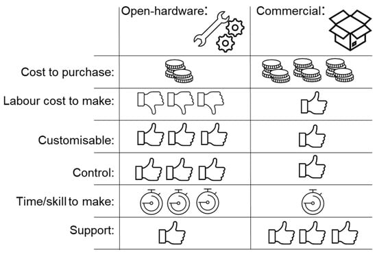
The integration of Raspberry Pi miniature computer systems with microfluidics has revolutionised the development of low-cost and customizable analytical systems in life science laboratories. This review explores the applications of Raspberry Pi in microfluidics, with a focus on imaging, including microscopy and automated image capture. By leveraging the low cost, flexibility and accessibility of Raspberry Pi components, high-resolution imaging and analysis have been achieved in direct mammalian and bacterial cellular imaging and a plethora of image-based biochemical and molecular assays, from immunoassays, through microbial growth, to nucleic acid methods such as real-time-qPCR. The control of image capture permitted by Raspberry Pi hardware can also be combined with onboard image analysis. Open-source hardware offers an opportunity to develop complex laboratory instrumentation systems at a fraction of the cost of commercial equipment and, importantly, offers an opportunity for complete customisation to meet the users’ needs. However, these benefits come with a trade-off: challenges remain for those wishing to incorporate open-source hardware equipment in their own work, including requirements for construction and operator skill, the need for good documentation and the availability of rapid prototyping such as 3D printing plus other components. These advances in open-source hardware have the potential to improve the efficiency, accessibility, and cost-effectiveness of microfluidic-based experiments and applications. Full article
Show Figures

Figure 1

2022

Jump to: 2023, 2021, 2020

11 pages, 2606 KiB  
Article
On-Chip Organoid Formation to Study CXCR4/CXCL-12 Chemokine Microenvironment Responses for Renal Cancer Drug Testing
by Adem Ozcelik, Burcin Irem Abas, Omer Erdogan, Evrim Cevik and Ozge Cevik
Biosensors 2022, 12(12), 1177; https://doi.org/10.3390/bios12121177 - 17 Dec 2022
Cited by 7 | Viewed by 2712
Abstract
Organoid models have gained importance in recent years in determining the toxic effects of drugs in cancer studies. Organoid designs with the same standardized size and cellular structures are desired for drug tests. The field of microfluidics offers numerous advantages to enable well-controlled [...] Read more.
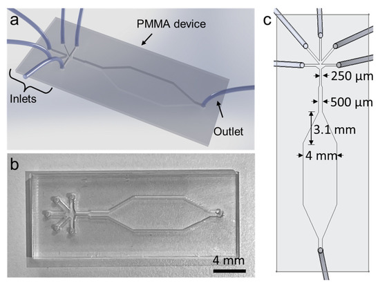
Organoid models have gained importance in recent years in determining the toxic effects of drugs in cancer studies. Organoid designs with the same standardized size and cellular structures are desired for drug tests. The field of microfluidics offers numerous advantages to enable well-controlled and contamination-free biomedical research. In this study, simple and low-cost microfluidic devices were designed and fabricated to develop an organoid model for drug testing for renal cancers. Caki human renal cancer cells and mesenchymal stem cells isolated from human umbilical cord were placed into alginate hydrogels. The microfluidic system was implemented to form size-controllable organoids within alginate hydrogels. Alginate capsules of uniform sizes formed in the microfluidic system were kept in cell culture for 21 days, and their organoid development was studied with calcein staining. Cisplatin was used as a standard chemotherapeutic, and organoid sphere structures were examined as a function of time with an MTT assay. HIF-1α, CXCR4 and CXCL-12 chemokine protein, and CXCR4 and CXCL-12 gene levels were tested in organoids and cisplatin responses. In conclusion, it was found that the standard renal cancer organoids made on a lab-on-a-chip system can be used to measure drug effects and tumor microenvironment responses. Full article
Show Figures

Figure 1

15 pages, 3162 KiB  
Review
CRISPR-Cas-Integrated LAMP
by Nazente Atçeken, Defne Yigci, Berin Ozdalgic and Savas Tasoglu
Biosensors 2022, 12(11), 1035; https://doi.org/10.3390/bios12111035 - 17 Nov 2022
Cited by 12 | Viewed by 4645
Abstract
Pathogen-specific point-of-care (PoC) diagnostic tests have become an important need in the fight against infectious diseases and epidemics in recent years. PoC diagnostic tests are designed with the following parameters in mind: rapidity, accuracy, sensitivity, specificity, and ease of use. Molecular techniques are [...] Read more.
Pathogen-specific point-of-care (PoC) diagnostic tests have become an important need in the fight against infectious diseases and epidemics in recent years. PoC diagnostic tests are designed with the following parameters in mind: rapidity, accuracy, sensitivity, specificity, and ease of use. Molecular techniques are the gold standard for pathogen detection due to their accuracy and specificity. There are various limitations in adapting molecular diagnostic methods to PoC diagnostic tests. Efforts to overcome limitations are focused on the development of integrated molecular diagnostics by utilizing the latest technologies available to create the most successful PoC diagnostic platforms. With this point of view, a new generation technology was developed by combining loop-mediated isothermal amplification (LAMP) technology with clustered regularly interspaced short palindromic repeat (CRISPR)-associated (CRISPR-Cas) technology. This integrated approach benefits from the properties of LAMP technology, namely its high efficiency, short turnaround time, and the lack of need for a complex device. It also makes use of the programmable function of CRISPR-Cas technology and the collateral cleavage activity of certain Cas proteins that allow for convenient reporter detection. Thus, this combined technology enables the development of PoC diagnostic tests with high sensitivity, specificity, and ease of use without the need for complicated devices. In this review, we discuss the advantages and limitations of the CRISPR/Cas combined LAMP technology. We review current limitations to convert CRISPR combined LAMP into pathogen-specific PoC platforms. Furthermore, we point out the need to design more useful PoC platforms using microfabrication technologies by developing strategies that overcome the limitations of this new technology, reduce its complexity, and reduce the risk of contamination. Full article
Show Figures

Figure 1

14 pages, 3768 KiB  
Article
Simple Staining of Cells on a Chip
by Fatma Betul Kosker, Omer Aydin and Kutay Icoz
Biosensors 2022, 12(11), 1013; https://doi.org/10.3390/bios12111013 - 13 Nov 2022
Cited by 2 | Viewed by 4147
Abstract
Simple staining of cells is a widely used method in basic medical diagnostics, education, and research laboratories. The stains are low-cost, but the extensive consumption results in excessive toxic waste generation. Thus, to decrease the amount of toxic waste resulting from the cell [...] Read more.
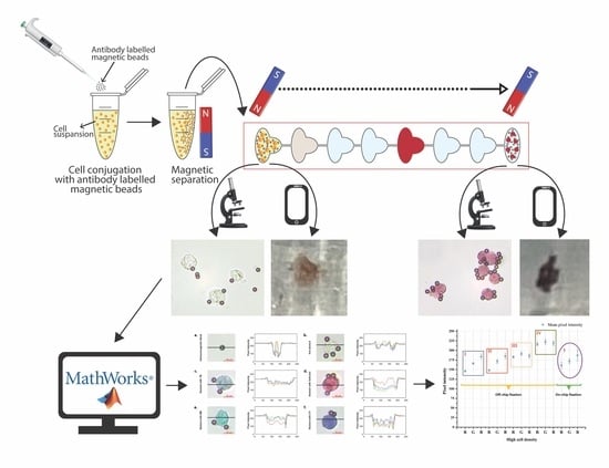
Simple staining of cells is a widely used method in basic medical diagnostics, education, and research laboratories. The stains are low-cost, but the extensive consumption results in excessive toxic waste generation. Thus, to decrease the amount of toxic waste resulting from the cell staining procedure is a need. In this study, we developed a magnetically driven and compartmentalized passive microfluidic chip to perform simple staining of human eukaryotic cells, K562 cells, and lymphocyte cells derived from patients. We demonstrated simple staining on cells with trypan blue, methylene blue, crystal violet, and safranin for high, medium, and low cell densities. The stained cells were imaged using a bright field optical microscope and a cell phone to count cells on the focal plane. The staining improved the color signal of the cell by 25-135-pixel intensity changes for the microscopic images. The validity of the protocol was determined using Jurkat and MDA-MB-231 cell lines as negative controls. In order to demonstrate the practicality of the system, lymphocyte cells derived from human blood samples were stained with trypan blue. The color intensity changes in the first and last compartments were analyzed to evaluate the performance of the chip. The developed method is ultra-low cost, significantly reduces the waste generated, and can be integrated with mobile imaging devices in terms of portability. By combining microfabrication technology with cell staining, this study reported a novel contribution to the field of microfluidic biosensors. In the future, we expect to demonstrate the detection of pathogens using this method. Full article
Show Figures

Graphical abstract

29 pages, 4146 KiB  
Review
Applications of Microfluidics and Organ-on-a-Chip in Cancer Research
by Sagar Regmi, Chetan Poudel, Rameshwar Adhikari and Kathy Qian Luo
Biosensors 2022, 12(7), 459; https://doi.org/10.3390/bios12070459 - 27 Jun 2022
Cited by 55 | Viewed by 8895
Abstract
Taking the life of nearly 10 million people annually, cancer has become one of the major causes of mortality worldwide and a hot topic for researchers to find innovative approaches to demystify the disease and drug development. Having its root lying in microelectronics, [...] Read more.
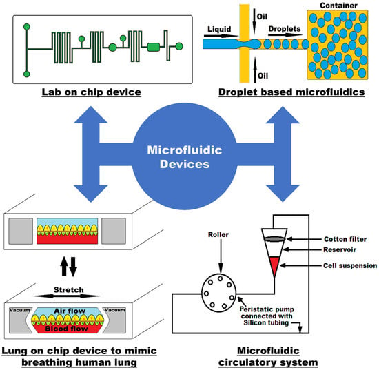
Taking the life of nearly 10 million people annually, cancer has become one of the major causes of mortality worldwide and a hot topic for researchers to find innovative approaches to demystify the disease and drug development. Having its root lying in microelectronics, microfluidics seems to hold great potential to explore our limited knowledge in the field of oncology. It offers numerous advantages such as a low sample volume, minimal cost, parallelization, and portability and has been advanced in the field of molecular biology and chemical synthesis. The platform has been proved to be valuable in cancer research, especially for diagnostics and prognosis purposes and has been successfully employed in recent years. Organ-on-a-chip, a biomimetic microfluidic platform, simulating the complexity of a human organ, has emerged as a breakthrough in cancer research as it provides a dynamic platform to simulate tumor growth and progression in a chip. This paper aims at giving an overview of microfluidics and organ-on-a-chip technology incorporating their historical development, physics of fluid flow and application in oncology. The current applications of microfluidics and organ-on-a-chip in the field of cancer research have been copiously discussed integrating the major application areas such as the isolation of CTCs, studying the cancer cell phenotype as well as metastasis, replicating TME in organ-on-a-chip and drug development. This technology’s significance and limitations are also addressed, giving readers a comprehensive picture of the ability of the microfluidic platform to advance the field of oncology. Full article
Show Figures

Figure 1

2021

Jump to: 2023, 2022, 2020

25 pages, 1620 KiB  
Review
PDMS Bonding Technologies for Microfluidic Applications: A Review
by Alexandra Borók, Kristóf Laboda and Attila Bonyár
Biosensors 2021, 11(8), 292; https://doi.org/10.3390/bios11080292 - 23 Aug 2021
Cited by 120 | Viewed by 21118
Abstract
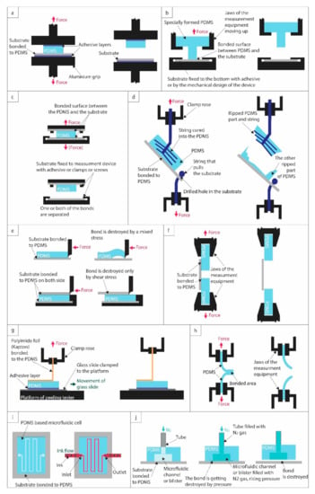
This review summarizes and compares the available surface treatment and bonding techniques (e.g., corona triggered surface activation, oxygen plasma surface activation, chemical gluing, and mixed techniques) and quality/bond-strength testing methods (e.g., pulling test, shear test, peel test, leakage test) for bonding PDMS (polydimethylsiloxane) [...] Read more.
This review summarizes and compares the available surface treatment and bonding techniques (e.g., corona triggered surface activation, oxygen plasma surface activation, chemical gluing, and mixed techniques) and quality/bond-strength testing methods (e.g., pulling test, shear test, peel test, leakage test) for bonding PDMS (polydimethylsiloxane) with other materials, such as PDMS, glass, silicon, PET (polyethylene terephthalate), PI (polyimide), PMMA (poly(methyl methacrylate)), PVC (polyvinyl chloride), PC (polycarbonate), COC (cyclic olefin copolymer), PS (polystyrene) and PEN (polyethylene naphthalate). The optimized process parameters for the best achievable bond strengths are collected for each substrate, and the advantages and disadvantages of each method are discussed in detail. Full article
Show Figures

Figure 1

13 pages, 10985 KiB  
Communication
3D-PAD: Paper-Based Analytical Devices with Integrated Three-Dimensional Features
by James S. Ng and Michinao Hashimoto
Biosensors 2021, 11(3), 84; https://doi.org/10.3390/bios11030084 - 17 Mar 2021
Cited by 9 | Viewed by 3973
Abstract
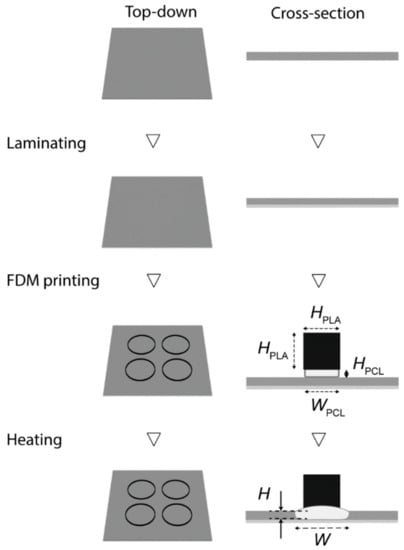
This paper describes the use of fused deposition modeling (FDM) printing to fabricate paper-based analytical devices (PAD) with three-dimensional (3D) features, which is termed as 3D-PAD. Material depositions followed by heat reflow is a standard approach for the fabrication of PAD. Such devices [...] Read more.
This paper describes the use of fused deposition modeling (FDM) printing to fabricate paper-based analytical devices (PAD) with three-dimensional (3D) features, which is termed as 3D-PAD. Material depositions followed by heat reflow is a standard approach for the fabrication of PAD. Such devices are primarily two-dimensional (2D) and can hold only a limited amount of liquid samples in the device. This constraint can pose problems when the sample consists of organic solvents that have low interfacial energies with the hydrophobic barriers. To overcome this limitation, we developed a method to fabricate PAD integrated with 3D features (vertical walls as an example) by FDM 3D printing. 3D-PADs were fabricated using two types of thermoplastics. One thermoplastic had a low melting point that formed hydrophobic barriers upon penetration, and another thermoplastic had a high melting point that maintained 3D features on the filter paper without reflowing. We used polycaprolactone (PCL) for the former, and polylactic acid (PLA) for the latter. Both PCL and PLA were printed with FDM without gaps at the interface, and the resulting paper-based devices possessed hydrophobic barriers consisting of PCL seamlessly integrated with vertical features consisting of PLA. We validated the capability of 3D-PAD to hold 30 μL of solvents (ethanol, isopropyl alcohol, and acetone), all of which would not be retained on conventional PADs fabricated with solid wax printers. To highlight the importance of containing an increased amount of liquid samples, a colorimetric assay for the formation of dimethylglyoxime (DMG)-Ni (II) was demonstrated using two volumes (10 μL and 30 μL) of solvent-based dimethylglyoxime (DMG). FDM printing of 3D-PAD enabled the facile construction of 3D structures integrated with PAD, which would find applications in paper-based chemical and biological assays requiring organic solvents. Full article
Show Figures

Figure 1

2020

Jump to: 2023, 2022, 2021

18 pages, 1329 KiB  
Review
Surface Modification Techniques for Endothelial Cell Seeding in PDMS Microfluidic Devices
by Fahima Akther, Shazwani Binte Yakob, Nam-Trung Nguyen and Hang T. Ta
Biosensors 2020, 10(11), 182; https://doi.org/10.3390/bios10110182 - 19 Nov 2020
Cited by 115 | Viewed by 19876
Abstract
Microfluidic lab-on-a-chip cell culture techniques have been gaining popularity by offering the possibility of reducing the amount of samples and reagents and greater control over cellular microenvironment. Polydimethylsiloxane (PDMS) is the commonly used polymer for microfluidic cell culture devices because of the cheap [...] Read more.
Microfluidic lab-on-a-chip cell culture techniques have been gaining popularity by offering the possibility of reducing the amount of samples and reagents and greater control over cellular microenvironment. Polydimethylsiloxane (PDMS) is the commonly used polymer for microfluidic cell culture devices because of the cheap and easy fabrication techniques, non-toxicity, biocompatibility, high gas permeability, and optical transparency. However, the intrinsic hydrophobic nature of PDMS makes cell seeding challenging when applied on PDMS surface. The hydrophobicity of the PDMS surface also allows the non-specific absorption/adsorption of small molecules and biomolecules that might affect the cellular behaviour and functions. Hydrophilic modification of PDMS surface is indispensable for successful cell seeding. This review collates different techniques with their advantages and disadvantages that have been used to improve PDMS hydrophilicity to facilitate endothelial cells seeding in PDMS devices. Full article
Show Figures

Graphical abstract

14 pages, 2882 KiB  
Article
Determination of the Empirical Electrokinetic Equilibrium Condition of Microorganisms in Microfluidic Devices
by Adriana Coll De Peña, Nicole Hill and Blanca H. Lapizco-Encinas
Biosensors 2020, 10(10), 148; https://doi.org/10.3390/bios10100148 - 19 Oct 2020
Cited by 12 | Viewed by 3296
Abstract
The increased concern regarding emerging pathogens and antibiotic resistance has drawn interest in the development of rapid and robust microfluidic techniques to analyze microorganisms. The novel parameter known as the electrokinetic equilibrium condition (EEEC) was presented in recent [...] Read more.
The increased concern regarding emerging pathogens and antibiotic resistance has drawn interest in the development of rapid and robust microfluidic techniques to analyze microorganisms. The novel parameter known as the electrokinetic equilibrium condition (EEEC) was presented in recent studies, providing an approach to analyze microparticles in microchannels employing unique electrokinetic (EK) signatures. While the EEEC shows great promise, current estimation approaches can be time-consuming or heavily user-dependent for accurate values. The present contribution aims to analyze existing approaches for estimating this parameter and modify the process into an accurate yet simple technique for estimating the EK behavior of microorganisms in insulator-based microfluidic devices. The technique presented here yields the parameter called the empirical electrokinetic equilibrium condition (eEEEC) which works well as a value for initial approximations of trapping conditions in insulator-based EK (iEK) microfluidic systems. A total of six types of microorganisms were analyzed in this study (three bacteria and three bacteriophages). The proposed approach estimated eEEEC values employing images of trapped microorganisms, yielding high reproducibility (SD 5.0–8.8%). Furthermore, stable trapping voltages (sTVs) were estimated from eEEEC values for distinct channel designs to test that this parameter is system-independent and good agreement was obtained when comparing estimated sTVs vs. experimental values (SD 0.3–19.6%). The encouraging results from this work were used to generate an EK library of data, available on our laboratory website. The data in this library can be used to design tailored iEK microfluidic devices for the analysis of microorganisms. Full article
Show Figures

Figure 1

19 pages, 2386 KiB  
Article
Combining Electrostatic, Hindrance and Diffusive Effects for Predicting Particle Transport and Separation Efficiency in Deterministic Lateral Displacement Microfluidic Devices
by Valentina Biagioni, Giulia Balestrieri, Alessandra Adrover and Stefano Cerbelli
Biosensors 2020, 10(9), 126; https://doi.org/10.3390/bios10090126 - 16 Sep 2020
Cited by 8 | Viewed by 2973
Abstract
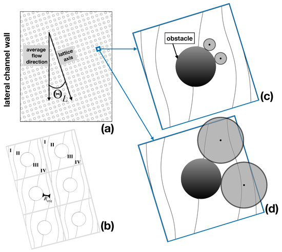
Microfluidic separators based on Deterministic Lateral Displacement (DLD) constitute a promising technique for the label-free detection and separation of mesoscopic objects of biological interest, ranging from cells to exosomes. Owing to the simultaneous presence of different forces contributing to particle motion, a feasible [...] Read more.
Microfluidic separators based on Deterministic Lateral Displacement (DLD) constitute a promising technique for the label-free detection and separation of mesoscopic objects of biological interest, ranging from cells to exosomes. Owing to the simultaneous presence of different forces contributing to particle motion, a feasible theoretical approach for interpreting and anticipating the performance of DLD devices is yet to be developed. By combining the results of a recent study on electrostatic effects in DLD devices with an advection–diffusion model previously developed by our group, we here propose a fully predictive approach (i.e., ideally devoid of adjustable parameters) that includes the main physically relevant effects governing particle transport on the one hand, and that is amenable to numerical treatment at affordable computational expenses on the other. The approach proposed, based on ensemble statistics of stochastic particle trajectories, is validated by comparing/contrasting model predictions to available experimental data encompassing different particle dimensions. The comparison suggests that at low/moderate values of the flowrate the approach can yield an accurate prediction of the separation performance, thus making it a promising tool for designing device geometries and operating conditions in nanoscale applications of the DLD technique. Full article
Show Figures

Figure 1

Back to TopTop