Cellular Senescence in Aging and Aging-Related Diseases

A special issue of Cells (ISSN 2073-4409). This special issue belongs to the section "Cellular Aging".

Deadline for manuscript submissions: closed (15 November 2022) | Viewed by 24703

Special Issue Editors


E-Mail Website
Guest Editor
Department of Biochemistry, Chungbuk National University College of Medicine, Cheongju 28644, Korea
Interests: aging; senescence; cell adhesion; fibrosis; neurodegeneration

E-Mail Website
Guest Editor
Department of Biochemistry, College of Medicine, Gachon University, Incheon 21999, Republic of Korea
Interests: aging; prostate cancer; vascular inflammation; cell adhesion; fibrosis

Special Issue Information

Dear Colleagues,

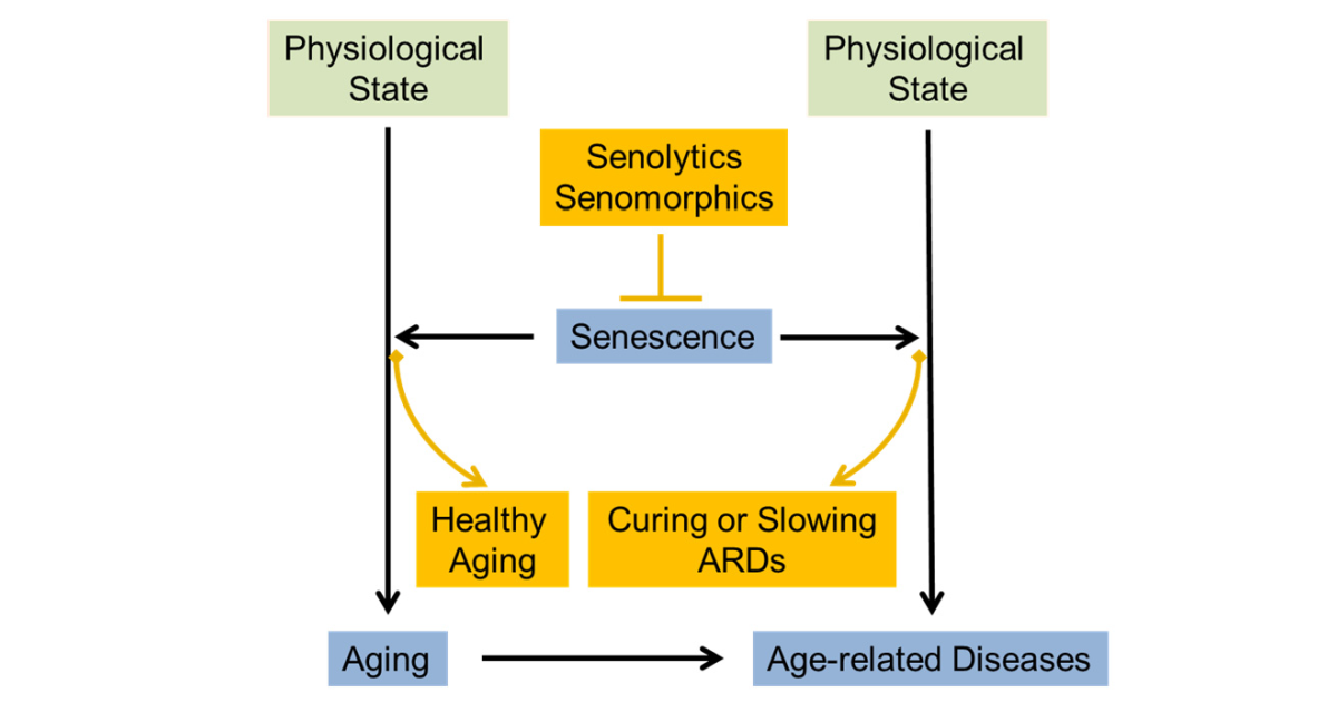
Diverse types of stress evoke cellular senescence, stress-induced premature senescence and replicative senescence, characterized by several features: a cell cycle arrest, increased senescence-associated beta-galactosidase activity, typical morphological changes and senescence-associated secretory phenotype (SASP). Hallmarks of aging include senescence; in this line, studies unraveling a causal effect of senescence on aging at the organism level are increasing. During the events of organismal aging, senescence results in both beneficial and detrimental consequences. Accumulated evidence indicates dysregulated senescence as a pathogenic mechanism in aging-related diseases (ARDs) such as cancer, diabetes, atherosclerosis, arthritis, fibrosis and neurodegenerative diseases. A couple of senolytics targeting these ARDs are currently under clinical trial. Senomorphics can also ameliorate ARDs by regulating the SASP components such as the pro-inflammatory cytokines and chemokines, and growth factors.  

This Special Issue recruits a wide range of studies that examine senescence regarding the issues from the basic mechanisms to the translational applications to clinics. In particular, we welcome the studies that address unresolved questions, for instance, the necessity of simple senescence biomarkers, context-dependent heterogeneity of senescence, single-cell level analysis and novel senolytics and senomorphics.

Prof. Dr. Eung-Gook Kim
Dr. Jaehong Kim 
Guest Editors

Manuscript Submission Information

Manuscripts should be submitted online at www.mdpi.com by registering and logging in to this website. Once you are registered, click here to go to the submission form. Manuscripts can be submitted until the deadline. All submissions that pass pre-check are peer-reviewed. Accepted papers will be published continuously in the journal (as soon as accepted) and will be listed together on the special issue website. Research articles, review articles as well as short communications are invited. For planned papers, a title and short abstract (about 100 words) can be sent to the Editorial Office for announcement on this website.

Submitted manuscripts should not have been published previously, nor be under consideration for publication elsewhere (except conference proceedings papers). All manuscripts are thoroughly refereed through a single-blind peer-review process. A guide for authors and other relevant information for submission of manuscripts is available on the Instructions for Authors page. Cells is an international peer-reviewed open access semimonthly journal published by MDPI.

Please visit the Instructions for Authors page before submitting a manuscript. The Article Processing Charge (APC) for publication in this open access journal is 2700 CHF (Swiss Francs). Submitted papers should be well formatted and use good English. Authors may use MDPI's English editing service prior to publication or during author revisions.

Keywords

  • senescence
  • SASP
  • aging
  • age-related disease
  • biomarker
  • senolytics
  • senomorphics

Benefits of Publishing in a Special Issue

  • Ease of navigation: Grouping papers by topic helps scholars navigate broad scope journals more efficiently.
  • Greater discoverability: Special Issues support the reach and impact of scientific research. Articles in Special Issues are more discoverable and cited more frequently.
  • Expansion of research network: Special Issues facilitate connections among authors, fostering scientific collaborations.
  • External promotion: Articles in Special Issues are often promoted through the journal's social media, increasing their visibility.
  • e-Book format: Special Issues with more than 10 articles can be published as dedicated e-books, ensuring wide and rapid dissemination.

Further information on MDPI's Special Issue polices can be found here.

Published Papers (6 papers)

Order results
Result details
Select all
Export citation of selected articles as:

Research

Jump to: Review

20 pages, 5788 KiB  
Article
ML216 Prevents DNA Damage-Induced Senescence by Modulating DBC1–BLM Interaction
by Feng Cui, Xueying Han, Xiaoqian Zhang, Siqi Wang, Na Liang, Qing Tan, Wuga Sha and Jun Li
Cells 2023, 12(1), 145; https://doi.org/10.3390/cells12010145 - 29 Dec 2022
Cited by 2 | Viewed by 2444
Abstract
DNA damage is the major cause of senescence and apoptosis; however, the manner by which DNA-damaged cells become senescent remains unclear. We demonstrate that DNA damage leads to a greater level of senescence rather than apoptosis in DBC1-deficient cells. In addition, we show [...] Read more.
DNA damage is the major cause of senescence and apoptosis; however, the manner by which DNA-damaged cells become senescent remains unclear. We demonstrate that DNA damage leads to a greater level of senescence rather than apoptosis in DBC1-deficient cells. In addition, we show that BLM becomes degraded during DNA damage, which induces p21 expression and senescence. DBC1 binds to and shields BLM from degradation, thus suppressing senescence. ML216 promotes DBC1–BLM interaction, which aids in the preservation of BLM following DNA damage and suppresses senescence. ML216 enhances pulmonary function by lowering the levels of senescence and fibrosis in both aged mice and a mouse model of bleomycin-induced idiopathic pulmonary fibrosis. Our data reveal a unique mechanism preventing DNA-damaged cells from becoming senescent, which may be regulated by the use of ML216 as a potential treatment for senescence-related diseases. Full article
(This article belongs to the Special Issue Cellular Senescence in Aging and Aging-Related Diseases)
Show Figures

Figure 1

17 pages, 24126 KiB  
Article
Captopril Alleviates Chondrocyte Senescence in DOCA-Salt Hypertensive Rats Associated with Gut Microbiome Alteration
by Lok Chun Chan, Yuqi Zhang, Xiaoqing Kuang, Mohamad Koohi-Moghadam, Haicui Wu, Theo Yu Chung Lam, Jiachi Chiou and Chunyi Wen
Cells 2022, 11(19), 3173; https://doi.org/10.3390/cells11193173 - 10 Oct 2022
Cited by 2 | Viewed by 2497
Abstract
Gut microbiota is the key controller of healthy aging. Hypertension and osteoarthritis (OA) are two frequently co-existing age-related pathologies in older adults. Both are associated with gut microbiota dysbiosis. Hereby, we explore gut microbiome alteration in the Deoxycorticosterone acetate (DOCA)-induced hypertensive rat model. [...] Read more.
Gut microbiota is the key controller of healthy aging. Hypertension and osteoarthritis (OA) are two frequently co-existing age-related pathologies in older adults. Both are associated with gut microbiota dysbiosis. Hereby, we explore gut microbiome alteration in the Deoxycorticosterone acetate (DOCA)-induced hypertensive rat model. Captopril, an anti-hypertensive medicine, was chosen to attenuate joint damage. Knee joints were harvested for radiological and histological examination; meanwhile, fecal samples were collected for 16S rRNA and shotgun sequencing. The 16S rRNA data was annotated using Qiime 2 v2019.10, while metagenomic data was functionally profiled with HUMAnN 2.0 database. Differential abundance analyses were adopted to identify the significant bacterial genera and pathways from the gut microbiota. DOCA-induced hypertension induced p16INK4a+ senescent cells (SnCs) accumulation not only in the aorta and kidney (p < 0.05) but also knee joint, which contributed to articular cartilage degradation and subchondral bone disturbance. Captopril removed the p16INK4a + SnCs from different organs, partially lowered blood pressure, and mitigated cartilage damage. Meanwhile, these alterations were found to associate with the reduction of Escherichia-Shigella levels in the gut microbiome. As such, gut microbiota dysbiosis might emerge as a metabolic link in chondrocyte senescence induced by DOCA-triggered hypertension. The underlying molecular mechanism warrants further investigation. Full article
(This article belongs to the Special Issue Cellular Senescence in Aging and Aging-Related Diseases)
Show Figures

Figure 1

21 pages, 6628 KiB  
Article
Mitoribosomal Deregulation Drives Senescence via TPP1-Mediated Telomere Deprotection
by Seongki Min, So Mee Kwon, Jiwon Hong, Young-Kyoung Lee, Tae Jun Park, Su Bin Lim and Gyesoon Yoon
Cells 2022, 11(13), 2079; https://doi.org/10.3390/cells11132079 - 30 Jun 2022
Cited by 3 | Viewed by 2817
Abstract
While mitochondrial bioenergetic deregulation has long been implicated in cellular senescence, its mechanistic involvement remains unclear. By leveraging diverse mitochondria-related gene expression profiles derived from two different cellular senescence models of human diploid fibroblasts, we found that the expression of mitoribosomal proteins (MRPs) [...] Read more.
While mitochondrial bioenergetic deregulation has long been implicated in cellular senescence, its mechanistic involvement remains unclear. By leveraging diverse mitochondria-related gene expression profiles derived from two different cellular senescence models of human diploid fibroblasts, we found that the expression of mitoribosomal proteins (MRPs) was generally decreased during the early-to-middle transition prior to the exhibition of noticeable SA-β-gal activity. Suppressed expression patterns of the identified senescence-associated MRP signatures (SA-MRPs) were validated in aged human cells and rat and mouse skin tissues and in aging mouse fibroblasts at single-cell resolution. TIN2- and POT1-interaction protein (TPP1) was concurrently suppressed, which induced senescence, accompanied by telomere DNA damage. Lastly, we show that SA-MRP deregulation could be a potential upstream regulator of TPP1 suppression. Our results indicate that mitoribosomal deregulation could represent an early event initiating mitochondrial dysfunction and serve as a primary driver of cellular senescence and an upstream regulator of shelterin-mediated telomere deprotection. Full article
(This article belongs to the Special Issue Cellular Senescence in Aging and Aging-Related Diseases)
Show Figures

Figure 1

Review

Jump to: Research

18 pages, 1089 KiB  
Review
Cellular Senescence, a Novel Area of Investigation for Metastatic Diseases
by Francesca Faggioli, Michael C. Velarde and Christopher D. Wiley
Cells 2023, 12(6), 860; https://doi.org/10.3390/cells12060860 - 10 Mar 2023
Cited by 3 | Viewed by 2806
Abstract
Metastasis is a systemic condition and the major challenge among cancer types, as it can lead to multiorgan vulnerability. Recently, attention has been drawn to cellular senescence, a complex stress response condition, as a factor implicated in metastatic dissemination and outgrowth. Here, we [...] Read more.
Metastasis is a systemic condition and the major challenge among cancer types, as it can lead to multiorgan vulnerability. Recently, attention has been drawn to cellular senescence, a complex stress response condition, as a factor implicated in metastatic dissemination and outgrowth. Here, we examine the current knowledge of the features required for cells to invade and colonize secondary organs and how senescent cells can contribute to this process. First, we describe the role of senescence in placentation, itself an invasive process which has been linked to higher rates of invasive cancers. Second, we describe how senescent cells can contribute to metastatic dissemination and colonization. Third, we discuss several metabolic adaptations by which senescent cells could promote cancer survival along the metastatic journey. In conclusion, we posit that targeting cellular senescence may have a potential therapeutic efficacy to limit metastasis formation. Full article
(This article belongs to the Special Issue Cellular Senescence in Aging and Aging-Related Diseases)
Show Figures

Figure 1

17 pages, 380 KiB  
Review
Cellular Senescence, Aging and Non-Aging Processes in Calcified Aortic Valve Stenosis: From Bench-Side to Bedside
by Andrea Ágnes Molnár, Dorottya Pásztor and Béla Merkely
Cells 2022, 11(21), 3389; https://doi.org/10.3390/cells11213389 - 27 Oct 2022
Cited by 8 | Viewed by 2418
Abstract
Aortic valve stenosis (AS) is the most common valvular heart disease. The incidence of AS increases with age, however, a significant proportion of elderly people have no significant AS, indicating that both aging and nonaging pathways are involved in the pathomechanism of AS. [...] Read more.
Aortic valve stenosis (AS) is the most common valvular heart disease. The incidence of AS increases with age, however, a significant proportion of elderly people have no significant AS, indicating that both aging and nonaging pathways are involved in the pathomechanism of AS. Age-related and stress-induced cellular senescence accompanied by further active processes represent the key elements of AS pathomechanism. The early stage of aortic valve degeneration involves dysfunction and disruption of the valvular endothelium due to cellular senescence and mechanical stress on blood flow. These cells are replaced by circulating progenitor cells, but in an age-dependent decelerating manner. When endothelial denudation is no longer replaced by progenitor cells, the path opens for focal lipid deposition, initiating subsequent oxidation, inflammation and micromineralisation. Later stages of AS feature a complex active process with extracellular matrix remodeling, fibrosis and calcification. Echocardiography is the gold standard method for diagnosing aortic valve disease, although computed tomography and cardiac magnetic resonance are useful additional imaging methods. To date, no medical treatment has been proven to halt the progression of AS. Elucidation of differences and similarities between vascular and valvular calcification pathomechanisms may help to find effective medical therapy and reduce the increasing health burden of the disease. Full article
(This article belongs to the Special Issue Cellular Senescence in Aging and Aging-Related Diseases)
13 pages, 664 KiB  
Review
Skin-Aging Pigmentation: Who Is the Real Enemy?
by Jin Cheol Kim, Tae Jun Park and Hee Young Kang
Cells 2022, 11(16), 2541; https://doi.org/10.3390/cells11162541 - 16 Aug 2022
Cited by 39 | Viewed by 10407
Abstract
Skin aging is induced and sustained by chronological aging and photoaging. Aging skin pigmentation such as mottled pigmentation (senile lentigo) and melasma are typical signs of photoaging. The skin, like other human organs, undergoes cellular senescence, and senescent cells in the skin increase [...] Read more.
Skin aging is induced and sustained by chronological aging and photoaging. Aging skin pigmentation such as mottled pigmentation (senile lentigo) and melasma are typical signs of photoaging. The skin, like other human organs, undergoes cellular senescence, and senescent cells in the skin increase with age. The crosstalk between melanocytes as pigmentary cells and other adjacent types of aged skin cells such as senescent fibroblasts play a role in skin-aging pigmentation. In this review, we provide an overview of cellular senescence during the skin-aging process. The discussion also includes cellular senescence related to skin-aging pigmentation and the therapeutic potential of regulating the senescence process. Full article
(This article belongs to the Special Issue Cellular Senescence in Aging and Aging-Related Diseases)
Show Figures

Figure 1

Back to TopTop