Exclusive Papers from Editorial Board Members and Invited Scholars in Cells
A topical collection in Cells (ISSN 2073-4409).
Viewed by 32116
Share This Topical Collection
Editor
 Dr. Alexander E. Kalyuzhny
 Dr. Alexander E. Kalyuzhny
E-Mail
Website
Collection Editor
Dental Basic Sciences, University of Minnesota, 308 Harvard St. SE, Minneapolis, MN 55455, USA
Interests: investigating the mechanisms underlying the constitutive induced heteromerization of opioid receptors
Special Issues, Collections and Topics in MDPI journals
Topical Collection Information
Dear Colleagues,
As the Editor-in-Chief of Cells, I am pleased to announce this Topical Collection (TC) titled “Exclusive Papers from Editorial Board Member and Invited Scholars in Cells”. It is quite different from our typical Special Issues, which have focused on either a selected area of research or special techniques. This TC will be a set of high-quality papers from our Editorial Board Members (EBMs) and leading researchers invited by the EBMs and the Editor-in-Chief.
This TC will not be limited to specific scientific topics and is intended to be a set of papers in any area of research within the scope of Cells. Our TC can be viewed as a celebration of Cells’ increased impact factor (IF) 2021, which we earned through many years of hard work, dedication, and commitment from our EBMs.
Both original research articles and comprehensive review papers are welcome. All papers will be published upon acceptance on an ongoing basis in full open access after peer review.
Prof. Dr. Alexander E. Kalyuzhny
Collection Editor
Manuscript Submission Information
Manuscripts should be submitted online at www.mdpi.com by registering and logging in to this website. Once you are registered, click here to go to the submission form. Manuscripts can be submitted until the deadline. All submissions that pass pre-check are peer-reviewed. Accepted papers will be published continuously in the journal (as soon as accepted) and will be listed together on the collection website. Research articles, review articles as well as short communications are invited. For planned papers, a title and short abstract (about 100 words) can be sent to the Editorial Office for announcement on this website.
Submitted manuscripts should not have been published previously, nor be under consideration for publication elsewhere (except conference proceedings papers). All manuscripts are thoroughly refereed through a single-blind peer-review process. A guide for authors and other relevant information for submission of manuscripts is available on the Instructions for Authors page. Cells is an international peer-reviewed open access semimonthly journal published by MDPI.
Please visit the Instructions for Authors page before submitting a manuscript.
The Article Processing Charge (APC) for publication in this open access journal is 2700 CHF (Swiss Francs).
Submitted papers should be well formatted and use good English. Authors may use MDPI's
English editing service prior to publication or during author revisions.
Published Papers (8 papers)
Open AccessEditor’s ChoiceArticle
Systematic Comparison of CRISPR and shRNA Screens to Identify Essential Genes Using a Graph-Based Unsupervised Learning Model
by
Yulian Ding, Connor Denomy, Andrew Freywald, Yi Pan, Franco J. Vizeacoumar, Frederick S. Vizeacoumar and Fang-Xiang Wu
Cited by 1 | Viewed by 2022
Abstract
Generally, essential genes identified using shRNA and CRISPR are not always the same, raising questions about the choice between these two screening platforms. To address this, we systematically compared the performance of CRISPR and shRNA to identify essential genes across different gene expression
[...] Read more.
Generally, essential genes identified using shRNA and CRISPR are not always the same, raising questions about the choice between these two screening platforms. To address this, we systematically compared the performance of CRISPR and shRNA to identify essential genes across different gene expression levels in 254 cell lines. As both platforms have a notable false positive rate, to correct this confounding factor, we first developed a graph-based unsupervised machine learning model to predict common essential genes. Furthermore, to maintain the unique characteristics of individual cell lines, we intersect essential genes derived from the biological experiment with the predicted common essential genes. Finally, we employed statistical methods to compare the ability of these two screening platforms to identify essential genes that exhibit differential expression across various cell lines. Our analysis yielded several noteworthy findings: (1) shRNA outperforms CRISPR in the identification of lowly expressed essential genes; (2) both screening methodologies demonstrate strong performance in identifying highly expressed essential genes but with limited overlap, so we suggest using a combination of these two platforms for highly expressed essential genes; (3) notably, we did not observe a single gene that becomes universally essential across all cancer cell lines.
Full article
►▼
Show Figures
Open AccessArticle
Evidence for Involvement of ADP-Ribosylation Factor 6 in Intracellular Trafficking and Release of Murine Leukemia Virus Gag
by
Hyokyun Kang, Taekwon Kang, Lauryn Jackson, Amaiya Murphy and Takayuki Nitta
Viewed by 1908
Abstract
Murine leukemia viruses (MuLVs) are simple retroviruses that cause several diseases in mice. Retroviruses encode three basic genes:
gag,
pol, and
env. Gag is translated as a polyprotein and moves to assembly sites where viral particles are shaped by cleavage of
[...] Read more.
Murine leukemia viruses (MuLVs) are simple retroviruses that cause several diseases in mice. Retroviruses encode three basic genes:
gag,
pol, and
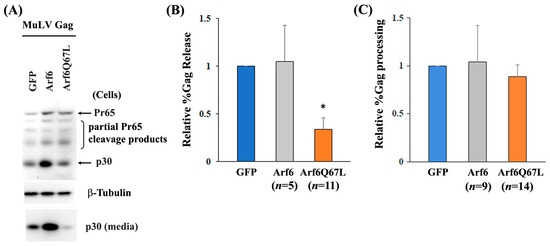
env. Gag is translated as a polyprotein and moves to assembly sites where viral particles are shaped by cleavage of poly-Gag. Viral release depends on the intracellular trafficking of viral proteins, which is determined by both viral and cellular factors. ADP-ribosylation factor 6 (Arf6) is a small GTPase that regulates vesicular trafficking and recycling of different types of cargo in cells. Arf6 also activates phospholipase D (PLD) and phosphatidylinositol-4-phosphate 5-kinase (PIP5K) and produces phosphatidylinositol-4,5-bisphosphate (PI(4,5)P
2). We investigated how Arf6 affected MuLV release with a constitutively active form of Arf6, Arf6Q67L. Expression of Arf6Q67L impaired Gag release by accumulating Gag at PI(4,5)P
2-enriched compartments in the cytoplasm. Treatment of the inhibitors for PLD and PIP5K impaired or recovered MuLV Gag release in the cells expressing GFP (control) and Arf6Q67L, implying that regulation of PI(4,5)P
2 through PLD and PIP5K affected MuLV release. Interference with the phosphoinositide 3-kinases, mammalian target of rapamycin (mTOR) pathway, and vacuolar-type ATPase activities showed further impairment of Gag release from the cells expressing Arf6Q67L. In contrast, mTOR inhibition increased Gag release in the control cells. The proteasome inhibitors reduced viral release in the cells regardless of Arf6Q67L expression. These data outline the differences in MuLV release under the controlled and overactivated Arf6 conditions and provide new insight into pathways for MuLV release.
Full article
►▼
Show Figures
Open AccessArticle
SUMOylation Modulates Reactive Oxygen Species (ROS) Levels and Acts as a Protective Mechanism in the Type 2 Model of Diabetic Peripheral Neuropathy
by
Nicolas Mandel, Michael Büttner, Gernot Poschet, Rohini Kuner and Nitin Agarwal
Cited by 7 | Viewed by 2768
Abstract
Diabetic peripheral neuropathy (DPN) is the prevalent type of peripheral neuropathy; it primarily impacts extremity nerves. Its multifaceted nature makes the molecular mechanisms of diabetic neuropathy intricate and incompletely elucidated. Several types of post-translational modifications (PTMs) have been implicated in the development and
[...] Read more.
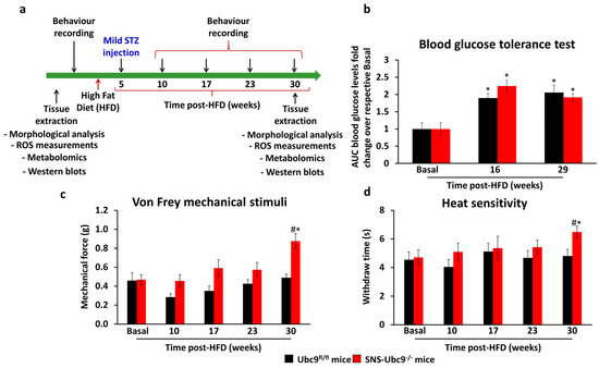
Diabetic peripheral neuropathy (DPN) is the prevalent type of peripheral neuropathy; it primarily impacts extremity nerves. Its multifaceted nature makes the molecular mechanisms of diabetic neuropathy intricate and incompletely elucidated. Several types of post-translational modifications (PTMs) have been implicated in the development and progression of DPN, including phosphorylation, glycation, acetylation and SUMOylation. SUMOylation involves the covalent attachment of small ubiquitin-like modifier (SUMO) proteins to target proteins, and it plays a role in various cellular processes, including protein localization, stability, and function. While the specific relationship between high blood glucose and SUMOylation is not extensively studied, recent evidence implies its involvement in the development of DPN in type 1 diabetes. In this study, we investigated the impact of SUMOylation on the onset and progression of DPN in a type 2 diabetes model using genetically modified mutant mice lacking SUMOylation, specifically in peripheral sensory neurons (SNS-Ubc9
−/−). Behavioural measurement for evoked pain, morphological analyses of nerve fibre loss in the epidermis, measurement of reactive oxygen species (ROS) levels, and antioxidant molecules were analysed over several months in SUMOylation-deficient and control mice. Our longitudinal analysis at 30 weeks post-high-fat diet revealed that SNS-Ubc9
−/− mice exhibited earlier and more pronounced thermal and mechanical sensation loss and accelerated intraepidermal nerve fibre loss compared to control mice. Mechanistically, these changes are associated with increased levels of ROS both in sensory neuronal soma and in peripheral axonal nerve endings in SNS-Ubc9
−/− mice. In addition, we observed compromised detoxifying potential, impaired respiratory chain complexes, and reduced levels of protective lipids in sensory neurons upon deletion of SUMOylation in diabetic mice. Importantly, we also identified mitochondrial malate dehydrogenase (MDH2) as a SUMOylation target, the activity of which is negatively regulated by SUMOylation. Our results indicate that SUMOylation is an essential neuroprotective mechanism in sensory neurons in type 2 diabetes, the deletion of which causes oxidative stress and an impaired respiratory chain, resulting in energy depletion and subsequent damage to sensory neurons.
Full article
►▼
Show Figures
Open AccessArticle
Sox9 Inhibits Cochlear Hair Cell Fate by Upregulating Hey1 and HeyL Antagonists of Atoh1
by
Mona Veithen, Aurélia Huyghe, Priscilla Van Den Ackerveken, So-ichiro Fukada, Hiroki Kokubo, Ingrid Breuskin, Laurent Nguyen, Laurence Delacroix and Brigitte Malgrange
Cited by 2 | Viewed by 2183
Abstract
It is widely accepted that cell fate determination in the cochlea is tightly controlled by different transcription factors (TFs) that remain to be fully defined. Here, we show that Sox9, initially expressed in the entire sensory epithelium of the cochlea, progressively disappears from
[...] Read more.
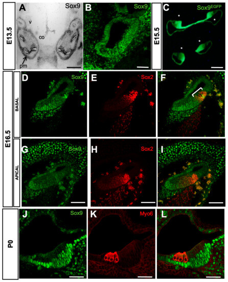
It is widely accepted that cell fate determination in the cochlea is tightly controlled by different transcription factors (TFs) that remain to be fully defined. Here, we show that Sox9, initially expressed in the entire sensory epithelium of the cochlea, progressively disappears from differentiating hair cells (HCs) and is finally restricted to supporting cells (SCs). By performing ex vivo electroporation of E13.5–E14.5 cochleae, we demonstrate that maintenance of Sox9 expression in the progenitors committed to HC fate blocks their differentiation, even if co-expressed with Atoh1, a transcription factor necessary and sufficient to form HC. Sox9 inhibits Atoh1 transcriptional activity by upregulating Hey1 and HeyL antagonists, and genetic ablation of these genes induces extra HCs along the cochlea. Although Sox9 suppression from sensory progenitors ex vivo leads to a modest increase in the number of HCs, it is not sufficient in vivo to induce supernumerary HC production in an inducible Sox9 knockout model. Taken together, these data show that Sox9 is downregulated from nascent HCs to allow the unfolding of their differentiation program. This may be critical for future strategies to promote fully mature HC formation in regeneration approaches.
Full article
►▼
Show Figures
Open AccessReview
Dendritic Cell Vaccines: A Shift from Conventional Approach to New Generations
by
Kyu-Won Lee, Judy Wai Ping Yam and Xiaowen Mao
Cited by 60 | Viewed by 7331
Abstract
In the emerging era of cancer immunotherapy, immune checkpoint blockades (ICBs) and adoptive cell transfer therapies (ACTs) have gained significant attention. However, their therapeutic efficacies are limited due to the presence of cold type tumors, immunosuppressive tumor microenvironment, and immune-related side effects. On
[...] Read more.
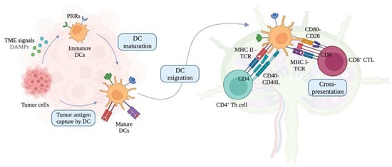
In the emerging era of cancer immunotherapy, immune checkpoint blockades (ICBs) and adoptive cell transfer therapies (ACTs) have gained significant attention. However, their therapeutic efficacies are limited due to the presence of cold type tumors, immunosuppressive tumor microenvironment, and immune-related side effects. On the other hand, dendritic cell (DC)-based vaccines have been suggested as a new cancer immunotherapy regimen that can address the limitations encountered by ICBs and ACTs. Despite the success of the first generation of DC-based vaccines, represented by the first FDA-approved DC-based therapeutic cancer vaccine Provenge, several challenges remain unsolved. Therefore, new DC vaccine strategies have been actively investigated. This review addresses the limitations of the currently most adopted classical DC vaccine and evaluates new generations of DC vaccines in detail, including biomaterial-based, immunogenic cell death-inducing, mRNA-pulsed, DC small extracellular vesicle (sEV)-based, and tumor sEV-based DC vaccines. These innovative DC vaccines are envisioned to provide a significant breakthrough in cancer immunotherapy landscape and are expected to be supported by further preclinical and clinical studies.
Full article
►▼
Show Figures
Open AccessCommunication
Generation of Conditional Knockout Alleles for PRUNE-1
by
Xiaoli Wu, Louise R. Simard and Hao Ding
Cited by 2 | Viewed by 2890
Abstract
PRUNE1 is a member of the aspartic acid-histidine-histidine (DHH) protein superfamily, which could display an exopolyphosphatase activity and interact with multiple cellular proteins involved in the cytoskeletal rearrangement. It is widely expressed during embryonic development and is essential for embryogenesis. PRUNE1 could also
[...] Read more.
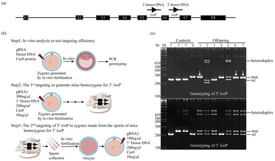
PRUNE1 is a member of the aspartic acid-histidine-histidine (DHH) protein superfamily, which could display an exopolyphosphatase activity and interact with multiple cellular proteins involved in the cytoskeletal rearrangement. It is widely expressed during embryonic development and is essential for embryogenesis. PRUNE1 could also be critical for postnatal development of the nervous system as it was found to be mutated in patients with microcephaly, brain malformations, and neurodegeneration. To determine the cellular function of PRUNE1 during development and in disease, we have generated conditional mouse alleles of the
Prune1 in which
loxP sites flank exon 6. Crossing these alleles with a ubiquitous Cre transgenic line resulted in a complete loss of PRUNE1 expression and embryonic defects identical to those previously described for
Prune1 null embryos. In addition, breeding these alleles with a Purkinje cell-specific Cre line (
Pcp2-Cre) resulted in the loss of Purkinje cells similar to that observed in patients carrying a mutation with loss of PRUNE1 function. Therefore, the
Prune1 conditional mouse alleles generated in this study provide important genetic tools not only for dissecting the spatial and temporal roles of PRUNE1 during development but also for understanding the pathogenic role of PRUNE1 dysfunction in neurodegenerative or neurodevelopmental disease. In addition, from this work, we have described an approach that allows one to efficiently generate conditional mouse alleles based on mouse zygote electroporation.
Full article
►▼
Show Figures
Open AccessArticle
Bone Marrow Mesenchymal Stromal/Stem Cell-Derived Extracellular Vesicles Promote Corneal Wound Repair by Regulating Inflammation and Angiogenesis
by
Gabriele Saccu, Valeria Menchise, Chiara Gai, Marina Bertolin, Stefano Ferrari, Cristina Giordano, Marta Manco, Walter Dastrù, Emanuela Tolosano, Benedetta Bussolati, Enzo Calautti, Giovanni Camussi, Fiorella Altruda and Sharmila Fagoonee
Cited by 31 | Viewed by 3725
Abstract
Severe corneal damage leads to complete vision loss, thereby affecting life quality and impinging heavily on the healthcare system. Current clinical approaches to manage corneal wounds suffer from severe drawbacks, thus requiring the development of alternative strategies. Of late, mesenchymal stromal/stem cell (MSC)-derived
[...] Read more.
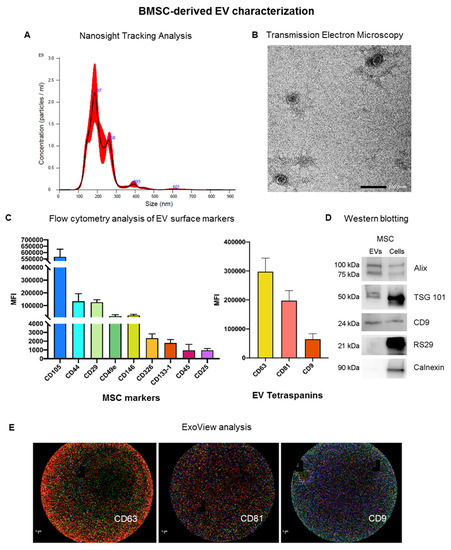
Severe corneal damage leads to complete vision loss, thereby affecting life quality and impinging heavily on the healthcare system. Current clinical approaches to manage corneal wounds suffer from severe drawbacks, thus requiring the development of alternative strategies. Of late, mesenchymal stromal/stem cell (MSC)-derived extracellular vesicles (EVs) have become a promising tool in the ophthalmic field. In the present study, we topically delivered bone-marrow-derived MSC-EVs (BMSC-EVs), embedded in methylcellulose, in a murine model of alkali-burn-induced corneal damage in order to evaluate their role in corneal repair through histological and molecular analyses, with the support of magnetic resonance imaging. Our data show that BMSC-EVs, used for the first time in this specific formulation on the damaged cornea, modulate cell death, inflammation and angiogenetic programs in the injured tissue, thus leading to a faster recovery of corneal damage. These results were confirmed on cadaveric donor-derived human corneal epithelial cells in vitro. Thus, BMSC-EVs modulate corneal repair dynamics and are promising as a new cell-free approach for intervening on burn wounds, especially in the avascularized region of the eye.
Full article
►▼
Show Figures
Open AccessEditor’s ChoiceReview
Molecular Regulators of Embryonic Diapause and Cancer Diapause-like State
by
Abdiasis M. Hussein, Nanditaa Balachandar, Julie Mathieu and Hannele Ruohola-Baker
Cited by 19 | Viewed by 7955
Abstract
Embryonic diapause is an enigmatic state of dormancy that interrupts the normally tight connection between developmental stages and time. This reproductive strategy and state of suspended development occurs in mice, bears, roe deer, and over 130 other mammals and favors the survival of
[...] Read more.
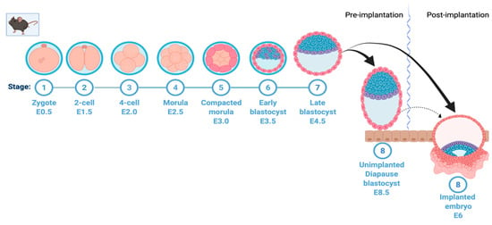
Embryonic diapause is an enigmatic state of dormancy that interrupts the normally tight connection between developmental stages and time. This reproductive strategy and state of suspended development occurs in mice, bears, roe deer, and over 130 other mammals and favors the survival of newborns. Diapause arrests the embryo at the blastocyst stage, delaying the post-implantation development of the embryo. This months-long quiescence is reversible, in contrast to senescence that occurs in aging stem cells. Recent studies have revealed critical regulators of diapause. These findings are important since defects in the diapause state can cause a lack of regeneration and control of normal growth. Controlling this state may also have therapeutic applications since recent findings suggest that radiation and chemotherapy may lead some cancer cells to a protective diapause-like, reversible state. Interestingly, recent studies have shown the metabolic regulation of epigenetic modifications and the role of microRNAs in embryonic diapause. In this review, we discuss the molecular mechanism of diapause induction.
Full article
►▼
Show Figures


