ijms-logo

Journal Browser

Journal Browser

Advances in Oxytocin

A special issue of International Journal of Molecular Sciences (ISSN 1422-0067). This special issue belongs to the section "Bioactives and Nutraceuticals".

Deadline for manuscript submissions: closed (30 September 2021) | Viewed by 39101

Special Issue Editor


E-Mail Website
Guest Editor
Department of Animal Environment and Health, Swedish University of Agricultural Sciences, 53223 Skara, Sweden
Interests: oxytocin; oxytocin agonists; release; effects; mechanisms of action in vitro; in vivo; clinical studies; behaviour; physiology; pharmacology; molecular biology; genetics
Special Issues, Collections and Topics in MDPI journals

Special Issue Information

Dear Colleagues,

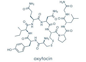
Oxytocin, originally considered to be a birth and breastfeeding hormone, is currently receiving a great deal of attention, as the effect profile of oxytocin is steadily growing. New data regarding behavioral/psychological effects as well as physiological functions and pharmacological effects of oxytocin are being published. The molecular and genetic mechanisms mediating and regulating the oxytocin-related effects are also being uncovered. It is difficult to get an overview of the literature on oxytocin, because it involves so many different research topics. This motivates a Special Issue in which data on oxytocin from different research fields can be included and published together. There is often a gap between all the experimental data published regarding oxytocin and a discussion regarding their relevance for the in vivo situation or for possible clinical effects in humans.

Data on oxytocin from any topic, obtained from experimental studies in vitro, from animal experiments, and from pre-clinical studies in humans are welcome in this Special Issue. We encourage the inclusion of discussions regarding the physiological or pre-clinical relevance of the data reported in the submitted articles. Additionally, studies reporting experimental regarding oxytocin agonists are welcome. Both original articles and review articles will be included in this Special Issue.

Prof. Dr. Kerstin Uvnäs-Moberg
Guest Editor

Manuscript Submission Information

Manuscripts should be submitted online at www.mdpi.com by registering and logging in to this website. Once you are registered, click here to go to the submission form. Manuscripts can be submitted until the deadline. All submissions that pass pre-check are peer-reviewed. Accepted papers will be published continuously in the journal (as soon as accepted) and will be listed together on the special issue website. Research articles, review articles as well as short communications are invited. For planned papers, a title and short abstract (about 100 words) can be sent to the Editorial Office for announcement on this website.

Submitted manuscripts should not have been published previously, nor be under consideration for publication elsewhere (except conference proceedings papers). All manuscripts are thoroughly refereed through a single-blind peer-review process. A guide for authors and other relevant information for submission of manuscripts is available on the Instructions for Authors page. International Journal of Molecular Sciences is an international peer-reviewed open access semimonthly journal published by MDPI.

Please visit the Instructions for Authors page before submitting a manuscript. There is an Article Processing Charge (APC) for publication in this open access journal. For details about the APC please see here. Submitted papers should be well formatted and use good English. Authors may use MDPI's English editing service prior to publication or during author revisions.

Keywords

  • oxytocin
  • oxytocin agonists
  • release
  • effects
  • mechanisms of action in vitro
  • in vivo
  • behaviour
  • physiology
  • pharmacology
  • molecular biology
  • genetics

Published Papers (7 papers)

Order results
Result details
Select all
Export citation of selected articles as:

Research

Jump to: Review, Other

15 pages, 3905 KiB  
Article
Co-Stimulation of Oxytocin and Arginine-Vasopressin Receptors Affect Hypothalamic Neurospheroid Size
by Mohammad Saied Salehi, Inga D. Neumann, Benjamin Jurek and Sareh Pandamooz
Int. J. Mol. Sci. 2021, 22(16), 8464; https://doi.org/10.3390/ijms22168464 - 6 Aug 2021
Cited by 7 | Viewed by 2224
Abstract
Oxytocin (OXT) is a neuropeptide involved in a plethora of behavioral and physiological processes. However, there is a prominent lack of 3D cell culture models that investigate the effects of OXT on a cellular/molecular level. In this study, we established a hypothalamic neuronal [...] Read more.
Oxytocin (OXT) is a neuropeptide involved in a plethora of behavioral and physiological processes. However, there is a prominent lack of 3D cell culture models that investigate the effects of OXT on a cellular/molecular level. In this study, we established a hypothalamic neuronal spheroid model to investigate the cellular response in a more realistic 3D setting. Our data indicate that the formation of spheroids itself does not alter the basic characteristics of the cell line and that markers of cellular morphology and connectivity are stably expressed. We found that both OXT and arginine vasopressin (AVP) treatment increase spheroid size (surface area and volume), as well as individual nucleus size, which serves as an indicator for cellular proliferation. The cellular response to both OXT and AVP seems mainly to be mediated by the AVP receptor 1a (V1aR); however, the OXT receptor (OXTR) contributes significantly to the observed proliferative effect. When we blocked the OXTR pharmacologically or knocked down the OXTR by siRNA, the OXT- or AVP-induced cellular proliferation decreased. In summary, we established a 3D cell culture model of the neuronal response to OXT and AVP and found that spheroids react to the treatment via their respective receptors but also via cross-talk between the two receptor types. Full article
(This article belongs to the Special Issue Advances in Oxytocin)
Show Figures

Figure 1

18 pages, 4427 KiB  
Article
Plasticity in Intrinsic Excitability of Hypothalamic Magnocellular Neurosecretory Neurons in Late-Pregnant and Lactating Rats
by Michael R. Perkinson, Rachael A. Augustine, Gregory T. Bouwer, Emily F. Brown, Isaiah Cheong, Alexander J. Seymour, Martin Fronius and Colin H. Brown
Int. J. Mol. Sci. 2021, 22(13), 7140; https://doi.org/10.3390/ijms22137140 - 1 Jul 2021
Cited by 4 | Viewed by 2722
Abstract
Oxytocin and vasopressin secretion from the posterior pituitary gland are required for normal pregnancy and lactation. Oxytocin secretion is relatively low and constant under basal conditions but becomes pulsatile during birth and lactation to stimulate episodic contraction of the uterus for delivery of [...] Read more.
Oxytocin and vasopressin secretion from the posterior pituitary gland are required for normal pregnancy and lactation. Oxytocin secretion is relatively low and constant under basal conditions but becomes pulsatile during birth and lactation to stimulate episodic contraction of the uterus for delivery of the fetus and milk ejection during suckling. Vasopressin secretion is maintained in pregnancy and lactation despite reduced osmolality (the principal stimulus for vasopressin secretion) to increase water retention to cope with the cardiovascular demands of pregnancy and lactation. Oxytocin and vasopressin secretion are determined by the action potential (spike) firing of magnocellular neurosecretory neurons of the hypothalamic supraoptic and paraventricular nuclei. In addition to synaptic input activity, spike firing depends on intrinsic excitability conferred by the suite of channels expressed by the neurons. Therefore, we analysed oxytocin and vasopressin neuron activity in anaesthetised non-pregnant, late-pregnant, and lactating rats to test the hypothesis that intrinsic excitability of oxytocin and vasopressin neurons is increased in late pregnancy and lactation to promote oxytocin and vasopressin secretion required for successful pregnancy and lactation. Hazard analysis of spike firing revealed a higher incidence of post-spike hyperexcitability immediately following each spike in oxytocin neurons, but not in vasopressin neurons, in late pregnancy and lactation, which is expected to facilitate high frequency firing during bursts. Despite lower osmolality in late-pregnant and lactating rats, vasopressin neuron activity was not different between non-pregnant, late-pregnant, and lactating rats, and blockade of osmosensitive ΔN-TRPV1 channels inhibited vasopressin neurons to a similar extent in non-pregnant, late-pregnant, and lactating rats. Furthermore, supraoptic nucleus ΔN-TRPV1 mRNA expression was not different between non-pregnant and late-pregnant rats, suggesting that sustained activity of ΔN-TRPV1 channels might maintain vasopressin neuron activity to increase water retention during pregnancy and lactation. Full article
(This article belongs to the Special Issue Advances in Oxytocin)
Show Figures

Figure 1

Review

Jump to: Research, Other

29 pages, 858 KiB  
Review
Role of Oxytocin and Vasopressin in Neuropsychiatric Disorders: Therapeutic Potential of Agonists and Antagonists
by Valeska Cid-Jofré, Macarena Moreno, Miguel Reyes-Parada and Georgina M. Renard
Int. J. Mol. Sci. 2021, 22(21), 12077; https://doi.org/10.3390/ijms222112077 - 8 Nov 2021
Cited by 15 | Viewed by 7038
Abstract
Oxytocin (OT) and vasopressin (AVP) are hypothalamic neuropeptides classically associated with their regulatory role in reproduction, water homeostasis, and social behaviors. Interestingly, this role has expanded in recent years and has positioned these neuropeptides as therapeutic targets for various neuropsychiatric diseases such as [...] Read more.
Oxytocin (OT) and vasopressin (AVP) are hypothalamic neuropeptides classically associated with their regulatory role in reproduction, water homeostasis, and social behaviors. Interestingly, this role has expanded in recent years and has positioned these neuropeptides as therapeutic targets for various neuropsychiatric diseases such as autism, addiction, schizophrenia, depression, and anxiety disorders. Due to the chemical-physical characteristics of these neuropeptides including short half-life, poor blood-brain barrier penetration, promiscuity for AVP and OT receptors (AVP-R, OT-R), novel ligands have been developed in recent decades. This review summarizes the role of OT and AVP in neuropsychiatric conditions, as well as the findings of different OT-R and AVP-R agonists and antagonists, used both at the preclinical and clinical level. Furthermore, we discuss their possible therapeutic potential for central nervous system (CNS) disorders. Full article
(This article belongs to the Special Issue Advances in Oxytocin)
Show Figures

Figure 1

19 pages, 1005 KiB  
Review
Signal Transduction of Mineralocorticoid and Angiotensin II Receptors in the Central Control of Sodium Appetite: A Narrative Review
by Michele Iovino, Tullio Messana, Giuseppe Lisco, Aldo Vanacore, Vito Angelo Giagulli, Edoardo Guastamacchia, Giovanni De Pergola and Vincenzo Triggiani
Int. J. Mol. Sci. 2021, 22(21), 11735; https://doi.org/10.3390/ijms222111735 - 29 Oct 2021
Cited by 1 | Viewed by 2354
Abstract
Sodium appetite is an innate behavior occurring in response to sodium depletion that induces homeostatic responses such as the secretion of the mineralocorticoid hormone aldosterone from the zona glomerulosa of the adrenal cortex and the stimulation of the peptide hormone angiotensin II (ANG [...] Read more.
Sodium appetite is an innate behavior occurring in response to sodium depletion that induces homeostatic responses such as the secretion of the mineralocorticoid hormone aldosterone from the zona glomerulosa of the adrenal cortex and the stimulation of the peptide hormone angiotensin II (ANG II). The synergistic action of these hormones signals to the brain the sodium appetite that represents the increased palatability for salt intake. This narrative review summarizes the main data dealing with the role of mineralocorticoid and ANG II receptors in the central control of sodium appetite. Appropriate keywords and MeSH terms were identified and searched in PubMed. References to original articles and reviews were examined, selected, and discussed. Several brain areas control sodium appetite, including the nucleus of the solitary tract, which contains aldosterone-sensitive HSD2 neurons, and the organum vasculosum lamina terminalis (OVLT) that contains ANG II-sensitive neurons. Furthermore, sodium appetite is under the control of signaling proteins such as mitogen-activated protein kinase (MAPK) and inositol 1,4,5-thriphosphate (IP3). ANG II stimulates salt intake via MAPK, while combined ANG II and aldosterone action induce sodium intake via the IP3 signaling pathway. Finally, aldosterone and ANG II stimulate OVLT neurons and suppress oxytocin secretion inhibiting the neuronal activity of the paraventricular nucleus, thus disinhibiting the OVLT activity to aldosterone and ANG II stimulation. Full article
(This article belongs to the Special Issue Advances in Oxytocin)
Show Figures

Figure 1

24 pages, 1994 KiB  
Review
Complementary Role of Oxytocin and Vasopressin in Cardiovascular Regulation
by Ewa Szczepanska-Sadowska, Agnieszka Wsol, Agnieszka Cudnoch-Jedrzejewska and Tymoteusz Żera
Int. J. Mol. Sci. 2021, 22(21), 11465; https://doi.org/10.3390/ijms222111465 - 24 Oct 2021
Cited by 19 | Viewed by 7829
Abstract
The neurons secreting oxytocin (OXY) and vasopressin (AVP) are located mainly in the supraoptic, paraventricular, and suprachiasmatic nucleus of the brain. Oxytocinergic and vasopressinergic projections reach several regions of the brain and the spinal cord. Both peptides are released from axons, soma, and [...] Read more.
The neurons secreting oxytocin (OXY) and vasopressin (AVP) are located mainly in the supraoptic, paraventricular, and suprachiasmatic nucleus of the brain. Oxytocinergic and vasopressinergic projections reach several regions of the brain and the spinal cord. Both peptides are released from axons, soma, and dendrites and modulate the excitability of other neuroregulatory pathways. The synthesis and action of OXY and AVP in the peripheral organs (eye, heart, gastrointestinal system) is being investigated. The secretion of OXY and AVP is influenced by changes in body fluid osmolality, blood volume, blood pressure, hypoxia, and stress. Vasopressin interacts with three subtypes of receptors: V1aR, V1bR, and V2R whereas oxytocin activates its own OXTR and V1aR receptors. AVP and OXY receptors are present in several regions of the brain (cortex, hypothalamus, pons, medulla, and cerebellum) and in the peripheral organs (heart, lungs, carotid bodies, kidneys, adrenal glands, pancreas, gastrointestinal tract, ovaries, uterus, thymus). Hypertension, myocardial infarction, and coexisting factors, such as pain and stress, have a significant impact on the secretion of oxytocin and vasopressin and on the expression of their receptors. The inappropriate regulation of oxytocin and vasopressin secretion during ischemia, hypoxia/hypercapnia, inflammation, pain, and stress may play a significant role in the pathogenesis of cardiovascular diseases. Full article
(This article belongs to the Special Issue Advances in Oxytocin)
Show Figures

Figure 1

43 pages, 6311 KiB  
Review
Oxytocin, Erectile Function and Sexual Behavior: Last Discoveries and Possible Advances
by Maria Rosaria Melis and Antonio Argiolas
Int. J. Mol. Sci. 2021, 22(19), 10376; https://doi.org/10.3390/ijms221910376 - 26 Sep 2021
Cited by 17 | Viewed by 11288
Abstract
A continuously increasing amount of research shows that oxytocin is involved in numerous central functions. Among the functions in which oxytocin is thought to be involved are those that play a role in social and sexual behaviors, and the involvement of central oxytocin [...] Read more.
A continuously increasing amount of research shows that oxytocin is involved in numerous central functions. Among the functions in which oxytocin is thought to be involved are those that play a role in social and sexual behaviors, and the involvement of central oxytocin in erectile function and sexual behavior was indeed one of the first to be discovered in laboratory animals in the 1980s. The first part of this review summarizes the results of studies done in laboratory animals that support a facilitatory role of oxytocin in male and female sexual behavior and reveal mechanisms through which this ancient neuropeptide participates in concert with other neurotransmitters and neuropeptides in this complex function, which is fundamental for the species reproduction. The second part summarizes the results of studies done mainly with intranasal oxytocin in men and women with the aim to translate the results found in laboratory animals to humans. Unexpectedly, the results of these studies do not appear to confirm the facilitatory role of oxytocin found in male and female sexual behavior in animals, both in men and women. Possible explanations for the failure of oxytocin to improve sexual behavior in men and women and strategies to attempt to overcome this impasse are considered. Full article
(This article belongs to the Special Issue Advances in Oxytocin)
Show Figures

Figure 1

Other

Jump to: Research, Review

25 pages, 21135 KiB  
Perspective
Biological Connection of Psychological Stress and Polytrauma under Intensive Care: The Role of Oxytocin and Hydrogen Sulfide
by Tamara Merz, Oscar McCook, Nicole Denoix, Peter Radermacher, Christiane Waller and Thomas Kapapa
Int. J. Mol. Sci. 2021, 22(17), 9192; https://doi.org/10.3390/ijms22179192 - 25 Aug 2021
Cited by 4 | Viewed by 4105
Abstract
This paper explored the potential mediating role of hydrogen sulfide (H2S) and the oxytocin (OT) systems in hemorrhagic shock (HS) and/or traumatic brain injury (TBI). Morbidity and mortality after trauma mainly depend on the presence of HS and/or TBI. Rapid “repayment [...] Read more.
This paper explored the potential mediating role of hydrogen sulfide (H2S) and the oxytocin (OT) systems in hemorrhagic shock (HS) and/or traumatic brain injury (TBI). Morbidity and mortality after trauma mainly depend on the presence of HS and/or TBI. Rapid “repayment of the O2 debt” and prevention of brain tissue hypoxia are cornerstones of the management of both HS and TBI. Restoring tissue perfusion, however, generates an ischemia/reperfusion (I/R) injury due to the formation of reactive oxygen (ROS) and nitrogen (RNS) species. Moreover, pre-existing-medical-conditions (PEMC’s) can aggravate the occurrence and severity of complications after trauma. In addition to the “classic” chronic diseases (of cardiovascular or metabolic origin), there is growing awareness of psychological PEMC’s, e.g., early life stress (ELS) increases the predisposition to develop post-traumatic-stress-disorder (PTSD) and trauma patients with TBI show a significantly higher incidence of PTSD than patients without TBI. In fact, ELS is known to contribute to the developmental origins of cardiovascular disease. The neurotransmitter H2S is not only essential for the neuroendocrine stress response, but is also a promising therapeutic target in the prevention of chronic diseases induced by ELS. The neuroendocrine hormone OT has fundamental importance for brain development and social behavior, and, thus, is implicated in resilience or vulnerability to traumatic events. OT and H2S have been shown to interact in physical and psychological trauma and could, thus, be therapeutic targets to mitigate the acute post-traumatic effects of chronic PEMC’s. OT and H2S both share anti-inflammatory, anti-oxidant, and vasoactive properties; through the reperfusion injury salvage kinase (RISK) pathway, where their signaling mechanisms converge, they act via the regulation of nitric oxide (NO). Full article
(This article belongs to the Special Issue Advances in Oxytocin)
Show Figures

Figure 1

Back to TopTop