

Orthodontic Treatment of Palatally Impacted Canines in Severe Non-Syndromic Oligodontia with the Use of Mini-Implants: A Case Report


Abstract
:1. Introduction
2. Materials and Methods
3. Results
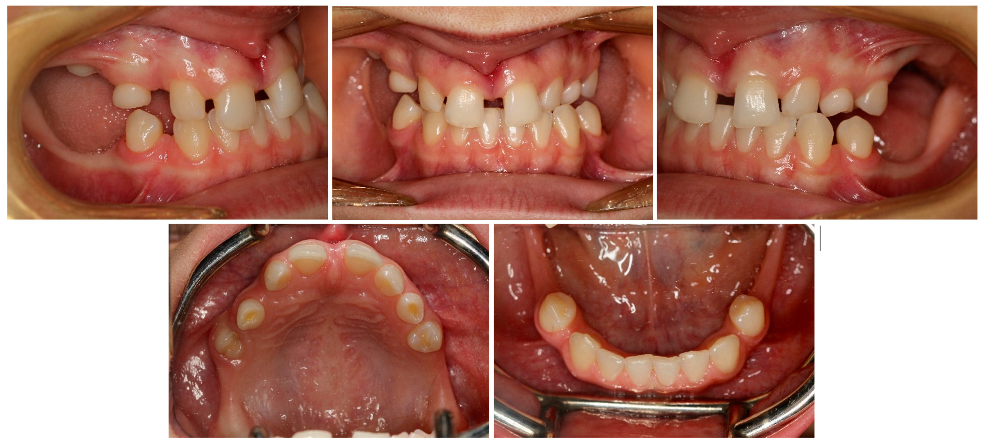
3.1. Treatment Process
3.2. Follow-Up
4. Discussion
5. Conclusions
Author Contributions
Funding
Institutional Review Board Statement
Informed Consent Statement
Data Availability Statement
Acknowledgments
Conflicts of Interest
References
- Letra, A.; Chiquet, B.; Hansen-Kiss, E.; Menezes, S.; Hunter, E. Nonsyndromic Tooth Agenesis Overview. In GeneReviews®; Adam, M.P., Everman, D.B., Mirzaa, G.M., Pagon, R.A., Wallace, S.E., Bean, L.J., Gripp, K.W., Amemiya, A., Eds.; University of Washington: Seattle, WA, USA, 1993. [Google Scholar]
- Bergendal, B. Orodental Manifestations in Ectodermal Dysplasia—A Review. Am. J. Med. Genet. A 2014, 164A, 2465–2471. [Google Scholar] [CrossRef]
- Tuna, E.B.; Koruyucu, M.; Kürklü, E.; Çifter, M.; Gençay, K.; Seymen, F.; Tüysüz, B. Oral and Craniofacial Manifestations of Ellis-van Creveld Syndrome: Case Series. J. Craniomaxillofac. Surg. 2016, 44, 919–924. [Google Scholar] [CrossRef] [PubMed]
- Tuna, E.B.; Sulun, T.; Rosti, O.; El Abdallah, F.; Kayserili, H.; Aktoren, O. Craniodentofacial Manifestations in Hallermann-Streiff Syndrome. Cranio 2009, 27, 33–38. [Google Scholar] [CrossRef] [PubMed]
- Ruiz-Heiland, G.; Lenz, S.; Bock, N.; Ruf, S. Prevalence of WNT10A Gene Mutations in Non-Syndromic Oligodontia. Clin. Oral Investig. 2019, 23, 3103–3113. [Google Scholar] [CrossRef]
- Zheng, J.; Yu, M.; Liu, H.; Cai, T.; Feng, H.; Liu, Y.; Han, D. Novel MSX1 Variants Identified in Families with Nonsyndromic Oligodontia. Int. J. Oral Sci. 2021, 13, 2. [Google Scholar] [CrossRef]
- Cai, Z.; Deng, X.; Jia, J.; Wang, D.; Yuan, G. Ectodysplasin A/Ectodysplasin A Receptor System and Their Roles in Multiple Diseases. Front. Physiol. 2021, 12, 788411. [Google Scholar] [CrossRef]
- Zhou, M.; Zhang, H.; Camhi, H.; Seymen, F.; Koruyucu, M.; Kasimoglu, Y.; Kim, J.-W.; Kim-Berman, H.; Yuson, N.M.R.; Benke, P.J.; et al. Analyses of Oligodontia Phenotypes and Genetic Etiologies. Int. J. Oral Sci. 2021, 13, 32. [Google Scholar] [CrossRef]
- Bishara, S.E. Impacted Maxillary Canines: A Review. Am. J. Orthod. Dentofac. Orthop. 1992, 101, 159–171. [Google Scholar] [CrossRef]
- Oliver, R.G.; Mannion, J.E.; Robinson, J.M. Morphology of the Maxillary Lateral Incisor in Cases of Unilateral Impaction of the Maxillary Canine. Br. J. Orthod. 1989, 16, 9–16. [Google Scholar] [CrossRef]
- Kim, Y.; Hyun, H.-K.; Jang, K.-T. The Position of Maxillary Canine Impactions and the Influenced Factors to Adjacent Root Resorption in the Korean Population. Eur. J. Orthod. 2012, 34, 302–306. [Google Scholar] [CrossRef]
- Becker, A.; Chaushu, S. Etiology of Maxillary Canine Impaction: A Review. Am. J. Orthod. Dentofac. Orthop. 2015, 148, 557–567. [Google Scholar] [CrossRef] [PubMed]
- Laganà, G.; Venza, N.; Borzabadi-Farahani, A.; Fabi, F.; Danesi, C.; Cozza, P. Dental Anomalies: Prevalence and Associations between Them in a Large Sample of Non-Orthodontic Subjects, a Cross-Sectional Study. BMC Oral Health 2017, 17, 62. [Google Scholar] [CrossRef] [PubMed]
- Parkin, N.; Furness, S.; Shah, A.; Thind, B.; Marshman, Z.; Glenroy, G.; Dyer, F.; Benson, P.E. Extraction of Primary (Baby) Teeth for Unerupted Palatally Displaced Permanent Canine Teeth in Children. Cochrane Database Syst. Rev. 2018, 2018, CD004621. [Google Scholar] [CrossRef] [PubMed]
- Parkin, N.; Benson, P.E.; Thind, B.; Shah, A.; Khalil, I.; Ghafoor, S. Open versus Closed Surgical Exposure of Canine Teeth That Are Displaced in the Roof of the Mouth. Cochrane Database Syst. Rev. 2017, 2017, CD006966. [Google Scholar] [CrossRef]
- Keener, D.J.; De Oliveira Ruellas, A.C.; Aliaga-Del Castillo, A.; Arriola-Guillén, L.E.; Bianchi, J.; Oh, H.; Gurgel, M.L.; Benavides, E.; Soki, F.; Rodríguez-Cárdenas, Y.A.; et al. Three-Dimensional Decision Support System for Treatment of Canine Impaction. Am. J. Orthod. Dentofac. Orthop. 2023, 164, 491–504. [Google Scholar] [CrossRef]
- Becker, A.; Chaushu, G.; Chaushu, S. Analysis of Failure in the Treatment of Impacted Maxillary Canines. Am. J. Orthod. Dentofac. Orthop. 2010, 137, 743–754. [Google Scholar] [CrossRef]
- Puricelli, E. Apicotomy: A Root Apical Fracture for Surgical Treatment of Impacted Upper Canines. Head Face Med. 2007, 3, 33. [Google Scholar] [CrossRef]
- Grisar, K.; Denoiseux, B.; Martin, C.; Hoppenreijs, T.; Calburean, F.; Politis, C.; Jacobs, R. Treatment for Critically Impacted Maxillary Canines: Clinical versus Scientific Evidence—A Systematic Review. J. Stomatol. Oral Maxillofac. Surg. 2022, 123, e12–e19. [Google Scholar] [CrossRef]
- Krasny, M.; Krasny, K.; Wojtowicz, A. Long Term Outcomes of En-Block Autotransplantation of a Tooth. Cell Tissue Bank. 2023, 24, 67–73. [Google Scholar] [CrossRef]
- Migliorati, M.; Drago, S.; Bocchino, T.; Michelotti, A.; D’Antò, V. Treatment of Palatally Displaced Canines Using Miniscrews for Direct or Indirect Anchorage: A Three-Dimensional Prospective Cohort Study on Tooth Movement Speed. Appl. Sci. 2022, 12, 10935. [Google Scholar] [CrossRef]
- Riley, D.S.; Barber, M.S.; Kienle, G.S.; Aronson, J.K.; von Schoen-Angerer, T.; Tugwell, P.; Kiene, H.; Helfand, M.; Altman, D.G.; Sox, H.; et al. CARE Guidelines for Case Reports: Explanation and Elaboration Document. J. Clin. Epidemiol. 2017, 89, 218–235. [Google Scholar] [CrossRef] [PubMed]
- Frost, H.M. The Regional Acceleratory Phenomenon: A Review. Henry Ford Hosp. Med. J. 1983, 31, 3–9. [Google Scholar]
- Levander, E.; Malmgren, O.; Stenback, K. Apical Root Resorption during Orthodontic Treatment of Patients with Multiple Aplasia: A Study of Maxillary Incisors. Eur. J. Orthod. 1998, 20, 427–434. [Google Scholar] [CrossRef] [PubMed]
- Preshaw, P. Detection and Diagnosis of Periodontal Conditions Amenable to Prevention. BMC Oral Health 2015, 15, S5. [Google Scholar] [CrossRef]
- Stasiak, M.; Wojtaszek-Słomińska, A.; Racka-Pilszak, B. Alveolar Bone Heights of Maxillary Central Incisors in Unilateral Cleft Lip and Palate Patients Using Cone-Beam Computed Tomography Evaluation. J. Orofac. Orthop. 2021, 82, 198–208. [Google Scholar] [CrossRef]
- Becker, A.; Chaushu, S. Success Rate and Duration of Orthodontic Treatment for Adult Patients with Palatally Impacted Maxillary Canines. Am. J. Orthod. Dentofac. Orthop. 2003, 124, 509–514. [Google Scholar] [CrossRef]
- Directorate-General for Energy (European Commission). Cone Beam CT for Dental and Maxillofacial Radiology: Evidence Based Guidelines; Publications Office of the European Union: Luxembourg, 2012; ISBN 978-92-79-24808-5. [Google Scholar]
- Hendee, W.R.; Edwards, F.M. ALARA and an Integrated Approach to Radiation Protection. Semin. Nucl. Med. 1986, 16, 142–150. [Google Scholar] [CrossRef] [PubMed]
- Zeno, K.G.; Mustapha, S.; Ayoub, G.; Ghafari, J.G. Effect of Force Direction and Tooth Angulation during Traction of Palatally Impacted Canines: A Finite Element Analysis. Am. J. Orthod. Dentofac. Orthop. 2020, 157, 377–384. [Google Scholar] [CrossRef] [PubMed]
- Motokawa, M.; Sasamoto, T.; Kaku, M.; Kawata, T.; Matsuda, Y.; Terao, A.; Tanne, K. Association between Root Resorption Incident to Orthodontic Treatment and Treatment Factors. Eur. J. Orthod. 2012, 34, 350–356. [Google Scholar] [CrossRef]
- Heboyan, A.; Avetisyan, A.; Margaryan, M.; Manrikyan, M.; Vardanyan, I. Tooth Root Resorption Conditioned by Orthodontic Treatment. Oral Health Dent. Sci. 2019, 3, 1–8. [Google Scholar] [CrossRef]
- Stasiak, M.; Adamska, P. Should Cone-Beam Computed Tomography Be Performed Prior to Orthodontic Miniscrew Placement in the Infrazygomatic Crest Area?—A Systematic Review. Biomedicines 2023, 11, 2389. [Google Scholar] [CrossRef] [PubMed]
- Wilmes, B.; Drescher, D. Impact of Bone Quality, Implant Type, and Implantation Site Preparation on Insertion Torques of Mini-Implants Used for Orthodontic Anchorage. Int. J. Oral Maxillofac. Surg. 2011, 40, 697–703. [Google Scholar] [CrossRef] [PubMed]
- Motoyoshi, M.; Yoshida, T.; Ono, A.; Shimizu, N. Effect of Cortical Bone Thickness and Implant Placement Torque on Stability of Orthodontic Mini-Implants. Int. J. Oral Maxillofac. Implant. 2007, 22, 779–784. [Google Scholar]
- Baumgaertel, S. Hard and Soft Tissue Considerations at Mini-Implant Insertion Sites. J. Orthod. 2014, 41 (Suppl. S1), S3–S7. [Google Scholar] [CrossRef]
- Wilmes, B.; Ludwig, B.; Vasudavan, S.; Nienkemper, M.; Drescher, D. The T-Zone: Median vs. Paramedian Insertion of Palatal Mini-Implants. J. Clin. Orthod. 2016, 50, 543–551. [Google Scholar]
- Angelieri, F.; Cevidanes, L.H.S.; Franchi, L.; Gonçalves, J.R.; Benavides, E.; McNamara, J.A., Jr. Midpalatal Suture Maturation: Classification Method for Individual Assessment before Rapid Maxillary Expansion. Am. J. Orthod. Dentofac. Orthop. 2013, 144, 759–769. [Google Scholar] [CrossRef]
- Pozzan, L.; Migliorati, M.; Dinelli, L.; Riatti, R.; Torelli, L.; Di Lenarda, R.; Contardo, L. Accuracy of the Digital Workflow for Guided Insertion of Orthodontic Palatal TADs: A Step-by-Step 3D Analysis. Prog. Orthod. 2022, 23, 27. [Google Scholar] [CrossRef]
- Wilmes, B.; Vasudavan, S.; Drescher, D. CAD-CAM-Fabricated Mini-Implant Insertion Guides for the Delivery of a Distalization Appliance in a Single Appointment. Am. J. Orthod. Dentofac. Orthop. 2019, 156, 148–156. [Google Scholar] [CrossRef] [PubMed]
- Mazhari, M.; Moradinejad, M.; Mazhary, M.; Rekabi, A.; Rakhshan, V. Effects of Rigid and Nonrigid Connections between the Miniscrew and Anchorage Tooth on Dynamics, Efficacy, and Adverse Effects of Maxillary Second Molar Protraction: A Finite Element Analysis. Biomed. Res. Int. 2022, 2022, 4714347. [Google Scholar] [CrossRef]
- McCabe, P.S.; Dummer, P.M.H. Pulp Canal Obliteration: An Endodontic Diagnosis and Treatment Challenge. Int. Endod. J. 2012, 45, 177–197. [Google Scholar] [CrossRef]
- Andreasen, J.O. Luxation of Permanent Teeth Due to Trauma. A Clinical and Radiographic Follow-up Study of 189 Injured Teeth. Scand. J. Dent. Res. 1970, 78, 273–286. [Google Scholar] [CrossRef] [PubMed]
- Ozan, O.; Orhan, K.; Aksoy, S.; Icen, M.; Bilecenoglu, B.; Sakul, B.U. The Effect of Removable Partial Dentures on Alveolar Bone Resorption: A Retrospective Study with Cone-Beam Computed Tomography. J. Prosthodont. 2013, 22, 42–48. [Google Scholar] [CrossRef] [PubMed]
- Brandão, R.C.B.; Brandão, L.B.C. Finishing Procedures in Orthodontics: Dental Dimensions and Proportions (Microesthetics). Dental Press J. Orthod. 2013, 18, 147–174. [Google Scholar] [CrossRef] [PubMed]


| Measurement | Norm | Deviation | Value |
|---|---|---|---|
| SNA | 82.0° | ± 3.0 | 82.9° |
| SNB | 80.0° | ± 3.0 | 83.6° |
| ANB | 2.0° | ± 2.0 | −0.7° |
| SNPg | 81.0° | ± 3.0 | 86.0° |
| GntgoAR | 122.0° | ± 7.0 | 121.8° |
| NL-NSL | 8.0° | ± 4.0 | 3.1° |
| ML-NSL | 28.0° | ± 5.0 | 26.2° |
| ML-NL | 20.0° | ± 7.0 | 23.1° |
| 1+:NA | 21.0° | ± 4.0 | 17.2° |
| 1+:NA (mm) | 3.7 mm | ± 2.0 | 3.2 mm |
| 1+:NL | 110° | ± 6.0 | 104° |
| 1−:NB | 24.0° | ± 4.0 | 10.5° |
| 1−:NB (mm) | 3.8 mm | ± 2.0 | 0.4 mm |
| 1−:ML | 94° | ± 7.0 | 79.2° |
| 1−:APg | 1.0 mm | ± 2.0 | −1.4 mm |
| 1+:1− | 133.0° | ± 8.0 | 153.5° |
| UL—“E” plane | −4.7 mm | ± 2.0 | −4.35 mm |
| LL—“E” plane | −2.0 mm | ± 2.0 | −4.45 mm |
| Treatment Period | Treatment Procedures |
|---|---|
| October 2017 | PDCs’ surgical exposure—open technique; brackets bonded on PDCs; MIs tomas®-pin SD 06 (Dentaurum, Ispringen, Germany) inserted in the palatal alveolar region; 0.016″ × 0.022″ TMA cantilevers activated in distal and downward direction (50 g) (Figure 6a,b). |
| November 2017 | Visible orthodontic movement—exclusion of primary ankylosis (Figure 6c). |
| December 2017 | Increased mobility and tenderness of PDCs, dismantlement of cantilevers, button on the buccal side of UL3 (derotation), power chains to move the teeth (Figure 6d). |
| January 2018 | Teeth stability improved; 0.017″ × 0.025″ TMA cantilever with activation in downward and buccal directions for UR3, power chain for distal movement and derotation of UL3. |
| February 2018 | Button on the buccal side of UR3 (derotation), power chains for distal movements and derotations (Figure 6e). |
| April 2018 | Extraction of the upper right deciduous canine due to the collision with movement of UR3; 0.017″ × 0.025″ TMA cantilever activated for buccal movement of UR3 (50 g) (Figure 6f). |
| June 2018 | Partial fixed upper SS appliance Equilibrium® 2 0.022″ in Roth prescription (Dentaurum, Ispringen, Germany), 0.016″ NiTi wire, continuous metal ligature to create space for UL3; cantilever activation (Figure 6g). |
| August 2018 | Bracket on tooth no. 24; 0.016″ NiTi wire and open coil spring to create space for UL3; extraction of the upper left deciduous canine. |
| October 2018 | New MI tomas®-pin SD 08 (Dentaurum, Ispringen, Germany) was inserted in the buccal surface of left alveolar ridge; 0.017″ × 0.025″ TMA cantilever for buccal movement of UL3 (50 g) (Figure 6h). |
| November 2018 | Mobility of the buccal alveolar MI, the miniscrew was tightened and left to stabilize for a month; tooth no. 55 was extracted due to progressive reinclusion. |
| December 2018 | Buccal alveolar MI was lost and new MI tomas®-pin SD 10 (Dentaurum, Ispringen, Germany) was placed in the IZC; 0.017″ × 0.025″ TMA cantilever with buccal activation for UL3 (50 g). |
| February 2019 | Inflammation and submucous abscess in the IZC; MI removal; antibiotic. |
| April 2019 | New MI tomas®-pin SD 10 (Dentaurum, Ispringen, Germany) was inserted in the palatal suture and used as a direct anchorage with 0.017″ × 0.025″ SS cantilever and power chain for buccal movement of UL3 (50 g); 0.016” SS wire and bend-out for UR3 (Figure 6i). |
| May 2019 | Overcorrection of UR3 transversal relationship, 0.018″ SS wire and bend-out for tooth no. 22, new power chain from cantilever to UL3 for its buccal movement. |
| July 2019 | Tooth no. 22 in correct sagittal relationship; MI in the palatal suture used as an indirect anchorage: 0.017″ × 0.025 SS connection wire with tooth no. 24, 0.017″ × 0.025″ cantilever with buccal activation for UL3 (50g); 0.017″ × 0.025″ Cooper NiTi wire (Figure 6j,k). |
| March 2020 | The correct position of UL3; tooth no. 24 showed significant mobility; control panoramic X-ray: root resorption of tooth no. 24 (Figure 7); no possibility to conduct control visits on a regular basis due to COVID-19 pandemic—next appointment took place in November 2020. |
| November 2020 | 0.019″ × 0.025″ SS wire, torque expression, closure of spaces with power chain. |
| March 2021 | Removal of MIs, 0.021″ × 0.025″ TMA wire for torque expression in the upper arch; Fixed lower SS appliance Dentaurum Equilibrium® 2 0.022″ in Roth prescription (Dentaurum, Ispringen, Germany); 0.016″ NiTi wire; elastics 4 ½ oz. from palatal buttons on teeth no. 12 and 22 to the lower arch to correct the anterior crossbite (Figure 6l). |
| April–June 2021 | Further alignment of the lower teeth by means 0.017″ × 0.025″ NiTi, and next 0.019″ × 0.025″ SS; intermaxillary elastics 4 ½ oz and offset bends on teeth no. 12 and 22 were used to correct the anterior crossbite; elastic power chains for space closure. |
| August 2021 | Open sinus lift surgery with porcine bone-derived grafting material (The Graft™ bone substitute cancellous granules (Purgo Biologics, Seongnam, Republic of Korea) and BioCover™ resorbable collagen membrane (Purgo Biologics, Seongnam, Republic of Korea)) was performed on the right side of the maxilla. |
| March 2022 | Two dental implants were placed: tooth no. 14—Axiom® PX 3.4 × 12 mm (Anthogyr, Sallanches, France), tooth no. 15—Axiom® PX 3.4 × 10 mm (Anthogyr, Sallanches, France). |
| May–August 2022 | Finishing; control panoramic X-ray (Figure 8); 1st canine relationships and midline consistency; debonding of the brackets; fixed upper and lower retainers’ placement (0.027″ × 0.011″ 8-strand braided SS), tooth no. 24 was not fixed to the retainer due to increased mobility. |
| September 2022 | E-max (lithium desilicated ceramic) veneers on upper teeth; individual implant abutments (titanium pre-milled abutments) and implant-supported blocked crowns (zirconia veneered with porcelain using the cut-back technique) on dental implants; removable thermoformable retainer. |
| March 2023 | Retention phase: 10-month follow-up. Stability of treatment results; minimal opening of the spaces mesially to teeth no. 34 and 44; proper mobility of tooth no. 24. Bleaching of the lower teeth. |
| Measurement | Pretreatment | Posttreatment | Difference |
|---|---|---|---|
| SNA | 82.9° | 83.4° | 0.5° |
| SNB | 83.6° | 85.0° | 1.4° |
| ANB | −0.7° | −1.6° | −0.9° |
| SNPg | 86.0° | 87.7° | 1.7° |
| GntgoAR | 121.8° | 116.3° | −5.5° |
| NL-NSL | 3.1° | 4.8° | 1.7° |
| ML-NSL | 26.2° | 22.0° | −4.2° |
| ML-NL | 23.1° | 17.2° | −5.9° |
| 1+:NA | 17.2° | 32.1° | 14.9° |
| 1+:NA (mm) | 3.2 mm | 5.4 mm | 2.2 mm |
| 1+:NL | 104° | 117.2° | 13.2° |
| 1−:NB | 10.5° | 13.7° | 3.2° |
| 1−:NB (mm) | 0.4 mm | −0.1 mm | −0.5 mm |
| 1−:ML | 79.2° | 85.7° | 6.5° |
| 1−:APg | −1.4 mm | −1.2 mm | 0.2 mm |
| 1+:1− | 153.5° | 135.8° | −17.7° |
| UL—“E” plane | −4.35 mm | −5.02 mm | −0.67 mm |
| LL—“E” plane | −4.45 mm | −3.62 mm | 0.83 mm |
| Tooth Number | 13 | 12 | 11 | 21 | 22 | 23 | 24 |
|---|---|---|---|---|---|---|---|
| Root length before treatment (mm) | 11.5 + 2.1 * | 12.9 | 11.1 | 10.8 | 12.8 | 14.6 | 9.4 |
| Root length after treatment (mm) | 11.0 | 12.3 | 10.5 | 9.5 | 10.9 | 12.4 | 8.5 |
Disclaimer/Publisher’s Note: The statements, opinions and data contained in all publications are solely those of the individual author(s) and contributor(s) and not of MDPI and/or the editor(s). MDPI and/or the editor(s) disclaim responsibility for any injury to people or property resulting from any ideas, methods, instructions or products referred to in the content. |
© 2023 by the authors. Licensee MDPI, Basel, Switzerland. This article is an open access article distributed under the terms and conditions of the Creative Commons Attribution (CC BY) license (https://creativecommons.org/licenses/by/4.0/).
Share and Cite
Stasiak, M.; Kołodziejska, A.; Racka-Pilszak, B. Orthodontic Treatment of Palatally Impacted Canines in Severe Non-Syndromic Oligodontia with the Use of Mini-Implants: A Case Report. Medicina 2023, 59, 2032. https://doi.org/10.3390/medicina59112032
Stasiak M, Kołodziejska A, Racka-Pilszak B. Orthodontic Treatment of Palatally Impacted Canines in Severe Non-Syndromic Oligodontia with the Use of Mini-Implants: A Case Report. Medicina. 2023; 59(11):2032. https://doi.org/10.3390/medicina59112032
Chicago/Turabian StyleStasiak, Marcin, Aleksandra Kołodziejska, and Bogna Racka-Pilszak. 2023. "Orthodontic Treatment of Palatally Impacted Canines in Severe Non-Syndromic Oligodontia with the Use of Mini-Implants: A Case Report" Medicina 59, no. 11: 2032. https://doi.org/10.3390/medicina59112032