Cytotoxic Tirucallane Triterpenoids from Melia azedarach Fruits


Abstract
:1. Introduction
2. Results and Discussion
2.1. Chemistry


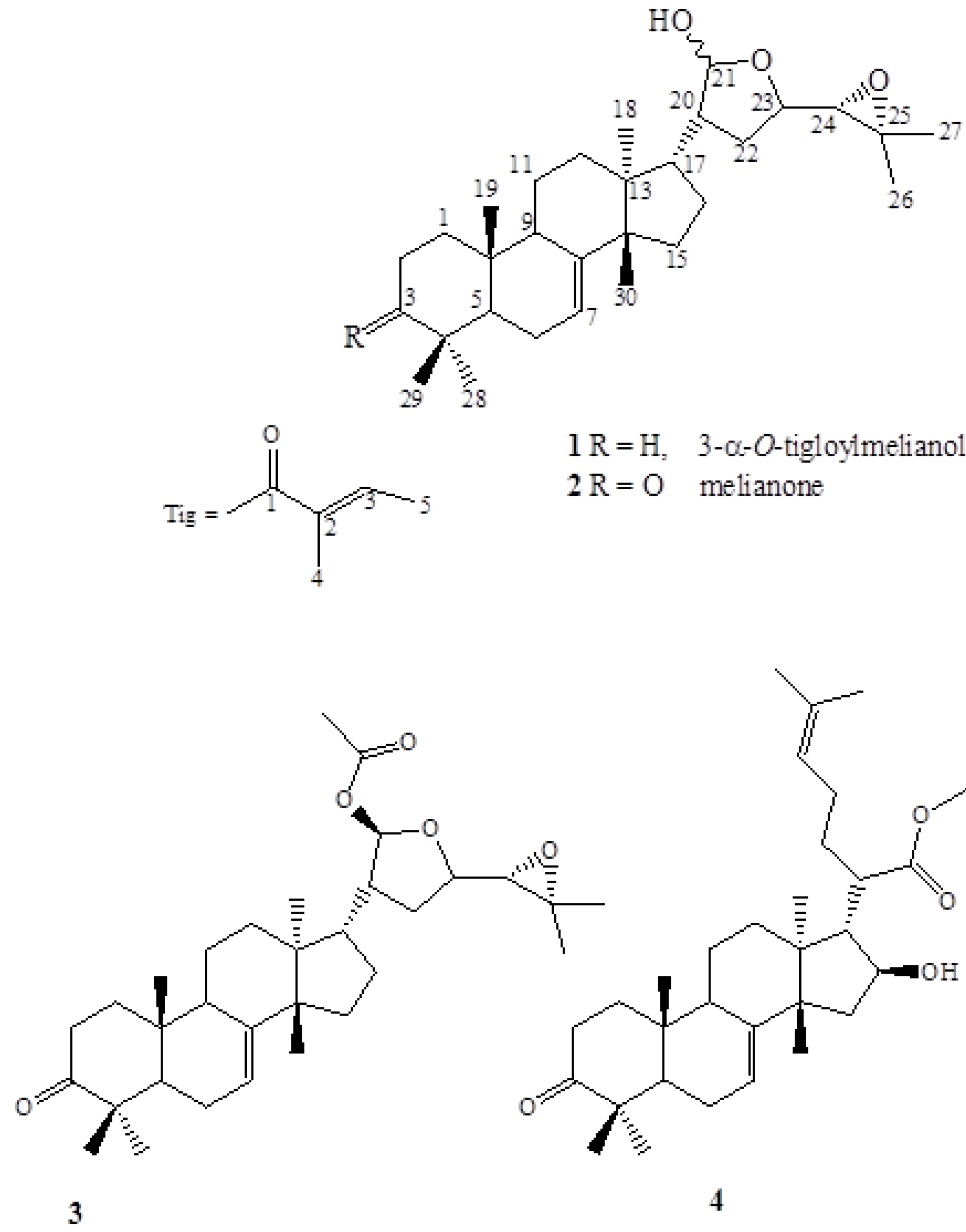
| Compound | Yield mg/kga (w/w) |
|---|---|
| Melianone | 130 |
| 21-β-Acetoxymelianone | 16 |
| Methyl kulonate | 35 |
| 3-α-Tigloylmelianol | 78 |

| Position | 1 | 2 | |||
|---|---|---|---|---|---|
| δH | δC | δH | δC | ||
| 1a | 1.39 (m) | 32.1 | 38.5 | ||
| 1b | 167 (m) | ||||
| 2 | 1.49 (m) | 27.5 | 2.24 ddd (14.3, 14, 6.4) | 35.1 | |
| 1.85 (m) | 2.75 ddd (14.3, 3.6, 3.4) | ||||
| 3 | 4.68 (s br) | 78.3 | 216.8; 216.7 | ||
| 4 | 36.9 | 47.9 | |||
| 5 | 1.79 (m) | 46.1 | 1.82 (m) | 52.5; 52.4 | |
| 6 | 1.73 (m) | 22.9 | 1.70 (m) | 23.3 | |
| 7 | 5.23 (d, 2.4) | 118.2; 118.1 | 5.28 br | 118.2; 118.1 | |
| 8 | 146.0; 145.9 | 145.8; 145.6 | |||
| 9 | 2.30 (t, 7.6); 2.2 (t, 7.6) | 49.7; 48.8 | 49.6; 48.4 | ||
| 10 | 34.9 | 34.9 | |||
| 11 | 1.54 (m) | 17.4 | 17.8 | ||
| 12a | 1.53 (m) | 35.3 | 35.2 | ||
| 12b | 1.98 (m) | ||||
| 13 | 43.8; 43.6 | 43.8; 43.6 | |||
| 4 | 50.9; 50.4 | 50.8; 50.5 | |||
| 15 | 1.55 (m) | 34.2 | 34.3 | ||
| 16 | 1.61 (m) | 27.5; 27.1 | 27.5; 27.3 | ||
| 17 | 2.01 (m); 2.04 (m) | 47.1; 45.2 | 2.03 (m); 2.06 (m) | 47.1; 45.2 | |
| 18 | 0.90 (s) | 23.9 | 0.80 (s); 085 (s) | 24.6 | |
| 19 | 0.76 (s) | 13.0 | 12.8 | ||
| 20 | 1.71 (m) | 33.8; 31.7 | 33.8; 31.7 | ||
| 21 | 5.33 (d, 2.8); 5.29 (d,2.8) | 101.8; 97.8 | 101.8; 97.8 | ||
| 22 | 1.98 (m) | 31.5; 31.3 | 31.5; 31.3 | ||
| 23 | 3.89 (m); 3.83 (m) | 78.5; 77.0 | 3.88 (m); 3.84 (m) | 78.5; 77.1 | |
| 24 | 2.82 (d, 7.6); 2.67(d,7.6) | 67.8; 65.3 | 2.81 (d, 7.6); 2.67 (d, 7.6) | 67.8; 65.4 | |
| 25 | 58.0; 57.2 | 58.0; 57.2 | |||
| 26a) | 1.29 (s); 1.28 (s) | 25.0; 24.9 | 1.29(s); 1.28 (s) | 25.0; 24.9 | |
| 27a) | 1.27 (s) | 19.4; 19.2 | 1.26 (s) | 19.5; 19.2 | |
| 28 | 0.81 (s) | 27.6 | 0.97 (s) | 24.4 | |
| 29 | 0.95 (s) | 21.4 | 0.98 (s) | 21.6 | |
| 30 | 0.98 (s); 0.96 (s) | 22.6; 22.3 | 1.00 (s); 1.07 (s) | 22.6 | |
| 1′ | 167.5 | ||||
| 2′ | 129.2 | ||||
| 3′ | 6.80 (qq, 6.8, 1.6) | 136.6 | |||
| 4′ | 1.75 (d, 6.8) | 14.4 | |||
| 5′ | 1.81 (s) | 12.2 | |||
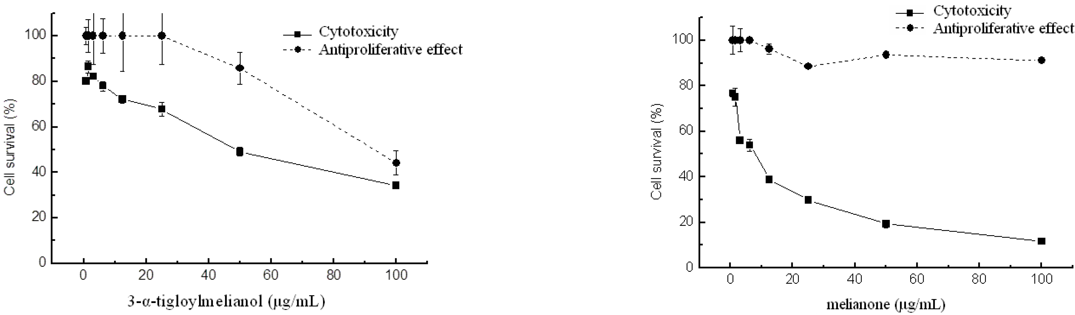
2.2. Cytotoxic and antiproliferative activities

2.3. Nematicidal activity
3. Experimental
3.1. General
3.2. Plant material
3.3. Extraction and isolation
3.4. Spectroscopic data
3.5. Test for in vitro cytotoxic activity
3.6. Test for in vitro antiproliferative activity
3.7. Second stage juveniles (J2) paralysis bioassays
3.8. Statistical analysis
4. Conclusions
Acknowledgements
References
- Isman, M.B. Botanical insecticides, deterrents, and repellents in modern agriculture and an increasingly regulated world. Annu. Rev. Entomol. 2006, 51, 45–66. [Google Scholar]
- Akhtar, Y.; Yeoung, Y.R.; Isman, M.B. Comparative bioactivity of selected extracts from Meliaceae and some commercial botanical insecticides against two noctuid caterpillars, Trichoplusia ni and Pseudaletia unipuncta. Phytochem. Rev. 2008, 7, 77–88. [Google Scholar]
- Carpinella, C.; Ferrayoli, C.; Valladares, G.; Defago, M.; Palacios, S. Potent limonoid insect antifeedant from Melia azedarach. Biosci. Biotechnol. Biochem. 2002, 66, 1731–1736. [Google Scholar]
- Coria, C.; Almiron, W.; Valladares, G.; Carpinella, C.; Ludueña, F.; Defago, M.; Palacios, S. Larvicide and oviposition deterrent effects of fruit and leaf extracts from Melia azedarach L. on Aedes aegypti (L.) (Diptera: Culicidae). Bioresource Technol. 2008, 99, 3066–3070. [Google Scholar] [CrossRef]
- Kamaraj, C.; Rahuman, A.A.; Bagavan, A.; Mohamed, M.J.; Elango, G.; Rajakumar, G.; Zahir, A.A.; Santhoshkumar, T.; Marimuthu, S. Unit Ovicidal and larvicidal activity of crude extracts of Melia azedarach against Haemonchus contortus (Strongylida). Parasitol. Res. 2010, 106, 1071–1077. [Google Scholar] [CrossRef]
- Carpinella, M.C.; Giorda, L.M.; Ferrayoli, C.G.; Palacios, S.M. Antifungal effects of different organic extracts from Melia azedarach L. on phytopathogenic fungi and their isolated active components. J. Agric. Food Chem. 2003, 51, 2506–2511. [Google Scholar]
- Carpinella, M.C.; Ferrayoli, C.G.; Palacios, S.M. Antifungal synergistic effect of scopoletin, a hydroxycoumarin isolated from Melia azedarach L. fruits. J. Agric. Food Chem. 2005, 53, 2922–2927. [Google Scholar]
- Ntalli, N.G.; Menkisoglu-Spiroudi, U.; Giannakou, I. Nematicidal activity of powder and extracts of Melia azedarach fruits against Meloidogyne incognita. Ann. Appl. Biol. 2010, 156, 309–317. [Google Scholar] [CrossRef]
- Vishnukanta, A.C.R. Melia azedarach: A phytopharmacological review. Pharmacogn. Rev. 2008, 2, 173–179. [Google Scholar]
- Bueno, C.A.; Alché, L.A.; Barquero, A.A. 1-Cinnamoyl-3,11-dihydroxymeliacarpinin delays glycoprotein transport restraining virus multiplication without cytotoxicity. Biochem. Biophys. Res. Commun. 2010, 393, 32–37. [Google Scholar] [CrossRef]
- Wu, S.B.; Ji, Y.P.; Zhu, J.J.; Zhao, Y.; Xia, G.; Hu, Y.H.; Hu, J.F. Steroids from the leaves of Chinese Melia azedarach and their cytotoxic effects on human cancer cell lines. Steroids 2009, 74, 761–765. [Google Scholar] [CrossRef]
- Mazumder, M.E.H.; Rahman, S. Pharmacological evaluation of Bangladeshi medicinal plants for antioxidant activity. Pharm. Biol. 2008, 46, 704–709. [Google Scholar] [CrossRef]
- Roy, A.; Saraf, S. Limonoids: Overview of significant bioactive triterpenes distributed in plants kingdom. Biol. Pharm. Bull. 2006, 29, 191–201. [Google Scholar] [CrossRef]
- Morgan, E.D. Azadirachtin, a scientific gold mine. Bioorg. Med. Chem. 2009, 17, 4096–4105. [Google Scholar] [CrossRef]
- Patil, B.S.; Jayaprakasha, G.K.; Murthy, K.N.C.; Vikram, A. Bioactive compounds: Historical perspectives, opportunities, and challenges. J. Agric. Food Chem. 2009, 57, 8142–8160. [Google Scholar]
- Zhang, B.; Wang, Z.F.; Tang, M.Z.; Shi, Y.L. Growth inhibition and apoptosis-induced effect on human cancer cells of toosendanin, a triterpenoid derivative from Chinese traditional medicine. Invest. New Drug. 2005, 23, 547–553. [Google Scholar] [CrossRef]
- Muregi, F.W.; Chhabra, S.C.; Njagi, E.N.M.; Lang'at-Thoruwa, C.C.; Njue, W.M.; Orago, A.S.S.; Omar, S.A.; Ndiege, I.O. Anti-plasmodial activity of some Kenyan medicinal plant extracts singly and in combination with chloroquine. Phytother. Res. 2004, 18, 379–384. [Google Scholar] [CrossRef]
- Madibela, O.R.; Kelemogile, K.M. Exposure of Melia azedarach fruits to Eimeria lowers oocyst output in yearling Tswana goats. Small Ruminant Res. 2008, 76, 207–210. [Google Scholar] [CrossRef]
- Khanavi, M.; Safavi, M.; Siavoshi, F.; Fallah Tafti, A.; Haji Mahmoodi, M.; Haji Akhoondi, A.; Rezazadeh Sh, R.; Foroumadi, A. Evaluation of anti Helicobacter pylori activity of methanol extracts of some species of Stachys and Melia. J. Med. Plants 2008, 7, 74–80. [Google Scholar]
- Peveling, R.; Ely, S.O. Side-effects of botanical insecticides derived from Meliaceae on coccinellid predators of the date palm scale. Crop Prot. 2006, 25, 1253–1258. [Google Scholar] [CrossRef]
- Matter, M.M.; Gesraha, M.A.; Ahmed, A.A.I.; Farag, N.A. Impact of neem and chinaberry fruit extracts on the pest/parasitoid (Pieris rapae/Hyposoter ebeninus) interactions. Anzeiger für Schädlingskunde 2002, 75, 13–18. [Google Scholar]
- Caboni, P.; Sarais, G.; Angioni, A.; Garcia, A.J.; Lai, F.; Dedola, F.; Cabras, P. Residues and persistence of neem formulations on strawberry after field treatment. J. Agric. Food Chem. 2006, 54, 10026–10032. [Google Scholar] [CrossRef]
- Sarais, G.; Angioni, A.; Lai, F.; Cabras, P.; Caboni, P. Persistence of two neem formulations on peach leaves and fruit: effect of the distribution. J. Agric. Food Chem. 2009, 57, 2457–2461. [Google Scholar]
- Phua, D.H.; Tsai, W.J.; Ger, J.; Deng, J.F.; Yang, C.C. Human Melia azedarach poisoning. Clin. Toxicol. 2008, 46, 1067–1070. [Google Scholar] [CrossRef]
- Nakanishi, T.; Inada, A.; Lavie, D. A new tirucallane-type triterpenoid derivative, lipomelianol from fruits of Melia toosendan SIEB. Et ZUCC. Chem. Pharm. Bull. 1986, 34, 100–104. [Google Scholar] [CrossRef]
- Joseph-Nathan, P.; Wesener, J.R.; Günther, H. A two-dimensional NMR study of angelic and tiglic acid. Magn. Res. Chem. 1984, 23, 190–191. [Google Scholar]
- Su, R.; Kim, M.; Kawaguchi, H.; Yamamoto, T.; Goto, K.; Taga, T.; Miwa, Y.; Kozuka, M.; Takahashi, S. triterpenoids from the fruits of Phellodendron chinense SCHNEID.: the stereostructure of niloticin. Chem. Pharm. Bull. 1990, 38, 1616–1619. [Google Scholar]
- Chiang, C.; Chang, F.C. Tetracyclic triterpenoids from Melia azedarach, L.-III. Tetrahedron 1973, 29, 1911–1929. [Google Scholar] [CrossRef]
- Itokawa, H.; Kishi, E.; Morita, H.; Takeya, K. Cytotoxic quassinoids and tirucallane-type triterpenes from woods of Eurycoma longifolia. Chem Pharm. Bull. 1992, 40, 1053–1055. [Google Scholar] [CrossRef]
- Zhou, H.; Hamazaki, A.; Fontana, J.D.; Takahashi, H.; Wandscheer, C.B.; Fukuyama, Y. Cytotoxic limonoids from Brazilian Melia azedarach. Chem. Pharm. Bull. 2005, 53, 1362–1365. [Google Scholar] [CrossRef]
- Zhou, H.; Hamazaki, A.; Fontana, J.D.; Takahashi, H.; Esumi, T.; Wandscheer, C.B.; Tsujimoto, H.; Fukuyama, Y. New ring C-seco limonoids from Brazilian Melia azedarach and their cytotoxic activity. J. Nat. Prod. 2004, 67, 1544–1547. [Google Scholar] [CrossRef]
- Tada, K.; Takido, M.; Kitanaka, S. Limonoids from fruit of Melia toosendan and their cytotoxic activity. Phytochemistry 1999, 51, 787–791. [Google Scholar]
- Pettit, G.R.; Numata, A.; Iwamoto, C.; Morito, H.; Yamada, T.; Goswami, A.; Clewlow, P.J.; Cragg, G.M.; Schmidt, J.M. Antineoplastic agents. 489. Isolation and structures of meliastatins 1-5 and related euphane triterpenes from the tree Melia dubia. J. Νat. Prod. 2002, 65, 1886–1891. [Google Scholar]
- Priyadarsini, R.V.; Murugan, R.S.; Sripriya, P.; Karunagaran, D.; Nagini, S. The neem limonoids azadirachtin and nimbolide induce cell cycle arrest and mitochondria-mediated apoptosis in human cervical cancer (HeLa) cells. Free Radical Res. 2010, 44, 624–634. [Google Scholar] [CrossRef]
- Biavatti, M.W.; Vieira, P.C.; da Silva, M.F.G.; Fernandes, J.B.; Albuquerque, S.; Magalhaes, C.M.I.; Pagnocca, F.C. Chemistry and bioactivity of Raulinoa echinata Cowan, an endemis Brazilian Rutaceae species. Phytomedicine 2001, 8, 121–124. [Google Scholar] [CrossRef]
- Kim, B.; Kim, H.; Choi, J.W.; Lee, C.K. The effects of meliae toosendan fructus on liver function. III. Effects of melianone and 28-deacetyl sendanin on drug metabolism and bile juice secretion. Korean J. Pharmacogn. 1996, 27, 47–52. [Google Scholar]
- Ntalli, N.G.; Menkissoglu-Spiroudi, U.; Giannakou, I.O.; Prophetou-Athanasiadou, D.A. Efficacy evaluation of a neem (Azadirachta indica A. Juss) formulation against root-knot nematodes Meloidogyne incognita. Crop Prot. 2009, 28, 489–494. [Google Scholar] [CrossRef]
- Denizot, F.; Lang, R. Rapid colorimetric assay for cell growth and survival. Modifications to the tetrazolium dye procedure giving improved sensitivity and reliability. J. Immunol. Methods 1986, 89, 271–277. [Google Scholar] [CrossRef]
- Hussey, R.S.; Barker, K.R. A comparison of methods of collecting inocula of Meloidogyne spp. including a new technique. Plant Dis. Rep. 1973, 57, 1025–1028. [Google Scholar]
- Puntener, W. Manual for Field Trials in Plant Protection, 2nd ed; Ciba Geigy Limited: Basel, Switzerland, 1981; p. 205. [Google Scholar]
- Sample Availability: Samples of the compounds are available from the authors.
© 2010 by the authors; licensee MDPI, Basel, Switzerland. This article is an open access article distributed under the terms and conditions of the Creative Commons Attribution license (http://creativecommons.org/licenses/by/3.0/).
Share and Cite
Ntalli, N.G.; Cottiglia, F.; Bueno, C.A.; Alché, L.E.; Leonti, M.; Vargiu, S.; Bifulco, E.; Menkissoglu-Spiroudi, U.; Caboni, P. Cytotoxic Tirucallane Triterpenoids from Melia azedarach Fruits. Molecules 2010, 15, 5866-5877. https://doi.org/10.3390/molecules15095866
Ntalli NG, Cottiglia F, Bueno CA, Alché LE, Leonti M, Vargiu S, Bifulco E, Menkissoglu-Spiroudi U, Caboni P. Cytotoxic Tirucallane Triterpenoids from Melia azedarach Fruits. Molecules. 2010; 15(9):5866-5877. https://doi.org/10.3390/molecules15095866
Chicago/Turabian StyleNtalli, Nikoletta G., Filippo Cottiglia, Carlos A. Bueno, Laura E. Alché, Marco Leonti, Simona Vargiu, Ersilia Bifulco, Urania Menkissoglu-Spiroudi, and Pierluigi Caboni. 2010. "Cytotoxic Tirucallane Triterpenoids from Melia azedarach Fruits" Molecules 15, no. 9: 5866-5877. https://doi.org/10.3390/molecules15095866