Article

Phenolic Compounds and Antioxidant Activities of Liriope muscari

1
National Institutes for Food and Drug Control, Beijing 100050, China
2
State Key Laboratory of Earth Surface Processes and Resource Ecology, Beijing Normal University, Beijing 100875, China
3
Department of Entomology, China Agricultural University, Beijing 100193, China
*
Authors to whom correspondence should be addressed.
Molecules 2012, 17(2), 1797-1808; https://doi.org/10.3390/molecules17021797
Submission received: 24 November 2011 / Revised: 24 January 2012 / Accepted: 31 January 2012 / Published: 10 February 2012

Abstract

:
Five phenolic compounds, namely N-trans-coumaroyltyramine (1), N-trans-feruloyltyramine (2), N-trans-feruloyloctopamine (3), 5,7-dihydroxy-8-methoxyflavone (4) and (3S)3,5,4′-trihydroxy-7-methoxy-6-methylhomoisoflavanone (5), were isolated from the fibrous roots of Liriope muscari (Liliaceae). Compounds 25 were isolated for the first time from the Liriope genus. Their in vitro antioxidant activities were assessed by the DPPH and ABTS scavenging methods with microplate assays. The structure-activity relationships of compounds 13 are discussed.

1. Introduction

Antioxidant activity usually means the ability of a compound to delay, inhibit, or prevent the oxidation of oxidizable materials by scavenging free radicals and reducing oxidative stress [1]. Antioxidants can scavenge ROS (reactive oxygen species) to protect the cells from damage caused by the latter. At present, the most commonly used antioxidants include vitamin C (VC), vitamin E, butylated hydroxyanisole (BHA), butylated hydroxytoluene (BHT), propyl gallate and tert-butyl hydroquinone. However, some chemically synthesized antioxidants like BHA and BHT are now being restricted by legislation because of doubts over their possible toxic and carcinogenic effects [2]. Therefore, there is a growing interest in finding antioxidants from natural sources [3,4]. Assays based on the use of 1,1-diphenyl-2-picryl-hydrazyl (DPPH) and 2,2′-azino-bis(3-ethylbenzthiazoline-6-sulfonic acid) (ABTS) radicals are among the most commonly used spectrophotometric methods for determination of the antioxidant capacity of foods, beverages, plant extracts and pure compounds due to the simple, rapid, sensitive, and reproducible procedures involved [5,6]. Phenolic compounds usually possess different antioxidant activity potentials because of their phenolic hydroxy groups which can act as a hydrogen or electron donor [7]. Phenolic acids, flavonoids and tannins are well-known potential natural antioxidants [1]. The hunt for effective and safe antioxidants from natural products is considered to be a shortcut [8].
Liriope muscari (Decne.) Bailey (Liliaceae) is locally known in China as duantingshanmaidong. In China, the roots of this species are used locally as a substitute for Radix Ophiopogonis (maidong in Chinese) [9], especially in Fujian Province. Maidong is a traditional herbal medicine widely used in China as a tonic agent. Modern pharmacological investigations suggest that maidong also has an positive effect on various inflammation-related diseases [10]. Previous studies indicated that the main components in L. muscari include polysaccharides and steroidal glycosides [11,12,13]. In this paper, five phenolic compounds (Figure 1), including three amides [N-trans-coumaroyltyramine (1), N-trans-feruloyltyramine (2) and N-trans-feruloyloctopamine (3)], one flavone [5,7-dihydroxy-8-methoxy-flavone (4)] and one homoisoflavanone [(3S)-3,5,4′-trihydroxy-7-methoxy-6-methylhomoiso-flavonone (5)] were isolated from L. muscari. Compounds 25 were isolated for the first time from the Liriope genus.
Figure 1. Structures of compounds isolated from L. muscari.
Molecules 17 01797 g001
To our knowledge, this is the first time that the phenolic components of L. muscari have been studied. Their in vitro antioxidant activities were assessed by the DPPH and ABTS scavenging method. N-trans-feruloyltyramine (IC50 28.7, 8.2 μg/mL) and N-trans-feruloyloctopamine (IC50 14.4, 7.6 μg/mL) showed potential antioxidant activities.

2. Results and Discussion

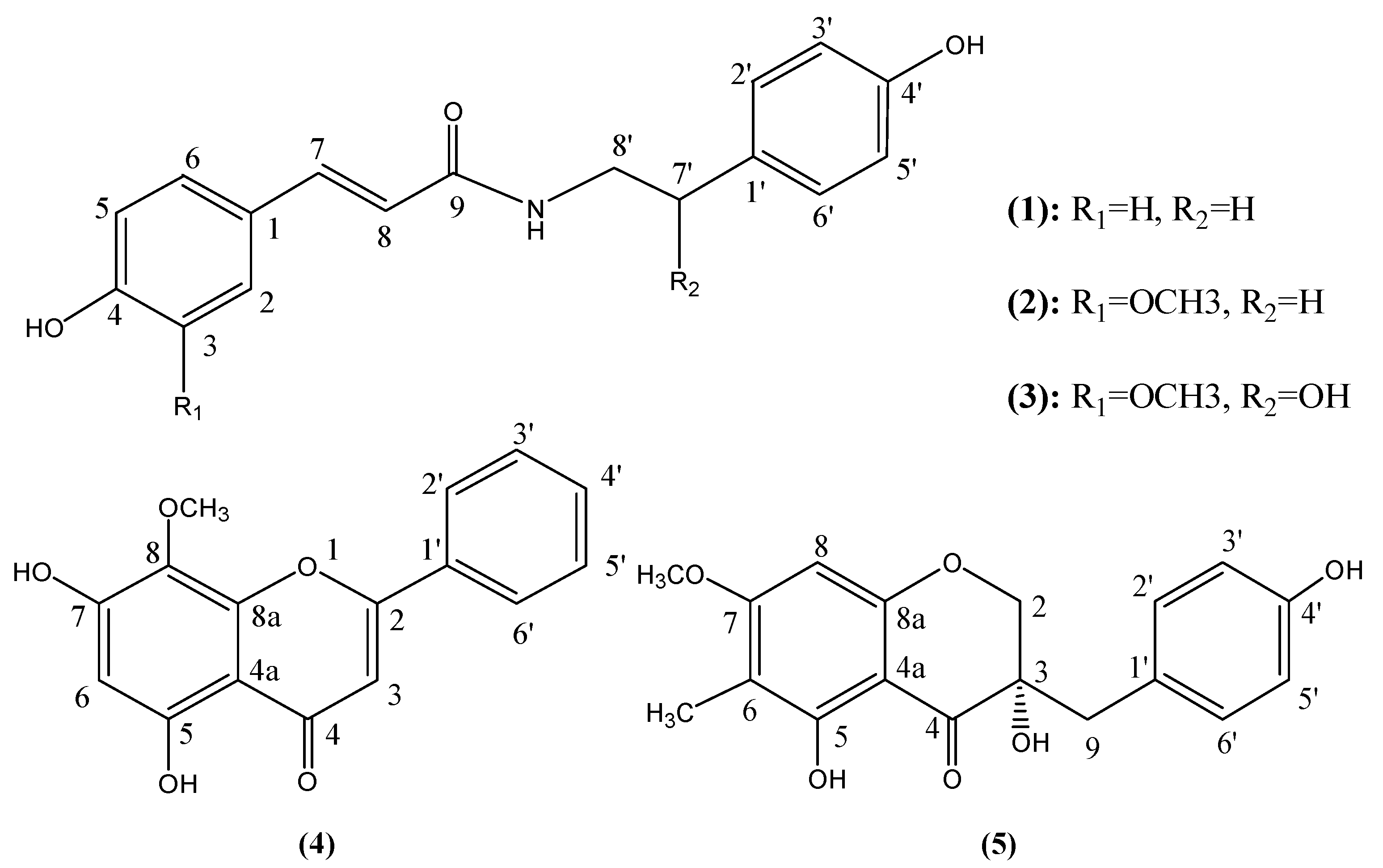
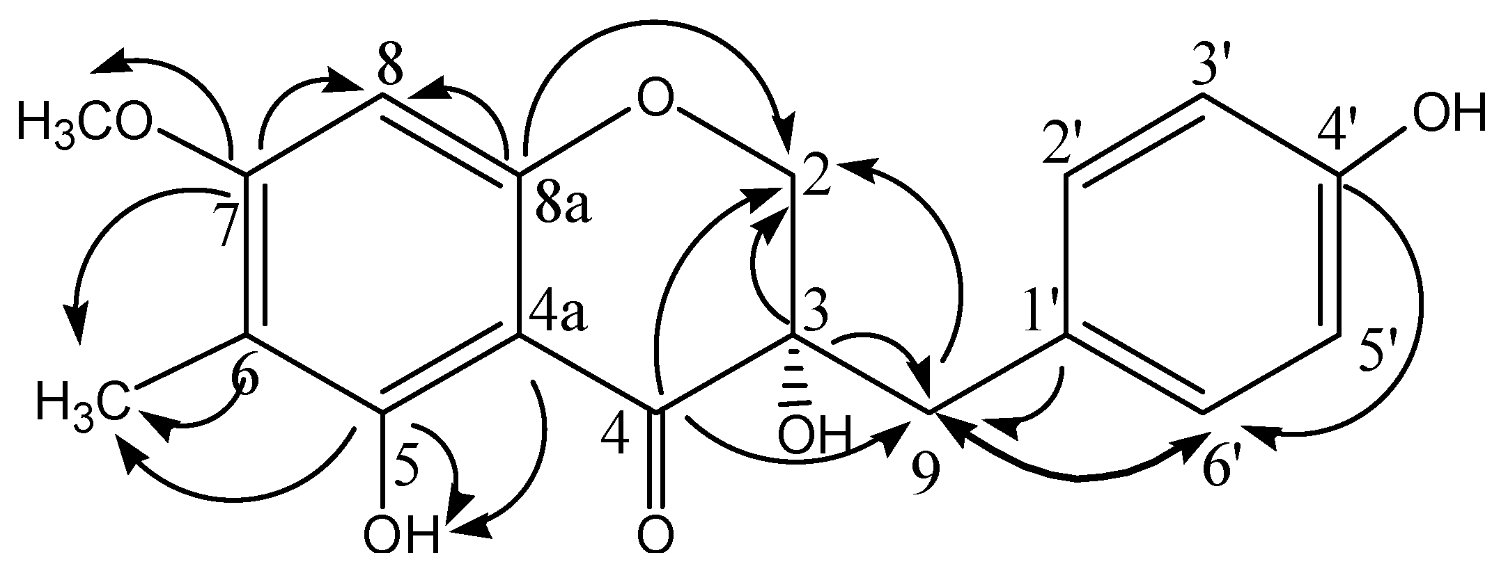
2.1. Isolation and Characterization of Compounds 15

The compounds were isolated using silica gel and Sephadex LH-20 gel column chromatography from 80% ethanol extract of L. muscari. The structures of compounds 14 were characterized by examination of their ESI-MS, NMR (1H- and 13C-) data and comparison with literature reports.
Compound 5 was first isolated in 1985 from Ophiopogonis [14] (Liliaceae), a closely linked genus that can be easily confused with Liriope. In the original paper, the planar structure of compound 5 was identified by comparing the 1H-NMR data with that of (3S)-3,5,7-trihydroxy-4′-methoxy homoisoflavonone (eucomol, a typical homoisoflavonone isolated from Eucomis bicolor BAK. (Liliaceae) [15,16]). In our study, the structure was further confirmed using 13C-NMR, APT and 2D-NMR techniques, including 1H-1HCOSY, HSQC, HMBC (Figure 2). The configuration at C-3 was determined to be (S), similar to that of eucomol, based on the positive sign of its specific rotation [16,17]. For a long time, it was believed that there were no homoisoflavones in Liriope, so this is the first time a homoisoflavone has been isolated from the Liriope genus.
Figure 2. Key HMBC correlations of compound 5.
Molecules 17 01797 g002

2.2. In Vitro Antioxidant Activity

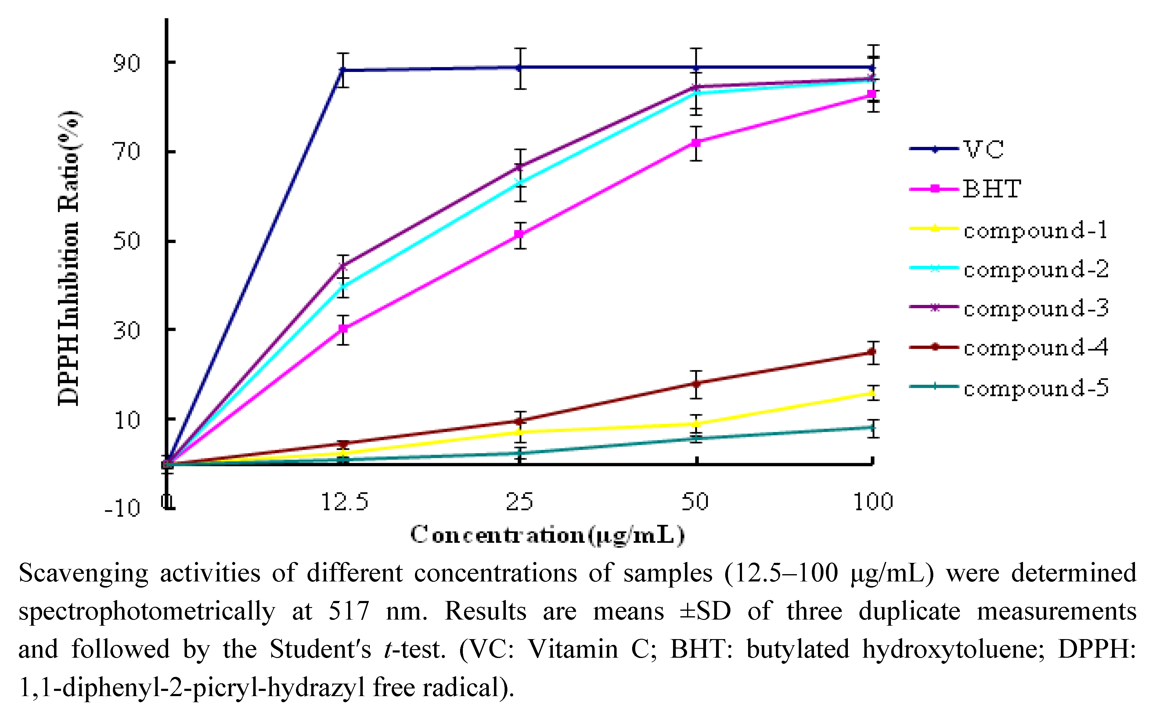
2.2.1. DPPH Scavenging Activity
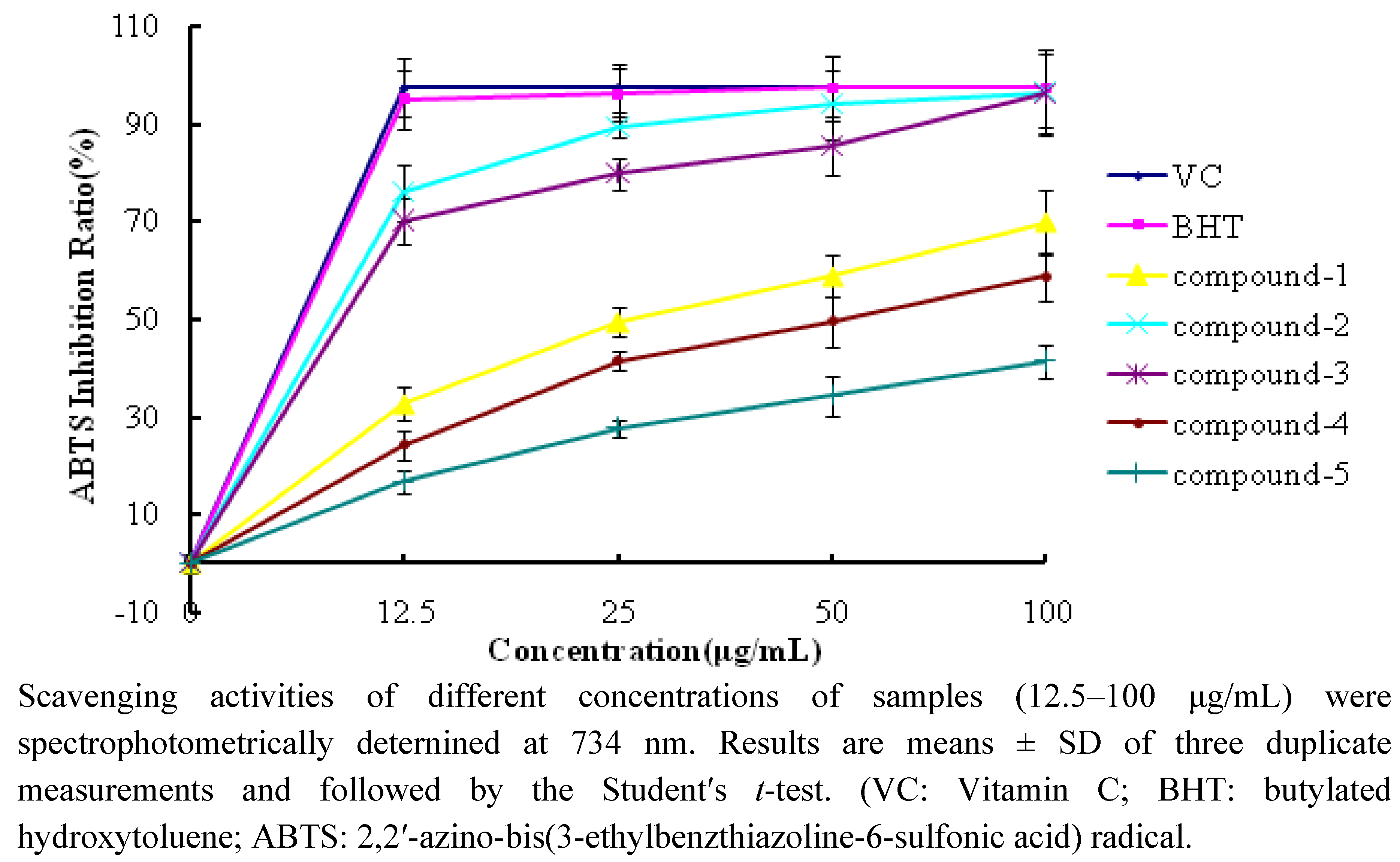
The 1,1-diphenyl-2-picrylhydrazyl radical (DPPH), which possesses an unpaired electron and exhibits a stable violet color in methanol solution (peak absorbance at 517 nm), is commonly used as a reagent for evaluation of the free radical scavenging activity of antioxidants [18]. The DPPH assay is based on the reduction of DPPH in methanol solution in the presence of a hydrogen-donating antioxidant due to the formation of the non-radical form (DPPH-H) in the reaction [19].
Figure 3 shows the DPPH scavenging activities of compounds 15 and reference antioxidants at different concentrations (12.5–100 μg/mL). The test compounds exhibited different DPPH scavenging activities in a concentration-dependent manner. The scavenging effects of compounds 15 and reference antioxidants on DPPH decreased in the following order: VC > compound 3 > compound 2 > BHT > compound 4 > compound 1 > compound 5. The inhibition ratios at a concentration of 25 μg/mL are listed in Table 1. Compounds 2 and 3 exhibited effective radical scavenging activity while compounds 1, 4, 5 showed very weak activity.
Figure 3. DPPHscavenging activity of compounds 15 and reference antioxidants.
Molecules 17 01797 g003
Table 1. The DPPH and ABTS inhibition ratio at the concentration of 25 μg/mL.
DPPH Inhibition Ratio (%)ABTS Inhibition Ratio (%)
Compound 17.2 ± 0.632.7 ± 1.6
Compound 263.2 ± 3.675.9 ± 2.0
Compound 366.6 ± 2.370.0 ± 3.2
Compound 49.6 ± 1.224.3 ± 1.9
Compound 52.6 ± 0.516.8 ± 1.7
VC88.9 ± 4.597.4 ± 5.0
BHT51.5 ± 3.195.1 ± 5.3

2.2.2. ABTS Scavenging Activity

In this assay, the 2,2′-azino-bis(3-ethylbenzthiazoline-6-sulfonic acid) (ABTS) radical, which has a peak absorbance at 734 nm, should be preformed by mixing ABTS and potassium persulfate (K2S2O8). When antioxidants were added, the ABTS radical, which has a blue-green color, is reduced to ABTS (no color). Different decoloration abilities indicate different ABTS scavenging activities [20].
Figure 4 shows the ABTS scavenging abilities of compounds 15 and reference standards. The test compounds also exhibited different radical scavenging activities in a concentration-dependent manner like in the DPPH assay. The scavenging effects of compounds 15 and reference antioxidants on ABTS·+ decreased in the following order: VC ≈ BHT > compound 2 > compound 3 > compound 1 > compound 4 > compound 5. The inhibition ratios at concentration of 25 μg/mL are listed in Table 1. Compounds 2 and 3 exhibited effective radical scavenging activity, while compounds 1, 4 and 5 showed relatively weak activity.
Figure 4. ABTS scavenging activity of compounds 15 and reference antioxidants.
Molecules 17 01797 g004
In both methods, compounds 2 [21] and 3 [22] showed potential activity, while compounds 1 [23,24] and 4 [25] showed very weak antioxidant activity, which is consistent with the reported results. Furthermore, for compound 4, quantitative structure-activity relationship analysis [26] also suggested it would not show effective activity because of the absence of 3-OH and o-dihydroxy structure in the B ring, and less free -OH groups (only two) in the structure, which are required for high antioxidant activity. As for compound 5, its antioxidant activity has been previously evaluated using an on-line HPLC-DAD-CL method based on hydrogen peroxide elimination [27]. However, the results cannot be compared with our data due to the absence of any mention of the concentration used in that study.
It is interesting to investigate the structure-activity relationship for compounds 13. These compounds have similar structures, but very different activities. Comparing their structures (Figure 1), the main differences are the substituents at C-3 (R1) and C-7′ (R2). Comparing the structures and activities of compounds 2 and 3 (p > 0.05, 25 μg/mL) it is inferred that the presence of methyl group at C-7′ seems to have some, but little influence on antioxidant activity. By comparing compounds 1 and 2 (p < 0.05, 25 μg/mL), it is inferred that the presence of methyl group at C-3 is the key factor that affects the activities, therefore, a radical scavenging activity mechanism represented by the reaction shown in Figure 5 is proposed, using DPPH as an example. Compounds 2 and 3 have two resonance structures A and B stabilizing the product, thereby exhibiting superior antioxidant activity than compound 1. As HPLC evaluation (Experimental section) indicates a relative purity between 90.1% and 96.7% for compounds 13, potential synergistic effects from minor impurities could also contribute to the observed activity.
Figure 5. The proposed reaction mechanism between DPPH· and compounds 13.
Molecules 17 01797 g005

3. Experimental

3.1. General

1H- and 13C-NMR spectra were recorded on Bruker Avance DRX 500 instrument using DMSO-d6 or CDCl3 as solvent with TMS as internal standard. An Agilent 6320 Ion TRAP LC/MS was employed for MS analysis. The specific rotation was recorded on AUTOPOL IV Automatic Polarimeter (Rudolph, Hackettstown, NJ, USA). For the microplate assay, a SpectraMax 190 Absorbance Microplate Reader (Molecular Devices, Sunnyvale, CA, USA) and 96 Well Cell Culture Cluster (Costar, Corning, NY, USA) were used. 1,1-diphenyl-2-picrylhydrazyl (DPPH) and 2,2′-azino-bis(3-ethylbenzthiazoline-6-sulfonic acid) (ABTS) were purchased from Sigma (Sigma-Aldrich GmbH, Stenheim, Germany). Sephadex LH-20 was purchased from Amersham Pharmacia Biotech AB (Uppsala, Sweden). Polyamide resin (100–200 mesh) was purchased from BeiJingZhongXiYuanDa Technical Co. Ltd. (Beijing, China). Silica gel (160–200 mesh, 200–300 mesh) for column chromatography was purchased from Qingdao Marine Chemical Plant (Shandong Province, China). All other chemicals were of analytical reagent grade and used without any further purification.

3.2. Plant Material

Fresh fibrous roots of L. muscari were collected from Quanzhou City, Fujian Province, China, in May 2010. The species was identified by Dr. Zhang. J. (National Institutes for Food and Drug Control, NIFDC for short). The voucher specimens were deposited at the herbarium of NIFDC. The roots were air-dried and ground to a powder using a grinding mill (Retsch Muhle, Haan, Germany).

3.3. Compound Isolation

In this part, there were mainly two procedures including enrichment and isolation. For the enrichment of phenolic compounds, polyamide resin was used. Polyamide is a commonly used stationary phase for the isolation of phenolic compounds. The enriching mechanism is based on the adsorption power from hydrogen bonds between carbonyl groups of the polyamide and the phenolic hydroxyl groups of target compounds or the polyamide amides and carbonyl groups of fatty acids, etc., The strength of adsorption is dependent on the number of phenolic hydroxyls exposed and their position in the molecule [28] and the power is strongest in water, while getting weaker when the ethanol concentration in the mobile phase is increased.
The detailed procedures are as follows: the powder (2 kg) was extracted three times with 80% hot ethanol (1 L), for 1 h each time. The extracts were concentrated to afford a syrup (1 kg), which was dissolved in 10% ethanol (4 L). Polyamide (1 kg) was added into the solution and stirred about 1 h to make sure the phenolic compounds were adsorbed on the polyamide to some extent. Then the polyamide was centrifuged to dryness (1,000× g, 10 min). Fresh water was used to rinse the polyamide several times till the water was nearly colorless. Then 95% ethanol was used to rinse the polyamide and the solution was collected. The ethanol solution was evaporated to dryness under reduced pressure to afford a solid residue (30 g). The solid residue was chromatographed over a silica gel (160–200 mesh) column (45 × 6.0 cm i.d.) with CHCl3/MeOH (20:1 to 8:1) to afford 30 fractions (F01–F30). Fraction F03 (2.2 g) was subjected to Sephadex LH-20 column (120 × 2.5 cm i.d.) chromatography with CHCl3/MeOH (10:1) to afford 11 subfractions (F0301–F0311). Then fraction F0308 (69 mg) was chromatographed over a silica gel column (200–300 mesh, 30 × 2.0 cm i.d.) with petroleum/EtOAc (P/E 6:1 to 3:1) to afford compound 4 (10 mg, crystal, P/E 6:1) and compound 5 (8 mg, crystal, P/E 3:1). The purities were 96.3% and 97.2%, respectively (HPLC, 254 nm with PDA detector). As for the isolation of compounds 13, fractions F10–F11 (500 mg), fractions F06-F08 (800 mg) and fractions F12–F14 (500 mg) were separated on a Sephadex LH-20 gel column (120 × 2.0 cm i.d.) with MeOH to afford 11 subfractions (F1001–F1011), 19 subfractions (F0601–F0619), and 17 subfractions (F1201–F1217), respectively. Then subfractions F1006 (40 mg), F0609 (100 mg), F1205 (120 mg) were chromatographed over a silica gel column (200–300 mesh, 30 × 2.0 cm i.d.) with P/E (1:1), P/E (3:2 to 1:1), P/E (1:1 to 1:2) to afford compounds 1 (10 mg), 2 (8 mg) and 3 (15 mg). The purity of compounds 1–3 was 96.6%, 90.1% and 91.1%, respectively (HPLC, 254 nm with PDA detector).
N-trans-coumaroyltyramine (1). White powder (petroleum/acetic ether). Rf 0.65 (acetic ether) ESI-MS: m/z 284 [M+H]+. C17H17NO3. 1H-NMR (DMSO-d6, 500 MHz) δ: 9.36 (-OH), 8.11 (1H, t, 5.0 Hz, -NH), 7.39 (2H, d, 8.5 Hz, H-2, 6), 7.30 (1H, d, 15.5 Hz, H-7), 7.01 (2H, d, 8.0 Hz, H-2′, 6′), 6.78 (2H, d, 8.5 Hz, H-3, 5), 6.67 (2H, d, 8.0 Hz, H-3′, 5′), 6.38 (1H, 15.5 Hz, H-8), 3.31 (2H, m, H-8′), 2.64 (2H, t, 7.5 Hz, H-7′). The 1H- and 13C-NMR (Table 2) spectral data are consistent with published data [29,30].
N-trans-feruloyltyramine (2). Colourless oil (petroleum/acetic ether). Rf 0.62 (acetic ether) ESI-MS: m/z 314 [M+H]+. C18H19NO4. 1H-NMR (DMSO-d6, 500 MHz) δ: 9.46, 9.21 (C4-OH, C4′-OH), 8.01 (1H, t, 5.0 Hz, -NH), 7.31 (1H, d, 16.0 Hz, H-7), 7.12 (1H, s, H-2), 7.01 (2H, d, 8.0 Hz, H-2′, 6′), 6.98 (1H, m, H-6), 6.78 (1H, d, 8.2 Hz, H-5), 6.68 (2H, d, 8.0 Hz, H-3′, 5′), 6.43 (1H, 15.5 Hz, H-8), 3.80 (3H, s, -OCH3), 3.33 (2H, m, H-8′), 2.64 (2H, t, 7.5 Hz, H-7′). The 1H- and 13C-NMR (Table 2) spectral data are consistent with published data [31,32].
N-trans-feruloyloctopamine (3). Colourless oil (petroleum/acetic ether). Rf 0.47 (acetic ether) ESI-MS: m/z 330 [M+H]+. C18H19NO5. 1H-NMR (DMSO-d6, 500 MHz) δ: 9.47, 9.32 (C4-OH, C4′-OH), 7.96 (1H, t, 5.5 Hz, -NH), 7.31 (1H, d, 15.5 Hz, H-7), 7.15 (2H, d, 8.5 Hz, H-2′, 6′), 7.12 (1H, s, H-2), 6.98 (1H, d, 7.5 Hz, H-6), 6.79 (1H, d, 8.5 Hz, H-5), 6.72 (2H, d, 8.5 Hz, H-3′, 5′), 6.55 (1H, 15.5 Hz, H-8), 4.54 (1H, m, H-7′), 3.80 (3H, s, -OCH3), 3.38, 3.18 (2H, m, H-8′). The 1H- and 13C-NMR (Table 2) spectral data are consistent with published data [32,33].
5,7-Dihydroxy-8-methoxyflavone (4). Yellow needle crystal (petroleum/acetic ether). Rf 0.40 (P/E 2:1) ESI-MS: m/z 283 [M−H]. C16H12O5. 1H-NMR (DMSO-d6, 500 MHz) δ:12.52 (1H, s, 5-OH), 10.87 (1H, s, 7-OH), 8.08 (2H, d, 6.5 Hz, H-2′, 6′), 7.62 (3H, m, H-3′, 4′, 5′), 7.02 (1H, s, H-3), 6.32 (1H, s, H-6), 3.86 (3H, s, -OCH3). 13C-NMR (DMSO-d6, 125 MHz) δ: 182.5 (C-4), 163.5 (C-2), 157.8 (C-7), 156.7 (C-5), 150.1 (C-8a), 132.6 (C-4′), 131.3 (C-1′), 129.8 (C-3′, 5′), 128.2 (C-8), 126.8 (C-2′, 6′), 105.5 (C-3), 104.2 (C-4a), 99.6 (C-6), 61.5 (-OCH3). The 1H- and 13C-NMR spectral data are consistent with published data [34,35].
(3S)-3,5,4′-trihydroxy-7-methoxy-6-methyl homoisoflavonone (5). Colorless needle crystal (CHCl3). Rf 0.36 (P/E=2:1) (c=0.0100, CHCl3) ESI-MS: m/z 329 [M−H]. C18H18O6. 1H-NMR (CDCl3, 500 MHz) δ: 7.09 (2H, d, 8.0 Hz, H-2′, 6′), 6.78 (2H, d, 8.0 Hz, H-3′, 5′), 6.10 (1H, s, H-8), 4.23 (1H, d, 11.0 Hz, H-2a), 4.06 (1H, d, 11.0 Hz, H-2b), 3.91 (3H, S, -OCH3), 2.96 (2H, dd, 14 Hz, 5.0 Hz), 2.06 (3H, s, -CH3). 13C-NMR (CDCl3, 125 MHz) δ: 198.3 (C-4), 166.5 (C-7), 161.1 (C-8a), 160.1 (C-5), 154.9 (C-4′), 131.8 (C-2′,6′), 126.2 (C-1′), 115.3 (C-3′,5′), 106.5 (C-6), 100.2 (C-4a), 91.0 (C-8), 72.3 (C-3), 71.9 (C-2), 56.0 (-OCH3), 40.8 (C-9), 6.9 (-CH3). The 1H-NMR spectral data are consistent with published data [14].
Table 2. 13C-NMR data of compounds 13 (DMSO-d6, 125 MHz).
PositionCompound 1Compound 2Compound 3
1126.3126.9126.9
2129.8111.2111.2
3116.2148.3148.3
4159.2148.7148.7
5116.2116.1116.1
6129.8122.0122.0
7139.3139.3139.4
8118.9119.5119.6
9166.1165.8166.0
1′129.1130.0134.5
2′130.0129.9127.6
3′115.6115.6115.2
4′156.0156.1156.9
5′115.6115.6115.2
6′130.0129.9127.6
7′34.734.971.6
8′41.241.147.5
-OCH3 56.055.9

3.4. Antioxidant Ability

3.4.1. DPPH Assay

In this method, a microplate reader and 96 well plate were used to carry out the determination of the spectral absorption values. This assay is based on the classic method developed by Blois in 1958 [19]. Various forms of this method are widely used [36,37]. Unlike the commonly used methods, which are labor and time-consuming and reagent and sample-wasting, this microplate assay method is much more rapid, sample-saving and environmentally-friendly. In this method, methanolic DPPH solutions (100 μg/mL, 50 μL) were added to samples of different concentration (200 μL, 12.5–100 μg/mL). These solutions were gently mixed and incubated in the dark for 30 min at room temperature. Then the absorbances of the resulting solutions were measured at 517 nm. For preparation of the standard curve, different concentrations of DPPH methanol solutions (5–50 μg/mL) were used. The DPPH concentration (μg/mL) in the reaction medium was calculated from the following calibration curve, determined by linear regression (r2: 0.9985):
Molecules 17 01797 i001
The scavenging capability of test compounds was calculated using the following equation:
Molecules 17 01797 i002
where λ517-C is absorbance of a control with no radical scavenger and λ517-S is absorbance of the remaining DPPH in the presence of scavenger.

3.4.2. ABTS Assay

The ABTS assay was carried out using a method based on the original and classic method developed by Miller in 1993 [38] with some modifications. The ABTS radical should be preformed by reacting equal volumes of 1.1 mg/mL aqueous ABTS and 0.68 mg/mL potassium persulfate (K2S2O8), and then storing in the dark for 6 h at room temperature, as described by Gülçin [6]. Then ABTS·+ solutions (50 μL) were added to samples of different concentrations (200 μL, 12.5–100 μg/mL). These solutions were gently mixed and incubated in the dark for 30 min at room temperature. Then the absorbances of the resulting solutions were measured at 734 nm. Different concentrations of ABTSradical solutions (55–220 μg/mL) were used to prepare the standard curve. The ABTS radical concentration (μg/mL) in the reaction medium was calculated from the following calibration curve, determined by linear regression (r2: 0.9985):
Molecules 17 01797 i003
The scavenging capability of test compounds was calculated using the following equation:
Molecules 17 01797 i004
where λ734-C is absorbance of a control with no radical scavenger and λ734-S is absorbance of the remaining ABTS in the presence of scavenger.

4. Conclusions

Phenolic components of L. muscari were studied for the first time. Three amides, one flavone and one homoisoflavonone were isolated and their antioxidant activities were evaluated using two microplate assay methods. N-trans-feruloyltyramine (2) and N-trans-feruloyloctopamine (3) showed effective activity and the structure-activity relation investigation indicates that the -OCH3 group at C-3 affects the antioxidant activity.

Supplementary Materials

Supplementary materials can be accessed at: https://www.mdpi.com/1420-3049/17/2/1797/s1.

Acknowledgements

This project was supported by National “Twelfth Five-Year” Plan for Science and Technology Program of China 2009BAI73B02. We thank Zhang. J. from National Institutes for Food and Drug Control, Beijing 100050, for the identification of the investigated medicinal herb.

Conflict of Interest

The authors declare no conflict of interest.
  • Sample Availability: Samples of the crude extracts and pure compounds are available from the authors.

References and Notes

  1. Dai, J.; Mumper, R.J. Plant Phenolics: Extraction, Analysis and Their Antioxidant and Anticancer Properties. Molecules 2010, 15, 7313–7352. [Google Scholar] [CrossRef]
  2. Alici, H.A.; Cesur, M. Determination of in vitro antioxidant and radical scavenging activities of propofol. Chem. Pharm. Bull. 2005, 53, 281–285. [Google Scholar] [CrossRef]
  3. Moure, A.; Cruz, J.M.; Franco, D. Natural antioxidants fromresidual sources. Food Chem. 2001, 72, 145–171. [Google Scholar] [CrossRef]
  4. Oktay, M.; Gülçin, İ.; Küfrevioğlu, Ö.İ. Determination of in vitro antioxidant activity of fennel (Foeniculum vulgare) seed extracts. LWT-Food Sci. Technol. 2003, 36, 263–271. [Google Scholar]
  5. Celik, B.; Lee, J.H.; Min, D.B. Effects of light, oxygen and pH on the 2,2-diphenyl-1-picrylhydra-zyl (DPPH) method to evaluate antioxidants. J. Food Sci. 2003, 68, 487–490. [Google Scholar] [CrossRef]
  6. Gülçin, İ. Antioxidant activity of l-adrenaline: A structure-activity insight. Chem. Biol. Interact. 2009, 179, 71–80. [Google Scholar]
  7. Havsteen, B.H. The biochemistry and medicinal significance of the flavonoids. Pharmacol. Ther. 2002, 96, 67–202. [Google Scholar] [CrossRef]
  8. Li, X.L.; Zhou, A.G.; Han, Y. Anti-oxidation and anti-microorganism activities of purification polysaccharide from Lygodium japonicum in vitro. Carbohydr. Polym. 2006, 66, 34–42. [Google Scholar] [CrossRef]
  9. Yu, B.Y.; Xu, G.J.; Jin, R.L.; Xu, L.S. Drug resources and identification of commercial drugs on Radix Ophiopogonis (in Chinese with English abstract). J. Chin. Pharm. Univ. 1991, 22, 150–153. [Google Scholar]
  10. Tian, Y.Q.; Kou, J.P.; Li, L.Z.; Yu, B.Y. Anti-inflammatory effects of aqueous extract from Radix Liriope muscari and its major active fraction and component. Chin. J. Nat. Med. 2011, 9, 222–226. [Google Scholar]
  11. Yu, B.Y.; Hirai, Y.; Shoji, J.Z.; Xu, G.J. Comparative studies on the constituents of ophiopogonis tuber and its congeners. VI. Studies on the constituents of the subterranean part of Liriope spicata var. prolifera and L. muscari. Chem. Pharm. Bull. 1990, 38, 1931–1935. [Google Scholar] [CrossRef]
  12. Cheng, Z.H.; Wu, T.; Yu, B.Y.; Xu, L.S. Studies on Chemical constituents of Liriope muscari (in Chinese). Zhong Cao Yao 2005, 36, 823–826. [Google Scholar]
  13. Cheng, Z.H.; Wu, T.; Guo, Y.L.; Yu, B.Y.; Xu, L.S. Two new steroidal glycosides from Liriope muscari. Chin. Chem. Lett. 2006, 17, 31–34. [Google Scholar]
  14. Watanabe, Y.; Sanada, S.; Ida, Y.; Shoji, J. Comparative studies on the constituents of ophiopogonis tuber and its congeners. IV. Studies on the homoisoflavonoids of the subterranean part of Ophiopogon ohwii Okuyama and O. jaburan (Kunth) Lodd. Chem. Pharm. Bull. 1985, 33, 5358–5363. [Google Scholar]
  15. Böhler, P.; Tamm, C. The homo-isoflavones, a new class of natural product. Isolation and structure of eucomin and eucomol. Tetrahedron Lett. 1967, 36, 3479–3483. [Google Scholar]
  16. Weber, H.P.; Heller, W.; Tamm, C. Homoisoflavanones. V. Crystal and molecular structure of (−)-7-O-(p-Bromophenacyl)-eucomol. The absolute configuration of (−)-eucomol). Helv. Chim. Acta 1977, 60, 1388–1392. [Google Scholar]
  17. Hernández, J.C.; León, F.; Estévez, F.; Quintana, J.; Bermejo, J. A homoisoflavonoid and a cytotoxic saponin from Dracaena draco. Chem. Biodivers. 2006, 3, 62. [Google Scholar] [CrossRef]
  18. Oyaizu, M. Studies on product of browning reaction prepared from glucose amine. Jpn. J. Nutr. 1986, 44, 307–315. [Google Scholar]
  19. Blois, M.S. Antioxidant determinations by the use of a stable free radical. Nature 1958, 26, 1199–1200. [Google Scholar]
  20. Re, R.; Pellegrini, N.; Proteggente, A. Antioxidant activity applying an improved ABTS radical cation decolourization assay. Free Radical Biol. Med. 1999, 26, 1231–1237. [Google Scholar] [CrossRef]
  21. Cheng, M.J.; Wu, M.D.; Chen, I.S.; Yuan, G.F. A new sesquiterpene isolated from the extracts of the fungus Monascus pilosus-fermented rice. Nat. Prod. Res. 2010, 24, 750–758. [Google Scholar] [CrossRef]
  22. Ichikawa, M.; Ryu, K.; Yoshida, J.; Ide, N.; Kodera, Y.; Sasaoka, T.; Rosen, R.T. Identification of six phenylpropanoids from garlic skin asmajor antioxidants. J. Agric. Food Chem. 2003, 51, 7313–7317. [Google Scholar] [CrossRef]
  23. Tomosaka, H.; Chin, Y.W.; Salim, A.A.; Keller, W.J.; Chai, H.; Kinghorn, A.D. Antioxidant and cytoprotective compounds from Berberis vulgaris (Barberry). Phytother. Res. 2008, 22, 979–981. [Google Scholar] [CrossRef]
  24. Li, Y.; Wang, C.L.; Wang, F.F.; Dong, H.L.; Guo, S.X.; Yang, J.S.; Xiao, P.G. Phenolic components and flavanones from Dendrobium candid (in Chinese with English abstract). Chin. Pharm. J. 2010, 45, 975–979. [Google Scholar]
  25. Sato, T.; Kawamoto, A.; Tawura, A.; Tatsumi, Y.; Fujii, T. Mechanism of antioxidant action of Pueraria Glycoside (PG-1) (an isoflavonoid) and Mangiferin (a xanthoniod). Chem. Pharm. Bull. 1992, 40, 721–724. [Google Scholar] [CrossRef]
  26. Lien, E.J.; Ren, S.; Bui, H.H.; Wang, R. Quantitative structure-activity relationship analysis of phenolic antioxidants. Free Radic. Biol. Med. 1999, 26, 285–294. [Google Scholar] [CrossRef]
  27. Wu, L.; Ding, X.P.; Zhu, D.N.; Yu, B.Y.; Yan, Y.Q. Study on the radical scavengers in the traditional Chinese medicine formula Shengmai San by HPLC-DAD coupled with chemiluminescence (CL) and ESI-MS/MS. J. Pharm. Biomed. Anal. 2010, 52, 438–445. [Google Scholar] [CrossRef]
  28. Gao, M.; Wang, X.L.; Gu, M. Separation of polyphenols using porous polyamide resin and assessment of mechanism of retention. J. Sep. Sci. 2011, 34, 1853–1858. [Google Scholar] [CrossRef]
  29. Holzbach, J.C.; Lopes, L.M.X. Aristolactams and alkamides of Aristolochia gigantean. Molecules 2010, 15, 9462–9472. [Google Scholar] [CrossRef]
  30. Yang, J.Q.; Wang, Y.; Yan, C.; Wang, N.N.; Hao, X.Y. Chemical constituents from Reineckia carnea Kunth (in Chinese with English abstract). Nat. Prod. Res. Dev. 2010, 22, 245–247. [Google Scholar]
  31. Munoz, O.; Piovano, M.; Garbarino, J.; Heuwing, V.; Breitmaier, E. Traopane alkaloids from Schizanthus Litoralis. Phytochemistry 1996, 43, 709–713. [Google Scholar]
  32. King, R.R.; Calhoun, L.A. Characterization of cross-linked hydroxycinnamic acid amides isolated from potato common scab lesions. Phytochemistry 2005, 66, 2468–2473. [Google Scholar] [CrossRef]
  33. Lee, D.G.; Park, Y.; Kim, M.R.; Jung, H.J.; Seu, Y.B.; Hahm, K.S.; Woo, E.R. Anti-fungal effects of phenolic amides isolated from the root bark of Lycium chinense. Biotechnol. Lett. 2004, 26, 1125–1130. [Google Scholar]
  34. Leslie, J.H.; Guat-Lee, S.; Keng-Yeow, S. 5,7-Dihydroxy-8-methoxyflavone from Tetracera indica. Planta Med. 1994, 60, 493–494. [Google Scholar]
  35. Huang, W.; Tan, G.S.; Xu, K.P.; Li, F.S.; Li, Z.K.; Liu, Y.P. Cytotoxic constituents from the root of Ardisia crisp (in Chinese with English abstract). Nat. Prod. Res. Dev. 2010, 22, 949–951. [Google Scholar]
  36. Gülçin, İ. Antioxidant and antiradical activities of l-carnitine. Life Sci. 2006, 78, 803–811. [Google Scholar]
  37. Shimada, K.; Fujikawa, K.; Yahara, K.; Nakamura, T. Antioxidative properties of xanthin on autoxidation of soybean oil in cyclodextrin emulsion. J. Agric. Food Chem. 1992, 40, 945–948. [Google Scholar] [CrossRef]
  38. Miller, N.J.; Rice-Evans, C.A.; Davies, M.J. A novel method for measuring antioxidant capacity and its application to monitoring the antioxidant status in premature neonates. Clin. Sci. 1993, 84, 407–412. [Google Scholar]

