Article

Identification of Compounds from the Water Soluble Extract of Cinnamomum cassia Barks and Their Inhibitory Effects against High-Glucose-Induced Mesangial Cells

1
State Key Laboratory of Phytochemistry and Plant Resources in West China, Kunming Institute of Botany, Chinese Academy of Sciences, Kunming 650201, Yunnan, China
2
College of Pharmacy, Chengdu University of Traditional Chinese Medicine, Chengdu 610075, Sichuan, China
3
College of Chinese Medicine, Guangdong Pharmaceutical University, Guangzhou 510006, Guangdong, China
4
College of Pharmaceutical Sciences, Dali University, Dali 671002, Yunnan, China
*
Author to whom correspondence should be addressed.
These authors contributed equally to this paper.
Molecules 2013, 18(9), 10930-10943; https://doi.org/10.3390/molecules180910930
Submission received: 16 July 2013 / Revised: 21 August 2013 / Accepted: 27 August 2013 / Published: 5 September 2013
(This article belongs to the Section Natural Products Chemistry)

Abstract

:
The difficulty of diabetic nephropathy (DN) treatment makes prevention the best choice. Cinnamomum cassia barks, known as Chinese cinnamon or Chinese cassia, is one of the most popular natural spices and flavoring agents in many parts of the World. Since previous reports indicated that Chinese cinnamon extract could be used for the treatment of diabetes, we proposed that this spice may be beneficial for the prevention of DN. However, the responsible compounds need to be further identified. In this study, we isolated three new phenolic glycosides, cinnacassosides A–C (1-3), together with fifteen known compounds from the water soluble extract of Chinese cinnamon. The structures of the new compounds were identified by comprehensive spectroscopic evidence. Eleven compounds (6-9, 11, 13-18) were isolated from this spice for the first time, despite extensive research on this species in the past, which added new facets for the chemical profiling of this spice. These isolates were purposely evaluated for their inhibitory effects on IL-6 and extracellular matrix production in mesangial cells which are definitely implicated in DN. The results showed that compounds 4-8 could inhibit over secretion of IL-6, collagen IV and fibronectin against high-glucose-induced mesangial cells at 10 µM, suggesting that Chinese cinnamon could be used as a functional food against DN.

1. Introduction

Diabetic nephropathy (DN) is a major complication of diabetes and a leading cause of end-stage renal disease (ESRD) [1]. For ESRD, the ultimate treatment is renal transplantation. However, due to the insufficient supply of kidneys, the patients who can benefit from renal transplantation are much less than those who should throughout the World. Quite a number of ESRD patients die during the surgery waiting period. Since DN is major factor of ESRD, retarding the progression of DN may be an alternative way to solve or alleviate the gap between kidney supply and demand. Unfortunately, the lack of efficient drugs or advanced technologies for DN in clinical practice makes it quite hard for DN patients to receive ideal treatment. Since it is very difficult to treat DN patients, earlier prevention appears to be more important to slow the progression of DN. Increasing clinical evidences showed that chronic inflammation, overproduction of extracellular matrix and reactive oxygen species in renal cells were implicated in the progression of DN [2,3]. Therefore, research targeting on these pathogenic factors may contribute to the prevention of DN.
For the prevention and control of chronic disease, functional foods will be the prior choice because they would be able to avoid adverse effects when taken regularly. Spices and a part of traditional herbs are considered to be important sources of functional foods. Spices have been used as flavoring agents across the World for thousands of years, indicating that spices are both common and safe food adjuncts [4]. Cinnamon, as a well-known spice comprising the types Ceylon cinnamon and cassia cinnamon (C. cassia) [5], is reported to be the second most important spice sold in the United States and European markets [6]. Due to the wide use of cinnamon, it has attracted much attention in the past [7]. Among these studies, many investigations on cinnamon have focused on type 2 diabetes. Polyphenols from cinnamon were reported to have in vitro and in vivo insulin-enhancing biological activity [8,9]. A human study showed that consuming cinnamon is beneficial for the health of patients with type 2 diabetes [10]. C. cassia (Lauraceae) is an evergreen tree originating in southern China (Guangdong, Guangxi and Yunnan Provinces), and now is widely cultivated there and southern and eastern Asia (Taiwan, Laos, Thailand, Vietnam, India, Indonesia and Malaysia) [11]. The dried stem bark of C. cassia, known as Chinese cinnamon or Chinese cassia is sold under the label cinnamon in the United States [12]. In China, it has been used as a spice, flavoring agent and preservative in food industry such as confectionery, desserts, pastries, and meat for a long history. Extensive phytochemical studies have been well conducted on C. cassia which revealed the presence of volatile oil, diterpenes [13,14], sesquiterpenoids [15], phenylpropanoids [16], flavonoids [17], and lignans [18]. The biological effects of C. cassia components, including antimicrobial [19], antioxidant [20], antiulcerogenic [21], anti-diabetic [22], and analgesic effects [23], are also well documented. We hypothesize that C. cassia could be very important for the prevention of chronic kidney diseases including DN according to its warm, spicy properties. Cinnamic aldehyde, the main constituent of C. cassia volatile oil, has been reported to be used to ameliorate metabolic disorder and relieve renal damage induced by diabetes via targeting Nrf2 activation [24], however, whether the other components, especially water soluble compounds in C. cassia, are beneficial for DN still remains unclear. As part of our continuous studies on the prevention and therapy of diabetic nephropathy, the water soluble fraction of the stem bark of C. cassia was investigated. Two new lignan glycosides (compounds 1 and 2), a new phenolic glycoside 3 (Figure 1), were obtained, together with 15 known compounds 418, and their inhibitory effects on DN were investigated using high-glucose-induced mesangial cells.
Figure 1. Structures of compounds 118.
Molecules 18 10930 g001

2. Results and Discussion

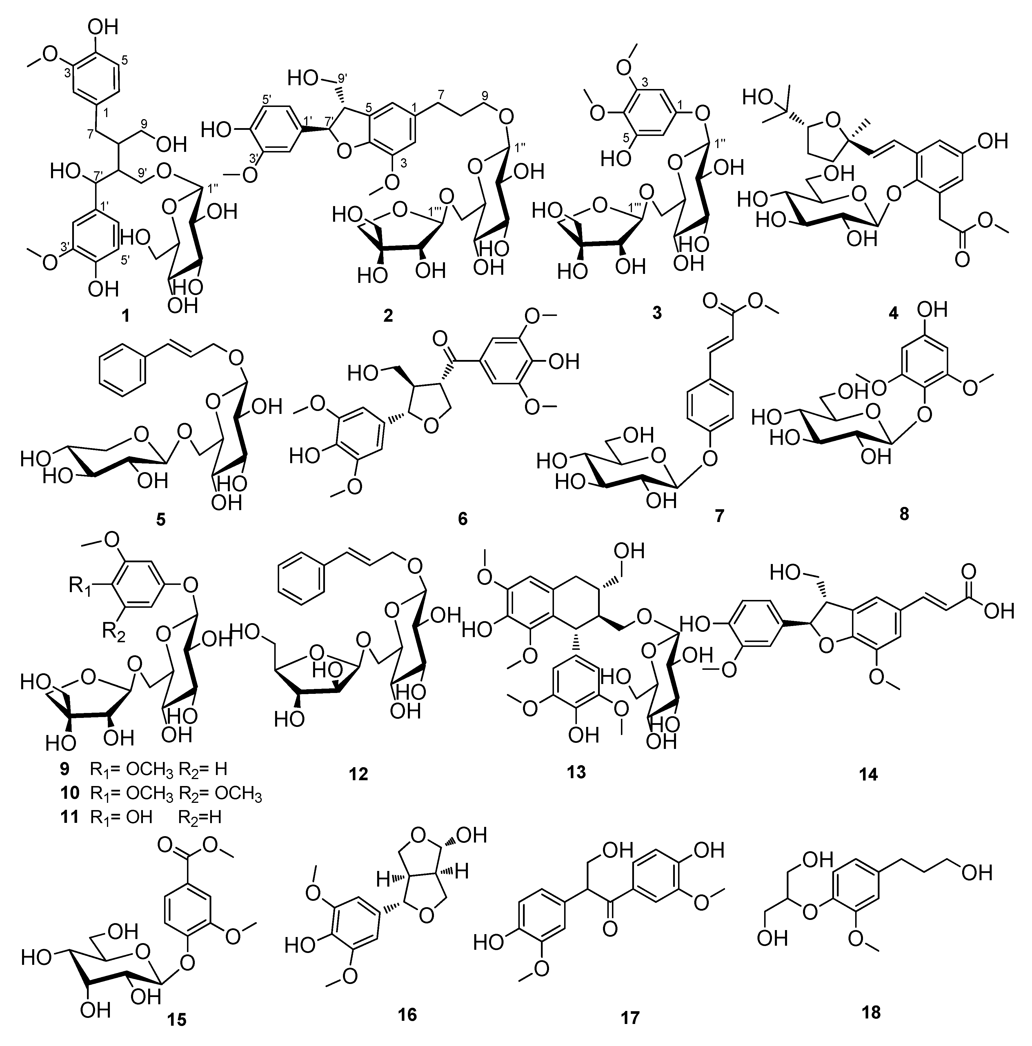
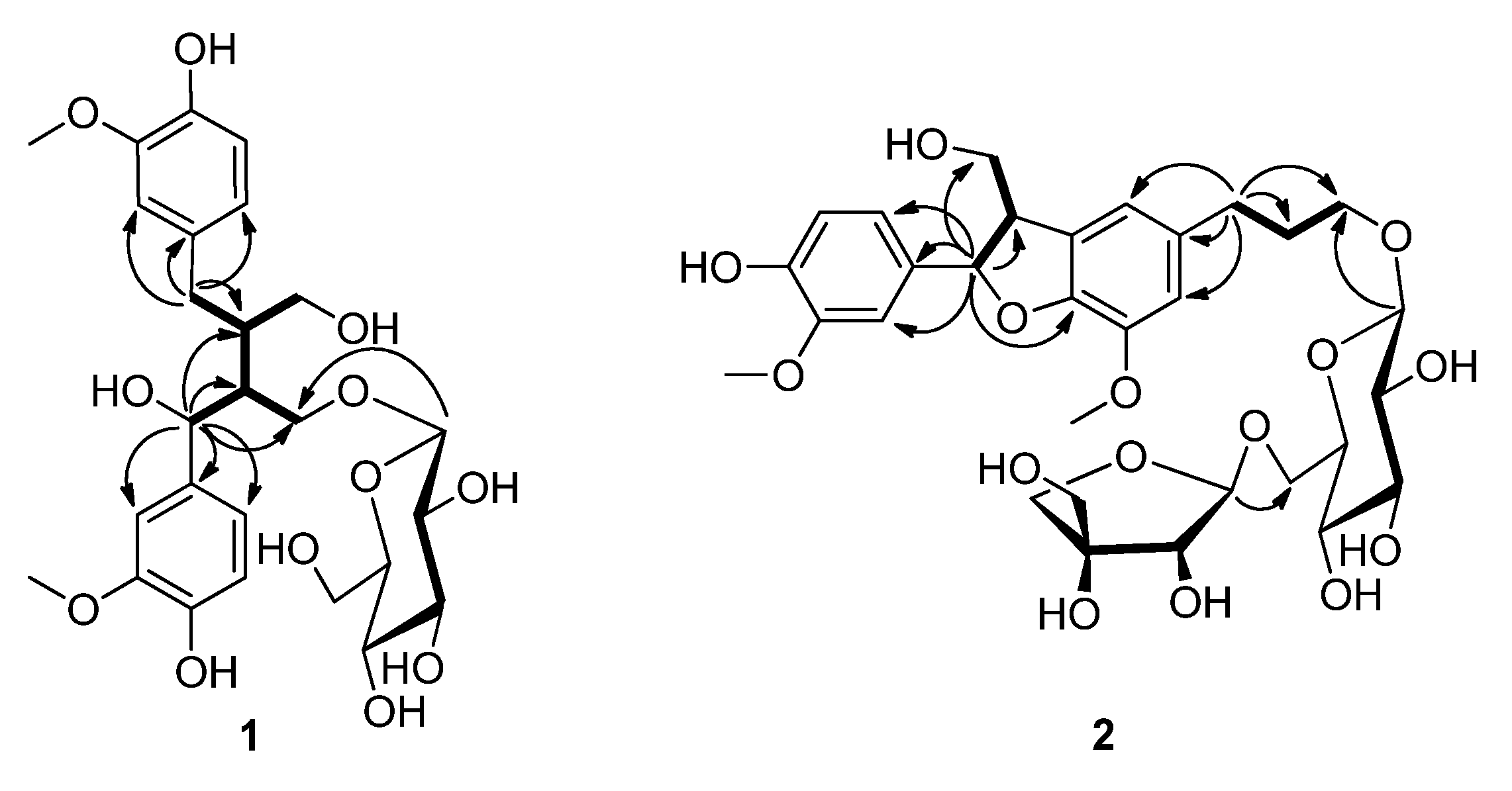
2.1. Structural Identification

Compound 1 was obtained as a white solid. Its molecular formula was assigned as C26H36O12, with nine degrees of unsaturation, based on the HREIMS (m/z 540.2180 [M]+, calcd. for C26H36O12, 540.2207). The 1H-NMR spectrum of compound 1 (Table 1) revealed six aromatic protons [δH 6.74 (1H, d, J = 1.5 Hz, H-2), 6.66 (1H, d, J = 8.0 Hz, H-5), and 6.57 (1H, dd, J = 8.0, 1.5 Hz, H-6); 6.84 (1H, s, H-2′), and 6.70 (2H, d, J = 8.0 Hz, H-5′, H-6′)] and two methoxyl [δH 3.74, 3.73 (6H, s, 3-OCH3, 3′-OCH3)] signals. The 13C-NMR spectrum (Table 1) of 1 displayed 26 carbon signals, including one glucopyranosyl moiety [δC 103.2 (C-1′′), 73.6 (C-2′′), 76.9 (C-3′′), 70.1 (C-4′′), 77.0 (C-5′′), and 61.2 (C-6′′)], two benzene ring, two methoxyl, three methylene (two oxygenated), and three methine (one oxygenated) groups. The 1H- and 13C-NMR data of 1 were thus characteristic of a lignan glycoside, and was highly similar to those of 7-hydroxy-9′-β-glucopyranosyloxyl secoisolariciresinol [25]. The two compounds have the same molecular formula, indicating they were isomers, the only difference was that the hydroxyl group at C-7 in 7-hydroxy-9′-β-glucopyranosyloxyl secoisolariciresinol was moved to C-7′ in compound 1, which was established by the HMBC correlations (Figure 2) from H-7′ (δH 4.72) to C-1′ (δC 134.5), C-2′ (δC 110.0), C-6′ (δC 118.3), C-8′ (δC 50.0), C-9′ (δC 66.8), and C-8 (δC 42.1) and correlations from H-7 (δH 2.83, 2.41) to C-1 (δC 131.9), C-2 (δC 112.7), C-6 (δC 120.6), C-8 (δC 42.1), and C-9′. The 1H-1H COSY correlations (Figure 2) of H-7′/H-8′, H-8′/H-9′, H-8′/H-8, H-8/H-7, and H-8/H-9 further confirmed this assumption. The HMBC correlation from H-1′′ (δH 4.17) to C-9′ confirmed the glucopyranosyl moiety was connected to C-9′. The presence of D-glucose was confirmed by TLC and optical rotation analyses. The configuration of glycosidic linkage of the glucopyranoside moiety in 1 was determined to be β on the basis of the J value of the anomeric proton signal at δH 4.17 (1H, d, J = 7.8 Hz, H-1′′). Although the ROESY correlations of H-8′/H-7a and H-7′/H2-7 were observed, however, it is not helpful for determining the relative configurations at C-8 and C-8′ because of the free rotation of the chain. Thus, the structure of 1 was established to be 7′-hydroxy-9′-β-glucopyranosyloxyl secoisolariciresinol, and the new compound was named cinnacassoside A.
Figure 2. Key HMBC (→) and 1H-1H COSY () correlations of compounds 1 and 2.
Molecules 18 10930 g002
Compound 2 gave a molecular formula of C31H42O15 from the HREIMS at m/z 654.2521 ([M]+, calcd for C31H42O15, 654.2524), corresponding to 11 degrees of unsaturation. The 1H-NMR spectrum of 2 (Table 1) showed an ABX coupling system [δH 6.95 (1H, d, J = 2.0 Hz, H-2′), 6.82 (1H, dd, J = 8.0, 2.0 Hz, H-6′), and 6.77 (1H, d, J = 8.0 Hz, H-5′)], two singlet aromatic protons [δH 6.76 (1H, s, H-2) and 6.75 (1H, s, H-6)], and two methoxyl [δH 3.86 (3H, s, 3-OCH3), 3.82 (3H, s, 3′-OCH3)] signals. The 1H- and 13C-NMR data (Table 1) of 2 showed 31 carbon signals, and were similar to those of symplolignanoside A [26], indicating a lignan glucoside with the same aglycon and saccharide moieties as the latter. Inspection of the 13C-NMR spectrum of compound 2 confirmed the presence of one glucopyranosyl [δC 104.7 (C-1′′), 75.3 (C-2′′), 77.0 (C-3′′), 71.8 (C-4′′), 78.2 (C-5′′), and 68.8 (C-6′′)] and one apiofuranosyl [δC 111.1 (C-1′′′), 78.1 (C-2′′′), 80.7 (C-3′′′), 75.1 (C-4′′′), and 65.6 (C-5′′′)] group in the molecule. The terminal apiofuranosyl unit was deduced to be attached at C-6′′ of the glucopyranosyl unit via oxygen on the basis of the downfield shift of C-6′′ (δC Δ = + 6.0 ppm) of the glucopyranosyl unit and the HMBC (Figure 2) correlation from H-6′′ (δH 3.98, 3.61) to C-1′′′. The glucopyranosyl unit was established to connect to C-9 of the aglycon by the HMBC correlation from H-1′′ (δH 4.24) to C-9 (δH 70.3). The two saccharide moieties were assigned to be β-form by the 1H-NMR spectroscopic data and the coupling constants of the anomeric proton signals at δH 4.24 (1H, d, J = 7.8 Hz, H-1′′) and 5.01 (1H, d, J = 2.4 Hz, H-1′′′). The HMBC correlations from H-7′ (δH 5.50) to C-1′ (δC 134.9), C-2′ (δC 110.6), C-6′ (δC 119.8), C-8′ (δC 55.6), C-9′ (δC 65.1), and C-4 (δC 147.6), and correlations from H-7 (δH 2.69) to C-1 (δC 137.1), C-2 (δC 114.2), C-6 (δC 118.1), C-8 (δC 33.2), and C-9′, along with the 1H-1H COSY correlations of H-7/H-8, H-8/H-9, H-7′/H-8′, and H-8′/H-9′ further confirmed the linkages of the two C3 units in compound 2 were the same as those in symplolignanoside A. The two methoxyl groups was deduced to be located at C-3 (δC 145.3) and C-3′ (149.2) by the HMBC correlations from 3-OCH3 to C-3 and from 3′-OCH3 to C-3′, respectively. The 3J value of H-7′ was 6.3 Hz, along with the ROESY correlation of H-7′/H-9′ indicated the trans-configuration of H-7 and H-8 [27,28]. The sugar moieties were assigned as d-glucose and d-apiose by TLC and optical rotation analyses. Thus, the structure of compound 2 was assigned as dihydrodehydrodiconiferyl alcohol-9-β-d-apiofuranosyl (1→6)-β-d-glucopyranoside, and it was named cinnacassoside B.
The molecular formula of compound 3 was deduced to be C19H28O13 by HREIMS at m/z 464.1519 ([M]+, calcd for C19H28O13, 464.1530). The 1H-NMR spectrum of 3 (Table 1) exhibited two overlapping single aromatic protons [δH 6.30 (2H, s, H-2, H-6)], two methoxyl [δH 3.80 (3H, s, 3-OCH3), 3.72 (3H, s, 4-OCH3)] and two anomeric protons [δH 4.75 (1H, d, J = 7.5 Hz, H-1′′), 4.98 (1H, d, J = 2.5 Hz, H-1′′′)] signals. The 13C-NMR spectrum (Table 1) of 3 displayed 19 carbon signals, including one benzene ring [δC 155.8 (C-1), 95.2 (C-2), 154.8 (C-3), 133.2 (C-4), 152.0 (C-5), and 99.1 (C-6)], one glucopyranosyl moiety [δC 103.0 (C-1′′), 74.9 (C-2′′), 78.0 (C-3′′), 71.7 (C-4′′), 77.1 (C-5′′) and 68.7 (C-6′′) and one apiofuranosyl group [δC 111.0 (C-1′′′), 78.0 (C-2′′′), 80.5 (C-3′′′), 75.0 (C-4′′′) and 65.7 (C-5′′′)], and two methoxyl [δC 56.5 (3-OCH3), 61.1 (4-OCH3)] signals. The 1H and 13C-NMR data of 3 were highly similar to those of 3,4-dimethoxyphenol β-d-apiofuranosyl (1→6)-β-d-glucopyranoside [26] except that a sp2 methine in 3,4-dimethoxyphenol β-d-apiofuranosyl (1→6)-β-d-glucopyranoside was replaced by a sp2 quaternary carbon in 3 and the downfield shift of C-5 (δC Δ = +37.8 ppm) and upfield shift of C-2 (δC Δ = −9.9 ppm), C-4 (δC Δ = −12.5 ppm) and C-6 (δC Δ = −12.4 ppm) in 3, indicating there was an additional hydroxyl group connected to C-5 in 3, in accordance with the molecular displayed by the HREIMS. The presence of glucose and apiose was confirmed by TLC and optical rotation comparison with authentic samples after acid hydrolysis and purification. Besides, the HMBC correlations from H-6 to C-1, C-4, and C-5 further confirmed this assumption. The HMBC correlations from δH 3.80 (3H, s, 3-OCH3) to C-3 and from δH 3.72 (3H, s, 4-OCH3) to C-4 indicated the two methoxyl groups were located at C-3 and C-4, respectively. The downfield shift of C-6′′ (δC 68.7), along with the HMBC correlations from H-1′′′ to C-6′′ indicated the apiofuranosyl moiety was connected to C-6′′ of the glucopyranosyl unit.
Table 1. The 1H and 13C-NMR spectroscopic data of 1-3.
Position123
δH (J in Hz)δC, multδH (J in Hz)δC, multδH (J in Hz)δC, mult
1 131.9, qC 137.1, qC 155.8, qC
26.74, d, 1.5112.7, CH26.76, s114.2, CH6.30, s95.2, CH
3 147.5, qC 145.3, qC 154.8, qC
4 144.6, qC 147.6, qC 133.2, qC
56.66, d, 8.0115.4, CH 129.9, qC 152.0, qC
66.57, dd, 8.0, 1.5120.6, CH6.75, s118.1, qC6.30, s99.1, CH
72.83, dd, 13.5, 4.432.2, CH22.69, dd, 13.8, 6.633.1, CH2
2.41, m
82.57, m42.1, CH1.91, m33.2, CH2
93.85, t, 7.472.0, CH23.90, m70.3, CH2
3.57, t, 7.4 3.54, m
1′ 134.5, qC 134.9, qC
2′6.84, d, 1.5110.0, CH26.95, d, 2.0110.6, CH
3′ 147.5, qC 149.2, qC
4′ 145.6, qC 147.6, qC
5′6.70, d, 8.0115.1, CH6.77, d, 8.0116.2, CH
6′6.70, d, 8.0118.3, CH6.82, dd, 8.0, 2.0119.8, CH
7′4.72, d, 6.081.9, CH5.50, d, 6.389.2, CH
8′2.30, m50.0, CH3.48, m55.6, CH
9′4.05, dd, 9.0, 7.866.8, CH23.86, m,65.1, CH2
3.43, m 3.76, m
1′′4.17, d, 7.8103.2, CH4.24, d, 7.8104.7, CH4.75, d, 7.5103.0, CH
2′′2.79, m73.6, CH3.19, m75.3, CH3.75, d, 9.874.9, CH
3′′3.10, m76.9, CH3.40, m77.0, CH3.92, m78.0, CH
4′′3.05, m70.1, CH3.28, m71.8, CH3.32, m71.7, CH
5′′3.12, m77.0, CH3.90, m78.2, CH3.56, m77.1, CH
6′′3.66, d,10.361.2, CH23.98, dd, 11.2, 1.868.8, CH24.03, d, 10.768.7, CH2
3.43, m 3.61, m 3.60, m
1′′′ 5.01, d, 2.4111.1, CH24.98, d, 2.5111.0, CH
2′′′ 3.33, m78.1, CH23.42, d, 8.678.0, CH
3′′′ 80.7, qC 80.5, CH
4′′′ 3.96, d, 9.775.1, CH23.98, d, 9.675.0, CH2
3.75, m 3.76, d, 7.4
5′′′ 3.56, m65.6, CH23.58, 2H, m65.7, CH2
3-OCH33.74, 3H, s55.6, CH33.82, s56.5, CH33.80, s56.5, CH3
3′-OCH33.73, 3H, s55.6, CH33.86, s56.9, CH3
4-OCH3 3.72, s61.1, CH3
In the HMBC spectrum, the correlations from H-1′′ to C-1 indicated the glucopyranosyl unit was linked to C-1. The β-configurations of the two saccharide moieties were assigned by the coupling constants of the anomeric proton signals at δH 4.75 (1H, d, J = 7.5 Hz, H-1′′) and 4.98 (1H, d, J = 2.5 Hz, H-1′′′). Thus, the structure of 3 was determined as 3,4-dimethoxy- 5-hydroxyphenol β-d-apiofuranosyl (1→6)-β-d-glucopyranoside, named as cinnacassoside C.
The known compounds were identified as cinnacasside C (4) [29], rosavin (5) [30], (−)-(7′S,8S,8′R)-4,4′-dihydroxy-3,3′,5,5′-tetramethoxy-7′,9-epoxylignan-9′-ol-7-one (6) [31], lino- cinnamarin (7) [32], leonuriside (8) [33], 3,4-dimethoxyphenol β-d–apiofuranosyl (1→6)- β-d-glucopyranoside (9) [26], 3,4,5-trimethoxyphenol β-d–apiofuranosyl (1→6)-β-d-glucopyranoside (10) [21], 3-trimethoxy-4- hydroxyphenol β-d–apiofuranosyl (1→6)-β-d-glucopyranoside (11) [34],rosarin (12) [30], (−)-lyoniresinol 3α-O-β-d-glucopyranoside (13) [35], spicatolignan B (14) [36], methyl 3-methoxy-4-(β-d-allopyranosyloxy)benzoate (15) [37], 6-hydroxy-2-(4-hydroxy- 3,5-dimethoxyphenyl)-3,7-dioxabicyclo-[3.3.0]-octane (16) [38], evofolin B (17) [39], and 2-[4-(3-hydroxypropyl)-2-methoxyphenoxy]-1,3-propanediol (18) [40] by comparison of their spectroscopic data with those in the corresponding literature.

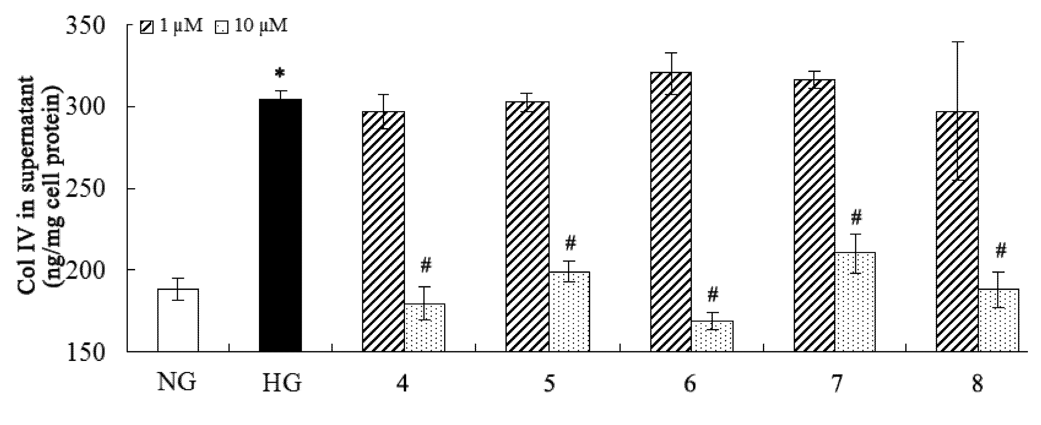
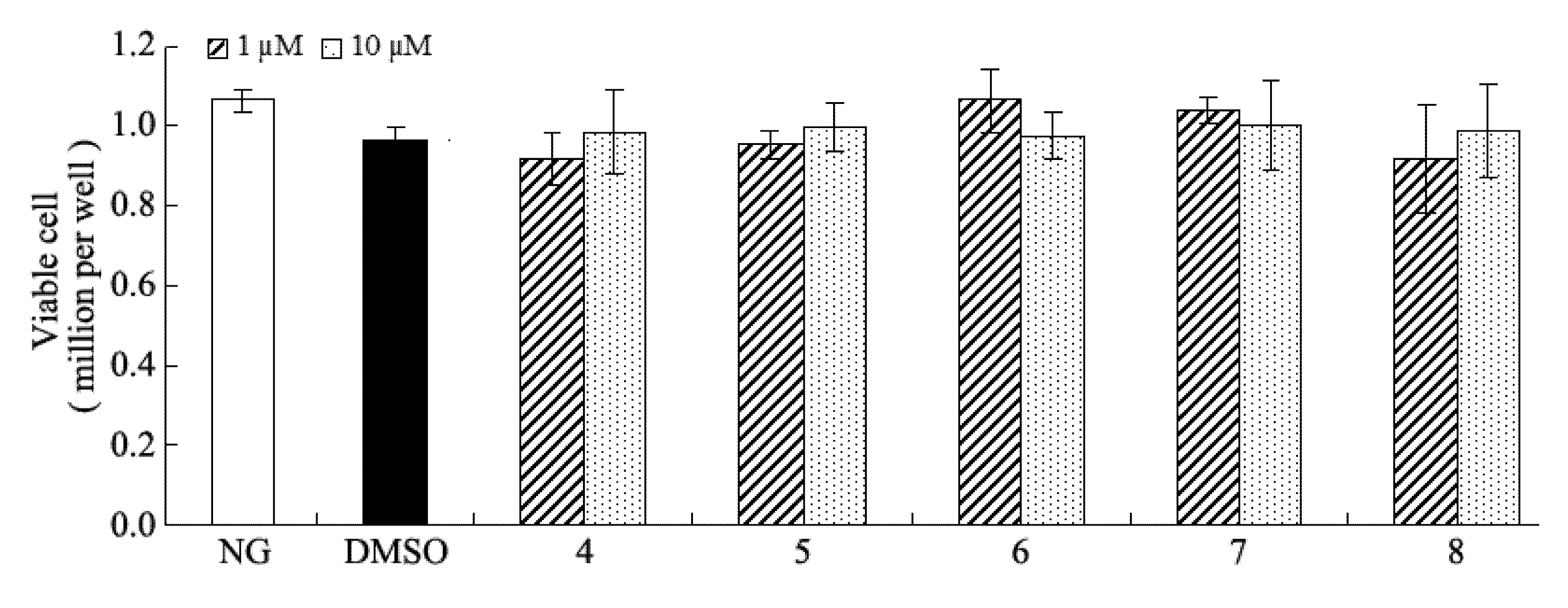
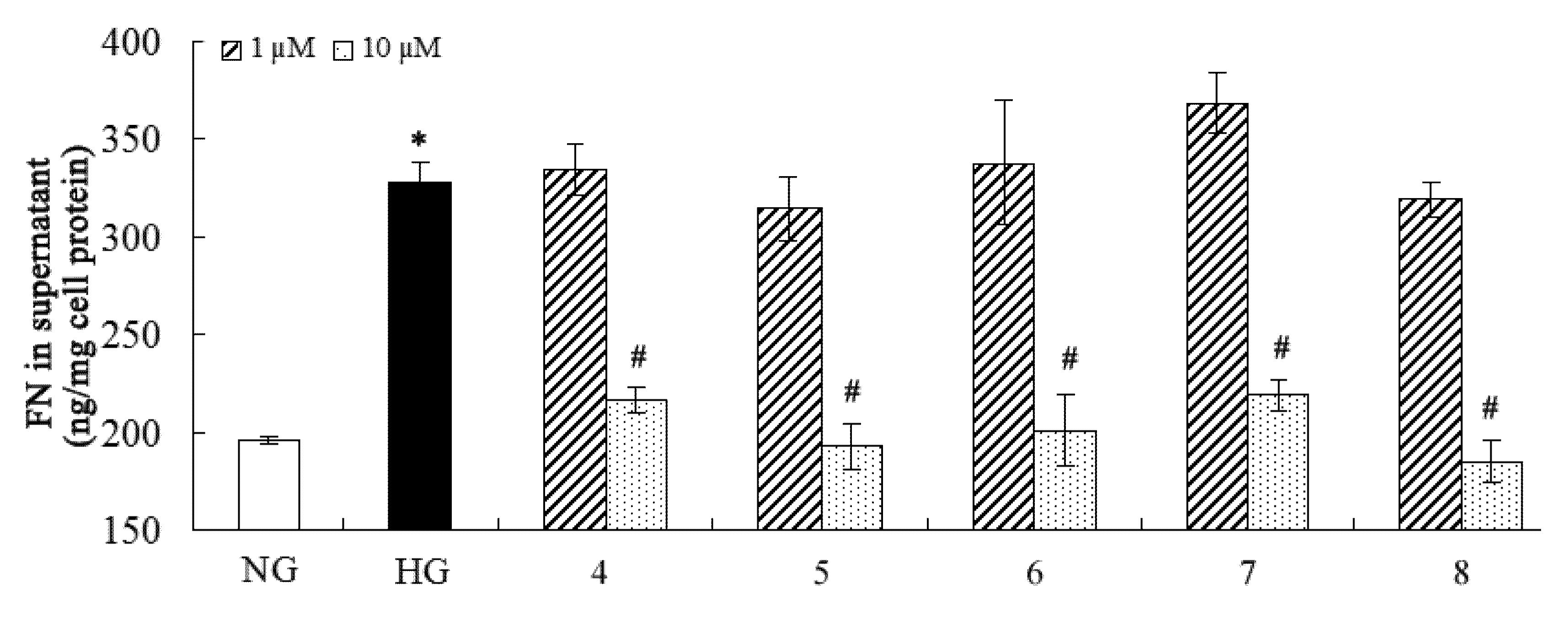
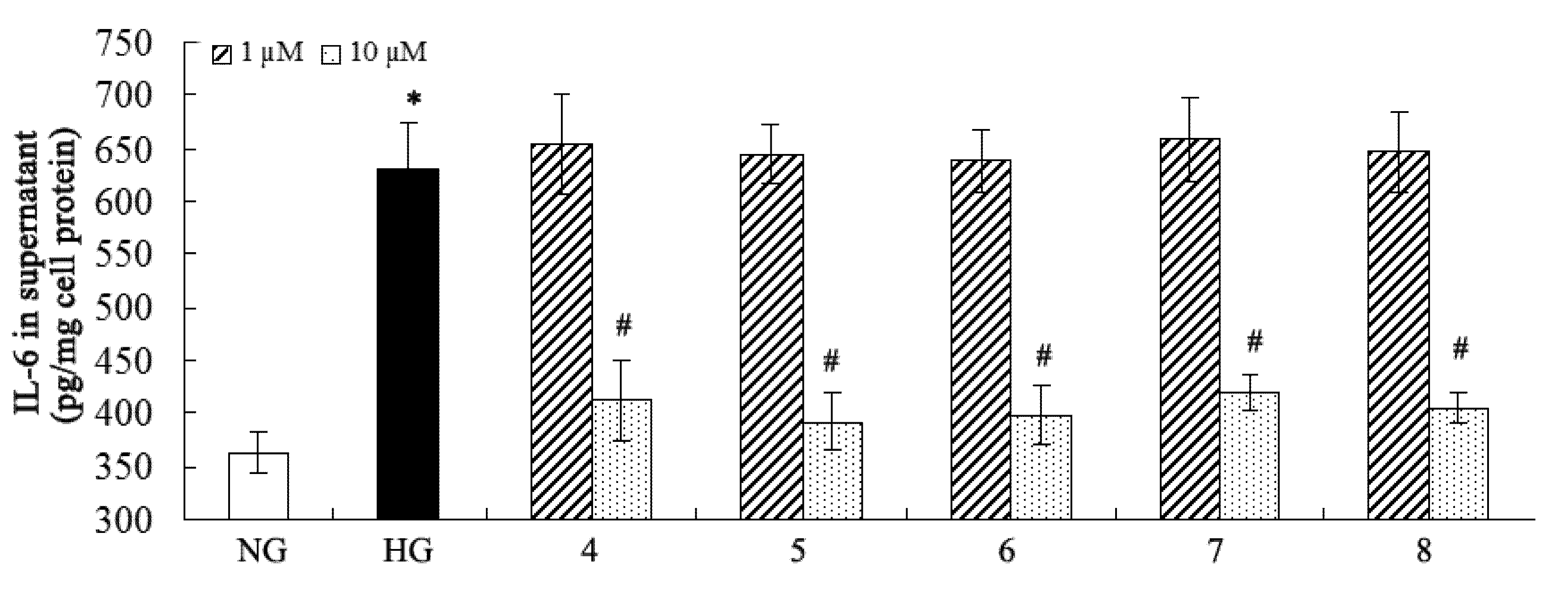
2.2. Inhibition of Fibronectin, Collagen IV, and IL-6 Secretion

Many pathogenic factors are involved in DN, and overproduction of extracellular matrix (such as fibronectin and collagen IV) and proinflammatory factors (such as IL-6) are distinctly implicated in DN [2,41]. Considering the traditional medical applications of C. cassia, the isolated compounds were purposely evaluated for their inhibitory effects on fibronectin, collagen IV, and IL-6 overproduction against high-glucose-induced mesangial cells (Figure 3, Figure 4 and Figure 5). The results showed that compounds 4-8 could inhibit overproduction of fibronectin, collagen IV, and IL-6 against high-glucose-induced mesangial cells at 10 µM, while all the other compounds were not active in this assay at the same concentration. Compound 4 is a novel natural hybrid with a unique geranylphenylacetate carbon skeleton which was previously isolated from the same material [29], however, its biological activities remained unknown yet.
Figure 3. Inhibitory effect of the compounds on fibronectin. * p < 0.05 vs. normal glucose; # p < 0.05 vs. high glucose.
Molecules 18 10930 g003
Figure 4. Inhibitory effect of the compounds on collagen IV. * p < 0.05 vs. normal glucose; # p < 0.05 vs. high glucose.
Molecules 18 10930 g004
Figure 5. Inhibitory effect of the compounds on IL-6. * p < 0.05 vs. normal glucose; # p < 0.05 vs. high glucose.
Molecules 18 10930 g005
This study revealed its possible role in DN. We noted that compounds 3, 811 are structurally analogues, however, only 8 was found to be active in this assay. Although compounds 5 and 12 differed only from the presence of apiose or glucose, their bioactivity is different, 5 is active and 12 is inactive. Actually, compound 5 exhibited a broad range of biological effects such as on antidepressant, nootropic, anticancer, neurotropic, immunostimulating and hepatoprotective properties which have attracted great research interest in the past [42,43,44,45,46]. Oxidative stress in also one of the major pathogenic factors of DN, in general, the other phenolic compounds should possess antioxidant activity which will be beneficial for DN, however, this need to be further investigated.

3. Experimental

3.1. Equipment

Optical rotations were taken on a Horiba SEAP-300 polarimeter. UV spectra were obtained on a Shimadzu UV-2401PC (Shimadzu, Kyoto, Japan) spectrophotometer. 1D and 2D NMR spectra were recorded on a Bruker AM-400 or DRX-500 or Avance III 600 NMR spectrometer. Unless otherwise specified, chemical shifts (δ) were expressed in ppm with reference to the solvent signals. Mass spectra were obtained on a VG Auto Spec-3000 mass spectrometer (VG, Manchester, UK). Column chromatography (CC) was performed on silica gel (200-300 mesh, Qingdao Marine Chemical Inc., People’s Republic of China), RP-18 (40-60 μm, Daiso Co., Japan), MCI gel CHP 20P (75-150 μm, Tokyo, Japan), and Sephadex LH-20 (Amersham Biosciences, Sweden). Semipreparative HPLC was carried out using an Agilent 1200 liquid chromatograph, the column used was a 250 mm × 4.6 mm i.d., 5 μm, Zorbax SB-C18.

3.2. Plant Material

The C. cassia barks were collected at Yunnan Xianghui Biological Technology Co. Ltd. (YNXHBT) in July, 2010. The sample was identified by Mr. Fu-Shou Xie at YNXHBT. A voucher specimen (CHYX0173) was preserved at the State Key Laboratory of Phytochemistry and Plant Resources in West China, Kunming Institute of Botany, Chinese Academy of Sciences, China.

3.3. Extraction and Isolation

Dried powders of C. cassia barks (50 kg) were extracted three times with 80% EtOH (3 × 90 L) under reflux to give a residue (7.5 kg) after removal of solvent under reduced pressure. The residue was dissolve in acetone and then filtered, the acetone solution was evaporated in vacuum to get the acetone-soluble extract (5.0 kg), which was suspended in water and followed by successive partition with petroleum ether and EtOAc. The water-soluble extract (1.5 kg) was subjected to silica gel column chromatography (CC) (20 × 120 cm, 7.5 kg) and eluted with a isocratic of CHCl3-MeOH-H2O (80:20:2.5, 25 L, v/v) to yield three fractions, A–C. Fraction A (3.1 g) was submitted to RP-18 CC (3.0 × 3.0 cm, 80 g) eluted with gradient aqueous MeOH (10:90, 30:70, 50:50, 70:30, 90:10, 1L, each, v/v) to yield two fractions, A1 and A2. Fraction A1 (400 mg) was chromatographed over MCI gel CHP 20P CC (2.0 × 50 cm, 100 g) eluted with MeOH/H2O (20:80, 30:70, 40:60, 50:50, 60:40, 70:30, 500 mL, each, v/v) to afford two portions, A1a and A1b. Fraction A1a (85 mg) was purified by preparative TLC eluting with CHCl3/MeOH (10:1, 100 mL, v/v) to afford compound 17 (2.6 mg). Fraction A2 (1 g) was subjected to MCI gel CHP 20P CC (2.0 × 50 cm, 100 g) eluted with MeOH/H2O, (30:70, 40:60, 50:50, 60:40, 70:30, 80:20, 700 mL, each, v/v) to yield three fractions, A2a-A2c. Fraction A2b (20 mg) was purified by preparative TLC eluting with CHCl3/MeOH (11:1, 100 mL, v/v) to give compound 16 (3.5 mg). Fraction A2c (300 mg) was purified by preparative TLC eluting with CHCl3/MeOH (11:1, 100 mL, v/v) to give compound 6 (19.7 mg).
Fraction B (155 g) was subjected to Sephadex LH-20 CC (6.0 × 125 cm, 500 g) (MeOH) to obtain two portions, B1 and B2. Fraction B1 (5.2 g) was separated by RP-18 CC (3.0 × 50 cm, 200 g) eluted with MeOH/H2O (10:90, 30:70, 50:50, 70:30, 90:10, 1 L, each, v/v) to provide three portions, B1a-B1c. Fraction B1b (462 mg) was purified by preparative TLC eluting with CHCl3/MeOH/H2O (3:1:0.05, 200 mL, v/v/v) to obtain compounds 12 (3.8 mg) and 5 (3.9 mg), the residue of fraction B1b was further purified using successive semi-preparative HPLC (MeOH/H2O, 33:77, 5 L, v/v) to yield compounds 1 (9.2 mg), 2 (10.5 mg), and 13 (3.9 mg). Fraction B1c (240 mg) was purified by preparative TLC eluting with CHCl3/MeOH/H2O (6:1:0.05, 150 mL, v/v/v) to yield compounds 9 (30.8 mg) and 10 (42.8 mg). Fraction B2 (5.5 g) was subjected to CC over MCI gel CHP 20P (3.0 × 3.0 cm, 200 g) eluted with gradient aqueous MeOH (20:80, 30:70, 50:50, 70:30, 80:20, 1 L, each, v/v) to obtain three fractions, B2a–B2c. Fraction B2a (105 mg) was purified by preparative TLC eluting with CHCl3/MeOH/H2O (3:1:0.05, 100 mL, v/v/v) to yield compounds 11 (4.8 mg) and 15 (2.4 mg). Fraction B2b (201 mg) was purified using successive semi-preparative HPLC (MeOH/H2O, 20:80, 2 L) to yield compound 14 (1.0 mg).
Fraction C (1 g) was subjected to CC over MCI gel CHP 20P (3.0 × 3.0 cm, 150 g) eluted with gradient aqueous MeOH (MeOH/H2O, 10:90, 30:70, 50:50, 70:30, 80:20, 1 L, each, v/v) to yield three fractions, C1-C3. Fraction C1 (70 mg) was purified by preparative TLC eluting with CHCl3/MeOH (5:1, 100 mL, v/v) and then followed by successive semi-preparative HPLC (MeOH/H2O, 20:80, 5L) to yield compounds 3 (9.3 mg), 8 (4.3 mg) and 4 (1.1 mg). Fraction C2 (100 mg) was purified by preparative TLC eluting with CHCl3/MeOH (8:1, 150 mL, v/v) to provide compound 7 (3 mg). Fraction C3 (20 mg) was purified by preparative TLC eluting with CHCl3/MeOH (9:1, 100 mL, v/v) to obtain compound 18 (1.5 mg).
Cinnacassoside A (1). White solid; Molecules 18 10930 i001 −31.9 (c 0.2, MeOH); UV (MeOH) λmax (log ε): 203 (4.65), 226 (4.06), 281 (3.64) nm;1H (DMSO-d6, 600 MHz) and 13C-NMR (DMSO-d6, 150 MHz) data, see Table 1; EI-MS: m/z 540 [M]+; HREIMS: m/z 540.2180 [M]+ (calcd for C26H36O12, 540.2207).
Cinnacassoside B (2). Yellow solid; Molecules 18 10930 i001 −38.0 (c 0.2, MeOH); UV (MeOH) λmax (log ε): 205 (4.74), 283 (3.72) nm;1H (CD3OD, 600 MHz) and 13C-NMR (CD3OD, 150 MHz) data, see Table 1; EIMS: m/z 654 [M]+; HREIMS: m/z 654.2521 [M]+, calcd for C31H42O15, 654.2524).
Cinnacassoside C (3). Yellow solid; Molecules 18 10930 i001 −82.6 (c 0.2, MeOH); UV (MeOH) λmax (log ε): 204 (4.54), 271 (3.01) nm;1H (CD3OD, 600 MHz) and 13C-NMR (CD3OD, 125 MHz) data, see Table 1; EI-MS: m/z 464 [M]+; HREIMS: m/z 464.1519 [M]+ (calcd for C19H28O13, 464.1530).

3.4. Acid Hydrolysis of 1-3

A solution of 1 (6 mg), 2 (8 mg), or 3 (8 mg) in 2 N HCl was heated at 90 °C on a water bath for 2 h. After cooling, the reaction mixture was extracted by EtOAc. The aqueous layer was neutralized with 2 N NaOH and dried by freeze-drying. The residue was purified by semipreparative HPLC with gradient aqueous MeOH to afford pure sugars. They were identified as d-glucose and d-apiose by comparison with authentic samples on TLC and measuring their optical rotations [glucose: EtOAC–MeOH–H2O (4:1:0.1), Rf 0.2, Molecules 18 10930 i002 +37 (c 0.06, H2O); apiose: EtOAC–MeOH–H2O (4:1:0.1), Rf 0.5, Molecules 18 10930 i002 +5(c 0.05, H2O)].

3.5. Inhibition of Fibronectin, Collagen IV, and IL-6 Secretion

Rat mesangial cells were grown in Dulbecco’s modified Eagle’s medium (Invitrogen, Carlsbad, CA, USA) containing 5.6 mM D-glucose (pH 7.4; Sigma Chemical Co., St. Louis, MO, USA), supplemented with 20% fetal calf serum (FCS; Invitrogen), 100 U/mL penicillin, 100 μg/mL streptomycin, and 10 mM HEPES. After the mesangial cells reached 80% confluence, their growth was arrested in 0.5% FCS for 24 h. Exposure of the mesangial cells to medium containing high-concentration glucose induced the overproduction of fibronectin (FN), collagen IV (Col IV), and IL-6, as described in the previous reports [47,48]. To determine whether the compounds inhibited the FN, collagen IV, and IL-6 overproduction triggered by high glucose, the mesangial cells were pretreated with 1 or 10 μM of each compound for 1 h and then stimulated with high glucose (25 mM) for 24 h. The levels of supernatant FN, collagen IV, and IL-6 were measured with a solid-phase quantitative sandwich enzyme-linked immunosorbent assay (ELISA) kit for FN, collagen IV, and IL-6 (Uscn Life Science Inc., Wuhan, China). The concentration in the culture supernatant was normalized to the total amount of cell protein, quantified with the BCA method [41].

3.6. Statistics

The differences were tested using ANOVA. All values are expressed as mean ± S.D., and statistical significance was defined as p < 0.05.

4. Conclusions

In conclusion, as a well-known spice, C. cassia barks have been well investigated in the past years. However, whether it could be used for the prevention of DN still needing further investigation. This study resulted in the isolation and identification of three new phenolic glycosides, along with 15 known compounds from the polar extract, of which 11 were isolated for the first time in this species. Compounds 4-8 could significantly inhibit over secretion of fibronectin, collagen IV, and IL-6 against high-glucose-induced mesangial cells at 10 µM, while the other compounds were inactive at 10 µM. Moreover, no cytoxicity was observed at the same concentration by using the MTT assay (Figure 6). These results suggested that C. cassia barks contains anti-diabetic nephropathy agents which may be beneficial for DN patients.
Figure 6. Effect of the compounds on cell viability at 1 or 10 µM.
Molecules 18 10930 g006

Supplementary Materials

Supplementary materials can be accessed at: https://www.mdpi.com/1420-3049/18/9/10930/s1.

Acknowledgments

This work was financially supported by the Open Research Fund of State Key Laboratory Breeding Base of Systematic Research, Development and Utilization of Chinese Medicine Resources, and Project of Natural Compound Library Construction from Chinese Academy of Sciences (KSCX2-EW-R-15).

Conflicts of Interest

The authors declare no conflict of interest.

References and Notes

  1. Ha, H.; Hwang, I.A.; Park, J.H.; Lee, H.B. Role of reactive oxygen species in the pathogenesis of diabetic nephropathy. Diabetes Res. Clin. Pract. 2008, 82s, s42–s45. [Google Scholar]
  2. Navarro-González, J.F.; Mora-Fernández, C. The role of inflammatory cytokines in diabetic nephropathy. J. Amer. Soc. Nephrol. 2008, 19, 433–442. [Google Scholar] [CrossRef]
  3. Sriniva Navarro, J.F.; Mora, C. Role of inflammation in diabetic complications. Nephrol. Dialysis Transplant. 2005, 20, 2601–2604. [Google Scholar] [CrossRef]
  4. San, K. Role of spices beyond food flavoring: Nutraceuticals with multiple health effects. Food Rev. Int. 2005, 21, 167–188. [Google Scholar] [CrossRef]
  5. Woehrlin, F.; Fry, H.; Abraham, K.; Preiss-Weigert, A. Quantification of flavoring constituents in Cinnamon: High variation of coumarin in cassia bark from the German retail market and in authentic samples from Indonesia. J. Agric. Food Chem. 2010, 58, 10568–10575. [Google Scholar] [CrossRef]
  6. Ravindran, P.N.; Nirmal Babu, K. Introduction. In Cinnamon and Cassia: The Genus Cinnamomum; CRC Press: Boca Raton, FL, USA, 2004; pp. 1–13. [Google Scholar]
  7. Chen, L.; Sun, P.; Wang, T.; Chen, K.X.; Jia, Q.; Wang, H.Y.; Li, Y.M. Diverse mechanisms of antidiabetic effects of the different procyanidin oligomer types of two different cinnamon species on db/db mice. J. Agric. Food Chem. 2012, 60, 9144–9150. [Google Scholar]
  8. Cheng, D.M.; Kuhn, P.; Poulev, A.; Rojo, L.E.; Lila, M.A.; Raskin, I. In vivo and in vitro antidiabetic effects of aqueous cinnamon extract and cinnamon polyphenol-enhanced food matrix. Food Chem. 2012, 135, 2994–3002. [Google Scholar] [CrossRef]
  9. Anderson, R.A.; Broadhurst, C.L.; Polansky, M.M.; Schmidt, W.F.; Khan, A.; Flanagan, V.P.; Schoene, N.W.; Graves, D. Isolation and characterization of polyphenol type-A polymers from cinnamon with insulin-like biological activity. J. Agric. Food Chem. 2004, 52, 65–70. [Google Scholar] [CrossRef]
  10. Khan, A.; Safdar, M.; Khan, M.H.A.; Khattak, K.N.; Anderson, R.A. Cinnamon improves glucose and lipids of people with type 2 diabetes. Diabetes Care 2003, 26, 3215–3218. [Google Scholar] [CrossRef]
  11. Li, X.W.; Li, J.; Werff, H. “Cinnamomum cassia”. Flora of China; Missouri Botanical Garden: St. Louis, MO, USA; Harvard University Herbaria: Cambridge, MA, USA, 2013. [Google Scholar]
  12. U.S. Food and Drug Administration. Inspections, Compliance, Enforcement, and 347 Criminal Investigations: CPG Sec. 525.750 Spices – Definitions: Cinnamon (Cassia). 348 1980.
  13. Nohara, T.; Kashiwada, Y.; Nishioka, I.; Cinncassiol, E. A diterpene from the bark of Cinnamomum cassia. Phytochemistry 1985, 24, 1849–1850. [Google Scholar]
  14. Nohara, T.; Kashiwada, Y.; Tomimatsu, T.; Nishioka, I. Two novel diterpenes from bark of Cinnamomum cassia. Phytochemistry 1982, 21, 2130–2132. [Google Scholar]
  15. Lin, R.J.; Cheng, M.J.; Huang, J.C.; Lo, W.L.; Yeh, Y.T.; Yen, C.M.; Lu, C.M.; Chen, C.Y. Cytotoxic compounds from the stems of Cinnamomum tenuifolium. J. Nat. Prod. 2009, 72, 1816–1824. [Google Scholar] [CrossRef]
  16. Ngoc, T.M.; Lee, I.; Ha, D.T.; Kim, H.; Min, B.; Bae, K. Tyrosinase-inhibitory constituents from the twigs of Cinnamomum cassia. J. Nat. Prod. 2009, 72, 1205–1208. [Google Scholar] [CrossRef]
  17. Prasad, K.N.; Yang, B.; Dong, X.; Jiang, G.; Zhang, H.; Xie, H.; Jiang, Y. Flavonoid contents and antioxidant activities from Cinnamomum species. Innov. Food Sci. Emerg. Technol. 2009, 10, 627–632. [Google Scholar] [CrossRef]
  18. Miyamura, M.; Nohara, T.; Tomimatsu, T.; Nishiokat, I. Seven aromatic compounds from bark of Cinnamomum cassia. Phytochemistry 1983, 22, 215–218. [Google Scholar]
  19. Ooi, L.S.; Li, Y.; Kam, S.L.; Wang, H.; Wong, E.Y.; Ooi, V.E. Antimicrobial activities of cinnamon oil and cinnamaldehyde from the Chinese medicinal herb Cinnamomum cassia Blume. Am. J. Chin. Med. 2006, 34, 511–522. [Google Scholar] [CrossRef]
  20. Lin, C.C.; Wu, S.J.; Chang, C.H.; Ng, L.T. Antioxidant activity of Cinnamomum cassia. Phytother. Res. 2003, 17, 726–730. [Google Scholar] [CrossRef]
  21. Shiraga, Y.; Okano, K.; Akira, T.; Fukaya, C.; Yokoyama, K.; Tanaka, S.; Fukui, H.; Tabata, M. Structures of potent antiulcerogenic compounds from Cinnamomum cassia. Tetrahedron 1988, 44, 4703–4711. [Google Scholar] [CrossRef]
  22. Verspohl, E.J.; Bauer, K.; Neddermann, E. Antidiabetic effect of Cinnamomum cassia and Cinnamomum zeylanicum in vivo and in vitro. Phytother. Res. 2005, 19, 203–206. [Google Scholar] [CrossRef]
  23. Nagai, H.; Shimazawa, T.; Matsuura, N.; Koda, A. Immunopharmacological studies of the aqueous extract of Cinnamomum cassia (CCAq). I. Anti-allergic action. Jpn. J. Pharmacol. 1982, 32, 813. [Google Scholar] [CrossRef]
  24. Zheng, H.T.; Whitman, S.A.; Wu, W.; Wondrak, G.T.; Wong, P.K.; Fang, D.Y.; Zhang, D.D. Therapeutic potential of Nrf2 activators in streptozotocin-induced diabetic nephropathy. Diabetes 2011, 60, 3055–3066. [Google Scholar] [CrossRef]
  25. Wu, B.; Zhang, X.D.; Wu, X.D. New lignan glucosides with tyrosinase inhibitory activities from exocarp of Castanea henryi. Carbohyd. Res. 2012, 335, 45–49. [Google Scholar]
  26. Jiang, J.; Feng, Z.; Wang, Y.; Zhang, P. New phenolics from the roots of Symplocos caudata Wall. Chem. Pharm. Bull. 2005, 53, 110–113. [Google Scholar] [CrossRef]
  27. Fang, J.M.; Lee, C.K.; Cheng, Y.S. Lignans from leaves of Juniperus chinensis. Phytochemistry 1992, 31, 3659–3661. [Google Scholar] [CrossRef]
  28. Liu, J.F.; Jiang, Z.Y.; Geng, C.A.; Zhang, Q.; Shi, Y.; Ma, Y.B.; Zhang, X.M.; Chen, J.J. Two new lignans and anti-HBV constituents from Illicium henryi. Chem. Biodivers. 2011, 8, 692–698. [Google Scholar] [CrossRef]
  29. Liao, S.G.; Yuan, T.; Zhang, C.; Yang, S.P.; Wu, Y.; Yue, J.M. Cinnacassides A-E, five geranylphenylacetate glycosides from Cinnamomum cassia. Tetrahedron 2009, 65, 883–887. [Google Scholar] [CrossRef]
  30. Tolonen, A.; Pakonen, M.; Hohtola, A.; Jalonen, J. Phenylpropanoid glycosides from Rhodiola rosea. Chem. Pharm. Bull. 2003, 51, 467–470. [Google Scholar] [CrossRef]
  31. Xiong, L.; Zhu, C.; Li, Y.; Tian, Y.; Lin, S.; Yuan, S.; Hu, J.; Hou, Q.; Chen, N.; Yang, Y. Lignans and neolignans from Sinocalamus affinis and their absolute configurations. J. Nat. Prod. 2011, 74, 1188–1200. [Google Scholar] [CrossRef]
  32. Luyengi, L.; Pezzuto, J.M.; Waller, D.P.; Beecher, C.W.; Fong, H.H.; Che, C.T.; Bowen, P.E. Linusitamarin, a new phenylpropanoid glucoside from Linum usitatissimum. J. Nat. Prod. 1993, 56, 2012–2015. [Google Scholar] [CrossRef]
  33. Luecha, P.; Umehara, K.; Miyase, T.; Noguchi, H. Antiestrogenic constituents of the Thai medicinal plants Capparis flavicans and Vitex glabrata. J. Nat. Prod. 2009, 72, 1954–1959. [Google Scholar] [CrossRef]
  34. Kanchanapoom, T.; Kasai, R.; Yamasaki, K. Iridoid and phenolic diglycosides from Canthium berberidifolium. Phytochemistry 2002, 61, 461–464. [Google Scholar] [CrossRef]
  35. Achenbach, H.; Löwel, M.; Waibel, R.; Gupta, M.; Solis, P. New lignan glucosides from Stemmadenia minima. Planta Med. 2007, 58, 270–272. [Google Scholar]
  36. Zheng, J.; Chen, G.T.; Gao, H.Y.; Wu, B.; Wu, L.J. Two new lignans from Mentha spicata L. J. Asian Nat. Prod. Res. 2007, 9, 431–435. [Google Scholar] [CrossRef]
  37. Li, Y.; Sun, J.; Gong, Y.; Yu, B. Synthesis of oligomeric 4-(glycosyloxy) benzoate macrocyclic glycosides. J. Org. Chem. 2011, 76, 3654–3663. [Google Scholar] [CrossRef]
  38. Huang, Y.; Zeng, Q.; Fu, J.J.; Kou, Z.J.; Chang, R.J.; Jin, H.Z.; Zhang, W.D. Chemical constituents from Tsoongiodendron odorum Chun. Biochem. Syst. Ecol. 2011, 39, 209–212. [Google Scholar] [CrossRef]
  39. Wu, T.S.; Yeh, J.H.; Wu, P.L. The heartwood constituents of tetradium glabrifolium. Phytochemistry 1995, 40, 121–124. [Google Scholar]
  40. Kouno, I.; Yukari, Y.G.; Satomi, S.M.; Miki, S.; Yang, C.S. Phenylpropanoids from the Barks of Illicium difengpi. Chem. Pharm. Bull. 1992, 40, 2461–2464. [Google Scholar] [CrossRef]
  41. Kolset, S.O.; Reinholt, F.P.; Jenssen, T. Diabetic nephropathy and extracellular matrix. J. Histochem. Cytochem. 2012, 60, 976–986. [Google Scholar] [CrossRef]
  42. Kurkin, V.A.; Dubishchev, A.V.; Ezhkov, V.N.; Titova, I.N.; Avdeeva, E.V. Antidepressant activity of some phytopharmaceuticals and phenylpropanoids. Pharm. Chem. J. 2006, 40, 614–619. [Google Scholar] [CrossRef]
  43. Sokolov, S.Y.; Ivashin, V.M.; Zapesochnaya, G.G.; Kurkin, V.A.; Shchavlinskii, A.N. Studies of neurotropic activity of newcompounds isolated from Rhodiola rosea. Clin. Pharm. J. 1985, 19, 1367–1371. [Google Scholar]
  44. Ewa, S.R.; Malgorzata, H.; Andrzej, K.S.; Aleksander, W.; Ewa, S.; Michal, M.; Janusz, B.; Henryk, S. The influence of Rhodiola rosea extracts and rosavin on cutaneous angiogenesis induced in mice after grafting of syngeneic tumor cells. Cent. Eur. J. Immunol. 2008, 33, 102–107. [Google Scholar]
  45. Kurkin, V.A. Phenylpropanoids from medicinal plants: Distribution, classification, structural analysis, and Biological activity. Chem. Nat. Compd. 2003, 39, 123–153. [Google Scholar] [CrossRef]
  46. Kurkin, V.A.; Dubishchev, A.V.; Titova, I.N.; Volotsueva, A.V.; Petrova, E.S.; Zhestkova, N.V.; Klimova, I.Y. Neurotropic properties of certain plant extracts containing phenylpropanoids. Rastit. Resur. 2003, 39, 115–122. [Google Scholar]
  47. Min, D.; Lyons, J.G.; Bonner, J.; Twigg, S.M.; Yue, D.K.; McLennan, S.V. Mesangial cell-derived factors alter monocyte activation and function through inflammatory pathways: Possible pathogenic role in diabetic nephropathy. Amer. J. Physiol-Renal Physiol. 2009, 297, F1229–F1237. [Google Scholar] [CrossRef]
  48. Xia, L.; Wang, H.; Goldberg, H.; Munk, S.; Fantus, I.; Whiteside, C. Mesangial cell NADPH oxidase upregulation in high glucose is protein kinase C dependent and required for collagen IV expression. Amer. J. Physiol. Renal. Physiol. 2006, 290, F345–F356. [Google Scholar] [CrossRef]
  • Sample Availability: Samples of the compounds 1, 3, 9, and 10 are available from the authors.

