Review

Structural Probing and Molecular Modeling of the A3 Adenosine Receptor: A Focus on Agonist Binding

by
Antonella Ciancetta
and
Kenneth A. Jacobson
*
Molecular Recognition Section (MRS), Laboratory of Bioorganic Chemistry, National Institutes of Diabetes and Digestive and Kidney Diseases, National Institutes of Health, Bethesda, MA 20892, USA
*
Author to whom correspondence should be addressed.
Molecules 2017, 22(3), 449; https://doi.org/10.3390/molecules22030449
Submission received: 5 February 2017 / Revised: 2 March 2017 / Accepted: 6 March 2017 / Published: 11 March 2017
(This article belongs to the Special Issue Adenosine Receptors)

Abstract

:
Adenosine is an endogenous modulator exerting its functions through the activation of four adenosine receptor (AR) subtypes, termed A1, A2A, A2B and A3, which belong to the G protein-coupled receptor (GPCR) superfamily. The human A3AR (hA3AR) subtype is implicated in several cytoprotective functions. Therefore, hA3AR modulators, and in particular agonists, are sought for their potential application as anti-inflammatory, anticancer, and cardioprotective agents. Structure-based molecular modeling techniques have been applied over the years to rationalize the structure–activity relationships (SARs) of newly emerged A3AR ligands, guide the subsequent lead optimization, and interpret site-directed mutagenesis (SDM) data from a molecular perspective. In this review, we showcase selected modeling-based and guided strategies that were applied to elucidate the binding of agonists to the A3AR and discuss the challenges associated with an accurate prediction of the receptor extracellular vestibule through homology modeling from the available X-ray templates.

Graphical Abstract

1. Introduction

The nucleoside adenosine (1, Figure 1) is an endogenous modulator regulating numerous physiological functions through the activation of four adenosine receptor (AR) subtypes, termed A1, A2A, A2B and A3, that belong to the G protein-coupled receptor (GPCR) superfamily. Generally, ARs are involved in cytoprotective functions, although each AR subtype is characterized by distinct tissue localization and biochemical pathway activation that give rise to a unique pharmacological profile [1].
The human A3AR (hA3AR) is expressed at the highest levels in liver, lung and immune cells [2], but lower levels are also detected in other tissues, including the heart and brain [3]. Small molecule modulators targeting the A3AR have therefore been sought for their potential application as anti-inflammatory, anticancer, and cardioprotective agents. In particular, A3AR agonists based on the adenosine scaffold emerged as promising antinociceptive agents in different preclinical models of chronic pain [4] and in clinical trials as agents for treating rheumatoid arthritis, psoriasis and hepatocellular carcinoma [5]. A3AR antagonists based on several heterocyclic scaffolds were suggested as potential candidates for the treatment of glaucoma [6,7] and respiratory tract inflammation such as asthma [8]. The structures of selected agonists, antagonists, and positive allosteric modulators (PAMs) of the A3AR that will be discussed below are shown in Figure 1 and Figure 2.
From the structural point of view, the A3AR is a cell membrane receptor consisting of 318 amino-acid residues arranged in the typical GPCR architecture characterized by seven transmembrane α-helices (TMs) connected by three intracellular loops and three extracellular loops (ELs). The N-terminal and C-terminal domains face the extracellular and intracellular sides, respectively, and the C-terminal domain of the A3AR, to a greater extent than other ARs, presents multiple serine and threonine residues representing potential phosphorylation sites, which may play a role in receptor desensitization upon activation by an agonist [9].
Over the years, structure-based molecular modeling techniques, and in particular molecular docking, have been increasingly applied to: (1) rationalize the structure–activity relationships (SARs) of newly introduced ligands at the A3AR; (2) guide the subsequent lead optimization; and (3) decipher the role of specific residues in ligand binding and receptor activation through the interpretation of site-directed mutagenesis (SDM) experiments [10,11,12,13,14,15,16,17]. Recently, molecular dynamics (MD) analysis has been applied to elucidate molecular recognition at the A3AR [18,19,20]. As the three-dimensional A3AR structure has not yet been solved, the accuracy of the predictions made with these methodologies, especially in the decades preceding the first disclosure of the structure of the hA2AAR [10], a close homologue, has strongly relied upon the quality in terms of sequence identity and resolution of the available templates [11,12].
The availability of three-dimensional hA2AAR structures in complexes with both agonists and antagonists contributed to a greater understanding of ligand recognition at ARs, thus leading to increased application of structure-based drug design (SBDD) techniques to ARs. Nonetheless, as we will discuss in the following sections, accurate hA3AR modeling is still challenging due to the inherent difficulties in correctly envisaging the impact on ligand binding of the highly plastic extracellular vestibule and identifying hits selective for the intended subtype during virtual screening (VS) campaigns. In this review, we showcase selected modeling and modeling-guided strategies that have been applied over the years to address the abovementioned issues, with a particular emphasis on elucidating agonist binding, as the progress of molecular modeling techniques in describing antagonist binding has been recently and extensively reviewed elsewhere [13].
Most hA3AR antagonists (1723, Figure 2) were discovered empirically, by one of three methods: (1) structural modification of a known AR antagonist to bind selectively to the hA3AR (e.g., 17 and 22 from [1,2,4]triazolo[1,5-c]quinazolin-5-amine and 21a and 21b from 8-phenylxanthines); (2) VS of medium to large chemical libraries to identify and optimize binding hits (1,4-dihydropyridines 18 and 19, pyridine 20 and pyrimidine 23); and (3) modification of agonist ligands to reduce relative efficacy in receptor activation without losing binding affinity or A3AR selectivity (e.g., antagonist 4) [6,21,28,29,30]. Typically, radiolabeled 125I agonist I-AB-MECA is used in binding assays [14], but antagonist [3H]22 has also been utilized. The 125I nucleosides I-AB-MECA and MRS5127 16, as a 4′-truncated derivative [28], were found to bind to A3AR species homologues equipotently. However, a wide variation of affinities between species was noted for nonnucleoside A3AR ligands [6], including the prototypical AR antagonists, alkylxanthines, with affinity at hA3AR usually much greater than at rat A3AR. Among the antagonists shown in Figure 2, only the pyridine derivative MRS1523 20 is reported to bind with sub-µM affinity at the mouse and rat A3AR. Currently, there is an increasing use of SBDD techniques to identify new chemotypes as hA3AR ligands.

2. Structural Probing of the A3 Adenosine Receptor

2.1. Site-Directed Mutagenesis

Over the past 15 years, extensive SDM studies have been carried out at the hA3AR that led to the identification of residues playing a role in agonist and antagonist binding as well as in receptor activation. Collected mutational data are summarized in Table 1, while a graphical depiction, with a color code reflecting the effects of each mutation, is given in Figure 3.
The first hA3AR SDM study identified amino acid residues involved in the recognition of five agonists (as shown in Figure 1, adenosine derivatives 2-Cl-adenosine, 2, and 9; the North (N)-methanocarba derivative of 9 (MRS1898); and 5’-N-methyl 1,3-dibutylxanthine 7-β-d-ribofuronamide (DBXRM)) and four antagonists (N-[9-chloro-2-(2-furanyl)[1,2,4]triazolo[1,5-c]quinazolin-5-amine (CGS15943) and its N5-benzoyl derivative MRS1220; N-(2-aminoethyl)-2-[4-(2,3,6,7-tetrahydro-2,6-dioxo-1,3-dipropyl-1H-purin-8-yl)-phenoxy]-acetamide (XAC); and 4-methoxy-N-[2-(2-pyridinyl)quinazolin-4-yl]benzamide (VUF8504)) and dissected the role of two residues in TM6, namely Asn250 (6.55, using standard notation for numbering GPCR amino acid residues [32]) and Trp243 (6.48), that are conserved among all four ARs subtypes and in a variety of other GPCRs [16]. Asn250 (6.55) played an essential role for ligand recognition, as the Asn250Ala mutant receptor lost the ability to bind all the tested agonists and antagonists. Trp243 (6.48), which was proposed to act as a toggle switch in various other GPCRs as well as the A3AR [33], was critical for receptor activation and the accommodation of antagonists (but not agonists) in the orthosteric binding site (the site for the native ligand) through π-π stacking interactions. Compatibly with this hypothesis, the Trp243Ala mutation affected to a greater extent the binding of antagonists bearing phenyl substituents (MRS1220 and VUF8504) with no clear distinction among different chemotypes. Two histidine residues in TM3 and TM7, His95 (3.37) and His272 (7.43), were required for both antagonist and agonist binding with the effect of their mutations being related to the chemotype of the tested ligands: indeed, the His95Ala mutation affected the binding of 2-Cl-adenosine and (N)-methanocarba derivative MRS1898, whereas His272Glu prevented the binding of xanthine riboside DBXRM (agonist) and 8-phenylxanthine derivative XAC (antagonist). Interestingly, the binding of MRS1220, a triazolo-quinazolin-acetamide derivative bearing both a chloro and an N5-benzoyl substituent was reduced by 600-fold by the His95Ala mutation while being abolished by the His272Glu mutation. Another site affecting only antagonist binding with no distinction between chemotypes was identified in the Lys152Ala (EL2) mutation.
Another SDM study identified residues participating in the response of the hA3AR to pyridinylisoquinoline (24, 25) and imidazoquinolinamine (26) derivatives (Figure 2) that act as PAMs [14]. Recognition of these PAM classes was associated with a different set of residues, although with some overlap, with respect to those required for orthosteric ligands. A3AR PAMs slow the dissociation rate of agonist radioligands, which is the most reliable method to characterize and compare new PAMs [14]. Among the tested mutation sites, Phe182 (5.43) and Asn274 (7.45) were required for the modulation mediated by all PAMs. On the other hand, the different effect produced by the Asn30Ala (1.50), Asp58Asn (2.50), and Asp107Asn (3.49) mutations, all located in the intracellular portion of the TM bundle and more likely to affect ligand binding through indirect effects, suggested that the two PAM classes might adopt different binding modes at the hA3AR.
SDM was also exploited to identify amino acid residues in the conserved motif of the tripeptide sequence DRY (TM3) and in TM6 whose mutation produces hA3AR constitutively active mutants (CAMs) [15]. The Ala229Glu (6.34), Arg108Ala (3.50), and Arg108Lys (3.50) receptor mutants displayed enhanced agonist-independent activity, while binding agonists with an affinity equal to or greater than the wild-type receptor. These mutations presumably maintain the hA3AR in an active conformation able to couple with a G protein. Other tested mutations, such as Cys88Phe (3.30), Tyr109Phe (3.51), and Tyr282Phe (5.43), greatly decreased the potency of the agonist.

2.2. Homology Modeling

As mentioned in the introduction, an A3AR X-ray structure is not yet available. Until the recent determination of the structure of a closer homologue [10], A3AR homology models were mostly derived by using the 2.8 Å resolution structure of bovine rhodopsin as a template (Protein Data Bank (PDB) code: 1F88) [34]. The disclosure of the first X-ray structure of an AR family member [10], namely the hA2AAR, provided a more accurate template and contributed to an overall improved structural representation of all the ARs. Though characterized by a higher sequence identity with the hA3AR (~40%) than with the previously used templates, the hA2AAR X-ray structures present unique features that might impact the shape, as well as the physicochemical properties of the orthosteric binding site and of the homology models derived from it. Indeed, the hA2AAR features EL2 with three disulfide bridges (through Cys71–Cys159 and Cys74–Cys146 linking EL1 to EL2, and Cys259–Cys262 within EL3), in addition to the disulfide linkage between EL2 and TM3 that is conserved among most of the class A GPCR members [35,36]. The additional disulfide bridges contribute to form a rigid lid that exposes the hA2AAR orthosteric binding site to the solvent. The hA3AR, on the contrary, has only one cysteine residue in EL2 (Cys166), which is expected to form the conserved disulfide bridge to Cys83 (3.25) in TM3 (yellow sticks in Figure 3). It is therefore conceivable that the A3AR exhibits a higher plasticity in the EL region with respect to the hA2AAR, and that A3AR homology models might need a careful validation and refinement in order to ensure they effectively take into account this feature.
To date, the X-ray structures of the hA2AAR in complex with four different agonists have been solved (PDB codes: 2YDV, 2YDO, 3QAK and 4UG2) [10,35,36,37]. The first agonist-bound A2AAR structure reported was with UK432097 (3QAK) [35], which is an adenosine derivative (6-(2,2-diphenylethylamino)-9-((2R,3R,4S,5S)-5-(ethylcarbamoyl)-3,4-dihydroxytetrahydrofuran-2-yl)-N-(2-(3-(1-(pyridin-2-yl)piperidin-4-yl)ureido)ethyl)-9H-purine-2-carboxamide), with a NECA (2)-like ribose modification, that contains large, stabilizing N6 and C2 substituents. The UK432097 complex displayed greatly increased thermal stability (melting temperature (Tm) elevated by 7 °C compared to the complex with CGS21680, 2-[p-(2-carboxyethyl)-phenyl-ethylamino]-5′-N-ethylcar-boxamidoadenosine). This extra stabilization obviated the need for receptor stabilization through a mutagenesis approach known as Stabilized Receptor (StaR©), as was used for the complexes with adenosine (1, 2YDO) and 5′-N-ethylcarboxamidoadenosine (NECA) (2, 2YDV) [36]. The interaction pattern that anchors potent nonselective AR agonist NECA (2) in the hA2AAR co-crystallized structure shows characteristic contact with residues, which, in many cases, is conserved in other ARs. However, it has to be pointed out that two key interactions that coordinate NECA in the A2AAR [36,37], i.e., the H-bond between His (6.52) and the 5′-carbonyl and the one linking Glu (EL2) with the exocyclic amine, are absent as these two residues occur as Ser (6.52) and Val (EL2), respectively, in the A3AR (Figure 4a). It is therefore conceivable to expect that those two residues might play a role in determining the ligand selectivity profile based on the adenosine scaffold. Nonetheless, the predicted overall interaction pattern common to various nucleoside agonists at the A3AR is consistent with the placement of adenosine derivatives during a two-decade-long progression of knowledge of the AR binding site, based on empirical probing ligand SAR, molecular modeling, and mutagenesis analysis.
An early SAR study of the rat A3AR included a molecular model that was based on bacteriorhodopsin (PDB code: 1BRD), which correctly predicted the ribose ring to be deeply embedded in the binding site and in proximity to a conserved His in TM7 [38]. The existence of a sub-pocket surrounding the ribose moiety was suggested as indicative of a receptor activation switch to explain the conversion of a series of adenosine (riboside, 4′-CH2OH) derivatives with C2-(ar)-alkynyl chains, which were A3AR partial agonists, to full A3AR agonists by substitution or modification of a 5′-alkyluronamide. Thus, the hydrophilic and sp3 hybridized ribose moiety is required for activation of the A3AR by nucleosides and as such may be considered the message moiety for receptor activation, while the hydrophobic sp2 hybridized nucleobase constitutes the address moiety that determines receptor selectivity. The comparison between agonist- and antagonist-bound A2AAR structures shows that the ribose moiety displaces water molecules, which have been computationally predicted to be “unhappy” [39], from a deep sub-pocket surrounding the antagonist structure. The placement of the ribose moiety as predicted by docking at the A3AR homology models suggests that a similar phenomenon might occur in the A3AR as well. Nucleoside analogues that have portions of the ribose moiety truncated have progressively reduced A3AR efficacy, i.e., they are unable to accommodate, or induce, the receptor conformational change associated with full activation. For example, 16 displayed reduced efficacy as an antagonist in a functional assay of [35S]GTP-γ-S binding, however it retained agonist properties in adenylate cyclase inhibition [28]. Various isolated nucleobase analogues lacking the ribose moiety are known to bind with similar AR selectivity as the corresponding riboside but entirely lack the ability to activate the receptor [40]. Thus, the distinction of address and message portions of a GPCR ligand is as applicable to the small molecule adenosine, as to peptide hormones [41].
Consistent with the division of roles of the ribose and adenine moieties, certain ribose modifications consistently reduced the A3AR efficacy, such as removal of the 5′-amide NH in NECA-like agonists, either by N-alkylation to form a tertiary amide or by conversion of the amide to an ester, or by truncation of the amide entirely [7]. The NECA (2)-like 5′-alkylamide in agonists enhances A3AR affinity and enhances efficacy, and an N-methylamide is favored over N-ethyl- and N-cyclopropylamides. Although many ribose modifications reduce A3AR affinity, 4′-thionucleoside and (N)-methanocarba (bicyclo[3.1.0]hexane) modifications of ribose (e.g., 1216) are well tolerated at the A3AR [7]. The 4′-truncation of adenine nucleosides, both in the ribose and (N)-methanocarba series, reduces the efficacy of A3AR activation; thus, such truncated nucleosides such as 16 tend to be antagonists or low-efficacy partial agonists at the A3AR. Also, flexibility of the 5′-amide or CH2OH appears to be a requirement for A3AR activation, as steric constraint of the 5′-amide in the form of spirolactam 4 produced an antagonist that was a potent and selective antagonist the A3AR [6,21]. The A2AAR undergoes conformational tightening, driven by H-bonding, around the ribose moiety in the agonist-bound, active-like state [35]. Isomeric pairs of apioadenosine derivatives were µM in binding to the A3AR [22]. A comparison of the stereochemistry required for partial agonism at the receptor (an α-d-apio-l-furanoadenosine derivative, 5), compared to antagonism (an α-d-apio-d-furanoadenosine derivative, 6), correlated with the H-bonding ability of the ligand with Thr94 (3.36), as determined in docking to an A3AR homology model.
The ability to modify the nucleobase and usually still retain A3AR efficacy, either as partial or full agonist as long as an intact ribose is present, was demonstrated upon removal of the exocyclic amine or its replacement with an alkane or alkene, which is incapable of stabilizing the bidentate H-bond of N7 and N6H with Asn250 (6.55) [23]. The full A3AR agonist MRS5919 (13) (C6-CH3, Figure 5) docked in the receptor in the same orientation as the corresponding C6-NHCH3 analogue 14. Thus, the lack of an otherwise conserved H-bond to the adenine moiety had no effect on the efficacy, although the binding affinity was reduced by 7-fold. Note that there is no H-bond present with N1, consistent with the ability of 1-deaza nucleosides to bind to the A3AR with minimal affinity loss.
The earliest modeling studies of the hA3AR were based on homology with the high-resolution structure of bovine rhodopsin [14,16,24,33]. The model featured orthosteric agonist binding with the hydrophobic adenine moiety and the substituted N6-benzyl group of 9 and its congeners directed to the EL region at the opening of the binding site and the ribose moiety in a deeper, hydrophilic region of the TM binding domains. Smaller N6 groups, such as methyl, are tolerated better at the hA3AR than at the mouse or rat A3AR, while the effects N6-benzyl groups tend to be generally of high affinity across species. In support of that ligand placement, a neoceptor approach was utilized (see following section) [24]. Rhodopsin-based homology models [42,43] were also able to identify an H-bonding network surrounding the ribose moiety of adenosine and leading toward the cytosolic side of the A3AR. The model was used to explain the enhanced affinity of 2-cyano-adenosine derivatives and the interdependence between the hA3AR regions accommodating the N6 and C2 groups, suggesting that a favorable orientation of the C2 group in the binding site was driven by additional hydrophobic interactions established by the phenyl ring of the N6 substituent [42].

2.3. Receptor Reengineering

The accuracy of the A3AR homology models, especially before the determination of the hA2AAR X-ray structures, has been subject to uncertainties and limitations due to the low sequence identity of the available templates [43]. Moreover, as noted in the previous section, A3AR homology models derived using the close homologue hA2AAR as template still entail potential limitations in the description of receptor plasticity in the EL region. The neoceptor approach is a strategy that has been devised to verify the accuracy of homology models, and in particular of rhodopsin-based models, while envisioning a potential application for gene therapy [44]. This approach made complementary structural changes on the ligand and the receptor that were predicted to be in proximity when the agonist was bound. The finding of enhanced binding affinity of a strategically modified nucleoside in the mutated receptor validated its proposed position in the binding site [24]. In the first application of this approach, the ligand contained an unnatural amino group on the ribose moiety and the hA3AR receptor contained an unnatural carboxylic group, in the form of a Glu residue. Specifically, 3′-amino-3′-deoxyadenosine MRS1960 (3) bound to the His272Glu (7.43) mutant A3AR with a 7-fold affinity enhancement (Ki 75 nM), which was consistent with the spatial proximity of the paired ionizable groups, i.e., the now positively charged ribose moiety and the extra negatively charged receptor (Figure 4b) [24]. In the native A3AR, a His (bearing a partial positive change) present in vicinity of the adenosine 3′ substituent would disfavor an amino group at that position. The A3AR homology model based on an inactive GPCR structure (rhodopsin), still provided useful insights into agonist recognition. A larger than expected gap between His272 and the 3′-OH of agonists (4.21 Å) was speculated to be the site of an interposed water molecule [30]. Retrospectively, it is more likely that the conformational tightening of the receptor residues around the ribose moiety in the agonist-bound structure [35] would explain this gap in the previous model.
Subsequent studies introduced additional modified ligands that demonstrated the same spatial proximity in carboxy-mutated A3ARs [25]. Although the initial ligand affinity enhancement in a neoceptor was based on an electrostatic attraction with an amino-nucleoside 3, introducing new H-bonding groups was also found to be suitable [44]. Extended 3′-acetamide 7 bound to the His272Glu (7.43) neoceptor with a 700 nM Ki value (>20-fold enhancement compared to the wild-type A3AR). The 3′-Urea derivative MRS3481 8, which is capable of forming multiple H bonds, bound to the His272Glu (7.43) neoceptor with a Ki value of 220 nM (>100-fold enhancement compared to the wild-type A3AR). Thus, an enhancement of affinity of a strategically-modified ligand at a neoceptor need not be based on an electrostatic interaction. As a negative control experiment, introduction of a Glu residue at a more distant location of the A3AR, in TM3, produced no enhancement for 8. Compound 8 also induced a cardioprotective response typical of the A3AR in cardiomyocytes that were transfected with the His272Glu (7.43) neoceptor. Thus, the urea-modified nucleoside ligand retained orthogonal agonist properties in a mutant A3AR, but not the wild-type A3AR.

3. Molecular Modeling of the A3 Adenosine Receptor

3.1. Docking

Docking has been extensively used to provide binding hypotheses of nucleoside and nonnucleoside ligands at A3AR homology models with the aim of guiding subsequent SAR studies. However, molecular docking has also been applied in a less canonical way to provide useful insight into agonist-induced receptor activation.
In an earlier study [33], two rhodopsin-based homology models, namely a “pure” model and one in which a putative Meta I state was induced by altering the Trp243 (6.48) side chain conformation, were used to compare the local conformational changes induced by the binding of the agonist Cl-IB-MECA (8) and the antagonists PBS-11 (22) and MRE3008-F20 (23) and to propose a receptor activation mechanism. The analysis suggested that the activation of the A3AR required the destabilization of the H-bonding network involving Trp243 (6.48) and His272 (7.43) and the one anchoring the ribose ring to Thr94 (3.36), Ser271 (7.42) and His272 (7.43), along with a rotamer switch of Trp243 (6.48) and the inward movement of Phe182 (5.43). In a subsequent study [45], by using a homology model based on the hA2AAR, the docking results further suggested a shift of the Trp243 (6.48) from an inactive “vertical” conformation to an active “horizontal” one upon agonist binding. As we will discuss in the following section, this conformational switch of Trp243 (6.48) has not yet been observed experimentally.
More recently, a docking analysis compared the binding modes of adenosine and NECA derivatives bearing C2-(aryl-alkynyl) groups acting as hA3AR partial and full agonists, respectively [18]. Compounds 10 and 11 were docked at a hA3AR homology model based on an antagonist-bound hA2AAR template and the resulting poses were subjected to short MD refinement. The analysis suggested that the C2-(aryl-alkynyl) group plays a role in the correct orientation of the 5′-substituent and that partial and full agonism might be linked to the agonist being able to adopt a syn or anti conformation of the glycosidic bond, respectively.
Another docking analysis explained the high A3AR selectivity of C2-extended (N)-methanocarba nucleoside analogues, such as biphenyl derivative 12, with the hypothesis of receptor plasticity [26]. As emerged from the binding data, the A3AR, but not the A1AR or A2AAR, is highly permissive in a region extending toward the EL region that can accommodates a variety of C2-alkynyl and C2-(aryl-alkynyl) groups, as large as pyrenyl-alkynyl groups. A plausible structural explanation is that, to maintain the conserved H-bonding and π-π stacking interactions of the adenosine moiety, an outward movement of the upper portion of TM2 (with respect to its position based on an agonist-bound A2AAR template) is required. Upon overlaying the A2AAR structure with the helical orientations of the active states of the β2-adrenergic receptor and opsin, all the helices except for TM2 were closely approximated; however, TM2 was displaced outward. A hybrid model of the A3AR, with all helices except TM2 based on the A2AAR structure and TM2 based on opsin [26], was able to accommodate the C2-extended analogues without a steric clash. In the A2AAR, the presence of four disulfide bridges in the ELs would preclude such an outward movement of TM2, thus effectively preventing analogues like 8 from binding. The concept of receptor plasticity as well as the outward shift of TM2 has been also recently suggested to play a role in modulating signaling pathways [27]. A docking analysis of C2-functionalized (N)-methanocarba 5′-N-methyluronamides that were biased agonists favoring cell survival in comparison to other functional readouts, using hybrid A3AR models bearing different TM2 displacements, suggested that a progressive outward TM2 shift was required to accommodate the C2 substituents (Figure 6). Interestingly, the degree of signaling bias towards cell survival was dependent upon the extension of the C2 moiety (the longer the substituent, the greater the bias), and directly correlated with the proposed outward shift of TM2 within a 2–12 Å range [27]. It should be noted that docking of C2-extended A3AR nucleoside antagonists also required an outward TM2 movement [46]; thus, both agonist and antagonist-bound states require a hybrid A3AR model [26]. Notably, this hypothesis has been confirmed by the recent X-ray structure of the A1AR in complex with an irreversible antagonist, in which TM2 was displaced by about 5 Å with respect to the TM bundle [47].
A docking analysis also served the purpose to search for linkers to replace the alkyne group of the highly selective A3AR agonists such as MRS5644 (14), because of potential toxicity associated with certain arylalkynes. A variety of one- and two-carbon groups (methylene, alkene, etc.) and small cyclic linkers were compared, using docking in the hybrid A3AR homology model [48]. The objective was to retain the preferred geometry of the terminal alkyne with respect to the adenine moiety when bound in the A3AR. The triazole linker in compound MRS7110 (15), which was readily synthesized by a click reaction, was found to have a predicted overlap with the alkyne when docked in the receptor binding site (Figure 7). Compound 15 was found to have similar affinity (Ki at hA3AR = 0.96 nM) and A3AR selectivity with respect to the parent alkyne 14 (Ki at hA3AR = 0.85 nM).

3.2. Molecular Dynamics (MD)

With respect to other GPCRs, only very few studies describing MD analysis of A3AR have been reported in literature. The first MD simulation of an agonist-hA3AR complex embedded in a phospholipid bilayer [19] examined the conformational changes of conserved Trp243 (6.48). The study suggested that the agonist Cl-IB-MECA (9) but not the antagonist PBS-10 (21) stabilized the trans conformation of the Trp243 (6.48) residue, and that the conformational switch was presumably driven by a gain in the electrostatic interaction energy. Although from SDM experiments it is clear that this residue plays a role in receptor activation [33], and MD analyses conducted at both the hA3AR [19] and the hA2AAR [49] suggested that a conformational switch might be involved in the process, no side chain rotation has been detected in the active X-ray structure that was recently solved [50]. This might imply that either this residue has a different role in receptor activation or that the conformational switch might result in a transient species difficult to capture experimentally.
More recently, Supervised MD (SuMD) has been used to model the approach of the selective imidazoquinolinamine PAM LUF6000 (25) to its putative allosteric binding site at the hA3AR [20]. In the SuMD approach [51], a series of short unbiased MD simulations is performed, in which a tabu-like algorithm monitors the distance between the barycenters of the ligand and the binding site. Depending on the supervision outcome, i.e., if the ligand-binding site distance is shortening or not, the simulation is restarted either from the last coordinate set or from the original one, respectively. The supervision is repeated until the ligand-binding site distance is less than 5 Å and enables to visualize possible ligand–target approaching paths in a nanosecond simulation time scale. The system modeled in the SuMD simulation [20] considered LUF6000 approaching the hA3AR in complex with the endogenous agonist adenosine (1). Prior reaching the orthosteric site, the PAM interacted with a meta-binding site located at the interface between EL2 and the upper region of TM5 and TM6. The recruitment of Phe168 (EL2) to establish a π-π stacking interaction with LUF6000 pushed the agonist deeper in the orthosteric binding site, thus accommodating the PAM. In the final ternary complex (Figure 8), the agonist recovered the π-π stacking interaction with Phe168 (EL2), and LUF6000 established a bidentate H-bond interaction with Asn250 (6.55) and several hydrophobic contacts with residues in the upper region of the orthosteric binding site, while directly interacting with adenosine acting as “pocket cap”.

4. Library Screening to Discover Novel Ligands of the A3 Adenosine Receptor

Although to date VS campaigns focused on the identification of novel A3AR binders have not been conducted yet, diverse chemical library screening at the orthosteric binding sites of A2AAR X-ray structures and of homology models of closely related AR subtypes has resulted in the discovery of novel A3AR ligands (Figure 9).
Screening a library of 6.7 million commercially available compounds at the agonist-bound A2AAR X-ray structure identified compounds 27 and 28 as novel albeit non-selective A3AR antagonists [52]. Although the template used was based on an active-like state of the A2AAR (at least in the ligand vicinity), no nonnucleoside AR agonists were discovered by that docking approach.
Screening a library of 2.2 million lead-like compounds at four different variants of an A1AR homology model based on an A2AAR antagonist-bound structure identified 29 and 30 as novel A3AR-selective antagonists [53]. In addition, novel antagonists of mixed AR selectivity were discovered.
An alternative strategy to discover new AR agonists by VS conceptually separated the ribose and nucleobase moieties and screened a virtual library of ribosides at the agonist-bound A2AAR structure [54]. Thus, commercially-available nucleobase alternatives were screened in the orthosteric binding site by constraining the ribose conformation as observed in the X-ray complex. The nucleobases were selected by filtering out the ZINC library (http://zinc.docking.org) for nucleobase-sized heterocycles with an available nitrogen atom for condensing chemically to a ribose moiety. The resulting focused library contained ~7000 small molecules, i.e., nucleobases, thus greatly increasing the possible diversity in comparison to the limited number of preformed nucleosides available in the commercial library (~300). The selected hits were synthesized and subjected to binding assays at hARs. Of the 13 ribosides that were screened for binding activity at A1AR, A2AAR and A3AR, 9 compounds displayed significant activity at one or more of the ARs (69% success rate), and several of these represented atypical agonist scaffolds. This process led to the discovery of nucleosides 3134 as A3AR ligands. Nucleoside 31 was A3AR-selective in binding (Ki 800 nM), while 32 and 34 were full agonists at both A1AR and A3AR. Nucleoside 33 was a weak partial agonist at A3AR, and it also bound to A1AR.

5. Receptor Dimerization

Family A rhodopsin-like GPCRs are known to form dimers, either homodimers or heterodimers, which display characteristic pharmacology due to allosteric interactions between the receptor protomers. However, to date, the pharmacological relevance of receptor dimers is still not well understood. A common approach to investigate the structure and function of receptor dimers is through the design of bivalent ligands, i.e., ligands consisting of two pharmacophoric units, each targeting a protomer, linked by a proper spacer [55]. While serving as pharmacological tools to unravel the significance of receptor dimers, bivalent ligands hold promise to represent safer drug candidates. The existence of an A3AR homodimer in a single cell was demonstrated by measuring the dissociation kinetics of a fluorescent agonist, and negative cooperativity was shown to occur through the dimer interface [56]. In early molecular modeling studies on a rhodopsin-based model, TMs 4 and 5 were predicted to form the A3AR homodimer interface [57]. Notably, those TMs were involved in an extensive interface between two receptor units in the hA1AR X-ray structure recently solved [47]. Based on the hypothesis that A3AR might participate in heterodimer formation, tethered bivalent A1AR and A3AR agonists were synthesized, and the conjugates displayed high affinity in binding at both receptors and were cardioprotective as expected for each type of agonist [58]. However, despite this evidence, to date, the structural validation of bivalent ligands targeting A3AR dimers is mostly unexplored.

6. Conclusions

Over the last two decades, the interplay between structure-based molecular modeling techniques, SDM, and empirical SAR of ligands has provided useful insights on the A3AR activation mechanism. The SAR of nucleosides binding at the A3AR has been probed in great detail, as summarized in Figure 10. This has led to the rational design of potent and selective A3AR agonists, mainly based on the nucleoside scaffold, and has laid the foundation for an improved understanding of more complicated mechanisms such as allosteric modulation and biased agonism. Conversion of agonists into partial agonists and antagonists was accomplished mainly by structurally altering the ribose “message” moiety of the nucleosides.
Notwithstanding, there are still challenges that need to be overcome, such as the improvement of the ability of homology models to reconstruct loop regions that are subjected to a higher structural variability with respect to the conserved TM architecture, and the design of VS strategies able to achieve the desired subtype selectivity. It will also be informative to probe the determinants of ligand residence time [59] on the A3AR as a rational approach to enhancing clinical efficacy and explore potential applications of bivalent ligands binding at receptor heterodimers.
Hopefully, the availability of three-dimensional A3AR structures in complexes with different ligand types will reveal interesting and unexpected structural details and resolve some hypotheses that have been raised so far. The design of novel A3AR agonists and other ligands, using structural insights and their translational development, promises to fulfill unmet medical needs [5,29,60].

Acknowledgments

Support from the NIDDK Intramural Research Program is acknowledged. Stefano Moro (Molecular Modeling Section, University of Padova, Italy) is gratefully acknowledged for providing three-dimensional coordinates of the LUF6000-adenosine-hA3AR complex.

Author Contributions

A.C. and K.A.J. wrote the paper.

Conflicts of Interest

The authors declare no conflict of interest.

References

  1. Fredholm, B.B.; IJzerman, A.P.; Jacobson, K.A.; Linden, J.; Müller, C.E. International Union of Basic and Clinical Pharmacology. LXXXI. Nomenclature and classification of adenosine receptors—An update. Pharmacol. Rev. 2011, 63, 1–34. [Google Scholar] [CrossRef] [PubMed]
  2. Linden, J. Cloned adenosine A3 receptors: Pharmacological properties, species differences and receptor functions. Trends Pharmacol. Sci. 1994, 15, 298–306. [Google Scholar] [CrossRef]
  3. Lopes, L.V.; Rebola, N.; Pinheiro, P.C.; Richardson, P.J.; Oliveira, C.R.; Cunha, R.A. Adenosine A3 receptors are located in neurons of the rat hippocampus. Neuroreport 2003, 14, 1645–1648. [Google Scholar] [CrossRef] [PubMed]
  4. Janes, K.; Symons-Liguori, A.; Jacobson, K.A.; Salvemini, D. Identification of A3 adenosine receptor agonists as novel non-narcotic analgesics: A3 receptor agonists as non-narcotic analgesics. Br. J. Pharmacol. 2016, 173, 1253–1267. [Google Scholar] [CrossRef] [PubMed]
  5. Fishman, P.; Bar-Yehuda, S.; Liang, B.T.; Jacobson, K.A. Pharmacological and therapeutic effects of A3 adenosine receptor agonists. Drug Discov. Today 2012, 17, 359–366. [Google Scholar] [CrossRef] [PubMed]
  6. Yang, H.; Avila, M.Y.; Peterson-Yantorno, K.; Coca-Prados, M.; Stone, R.A.; Jacobson, K.A.; Civan, M.M. The cross-species A3 adenosine-receptor antagonist MRS 1292 inhibits adenosine-triggered human nonpigmented ciliary epithelial cell fluid release and reduces mouse intraocular pressure. Curr. Eye Res. 2005, 30, 747–754. [Google Scholar] [CrossRef] [PubMed]
  7. Wang, Z.; Do, C.W.; Avila, M.Y.; Peterson-Yantorno, K.; Stone, R.A.; Gao, Z.-G.; Joshi, B.; Besada, P.; Jeong, L.S.; Jacobson, K.A.; et al. Nucleoside-derived antagonists to A3 adenosine receptors lower mouse intraocular pressure and act across species. Exp. Eye Res. 2010, 90, 146–154. [Google Scholar] [CrossRef] [PubMed]
  8. Brown, R.A.; Spina, D.; Page, C.P. Adenosine receptors and asthma. Br. J. Pharmacol. 2008, 153 (Suppl. 1), S446–S456. [Google Scholar] [CrossRef] [PubMed]
  9. Borea, P.A.; Gessi, S.; Bar-Yehuda, S.; Fishman, P. A3 adenosine receptor: pharmacology and role in disease. Handb. Exp. Pharmacol. 2009, 297–327. [Google Scholar]
  10. Jaakola, V.-P.; Griffith, M.T.; Hanson, M.A.; Cherezov, V.; Chien, E.Y.T.; Lane, J.R.; IJzerman, A.P.; Stevens, R.C. The 2.6 angstrom crystal structure of a human A2A adenosine receptor bound to an antagonist. Science 2008, 322, 1211–1217. [Google Scholar] [CrossRef] [PubMed]
  11. Martinelli, A.; Tuccinardi, T. Molecular modeling of adenosine receptors: new results and trends. Med. Res. Rev. 2008, 28, 247–277. [Google Scholar] [CrossRef] [PubMed]
  12. Moro, S.; Morizzo, E.; Jacobson, K.A. Molecular modeling and reengineering of A3 adenosine receptors. In A3 Adenosine Receptors from Cell Biology to Pharmacology and Therapeutics; Borea, P.A., Ed.; Springer: Dordrecht, The Netherlands, 2010; pp. 149–161. [Google Scholar]
  13. Cheong, S.L.; Federico, S.; Venkatesan, G.; Mandel, A.L.; Shao, Y.-M.; Moro, S.; Spalluto, G.; Pastorin, G. The A3 adenosine receptor as multifaceted therapeutic target: Pharmacology, medicinal chemistry, and in silico approaches. Med. Res. Rev. 2013, 33, 235–335. [Google Scholar] [CrossRef] [PubMed]
  14. Gao, Z.-G.; Kim, S.-K.; Gross, A.S.; Chen, A.; Blaustein, J.B.; Jacobson, K.A. Identification of essential residues involved in the allosteric modulation of the human A3 adenosine receptor. Mol. Pharmacol. 2003, 63, 1021–1031. [Google Scholar] [CrossRef] [PubMed]
  15. Chen, A.; Gao, Z.-G.; Barak, D.; Liang, B.T.; Jacobson, K.A. Constitutive Activation of A3 Adenosine Receptors by Site-Directed Mutagenesis. Biochem. Biophys. Res. Commun. 2001, 284, 596–601. [Google Scholar] [CrossRef] [PubMed]
  16. Gao, Z.-G.; Chen, A.; Barak, D.; Kim, S.-K.; Müller, C.E.; Jacobson, K.A. Identification by site-directed mutagenesis of residues involved in ligand recognition and activation of the human A3 adenosine receptor. J. Biol. Chem. 2002, 277, 19056–19063. [Google Scholar] [CrossRef] [PubMed]
  17. Tchilibon, S.; Kim, S.-K.; Gao, Z.-G.; Harris, B.A.; Blaustein, J.B.; Gross, A.S.; Duong, H.T.; Melman, N.; Jacobson, K.A. Exploring distal regions of the A3 adenosine receptor binding site: Sterically constrained N6-(2-phenylethyl)adenosine derivatives as potent ligands. Bioorg. Med. Chem. 2004, 12, 2021–2034. [Google Scholar] [CrossRef] [PubMed]
  18. Dal Ben, D.; Buccioni, M.; Lambertucci, C.; Kachler, S.; Falgner, N.; Marucci, G.; Thomas, A.; Cristalli, G.; Volpini, R.; Klotz, K.-N. Different efficacy of adenosine and NECA derivatives at the human A3 adenosine receptor: Insight into the receptor activation switch. Biochem. Pharmacol. 2014, 87, 321–331. [Google Scholar] [CrossRef] [PubMed]
  19. Hallmen, C.; Wiese, M. Molecular dynamics simulation of the human adenosine A3 receptor: Agonist induced conformational changes of Trp243. J. Comput. Aided Mol. Des. 2006, 20, 673–684. [Google Scholar] [CrossRef] [PubMed]
  20. Deganutti, G.; Cuzzolin, A.; Ciancetta, A.; Moro, S. Understanding allosteric interactions in G protein-coupled receptors using Supervised Molecular Dynamics: A prototype study analysing the human A3 adenosine receptor positive allosteric modulator LUF6000. Bioorg. Med. Chem. 2015, 23, 4065–4071. [Google Scholar] [CrossRef] [PubMed]
  21. Gao, Z.-G.; Kim, S.-K.; Biadatti, T.; Chen, W.; Lee, K.; Barak, D.; Kim, S.G.; Johnson, C.R.; Jacobson, K.A. Structural determinants of A3 adenosine receptor activation: Nucleoside ligands at the agonist/antagonist boundary. J. Med. Chem. 2002, 45, 4471–4484. [Google Scholar] [CrossRef] [PubMed]
  22. Toti, K.S.; Moss, S.M.; Paoletta, S.; Gao, Z.-G.; Jacobson, K.A.; van Calenbergh, S. Synthesis and evaluation of N6-substituted apioadenosines as potential adenosine A3 receptor modulators. Bioorg. Med. Chem. 2014, 22, 4257–4268. [Google Scholar] [CrossRef] [PubMed] [Green Version]
  23. Tosh, D.K.; Ciancetta, A.; Warnick, E.; O’Connor, R.; Chen, Z.; Gizewski, E.; Crane, S.; Gao, Z.-G.; Auchampach, J.A.; Salvemini, D.; et al. Purine (N)-methanocarba nucleoside derivatives lacking an exocyclic amine as selective A3 adenosine receptor agonists. J. Med. Chem. 2016, 59, 3249–3263. [Google Scholar] [CrossRef] [PubMed]
  24. Jacobson, K.A.; Gao, Z.-G.; Chen, A.; Barak, D.; Kim, S.-A.; Lee, K.; Link, A.; Rompaey, P.V.; van Calenbergh, S.; Liang, B.T. Neoceptor concept based on molecular complementarity in GPCRs: A mutant adenosine A3 receptor with selectively enhanced affinity for amine-modified nucleosides. J. Med. Chem. 2001, 44, 4125–4136. [Google Scholar] [CrossRef] [PubMed]
  25. Gao, Z.-G.; Duong, H.T.; Sonina, T.; Kim, S.-K.; van Rompaey, P.; van Calenbergh, S.; Mamedova, L.; Kim, H.O.; Kim, M.J.; Kim, A.Y.; et al. Orthogonal activation of the reengineered A3 adenosine receptor (neoceptor) using tailored nucleoside agonists. J. Med. Chem. 2006, 49, 2689–2702. [Google Scholar] [CrossRef] [PubMed]
  26. Tosh, D.K.; Deflorian, F.; Phan, K.; Gao, Z.-G.; Wan, T.C.; Gizewski, E.; Auchampach, J.A.; Jacobson, K.A. Structure-guided design of A3 adenosine receptor-selective nucleosides: Combination of 2-arylethynyl and bicyclo[3.1.0]hexane substitutions. J. Med. Chem. 2012, 55, 4847–4860. [Google Scholar] [CrossRef]
  27. Baltos, J.-A.; Paoletta, S.; Nguyen, A.T.N.; Gregory, K.J.; Tosh, D.K.; Christopoulos, A.; Jacobson, K.A.; May, L.T. Structure-activity analysis of biased agonism at the human adenosine A3 receptor. Mol. Pharmacol. 2016, 90, 12–22. [Google Scholar] [CrossRef] [PubMed]
  28. Auchampach, J.A.; Gizewski, E.; Wan, T.C.; de Castro, S.; Brown, G.G.; Jacobson, K.A. Synthesis and pharmacological characterization of [125I]MRS5127, a high affinity, selective agonist radioligand for the A3 adenosine receptor. Biochem. Pharmacol. 2010, 79, 967–973. [Google Scholar] [CrossRef] [PubMed]
  29. Müller, C.; Jacobson, K.A. Recent developments in adenosine receptor ligands and their potential as novel drugs. Biochim. Biophys. Acta Biomembr. 2011, 1808, 1290–1308. [Google Scholar] [CrossRef] [PubMed]
  30. Yaziji, V.; Rodríguez, D.; Gutiérrez-de-Terán, H.; Coelho, A.; Caamaño, O.; García-Mera, X.; Brea, J.; Loza, M.I.; Cadavid, M.I.; Sotelo, E. Pyrimidine derivatives as potent and selective A3 adenosine receptor antagonists. J. Med. Chem. 2011, 54, 457–471. [Google Scholar] [CrossRef] [PubMed]
  31. Jacobson, K.A.; Gao, Z.G. Allosteric modulators of adenosine, P2Y and P2X receptors. In Allosterism in Drug Discovery; Doller, D., Ed.; RSC Drug Discovery Series No. 56; Royal Society of Chemistry: London, UK, 2017; Chapter 11; pp. 247–270. [Google Scholar]
  32. Ballesteros, J.A.; Weinstein, H. [19] Integrated methods for the construction of three-dimensional models and computational probing of structure-function relations in G protein-coupled receptors. Methods Neurosci. 1995, 25, 366–428. [Google Scholar]
  33. Kim, S.-K.; Gao, Z.-G.; Jeong, L.S.; Jacobson, K.A. Docking studies of agonists and antagonists suggest an activation pathway of the A3 adenosine receptor. J. Mol. Graph. Model. 2006, 25, 562–577. [Google Scholar] [CrossRef] [PubMed]
  34. Palczewski, K.; Kumasaka, T.; Hori, T.; Behnke, C.A.; Motoshima, H.; Fox, B.A.; Le Trong, I.; Teller, D.C.; Okada, T.; Stenkamp, R.E.; et al. Crystal structure of rhodopsin: A G protein-coupled receptor. Science 2000, 289, 739–745. [Google Scholar] [CrossRef] [PubMed]
  35. Xu, F.; Wu, H.; Katritch, V.; Han, G.W.; Jacobson, K.A.; Gao, Z.-G.; Cherezov, V.; Stevens, R.C. Structure of an agonist-bound human A2A adenosine receptor. Science 2011, 332, 322–327. [Google Scholar] [CrossRef] [PubMed]
  36. Lebon, G.; Warne, T.; Edwards, P.C.; Bennett, K.; Langmead, C.J.; Leslie, A.G.W.; Tate, C.G. Agonist-bound adenosine A2A receptor structures reveal common features of GPCR activation. Nature 2011, 474, 521–525. [Google Scholar] [CrossRef] [PubMed]
  37. Lebon, G.; Edwards, P.C.; Leslie, A.G.W.; Tate, C.G. Molecular determinants of CGS21680 binding to the human adenosine A2A receptor. Mol. Pharmacol. 2015, 87, 907–915. [Google Scholar] [CrossRef] [PubMed]
  38. van Galen, P.J.; van Bergen, A.H.; Gallo-Rodriguez, C.; Melman, N.; Olah, M.E.; IJzerman, A.P.; Stiles, G.L.; Jacobson, K.A. A binding site model and structure-activity relationships for the rat A3 adenosine receptor. Mol. Pharmacol. 1994, 45, 1101–1111. [Google Scholar] [PubMed]
  39. Higgs, C.; Beuming, T.; Sherman, W. Hydration site thermodynamics explain SARs for triazolylpurines analogues binding to the A2A receptor. ACS Med. Chem. Lett. 2010, 1, 160–164. [Google Scholar] [CrossRef] [PubMed]
  40. Jacobson, K.A.; Gao, Z.-G.; Paoletta, S.; Kiselev, E.; Chakraborty, S.; Jayasekara, P.S.; Balasubramanian, R.; Tosh, D.K. John Daly Lecture: Structure-guided drug design for adenosine and P2Y receptors. Comput. Struct. Biotechnol. J. 2015, 13, 286–298. [Google Scholar] [CrossRef] [PubMed]
  41. Schwyzer, R. ACTH: A short introductory review. Ann. New York Acad. Sci. 1977, 297, 3–26. [Google Scholar] [CrossRef]
  42. Ohno, M.; Gao, Z.-G.; van Rompaey, P.; Tchilibon, S.; Kim, S.-K.; Harris, B.A.; Gross, A.S.; Duong, H.T.; van Calenbergh, S.; Jacobson, K.A. Modulation of adenosine receptor affinity and intrinsic efficacy in adenine nucleosides substituted at the 2-position. Bioorg. Med. Chem. 2004, 12, 2995–3007. [Google Scholar] [CrossRef] [PubMed]
  43. Morizzo, E.; Federico, S.; Spalluto, G.; Moro, S. Human A3 adenosine receptor as versatile G protein-coupled receptor example to validate the receptor homology modeling technology. Curr. Pharm. Des. 2009, 15, 4069–4084. [Google Scholar] [CrossRef] [PubMed]
  44. Jacobson, K.A.; Gao, Z.-G.; Liang, B.T. Neoceptors: Reengineering GPCRs to recognize tailored ligands. Trends Pharmacol. Sci. 2007, 28, 111–116. [Google Scholar] [CrossRef] [PubMed]
  45. Kim, S.-K.; Riley, L.; Abrol, R.; Jacobson, K.A.; Goddard, W.A. Predicted structures of agonist and antagonist bound complexes of adenosine A3 receptor. Proteins 2011, 79, 1878–1897. [Google Scholar] [CrossRef] [PubMed]
  46. Tosh, D.K.; Paoletta, S.; Phan, K.; Gao, Z.G.; Jacobson, K.A. Truncated nucleosides as A3 adenosine receptor ligands: Combined 2-arylethynyl and bicyclohexane substitutions. ACS Med. Chem. Lett. 2012, 3, 596–601. [Google Scholar] [CrossRef] [PubMed]
  47. Glukhova, A.; Thal, D.M.; Nguyen, A.T.; Vecchio, E.A.; Jörg, M.; Scammells, P.J.; May, L.T.; Sexton, P.M.; Christopoulos, A. Structure of the adenosine A1 receptor reveals the basis for subtype selectivity. Cell 2017, 168, 867.e13–877.e13. [Google Scholar] [CrossRef] [PubMed]
  48. Tosh, D.K.; Paoletta, S.; Chen, Z.; Crane, S.; Lloyd, J.; Gao, Z.-G.; Gizewski, E.T.; Auchampach, J.A.; Salvemini, D.; Jacobson, K.A. Structure-based design, synthesis by click chemistry and in vivo activity of highly selective A3 adenosine receptor agonists. Med. Chem. Commun. 2015, 6, 555–563. [Google Scholar] [CrossRef] [PubMed]
  49. Li, J.; Jonsson, A.L.; Beuming, T.; Shelley, J.C.; Voth, G.A. Ligand-dependent activation and deactivation of the human adenosine A2A receptor. J. Am. Chem. Soc. 2013, 135, 8749–8759. [Google Scholar] [CrossRef] [PubMed]
  50. Carpenter, B.; Nehmé, R.; Warne, T.; Leslie, A.G.; Tate, C.G. Structure of the adenosine A2A receptor bound to an engineered G protein. Nature 2016, 536, 104–107. [Google Scholar] [CrossRef] [PubMed]
  51. Cuzzolin, A.; Sturlese, M.; Deganutti, G.; Salmaso, V.; Sabbadin, D.; Ciancetta, A.; Moro, S. Deciphering the complexity of ligand-protein recognition pathways using Supervised Molecular Dynamics (SuMS) simulations. J. Chem. Inf. Model. 2016, 56, 687–705. [Google Scholar] [CrossRef] [PubMed]
  52. Rodríguez, D.; Gao, Z.-G.; Moss, S.M.; Jacobson, K.A.; Carlsson, J. Molecular docking screening using agonist-bound GPCR structures: Probing the A2A adenosine receptor. J. Chem. Inf. Model. 2015, 55, 550–563. [Google Scholar] [CrossRef] [PubMed]
  53. Kolb, P.; Phan, K.; Gao, Z.-G.; Marko, A.C.; Sali, A.; Jacobson, K.A. Limits of ligand selectivity from docking to models: In silico screening for A1 adenosine receptor antagonists. PLoS ONE 2012, 7, e49910. [Google Scholar] [CrossRef] [PubMed]
  54. Rodríguez, D.; Chakraborty, S.; Warnick, E.; Crane, S.; Gao, Z.-G.; O’Connor, R.; Jacobson, K.A.; Carlsson, J. Structure-based screening of uncharted chemical space for atypical adenosine receptor agonists. ACS Chem. Biol. 2016, 11, 2763–2772. [Google Scholar] [CrossRef] [PubMed]
  55. Hiller, C.; Kühorn, J.; Gmeiner, P. Class A G-Protein-Coupled Receptor (GPCR) Dimers and Bivalent Ligands. J. Med. Chem. 2013, 56, 6542–6559. [Google Scholar] [CrossRef] [PubMed]
  56. May, L.T.; Bridge, L.J.; Stoddart, L.A.; Briddon, S.J.; Hill, S.J. Allosteric interactions across native adenosine-A3 receptor homodimers: Quantification using single-cell ligand-binding kinetics. FASEB J. 2011, 25, 3465–3476. [Google Scholar] [CrossRef] [PubMed]
  57. Kim, S.-K.; Jacobson, K.A. Computational prediction of homodimerization of the A3 adenosine receptor. J. Mol. Graph. Model. 2006, 25, 549–561. [Google Scholar] [CrossRef] [PubMed]
  58. Jacobson, K.A.; Xie, R.; Young, L.; Chang, L.; Liang, B.T. A novel pharmacological approach to treating cardiac ischemia: Binary conjugates of A1 and A3 adenosine receptor agonists. J. Biol. Chem. 2000, 275, 30272–30279. [Google Scholar] [CrossRef] [PubMed]
  59. Guo, D.; Heitman, L.H.; IJzerman, A.P. Kinetic aspects of the interaction between ligand and G protein-coupled receptor: The case of the adenosine receptors. Chem. Rev. 2017, 117, 38–66. [Google Scholar] [CrossRef] [PubMed]
  60. Borea, P.A.; Gessi, S.; Merighi, S.; Varani, K. Adenosine as a multi-signalling guardian angel in human diseases: When, where and how does it exert its protective effects? Trends Pharmacol. Sci. 2016, 37, 419–434. [Google Scholar] [CrossRef] [PubMed]
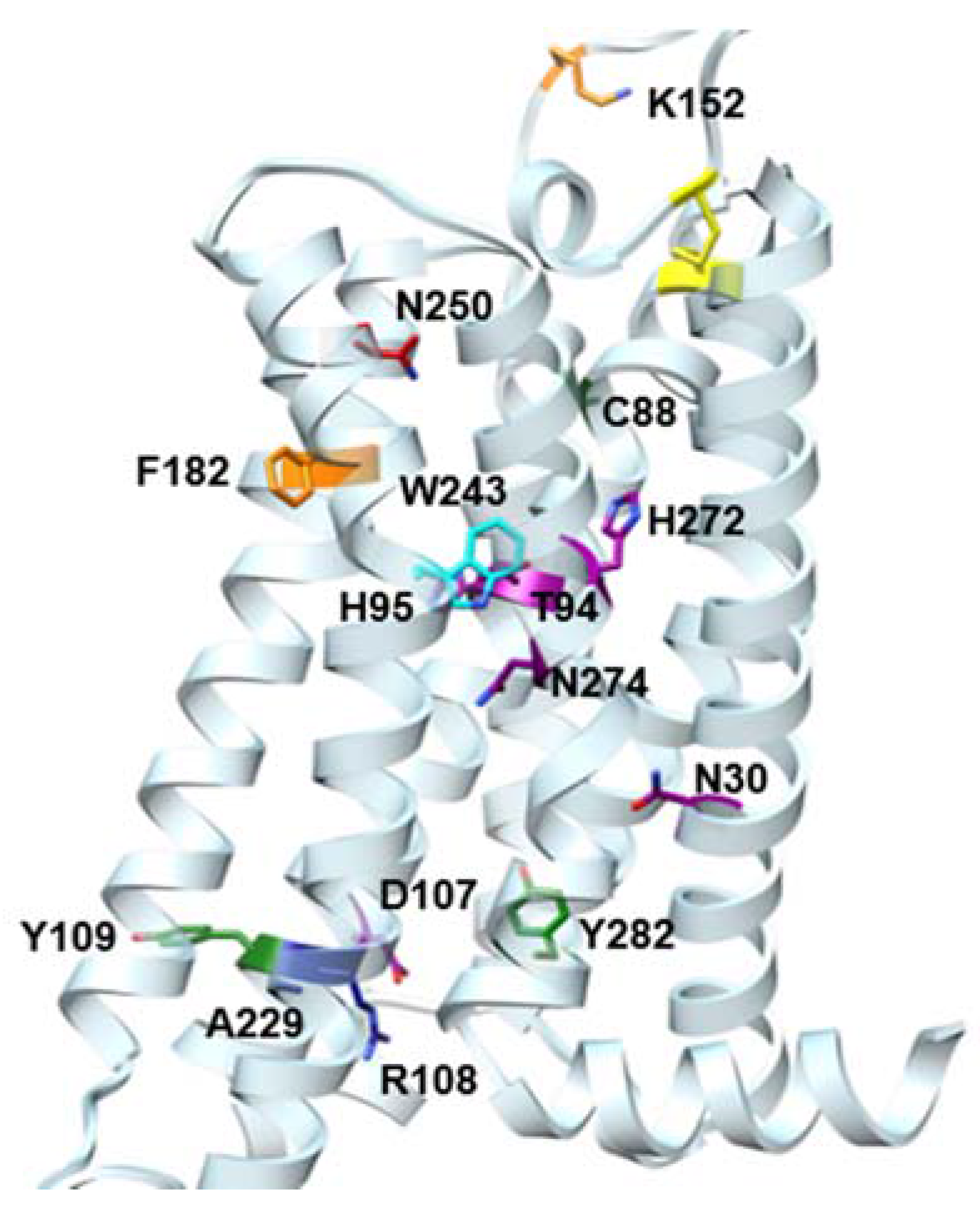
Figure 1. Selected representative adenosine receptor subtype A3 (A3AR) nucleoside ligands, including ribose, modified ribose and (N)-methanocarba derivatives (containing a bicyclic substitution of ribose in 1216 that locks the conformation). Ki values in the wild-type human A3AR (hA3AR) are shown [14,18,21,22,23,24,25,26,27,28,29]. Most are agonists, but 46 and 16 display less than full efficacy.
Molecules 22 00449 g001
Figure 2. Selected representative nonnucleoside A3AR antagonists 1723 and allosteric enhancers 2426. Antagonist Ki values in the hA3AR are shown [1,6,29,30,31].
Molecules 22 00449 g002
Figure 3. Summary of available hA3AR mutagenesis studies [14,15,16]. The color code is: green, mutation site affecting only agonist binding; orange, mutation site affecting only antagonist binding; purple, mutation site affecting both agonist and antagonist binding; blue, mutation site producing constitutively active mutants; red, Asn250 (6.55); cyan, Trp243 (6.48). The conserved TM3-EL2 disulfide bridge is also highlighted (yellow sticks). The residue Ballesteros–Weinstein notation [32] is: Asn30 (1.50), Cys88 (3.30), Thr94 (3.36), His95 (3.37), Asp107 (3.49), Arg108 (3.50), Tyr109 (3.51), Lys152 (EL2), Phe168 (EL2), Phe182 (5.43), Ala229 (6.34), Trp243 (6.48), Asn250 (6.55), His272 (7.43), Asn274 (7.45), and Tyr282 (7.53). TM, transmembrane α-helix.
Molecules 22 00449 g003
Figure 4. (a) Contacts of NECA 2 when docked in the hA3AR (using Ballesteros–Weinstein notation [32]). The residue numbers are Thr94 (3.36), Asn250 (6.55), His272 (7.43) and Ser271 (7.42); (b) Schematic depiction of the neoceptor approach at the A3AR: comparison of the binding of Cl-IB-MECA 9 at the wild-type hA3AR (left) with the binding of neoligand MRS1960 3 at the His7.43Glu mutant A3AR (right) [24]. Orange arrows denote H-bonds, while blue lines π-π stacking interactions.
Molecules 22 00449 g004
Figure 5. Docking pose of 6-Me analogue MRS5919 (13, magenta) at the hA3AR (hybrid homology model, A2AAR-β2 template) [23]. H-bond interactions are represented as orange solid lines. The residue Ballesteros–Weinstein notation [18] is: Thr94 (3.36), Phe168 (EL2), Asn250 (6.55), His272 (7.43), Ser271 (7.42) and His272 (7.43).
Molecules 22 00449 g005
Figure 6. Docking pose of MRS5679 (12, violet sticks) at the hA3AR model based on a hybrid A2AAR-opsin template where TM2 (violet ribbon) is shifted outward from the binding site with respect to the model based on the hybrid A2AAR-β2 template (purple ribbon) and the one based entirely on the human A2AAR (red ribbon) [27]. H-bond interactions are represented as orange solid lines and TM1 is omitted to aid visualization. The residue Ballesteros–Weinstein notation [32] is: Thr94 (3.36), Phe168 (EL2), Asn250 (6.55), His272 (7.43), Ser271 (7.42) and His272 (7.43).
Molecules 22 00449 g006
Figure 7. Comparison of the docking of a 2-(phenylethynyl) agonist MRS5644 (14, magenta) and the corresponding 2-(phenyl-triazolyl) agonist MRS7110 (15, purple) at the hybrid hA3AR model based on a A2AAR-β2 template [48]. The positions of the terminal phenyl rings overlap to a large degree. The residue Ballesteros–Weinstein notation is: Thr94 (3.36), Phe168 (EL2), Asn250 (6.55), His272 (7.43), Ser271 (7.42) and His272 (7.43).
Molecules 22 00449 g007
Figure 8. Representation of the ternary complex between hA3AR, the endogenous agonist adenosine (1, magenta) and the positive allosteric modulator LUF6000 (25, cyan) as obtained by supervised molecular dynamics (MD) simulation [20]: side view (a) and top view (b). The residue Ballesteros–Weinstein notation [32] is: Phe168 (EL2), Asn250 (6.55), and Ser181 (5.42).
Molecules 22 00449 g008
Figure 9. Structures of novel nonnucleosides (2730) and nucleosides (3134) found to bind to the hA3AR (Ki values given) as a result of structure-based virtual screening (VS) [52,53,54].
Molecules 22 00449 g009
Figure 10. A general SAR summary of nucleosides at the hA3AR, including effects on affinity, efficacy, selectivity and receptor conformation.
Molecules 22 00449 g010
Table 1. Summary of Site-Directed Mutagenesis Analyses at the hA3AR.
ResidueResidue Number 1MutationEffect on Binding of 2Reference
AgonistAntagonist
Asn301.50AlaDecreasedDecreased[14]
Asn582.50AsnNo changesNo Changes[14]
Cys883.30PheDecreasedNo effect[15]
Thr943.36AlaDecreasedDecreased[14]
His953.37AlaDecreasedDecreased[14]
Asp1073.49AsnMinor changesDecreased[14]
Arg1083.50Arg,Lys,Asn, Glu,HisMinor changesNo effect[15]
Tyr1093.51PheDecreasedNo effect[15]
Lys152(EL2)AlaNo effectDecrease[16]
Phe1825.43AlaNo effectDecreased[14]
Trp2436.48Ala,PheNo effect 3Decreased[16]
Asn2506.55AlaNo bindingNo binding[16]
His2727.43GluDecreasedDecreased[16,17]
Asn2747.45AlaDecreasedDecreased[14]
Tyr2827.53PheDecreasedNo effect[14]
1 [32]; 2 See text for chemotypes affected; 3 The ability of nucleosides to activate the receptor was progressively lost upon mutation to smaller residues. EL, extracellular loop.

