Communication

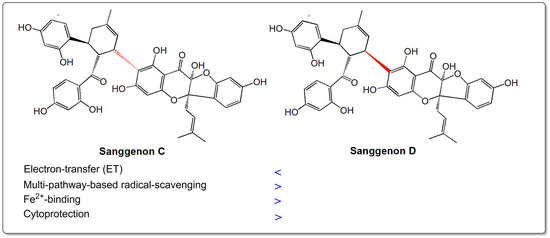
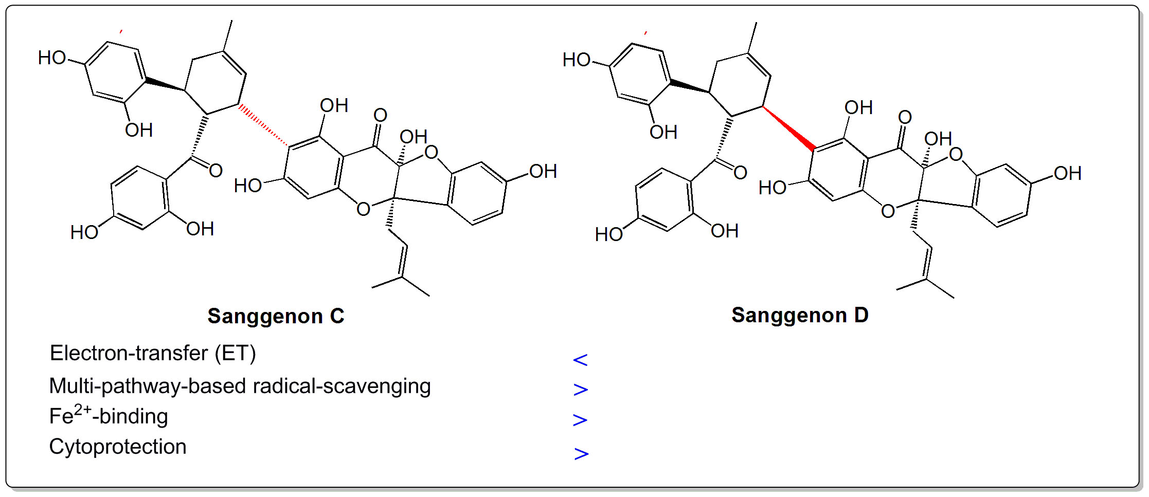
Steric Effect of Antioxidant Diels-Alder-Type Adducts: A Comparison of Sanggenon C with Sanggenon D

1
School of Chinese Herbal Medicine, Guangzhou University of Chinese Medicine, Guangzhou 510006, China
2
Innovative Research & Development Laboratory of TCM, Guangzhou 510006, China
3
School of Basic Medical Science, Guangzhou University of Chinese Medicine, Guangzhou 510006, China
4
The Research Center of Basic Integrative Medicine, Guangzhou University of Chinese Medicine, Guangzhou 510006, China
5
School of Biomedical Sciences, Faculty of Medicine, The Chinese University of Hong Kong, Sha Tin, Hong Kong 999077, China
*
Authors to whom correspondence should be addressed.
These authors contributed equally to this work.
Molecules 2018, 23(10), 2610; https://doi.org/10.3390/molecules23102610
Submission received: 18 July 2018 / Revised: 8 October 2018 / Accepted: 10 October 2018 / Published: 11 October 2018
(This article belongs to the Special Issue Antioxidants Chemistry and Applications)

Abstract

:
Sanggenons C and D are two Diels-Alder-type adducts from Chinese crude drug Sang-bai-pi. Structurally, both sanggenons construct stereoisomers. In the study, they were comparatively determined using four antioxidant assays, including ferric ion reducing antioxidant power (FRAP) assay, Cu2+-reducing assay, 1,1-diphenyl-2-picryl-hydrazl (DPPH•)-scavenging assay, and 2,2′-azino-bis (3-ethylbenzo-thiazoline-6-sulfonic acid radical (ABTS•+)-scavenging assay. Their Fe2+-binding reactions were explored using UV-Vis spectra. Finally, their cytoprotective effects were evaluated using flow cytometry. In electron transfer (ET)-based FRAP and Cu2+-reducing assays, sanggenon D was found to have lower IC50 values than sanggenon C; however, in multi-pathway-based DPPH•-scavenging and ABTS•+-scavenging assays, sanggenon C possessed lower IC50 values than sanggenon D. UV-Vis spectra suggested that sanggenon C generated a bathochromic-shift (286 nm → 302 nm) and displayed stronger UV absorption than sanggenon D. In flow cytometry, sanggenon C and sanggenon D, respectively, exhibited 31.1% and 42.0% early apoptosis-percentages towards oxidative-stressed mesenchymal stem cells (MSCs). In conclusion, both sanggenons may undergo multiple pathways (e.g., ET and Fe2+-binding) to protect MSCs against oxidative stress. In the mere ET aspect, sanggenon D possesses a higher level than sanggenon C, while in multi-pathway-based radical-scavenging, Fe2+-binding, and cytoprotection aspects, sanggenon C is more active than sanggenon D. These discrepancies can conclusively be attributed to the steric effect.

Graphical Abstract

1. Introduction

Diels-Alder-type adducts are unusual natural products occurring in plants. Until now, about 60 Diels-Alder-type adducts have already been obtained from plants, especially from the Morus family [1]. Two earliest-found Diels-Alder-type adducts sanggenons C and D are typical examples (Figure 1) [2,3]. Recent studies indicated that sanggenon C had various bioactivities, such as anti-cancer [4,5], anti-inflammatory [6], and cytoprotective effects against hypoxia injury in cardiac cells [6]. To the best of our knowledge, no cutting-edge biotechnology, such as stem cell technology, has been used to study Diels-Alder-type adducts until now.
Stem cell technology is undoubtedly one of the most attractive therapeutic tools in recent decades. This technology brings hope to the treatment of human diseases through tissue transplantation and regeneration. As ideal seed cells of tissue transplantation and regeneration, mesenchymal stem cells (MSCs) are usually used for clinical or experimental studies, owing to several advantages (such as differentiation potential [7,8], easy access [9], expansion [9], and proliferation [10,11]).
However, in the process of MSC expansion and proliferation, oxidative stress greatly lowers their survival and limits their clinical applications [11]. The so-called oxidative stress is from various reactive oxygen species (ROS) or reactive nitrogen species (RNS), such as hydroxyl radicals (•OH), peroxide hydrogen molecules (H2O2), and nitric oxide (•NO). Particularly, when H2O2 encounters ferrous (Fe2+), it can undergo a Fenton reaction (Fe2++ H2O2 → Fe3++•OH + OH) to give rise to hydroxyl radicals (•OH radicals) [12]. Hydroxyl radicals are known as the most powerful ROS form and can attack biomolecules (especially DNA [13]) to cause cellular damage [12]. Hence, supplementing phenolic Diels-Alder-type adducts is expected to resolve this bottleneck of cutting-edge stem cell technologies. As seen in Figure 1, both sanggenons C and D bear several phenolic –OHs and belong to phenolic Diels-Alder-type adducts. Thus, they are believed to act as antioxidants to protect MSCs from oxidative stress.
However, from the point of stereochemistry, two sanggenons are different: sanggenon C bears a (2R, 3S, 3″S, 4″R, 5″S) configuration, while sanggenon D displays a (3″R, 4″R, 5″S) configuration (Figure 1). The configurations of 2-C and 3-C in sanggenon D have remained unclear until now [14,15]. Nevertheless, sanggenons C and D are undoubtedly two steric isomers. In fact, a similar situation can also be observed in other Diels-Alder-type adducts, such as cathayanons A and B [16]. To our knowledge, no relevant study has discussed the possible steric effect in Diels-Alder-type adducts (and other phenolics). Thus, the present study tries to provide relevant evidence. The evidence is believed to be helpful to explain the steric effects in other Diels-Alder-type adducts, and to design new cytoprotector for stem cell technology.

2. Results and Discussion

In the study, FRAP assay has been fulfilled under pH 3.6. As seen in Figure S1A, the concentration dependency of sanggenons C and D increased the FRAP percentages. FRAP assay, however, is documented as a mere ET process [17,18], because such an acidic condition (pH 3.6) has remarkably suppressed H+ ionization. This implies that, when sanggenons C and D act as antioxidants, they may undergo ET pathways to exert their antioxidant abilities.
Unlike FRAP assay, Cu2+-reducing assay was conducted in pH 7.4 buffer, in which both sanggenons exhibited potent metal-reducing activity in a dose dependent manner (Figure S1B). It means that, under physiological pH 7.4, both sanggenons still have the ET potential. However, the ET potential of sanggenon D is higher than that of sanggenon C, according to the IC50 values (Table 1). Apparently, the difference in ET potential between sanggenons C and D can be attributed to the steric configuration of chiral atoms.
After all, antioxidation is not identical with a mere reducing reaction. In the antioxidant process, ET is usually accompanied by a proton (H+) transfer to give rise to a stable (semi) quinone form [13]. Detailed mechanisms are suggested to include hydrogen atom transfer (HAT) [19,20,21], sequential proton loss single electron transfer (SPLET) [22,23], proton coupled electron-transfer (PCET) [20,24], and sequential electron proton transfer (SEPT) [19,20]. For example, ABTS•+-scavenging, a single electron transfer reaction [25], has also been proven to be affected by H+ levels [26]. Thus, ABTS•+-scavenging is actually a multi-pathways-based antioxidant assay [18,27]. As seen in Figure S1C, sanggenons could dose-dependently increase the ABTS•+-scavenging percentages, suggesting that they could also undergo multiple pathways to exert the antioxidant action.
Evidence from DPPH•-scavenging assay further confirmed the above hypothesis based on ABTS•+-scavenging assay (Supplementary File S2D), because DPPH•-scavenging has also been demonstrated to occur via multi-pathways, including HAT [19,20,21], SPLET [22,23], SEPT [19,20], PCET [20,24], and ET [28,29]. However, the quantitative analysis of IC50 values (Table 1) indicated that, in multi-pathway-based ABTS•+-scavenging and DPPH•-scavenging aspects, sanggenon C is superior to its stereoisomer sanggenon D.
It should be noted that, some ROS can also be transformed through transition-metal (especially Fe2+) catalysis. For example, in the Fenton reaction, H2O2 molecules can be catalyzed by Fe2+ to produce •OH radicals. Therefore, the decrease of Fe2+ levels through a binding reaction can successfully reduce the •OH radicals to release cellular oxidative stress. Hence, iron binding by antioxidants has been developed to be an effective therapeutic approach for some oxidative stress diseases [30].
In the present study, two stereoisomers sanggenons C and D were found to be able to bind Fe2+. As illustrated in Figure 2A,B, within the initial 3 min, sanggenons C and D presented similar UV-peak intensities; however, in the subsequent 20 min, sanggenon C continued to increase the UV-peak intensity, while sanggenon D basically stopped the tendency. At the last stage of scanning (at 24 min), the UV peaks of sanggenon C were generally higher than those of sanggenon D. In the aspect of peak site, sanggenon C apparently produced a bathochromic shift (286 nm → 302 nm), and sanggenon D gave no bathochromic shift (289 nm → 289 nm). The data of UV-peak intensity and bathochromic shift showed that sanggenon C possessed stronger Fe2+-binding ability than sanggenon D.
The above difference can only be attributed to the steric configurations. As seen in Figure 1, in sanggenon C, 3″-substituent group is on one side together with 4″-substituent group. It provides a possibility that 5-OH, 7-OH, 8″-carbonyl group, and 10″-OH jointly bind Fe2+ to form a big cyclic complex. However, in sanggenon D, 3″-substituent group is on the reverse side with 4″-substituent group. It is impossible for the above phenolic -OH and carbonyl groups to jointly bind Fe2+ to form a big cyclic complex. As a result, sanggenon C is more active than sanggenon D in the Fe2+-binding reaction.
As mentioned above, the steric configurations of 2-C and 3-C atoms in sanggenon D are unknown. If the steric configurations of 2-C and 3-C atoms in sanggenon D are identical with those in sanggenon C; The difference in Fe2+-binding reaction can solely be attributed to the steric configuration of 3″-C. However, if the steric configurations of 2-C and 3-C atoms in sanggenon D are not identical with those in sanggenon C. The Fe2+-binding difference can be attributed to the steric configurations of 3″-C, 2-C, and 3-C atoms.
As seen in Figure 1, 2-C and 3-C atoms are far from the Fe2+-binding reaction center; their effects may be very limited in Fe2+-binding reaction.
In short, the difference in the Fe2+-binding reaction is seemingly from the difference in steric configuration of chiral atoms (especially 3″-C); however, the steric configurations (R/S) can change the chemical environment of these huge substituent groups and then affect molecular reactivity (especially metal-binding). This may be caused by the steric effect, which is usually ignored by chemists and pharmacists.
The steric effect can lead to different cytoprotective effects. As indicated by flow cytometry assay (Figure 3), the MSCs in the control, model, sanggenon C, and sanggenon D groups exhibited 4.37 ± 0.05%, 42.6 ± 1.65%, 31.3 ± 1.51%, and 42.0 ± 1.12% early apoptosis percentages, respectively. The results revealed that sanggenon C could effectively inhibit apoptosis of MSCs under oxidative stress and protect them from oxidative stress. However, sanggenon D was basically inactive. The difference from the cellular model is roughly parallel to those from multi-pathway-based radical-scavenging and Fe2+-binding assays.

3. Materials and Methods

3.1. Animals and Chemicals

Sprague-Dawley (SD) rats of 4 weeks of age were obtained from the Animal Center of Guangzhou University of Chinese Medicine. The protocol of this experiment was performed under the supervision of the Institutional Animal Ethics Committee in Guangzhou University of Chinese. Sanggenon C (CAS number: 80651-76-9, C40H36O12, M.W. 708.7, 98%, Supplementary File S2) and sanggenon D (CAS number: 81422-93-7, C40H36O12, M.W. 708.7, 97%) were obtained from Chengdu Biopurify Phytochemicals Ltd. (Chengdu, China, Supplementary File S3). The 1,1-diphenyl-2-picryl-hydrazl radical (DPPH•), (±)-6-hydroxyl-2,5,7,8-tetramethlychromane-2-carboxylic acid (Trolox), 2,9-dimethyl-1,10-phenanthroline (neocuproine), and 2,4,6-tripyridyltriazine (TPTZ) were purchased from Sigma-Aldrich Shanghai Trading Co. (Shanghai, China). (NH4)2ABTS [2,2′-azino-bis (3-ethylbenzo-thiazoline-6-sulfonic acid diammonium salt)] was purchased from Amresco Chemical Co. (Solon, OH, USA). Fetal bovine serum (FBS), Dulbecco’s modified Eagle’s medium (DMEM), and trypsin were from Gibco (Grand Island, NY, USA). Annexin V/propidium iodide (PI) assay kit was obtained from Abcam (Cambridge, UK). All other reagents were of analytical grade.

3.2. FRAP Assay

The FRAP assay was carried out according to the method of Benzie and Strain [17,31]. Briefly, the FRAP reagent was prepared freshly by mixing 10 mM TPTZ, 20 mM FeCl3, and 0.25 M acetate buffer at 1:1:10 at pH 3.6. The test sample (x = 2–10 μL, 0.5 mg/mL) was added to (20 − x) μL of 95% ethanol followed by 80 μL of FRAP reagent. The absorbance was measured at 595 nm after a 15 min incubation at room temperatures using distilled water as the blank. The relative reducing power of the sample was calculated using the formula:
R e l a t i v e   r e d u c i n g   e f f e c t   %   = A A m i n A m a x A m i n × 100 %
in which Amax is the maximum absorbance at 595 nm and Amin is the minimum absorbance at 595 nm in the test. A is the absorbance at 595 nm of sample.

3.3. Cu2+-Reducing Power Assay

The Cu2+-reducing power assay was performed based on a previously published method [19]. Briefly, 12 μL of CuSO4 aqueous solution (10 mmol/L), 12 μL of neocuproine ethanolic solution (7.5 mmol/L), and (75 − x) μL of NH4OAc buffer solution (0.1 mol/L, pH 7.5) were added to wells with different volumes of sample (0.1 mg/mL, 2–10 μL). The absorbance at 450 nm after 30 min was determined on a microplate reader (Multiskan FC, Thermo Scientific, Shanghai, China). The relative Cu2+-reducing power was calculated using the formula described above in Section 3.2

3.4. ABTS•+-Scavenging Assay

The ABTS•+-scavenging activity was assessed according to the method [32,33]. The ABTS• + was produced by mixing 0.2 mL of (NH4)2ABTS (7.4 mmol/L) with 0.35 mL of K2S2O8 (2.6 mmol/L). The mixture was kept in the dark at room temperature for 12 h and then diluted with distilled water (about 1:20), so that its absorbance at 734 nm was measured on a microplate reader (Multiskan FC, Thermo Scientific, Shanghai, China). To determine the scavenging activity, the test sample (x = 2–10 μL, 0.1 mg/mL) was added to (20 − x) μL of distilled water followed by 80 μL of ABTS• + reagent, and the absorbance at 734 nm was measured 3 min after the initial mixing, using distilled water as the blank. The percentage inhibition of the samples was calculated as follows:
S c a v e n g i n g   %   = A 0 A A 0 × 100 %
in which A0 indicates the absorbance of the blank and A indicates the absorbance of the sample, sanggenons C and D.

3.5. DPPH•-Scavenging Assay

DPPH• radical-scavenging activity was determined as previously described [34]. Briefly, 80μL of DPPH• solution (0.1 mol/L) was mixed with the indicated concentrations of sample (0.5 mg/mL, 4–20 μL) dissolved in methanol. The mixture was maintained at room temperature for 30 min, and the absorbance was measured at 519 nm on a microplate reader (Multiskan FC, Thermo Scientific, Shanghai, China). The DPPH• scavenging percentage was calculated based on the formula presented in Section 3.4.

3.6. UV-Vis-Spectra Analysis of Fe2+-Binding With Sanggenons

The sanggenon-Fe2+ complex was evaluated by UV-Vis-spectroscopy method [35]. For these experiments, 300 μL of a methanolic solution of sanggenons C and D (1 mg/mL) and 100 μL of an aqueous solution of FeCl2·4H2O (100 mg/mL) was added to 600 μL of an aqueous of the mixture of distilled water and methanol (1:1). The solution was then mixed vigorously. Subsequently, the mixture was diluted 20-fold, and the spectrum was obtained using a UV-Vis spectrophotometer every 3 min (Unico 2600A, Shanghai, China) from 200–400 nm.

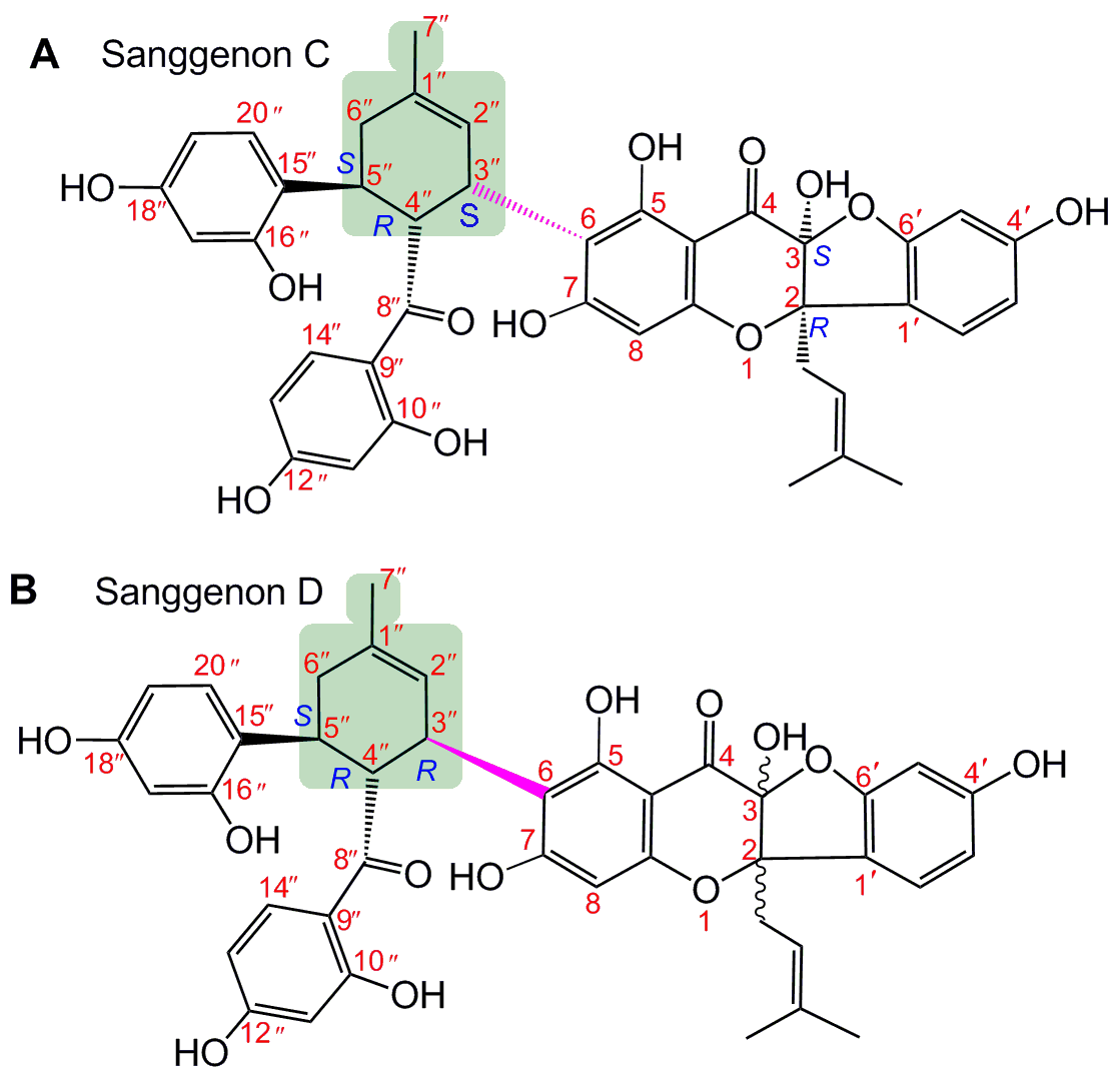
3.7. Flow Cytometry Assay

The MSCs were cultured according to our previous report with slight modifications [10,36]. In brief, bone marrow was obtained from the femur and tibia of rat. The marrow samples were diluted with DMEM (LG: low glucose) containing 10% FBS. MSCs were prepared by gradient centrifugation at 900 g for 30 min on 1.073 g/mL Percoll. The prepared cells were detached by treatment with 0.25% trypsin and passenged into cultural flasks at the density of 1 × 10⁴/cm2. The MSCs at passage 3 were evaluated for cultured cell homogeneity using detection of CD44 by flow cytometry and used for the investigation. The prepared cells were detached by treatment with 0.25% trypsin and passaged in culture flasks at the density of 1 × 10⁴/cm2. In the control group, MSCs were incubated in DMEM. In the model and sample groups, MSCs were treated with H2O2 (200 μM). After incubation for 3 h, MSCs in the control group and model group were incubated for 24 h in DMEM, while MSCs in the sample groups were incubated for 24 h in DMEM with sanggenons C and D.
The cells were then analyzed using flow cytometry assay described as the literature [37]. Briefly, the cultured cells were harvested by trypsin (0.05%) digestion in phosphate-buffered saline (PBS). The cells (5 × 10⁵/cm2) were collected by centrifugation, then re-suspended cells in 500 μL of 1X Binding Buffer; at the same time, 5 μL of Annexin V-FITC and 5 μL of propidium iodide were added. After incubating at room temperature for 5 min in the dark, fluorescence was measured using flow cytometry (Accuri C6, BD, USA) with standard software.

3.8. Statistical Analysis

The analysis was made in triplicate and the data were recorded as mean ± SD (standard deviation). The dose response curves were plotted using Origin 8.0 professional software (Origin Lab, Northampton, MA, USA). The IC50 value was defined as the final concentration of 50% radical inhibition (or relative metal-reducing power). Statistical comparisons were made by one-way ANOVA to detect significant difference using SPSS 13.0 (SPSS Inc., Chicago, IL, USA) for windows. p < 0.05 was considered statistically significant.

4. Conclusions

As two Diels-Alder-type adduct stereoisomers, sanggenons C and D may undergo an antioxidant approach to protect MSCs from oxidative stress. However, in the mere ET potential, sanggenon D is more effective than sanggenon C. In multi-pathway-based radical-scavenging, Fe2+-binding, and cytoprotection potentials, sanggenon C is more effective than sanggenon D. These differences can be attributed to the steric configurations from chiral atoms (especially 3″-C).

Supplementary Materials

Supplementary materials are available online. Supplementary File S1: Dose response curves of FRAP, Cu2+ reducing, ABTS, and DPPH assays, Supplementary File S2: Certificate of analysis of sanggenon C; Supplementary File S3: Certificate of analysis of sanggenon D.

Author Contributions

X.L. and D.C. conceived and designed the experiments; Z.R., Z.W., Z.F., and L.D. performed the experiments; H.X. analyzed the data; X.L. wrote the paper; and X.J. revised the paper. All authors read and approved the final manuscript.

Funding

This research was funded by Guangdong Science and Technology Project (2017A050506043, 2017A030312009), Guangdong Provincial Education Office Science and Technology Project (2017KCXTD007), and National Nature Science Foundation of China (81573558).

Conflicts of Interest

The authors declare that they have no competing interests.

Ethics Approval and Consent to Participate

The Institution Animal Ethics Committee in Guangzhou University of Chinese Medicine (Guangzhou, China) approved the protocols used in this study.

Abbreviations

The following abbreviations are used in this manuscript:
ABTS2,2′-azino-bis(3-ethylbenzo-thiazoline-6-sulfonic acid diammonium) salt
bmMSCsbone marrow-derived mesenchymal stem cells
DMEMDulbecco’s modified Eagle’s medium
DPPHDulbecco’s modified Eagle’s medium
ETelectron transfer
FBSfetal bovine serum
FRAPferric reducing antioxidant power
HAThydrogen atom transfer
PCETproton-coupled electron transfer
ROSreactive oxygen species
RNSreactive nitrogen species
SDstandard deviation
SEPTsequential electron proton transfer
SPLETsequential proton loss single electron transfer
TPTZ2,4,6-tris (2-pyridyl-s-triazine)
Trolox(±)-6-hydroxyl-2,5,7,8-tetramethlychromane-2-carboxylic acid

References

  1. Dai, S.J.; Lu, Z.M.; Chen, R.Y.; Yu, D.Q. Structure and spectral characteristics of Diels-Alder-type adducts from Morus. Yao Xue Xue Bao 2005, 40, 876–881. [Google Scholar] [PubMed]
  2. Nomura, T.; Fukai, T.; Hano, Y.; Uzawa, J. Structure of sanggenon D, a natural hypotensive Diels-Alder adduct from Chinese crude drug Sang-bai-pi. Heterocycles 1982, 17, 381–389. [Google Scholar] [CrossRef]
  3. Nomura, T.; Fukai, T.; Hano, Y.; Uzawa, J. Structure of sanggenon C, a natural hypotensive Diels-Alder adduct from Chinese crude drug Sang-bai-pi. Heterocycles 1981, 16, 2141–2148. [Google Scholar] [CrossRef]
  4. Chen, L.D.; Liu, Z.H.; Zhang, L.F.; Yao, J.N.; Wang, C.F. Sanggenon C induces apoptosis of colon cancer cells via inhibition of NO production, iNOS expression and ROS activation of the mitochondrial pathway. Oncol. Rep. 2017, 38, 2123–2131. [Google Scholar] [CrossRef] [PubMed]
  5. Huang, H.; Liu, N.; Zhao, K.; Zhu, C.; Lu, X.; Li, S.; Lian, W.; Zhou, P.; Dong, X.; Zhao, C.; et al. Sanggenon C decreases tumor cell viability associated with proteasome inhibition. Front. Biosci. 2011, 3, 1315–1325. [Google Scholar] [CrossRef]
  6. Gu, Y.; Gao, L.; Chen, Y.; Xu, Z.; Yu, K.; Zhang, D.; Zhang, G.; Zhang, X. Sanggenon C protects against cardiomyocyte hypoxia injury by increasing autophagy. Mol. Med. Rep. 2017, 16, 8130–8136. [Google Scholar] [CrossRef] [PubMed] [Green Version]
  7. Narakornsak, S.; Aungsuchawan, S.; Pothacharoen, P.; Markmee, R.; Tancharoen, W.; Laowanitwattana, T.; Thaojamnong, C.; Peerapapong, L.; Boonma, N.; Tasuya, W.; et al. Encouraging effects on chondrogenic differentiation of human amniotic fluid-derived mesenchymal stem cells. Acta Histochem. 2017, 119, 451–461. [Google Scholar] [CrossRef] [PubMed]
  8. Li, X.C.; Wei, G.; Wang, X.Z.; Liu, D.H.; Deng, R.D.; Li, H.; Zhou, J.H.; Li, Y.W.; Zeng, H.P.; Chen, D.F. Targeting of the sonic hedgehog pathway by atractylenolides promotes chondrogenic differentiation of mesenchymal stem cells. Biol. Pharm. Bull. 2012, 35, 1328–1335. [Google Scholar] [CrossRef] [PubMed]
  9. Matthay, M.A.; Pati, S.; Lee, J.W. Concise Review: Mesenchymal Stem (Stromal) Cells: Biology and Preclinical Evidence for Therapeutic Potential for Organ Dysfunction Following Trauma or Sepsis. Stem Cells 2017, 35, 316–324. [Google Scholar] [CrossRef] [PubMed] [Green Version]
  10. Chen, D.F.; Li, X.; Xu, Z.; Liu, X.; Du, S.H.; Li, H.; Zhou, J.H.; Zeng, H.P.; Hua, Z.C. Hexadecanoic Acid from Buzhong Yiqi Decoction Induces Proliferation of Bone Marrow Mesenchymal Stem Cells. J. Med. Food 2010, 13, 967–970. [Google Scholar] [CrossRef] [PubMed]
  11. Wang, G.; Li, X.; Zeng, H. Synthesis, Antioxidation Activity of (E)-9-p-Tolyl-3-[2-(8-hydroxy-quinol-2-yl)vinyl]-carbazole and (E)-9-(p-Anisyl)-3-[2-(8-hydroxy-quinol-2-yl)vinyl]-carbazole and Their Induction Proliferation of Mesenchymal Stem Cells. Acta Chim. Sin. 2009, 67, 974–982. [Google Scholar]
  12. Li, X.C.; Hu, Q.P.; Jiang, S.X.; Li, F.; Lin, J.; Han, L.; Hong, Y.L.; Lu, W.B.; Gao, Y.X.; Chen, D.F. Flos chrysanthemi indici protects against hydroxyl-induced damages to DNA and MSCs via antioxidant mechanism. J. Saudi Chem. Soc. 2015, 19, 454–460. [Google Scholar] [CrossRef]
  13. Li, X.C.; Mai, W.Q.; Chen, D.F. Chemical study on protective effect against hydroxyl-induced DNA damage and antioxidant mechanism of myricitrin. J. Chin. Chem. Soc. 2014, 61, 383–391. [Google Scholar] [CrossRef]
  14. Kim, H.J.; Baburin, I.; Zaugg, J.; Ebrahimi, S.N.; Hering, S.; Hamburger, M. HPLC-based activity profiling-discovery of sanggenons as gabaa receptor modulators in the traditional Chinese drug sang bai pi (morus alba root bark). Planta Med. 2012, 78, 440–447. [Google Scholar] [CrossRef] [PubMed]
  15. Shi, Y.Q.; Fukai, T.; Sakagami, H.; Chang, W.J.; Yang, P.Q.; Wang, F.P.; Nomura, T. Cytotoxic Flavonoids with Isoprenoid Groups from Morus mongolica. J. Nat. Prod. 2001, 64, 181–188. [Google Scholar] [CrossRef] [PubMed]
  16. Qin, H.L.; Yu, D.O. 1H-NMR Spectroscopic Databook of Natural Products, 1st ed.; Chemical Industry Press: Beijing, China, 2011; pp. 1046–1051. [Google Scholar]
  17. Benzie, I.F.; Strain, J.J. The ferric reducing ability of plasma (FRAP) as a measure of “antioxidant power”: The FRAP assay. Anal. Biochem. 1996, 239, 70–76. [Google Scholar] [CrossRef] [PubMed]
  18. Gülçin, İ. Antioxidant activity of food constituents: An overview. Arch. Toxicol. 2012, 86, 345–391. [Google Scholar]
  19. Li, X.C.; Han, W.; Mai, W.; Wang, L. Antioxidant Activity and mechanism of tetrahydroamentoflavone in vitro. Nat. Prod. Commun. 2013, 8, 787–789. [Google Scholar]
  20. Li, X.; Gao, Y.; Li, F.; Liang, A.; Xu, Z.; Bai, Y.; Mai, W.; Han, L.; Chen, D. Maclurin protects against hydroxyl radical-induced damages to mesenchymal stem cells: Antioxidant evaluation and mechanistic insight. Chem. Biol. Interact. 2014, 219, 221–228. [Google Scholar] [CrossRef] [PubMed]
  21. Lin, J.; Li, X.; Chen, L.; Lu, W.; Chen, X.; Han, L.; Chen, D. Protective effect against hydroxyl radical-induced DNA damage and antioxidant mechanism of [6]-gingerol: A Chemical Study. Bull. Korean Chem. Soc. 2014, 35, 1633–1638. [Google Scholar] [CrossRef]
  22. Li, X.; Liu, J.; Lin, J.; Wang, T.; Huang, J.; Lin, Y.; Chen, D. Protective effects of dihydromyricetin against •OH-induced mesenchymal stem cells damage and mechanistic chemistry. Molecules 2016, 21, 604. [Google Scholar] [CrossRef] [PubMed]
  23. López-Munguía, A.; Hernández-Romero, Y.; Pedraza-Chaverri, J.; Miranda-Molina, A.; Regla, I.; Martínez, A.; Castillo, E. Phenylpropanoid glycoside analogues: Enzymatic synthesis, antioxidant activity and theoretical study of their free radical scavenger mechanism. PloS ONE 2011, 6, e20115. [Google Scholar] [CrossRef] [PubMed]
  24. Smith, P.J.; Goeltz, J.C. Proton-Coupled Electron Transfer and Substituent Effects in Catechol-Based Deep Eutectic Solvents: Gross and Fine Tuning of Redox Activity. J. Phys. Chem. B 2017, 121, 10974–10978. [Google Scholar] [CrossRef] [PubMed]
  25. Lee, C.; Yoon, J. UV direct photolysis of 2,2′-azino-bis(3-ethylbenzothiazoline-6-sulfonate) (ABTS) in aqueous solution: Kinetics and mechanism. J. Photoch. Photobiol. A 2008, 197, 232–238. [Google Scholar] [CrossRef]
  26. Aliaga, C.; Lissi, E.A. Reactions of the radical cation derived from 2,2′-azinobis(3-ethylbenzothiazoline-6-sulfonic acid) (ABTS·+) with amino acids. Kinetics and mechanism. Can. J. Chem. 2000, 78, 1052–1059. [Google Scholar] [CrossRef]
  27. Osman, A.M.; Wong, K.; Fernyhough, A. ABTS radical-driven oxidation of polyphenols: Isolation and structural elucidation of covalent adducts. Biochem. Biophys. Res. Commun. 2006, 34, 6321–6329. [Google Scholar] [CrossRef] [PubMed]
  28. Osman, A.M. Multiple pathways of the reaction of 2,2-diphenyl-1-picrylhydrazyl radical (DPPH) with (+)-catechin: Evidence for the formation of a covalent adduct between DPPH and the oxidized form of the polyphenol. Biochem. Biophys. Res. Commun. 2011, 412, 473–478. [Google Scholar] [CrossRef] [PubMed]
  29. Li, X.C. 2-phenyl-4,4,5,5-tetramethylimidazoline-1-oxyl 3-oxide (PTIO•) radical-scavenging: A new and simple antioxidant assay in vitro. J. Agric. Food Chem. 2017, 65, 6288–6297. [Google Scholar] [CrossRef] [PubMed]
  30. Perron, N.R.; Brumaghim, J.L. A review of the antioxidant mechanisms of polyphenol compounds related to iron chelating. Cell Biochem. Biophys. 2009, 53, 75–100. [Google Scholar] [CrossRef] [PubMed]
  31. Li, X.C.; Jiang, Q.; Wang, T.T.; Liu, J.J.; Chen, D.F. Comparison of the Antioxidant Effects of Quercitrin and Isoquercitrin: Understanding the Role of the 6″-OH Group. Molecules 2016, 21, 1246. [Google Scholar] [CrossRef] [PubMed]
  32. Re, R.; Pellegrini, N.; Proteggente, A.; Pannala, A. Antioxidant activity applying an improved ABTS radical cation decolorization assay. Free Radic. Biol. Med. 1999, 26, 1231–1237. [Google Scholar] [CrossRef]
  33. Li, X.; Chen, D.; Mai, Y.; Wen, B.; Wang, X. Concordance between antioxidant activities in vitro and chemical components of Radix Astragali (Huangqi). Nat. Prod. Res. 2012, 26, 1050–1053. [Google Scholar] [CrossRef] [PubMed]
  34. Wang, T.T.; Zeng, G.C.; Li, X.C.; Zeng, H.P. In vitro studies on the antioxidant and protective effect of 2-substituted -8-hydroxyquinoline derivatives against H2O2-induced oxidative stress in BMSCs. Chem. Biol. Drug Des. 2010, 75, 214–222. [Google Scholar] [CrossRef] [PubMed]
  35. Liu, J.J.; Li, X.C.; Lin, J.; Li, Y.R.; Wang, T.T.; Jiang, Q.; Chen, D.F. Sarcandra glabra (Caoshanhu) protects mesenchymal stem cells from oxidative stress: A bioevaluation and mechanistic chemistry. BMC Complement. Altern. Med. 2016, 16, 423–432. [Google Scholar] [CrossRef] [PubMed]
  36. Jiang, Q.; Li, X.C.; Tian, Y.G.; Lin, Q.; Xie, H.; Lu, W.B.; Chi, Y.G.; Chen, D.F. Lyophilized aqueous extracts of Mori Fructus and Mori Ramulus protect Mesenchymal stem cells from •OH–treated damage: Bioassay and antioxidant mechanism. BMC Complement. Altern. Med. 2017, 17, 242. [Google Scholar] [CrossRef] [PubMed]
  37. Li, X.C.; Han, L.; Li, Y.R.; Zhang, J.; Chen, J.M.; Lu, W.B.; Zhao, X.J.; Lai, Y.T.; Chen, D.F.; Wei, G. Protective Effect of Sinapine against Hydroxyl Radical-Induced Damage to Mesenchymal Stem Cells and Possible Mechanisms. Chem. Pharm. Bull. 2016, 64, 319–325. [Google Scholar] [CrossRef] [PubMed]
Sample Availability: Sample of sanggenon D is available from the authors.
Figure 1. The structures of sanggenons C (A) and sanggenons D (B) (the Diels-Alder-type adduct skeletons are in light green).
Molecules 23 02610 g001
Figure 2. (A) UV spectra of reaction mixture of Fe2+ (50 mmol/L) with sanggenon C (0.07 mmol/L) within the initial 0–24 min; (B) UV spectra of reaction mixture of Fe2+ (50 mmol/L) with sanggenon D (0.07 mmol/L) within the initial 0–24 min; (C) the comparison of UV-vis spectra of sanggenon C and sanggenon D in Fe2+-binding reaction at 24 min.
Molecules 23 02610 g002
Figure 3. Cytoprotective effect of sanggenons C and D towards MSCs under oxidative stress based on flow cytometry assay (left upper, control; right upper, model; left lower, sanggenon C; right lower, sanggenon D). (MSCs, bone marrow-derived mesenchymal stem cells; each value is expressed as the mean ± SD, n = 3; * p < 0.05 vs. model.).
Molecules 23 02610 g003
Table 1. The IC50 values of sanggenons C and D in various antioxidant assays.
AssaysSanggenon C
μg/mL (μM)
Sanggenon D
μg/mL (μM)
Trolox
µg/mL (μM)
FRAP285.2 ± 18.3
(402.5 ± 25.8 c)
151.4 ± 21.2
(213.7 ± 30.0 b)
20.8 ± 1.4
(83.1 ± 5.5 a)
Cu2+-reducing7.6 ± 0.2
(10.8 ± 0.3 b)
5.8 ± 0.0
(8.2 ± 0.0 a)
5.7 ± 0.0
(22.8 ± 0.0 c)
ABTS•+-scavenging3.8 ± 0.3
(5.4 ± 0.4 a)
7.2 ± 0.5
(10.2 ± 0.7 b)
5.9 ± 0.1
(23.7 ± 0.2 c)
DPPH•-scavenging81.4 ± 1.9
(114.9 ± 2.7 b)
104.0 ± 1.2
(146.8 ± 1.7 c)
4.8 ± 0.1
(19.4 ± 0.6 a)
The IC50 value (in μg/mL unit) refers to the final concentration of 50% radical inhibition or relative metal-reducing power. It was calculated by linear regression analysis, which was conducted by Origin 8.0 professional software and expressed as the mean ± SD (n = 3). The IC50 value (μg/mL) was conversed into μM and collected in brackets. The IC50 values in μM with different superscripts (a or b) in the same row are significantly different (p < 0.05). Trolox is the positive control.

