
Synthesis and Cytotoxic Evaluation of 3-Methylidenechroman-4-ones
by

Jacek Kędzia
1, 
Tomasz Bartosik
1,
Joanna Drogosz
2,
Anna Janecka
2,
Urszula Krajewska
3 and
Tomasz Janecki
1,* 1
Institute of Organic Chemistry, Lodz University of Technology, Żeromskiego 116, 90-924 Łódź, Poland
2
Department of Biomolecular Chemistry, Medical University of Łódź, Mazowiecka 6/8, 92-215 Łódź, Poland
3
Department of Pharmaceutical Biochemistry and Molecular Diagnostics, Faculty of Pharmacy, Medical University of Łódź, Muszyńskiego 1, 90-151 Łódź, Poland
*
Author to whom correspondence should be addressed.
Molecules 2019, 24(10), 1868; https://doi.org/10.3390/molecules24101868
Submission received: 26 April 2019
/
Revised: 9 May 2019
/
Accepted: 11 May 2019
/
Published: 15 May 2019
(This article belongs to the Special Issue Design and Synthesis of Organic Molecules as Antineoplastic Agents)
Abstract
:In the search for new anticancer agents, a library of variously substituted 3-methylidenechroman-4-ones was synthesized using Horner–Wadsworth–Emmons methodology. Acylation of diethyl methylphosphonate with selected ethyl salicylates furnished 3-diethoxyphosphorylchromen-4-ones which were next used as Michael acceptors in the reaction with various Grignard reagents. The adducts were obtained as the mixtures of trans and cis diastereoisomers along with a small amount of enol forms. Their relative configuration and preferred conformation were established by NMR analysis. The adducts turned up to be effective Horner–Wadsworth–Emmons reagents giving 2-substituted 3-methylidenechroman-4-ones, which were then tested for their possible cytotoxic activity against two leukemia cell lines, HL-60 and NALM-6, and against MCF-7 breast cancer cell line. All new compounds (14a–o) were highly cytotoxic for the leukemic cells and showed a moderate or weak effect on MCF-7 cells. Analog 14d exhibited the highest growth inhibitory activity and was more potent than carboplatin against HL-60 (IC50 = 1.46 ± 0.16 µM) and NALM-6 (IC50 = 0.50 ± 0.05 µM) cells. Further tests showed that 14d induced apoptosis in NALM-6 cells, which was mediated mostly through the extrinsic pathway.

1. Introduction
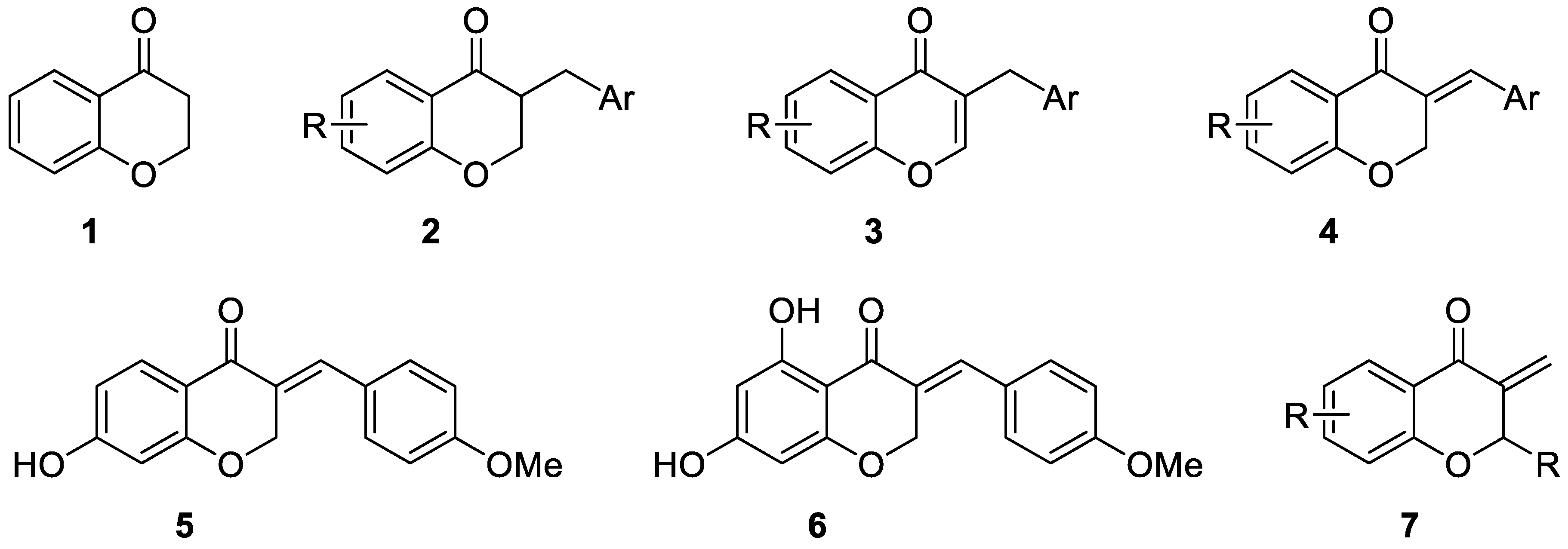
Chroman-4-one skeleton 1 is a core structure for a large group of plant metabolites called flavonoids, which possess many desirable biological activities including anticancer, antibacterial, and antioxidant properties [1,2]. One relatively small subgroup of flavonoids are homoisoflavonoids 2–4 which are characterized by the presence of arylmethyl or arylidene group in position 3 (Figure 1). A special place within this group belongs to 3-arylidenechroman-4-ones 4 which were found in many plants. For example, Bonducellin 5 was isolated from Caesalpina bonducella [3] and Eucomin 6 from Eucomis bicolor BAK (Liliaceae) [4].
Both natural and synthetic 3-arylidenechroman-4-ones 4 display valuable biological activities. They are potent and selective monoamine oxidase-B (MAO-B) inhibitors [5,6] and possess anti-cholinesterase activity [7,8], what makes them good candidates for the treatment of various neurological diseases such as Alzheimer’s or Parkinson’s disease. Furthermore, they show significant cytotoxicity for several cancer cell lines [9,10] and are cytochrome P450 aromatase inhibitors [11] being used for the treatment of advanced breast cancer. Also, their antifungal [12,13], antioxidant [14,15], and anti-inflammatory [16] activity was reported.
On the other hand, 3-methylidenechroman-4-ones 7, which are structurally closely related to 3-arylidenechroman-4-ones 4, have not been found in nature. Nevertheless, several syntheses of these compounds were reported. 2-Aryl-3-methylidenechroman-4-ones were obtained by Mannich reaction with 2-arylchroman-4-ones [17,18] and 2-alkyl(aryl)-3-methylidenechroman-4-ones were prepared by palladium-catalyzed tandem carbonylation-allene insertion reaction [19,20]. 3-Methylidenechroman-4-one was prepared by direct α-methylidenation mediated by diisopropylammonium trifluoroacetate [21] or by dehydration of 3-hydroxymethylchromen-4-one in the presence of methanesulfonylchloride [22]. Finally, 2-alkoxycarbonylmethyl-3-methylidenechroman-4-ones were obtained from enol silyl ethers of the corresponding chroman-4-ones [23,24]. Unfortunately, most of these methods have a very limited scope and/or are inefficient. For example, yields of the Mannich reactions were very low (3–22%) [17] or were not given because the obtained, crude 3-methylidenechroman-4-ones were used in further transformations [18]. On the other hand, palladium-catalyzed insertion reactions were more effective (23–90% yield) but required not readily available allenes as substrates. In turn the scope of direct α-methylidenation, dehydration of 3-hydroxymethylchromen-4-one or use the enol silyl ethers was limited to a single example in each case.
In contrast to 3-arylidenechroman-4-ones 4, biological activity of 3-methylidenechroman-4-ones 7 is poorly recognized. There is only one report describing their significant bacteriostatic activity against Gram-positive microorganisms [17]. However, an exo-cyclic methylidene bond conjugated with a carbonyl group present in 3-methylidenechroman-4-ones 7 is a structural motif found also in a large number of natural products, such as α-methylidene-γ- and δ-lactones [25,26] which react by the Michael-type addition with various bionucleophiles, disrupting key biological processes and are considered promising anticancer agents [27,28]. Consequently, we reasoned that also 3-methylidenechroman-4-ones 7 might have considerable cytotoxic activity.
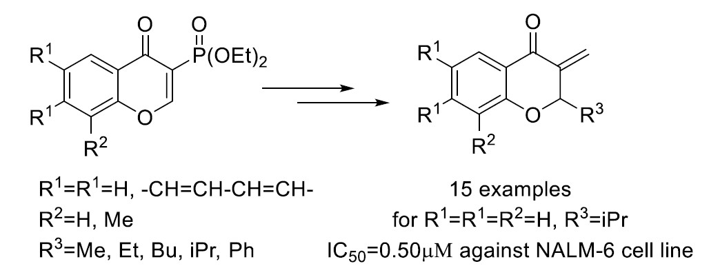
In this report, we present a new, general synthetic method for obtaining variously substituted 3-methylidenechroman-4-ones 14, based on the (well-recognized in our laboratory) Horner–Wadsworth–Emmons approach for the construction of exo-methylidene bond [29,30,31]. All obtained 3-methylidenechroman-4-ones 14 were evaluated in terms of their cytotoxic activity against three human cancer cell lines: promyelocytic leukemia HL-60, NALM-6, and breast adenocarcinoma cell line MCF-7. The most cytotoxic compound, 14d was selected for further experiments and its effect on the induction of apoptosis was investigated.
2. Results and Discussion
2.1. Chemistry
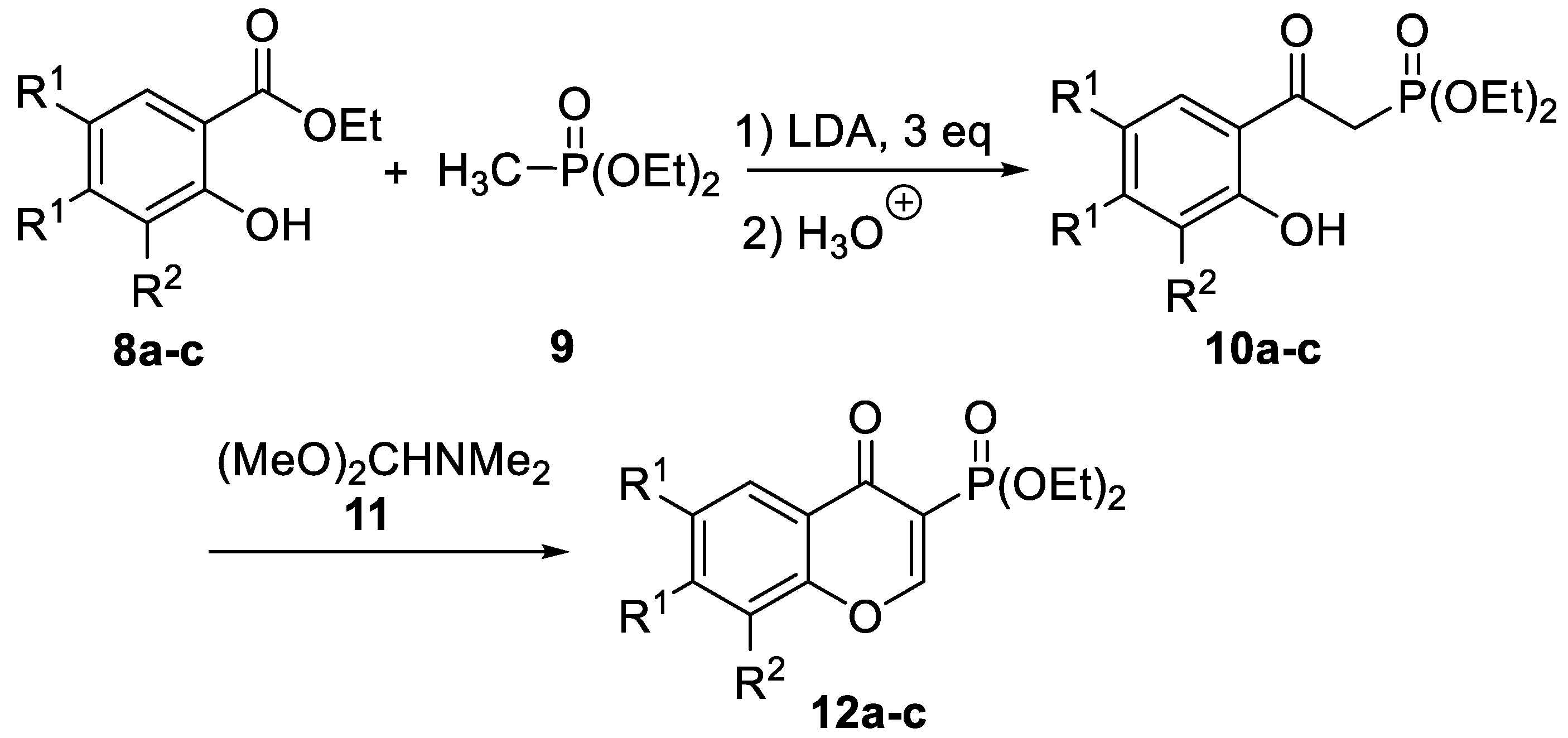
The first step was the synthesis of 3-diethoxyphosphorylchromen-4-ones 12a–c, which are crucial intermediates in our methodology. Literature search revealed that there is no efficient method for obtaining 2-unsubstituted 3-phosphorylchromen-4-ones. In the only report we have found, 3-diethoxyphosphorylchromen-4-one was formed in 5% yield, as a side product in free radical phosphorylation of chromen-4-one [32]. Therefore, we worked out a two-step procedure, which starts with the reaction of commercially available ethyl salicylates 8a–c with diethyl methylphosphonate 9 in the presence of three equivalents of LDA (Scheme 1). Using this stoichiometry, protection of the hydroxyl group is avoided and we believe this is the major improvement over the reported acylation of dimethyl methylphosphonate using benzyl protected methyl salicylate [33]. The standard work-up and column chromatography purification gave 2-(2-hydroxyphenyl)-2-oxoethylphoshonates 10a–c in good yields (Table 1). In the next step, reaction between phophonates 10a–c and dimethylformamide dimethyl acetal 11 gave, after purification by column chromatography, 3-diethoxyphosphorylchromen-4-ones 12a–c in high yields (Table 1 and Supplementary Materials).
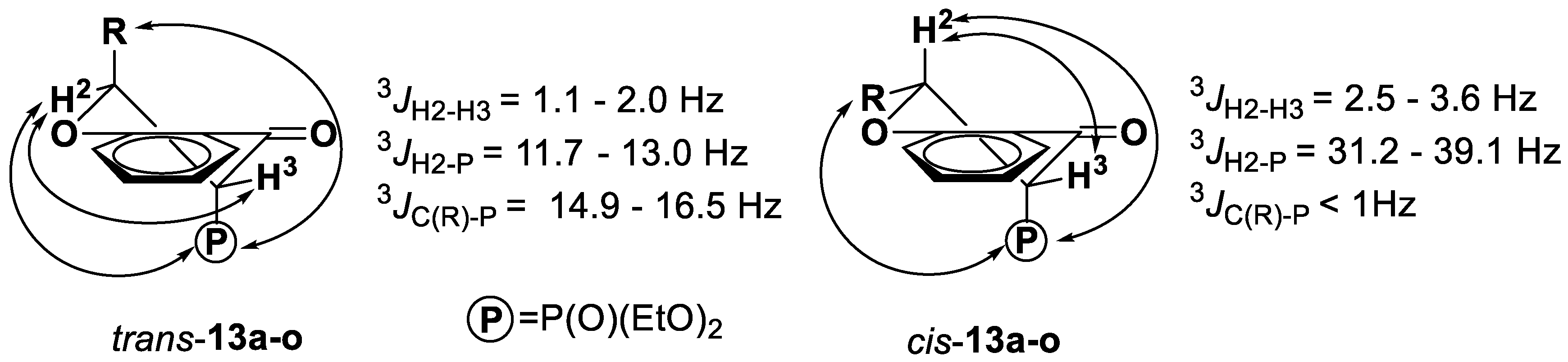
With 3-diethoxyphosphorylchromen-4-ones 12a–c in hand, we performed their reactions with various Grignard reagents (Scheme 2). In all cases, after standard work-up, we received adducts 13a–o, which were purified by column chromatography with yields given in Table 2. Interestingly, examination of the 1H, 13C and 31P NMR spectra revealed that all adducts 13 were formed as mixtures of trans and cis diastereoisomers (trans- or cis-13a–o), along with small amount of enol form (enol-13a–o), with trans diastereoisomers strongly predominating. Ratios, determined from the 31P NMR spectra of the crude reaction mixtures, are given in Table 2.
Careful analysis of the NMR spectra showed also that both trans- and cis-13a–o exist in the half-chair conformation and diethoxyphosphoryl group occupies the axial position (Figure 2). Simple application of Karplus correlation between corresponding dihedral angles and coupling constants 3JH2-H3, 3JH2-P and 3JC(R3)-P, determined from the 1H and 13C NMR spectra of trans- and cis-13a–o shows full agreement with the proposed configurations and conformations. Corresponding coupling constants are given in Figure 2. Similar half-chair conformation with diethoxyphosphoryl group in axial position was reported for 3-diethoxyphosphorylchroman-2-ones [34,35].
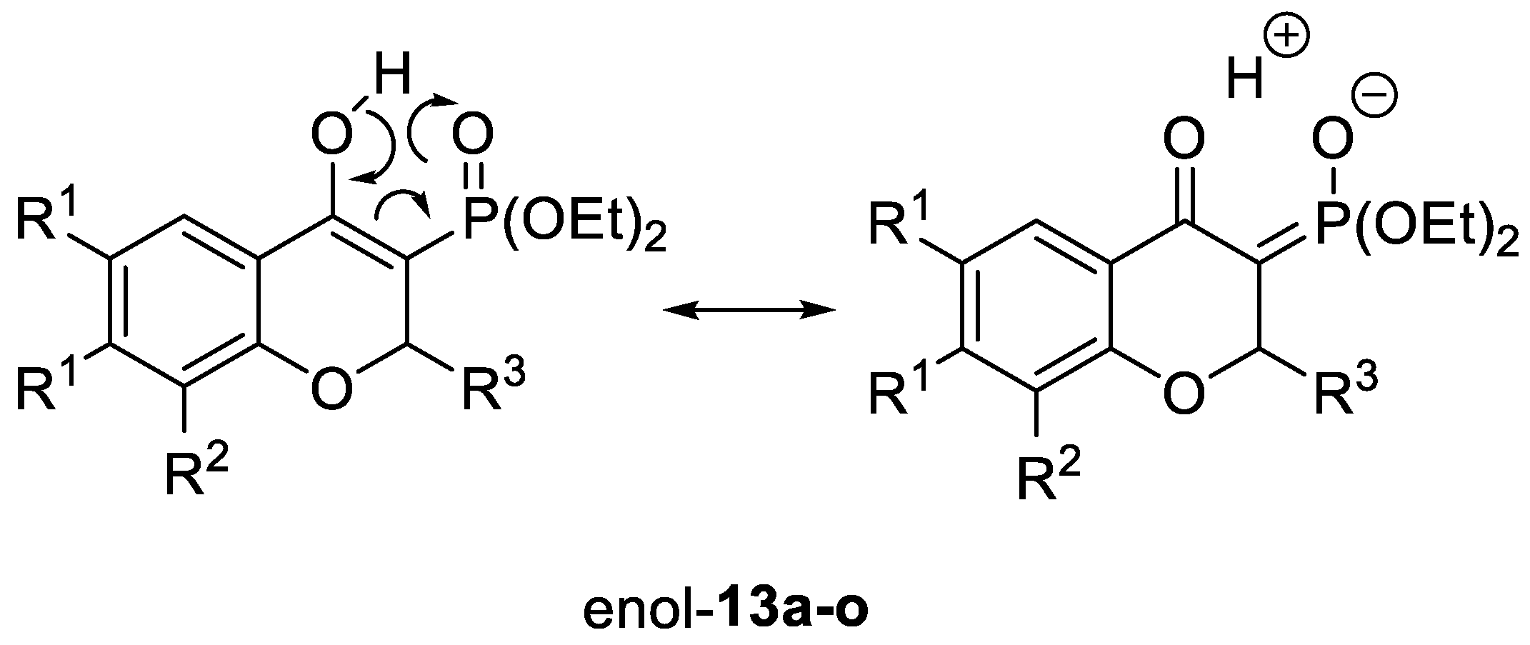
On the other hand, 1H NMR spectra of enols-13a–o revealed very characteristic doublets with coupling constant 4JP-H ~ 1 Hz and chemical shift in the range of 11–12 ppm, which can be assigned to the proton of the hydroxyl group. Coupling between this proton and phosphorus indicates the presence of resonance-assisted hydrogen bond (RAHB) [36] and can be visualized by resonance structures shown in Figure 3. Recently, we have reported on the existence of RAHB in 3-(dimenthoxyphosphoryl)-2-phenyl-1,2-dihydroquinolin-4-ol [37], what was the first example of this phenomenon in organophosphorus compounds. Now, we can confirm the existence of RAHB also in 3-diethoxyphosphoryl-2H-chromen-4-oles.
Finally, all adducts 13a-o were transformed into 3-methylidenechroman-4-ones 14a–o performing Horner–Wadsworth–Emmons olefination of formaldehyde. The best results were obtained with K2CO3 used as a base and formalin as a source of formaldehyde (Scheme 3). Standard work-up and purification by column chromatography furnished 3-methylidenechroman-4-ones 14a–o in moderate to good yields (Table 2).
2.2. Biology
2.2.1. In Vitro Cytotoxicity of New Analogs Against Three Cancer Cell Lines
All obtained 3-methylidenechroman-4-ones 14a–o were evaluated for their possible cytotoxic activity against three human cancer cell lines: leukemia HL-60 and NALM-6 and breast adenocarcinoma MCF-7 using the MTT assay (after 48 h incubation) (Table 3). Carboplatin served as a reference compound.
Analysis of the structure–activity relationship revealed that chromanones 14a–j were, in general, more potent than benzochromanones 14k–o, containing additional benzene ring ortho-fused with a chromanone skeleton. All compounds (14a–o) were much more cytotoxic for leukemia cells than for the solid tumor MCF-7 cells. In both series of chromanones, 14a–e (R2 = H) and 14f–j (R2 = Me), the most potent compounds against leukemic HL-60 and NALM-6 cells were these containing an i-propyl substituent in position 2, i.e., 14d and 14i, respectively. For HL-60 cells only 14d was more cytotoxic than the reference carboplatin. The highest cytotoxicity was observed against NALM-6 cells, with three analogs 14b, 14d, and 14i exhibiting lower half maximal inhibitory concentration values (IC50) than carboplatin. Analog 14d was the most cytotoxic chromanone for NALM-6 cells (IC50 of 0.5 ± 0.05 µM) and was selected for further investigation of its potential antineoplastic properties.
2.2.2. Apoptotic Cell Death Determination
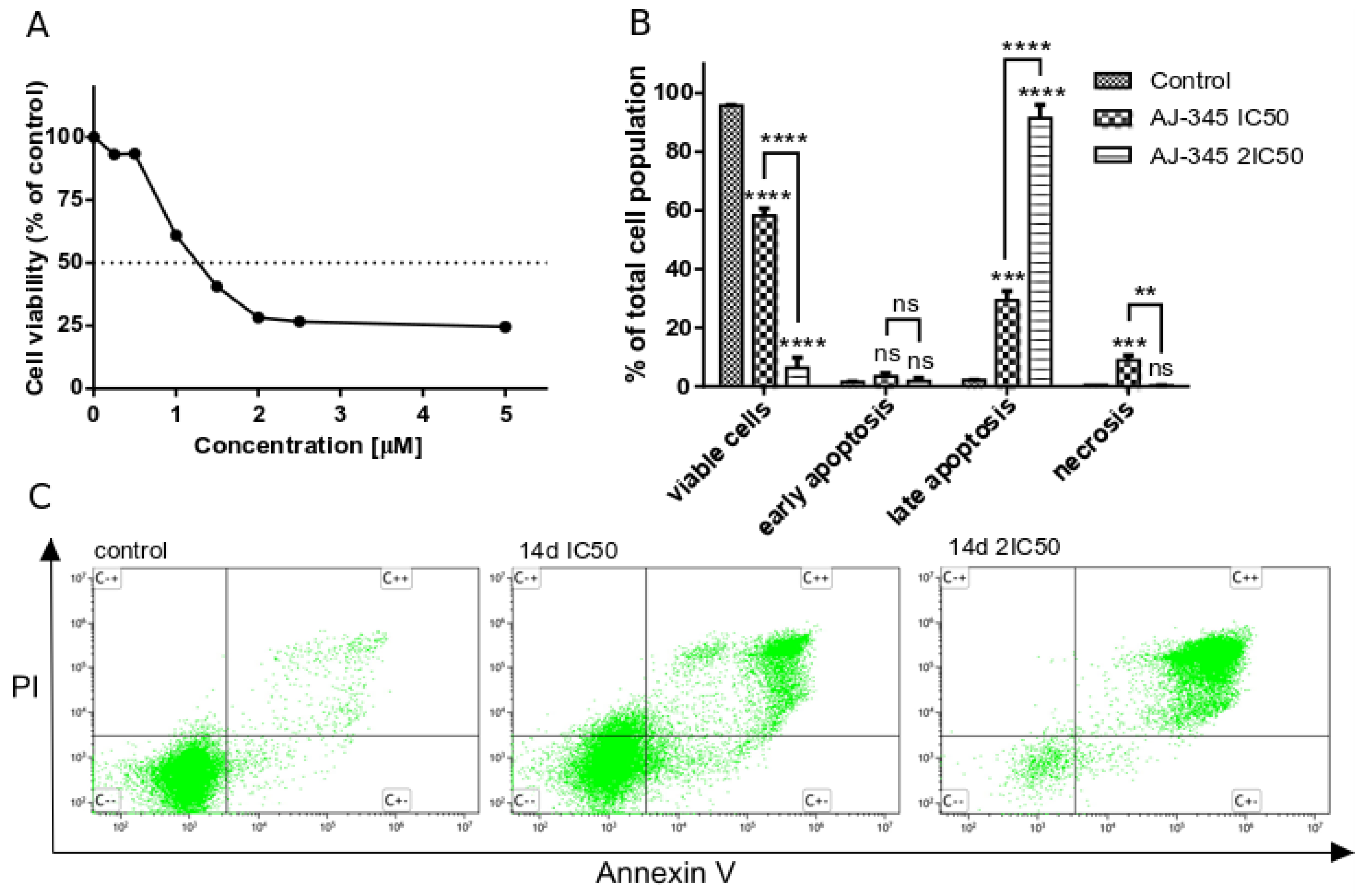
It is now well documented that most anticancer drugs induce apoptosis. One of the main characteristics of apoptosis, phosphatidylserine (PS) translocation to the outer surface of the cellular membrane, was investigated by double-staining with Annexin V and propidium iodide (PI). Annexin V is a protein exerting high affinity to PS exposed on the outer surface of the plasma membrane, enabling detection of even an early stage apoptosis. The late stage of apoptosis is characterized by loss of membrane integrity allowing permeation of a dye such as PI into the cells [38,39]. Treatment of NALM-6 cells for 24 h (Figure 4A) with the selected analog 14d at 1.25 µM (IC50) and 2.5 µM (2 IC50) concentrations led to the increase of Annexin V and PI-positive cells from 2.2% to 29.4% and 91.4%, respectively (Figure 4B,C), showing that 14d induced the late stage of apoptosis in NALM-6 cells.
Apoptosis occurs when caspases, which have proteolytic activity, cleave specific substrates, causing cell death. Depending on the initiator caspase involved in this process, the apoptosis may be mediated by the intrinsic (caspase 9) or extrinsic (caspase 8) pathway [40]. To investigate which caspases were involved in the apoptosis inducted by analog 14d in NALM-6 cells, the cells were treated with 14d at 1.25 µM and 2.5 µM concentrations for 6 h. Then, the activity of executioner caspase 3 and initiator caspases 8 and 9 was quantified using fluorogenic indicators. Results presented in Figure 5 indicate that the levels of caspase 3, 8 and 9 were significantly increased: 2.7- and 3.7-fold for caspase 3, 4.9- and 11.3-fold for caspase 8, and 1.25- and 5.7-fold for caspase 9 after treatment of the cells with 1.25 µM and 2.5 µM concentration of 14d, respectively. Activation of the extrinsic pathway was more prominent than the intrinsic pathway.
Presented results indicate that compound 14d is a potent cytotoxic agent that significantly inhibits metabolic activity of NALM-6 cells with IC50 value as low as 0.5 µM. Analog 14d also promotes apoptosis in the investigated cell line, which is mediated by the extrinsic and in a much lesser extend intrinsic pathway.
3. Materials and Methods
3.1. Chemistry
3.1.1. General Information
NMR spectra were recorded on a Bruker DPX 250 or Bruker Avance II instrument at 250.13 MHz or 700 MHz for 1H, 62.9 MHz or 176 MHz for 13C, and 101.3 MHz for 31P NMR with tetramethylsilane used as an internal and 85% H3PO4 as an external standard. 31P NMR spectra were recorded using broadband proton decoupling. IR spectra were recorded on a Bruker Alpha ATR spectrophotometer. Melting points were determined in open capillaries and are uncorrected. Optical rotations were measured on a Perkin-Elmer 241 polarimeter. The [α]D values are given in deg·cm2·g−1 and concentration c in g·(100 mL)−1. Column chromatography was performed on silica gel 60 (230–400 mesh) (Aldrich, Steinheim, Germany). Thin-layer chromatography was performed on the pre-coated TLC sheets of silica gel 60 F254 (Aldrich, Steinheim, Germany). The purity of the synthesized compounds was confirmed by the combustion elemental analyses (CHN, elemental analyzer EuroVector 3018, Elementar Analysensysteme GmbH (Langenselbold, Germany). MS spectra were recorded on Waters 2695-Waters ZQ 2000 LC/MS apparatus (Waters Corporation, Milford, MA, USA). All reagents and starting materials were purchased from commercial vendors and used without further purification. Organic solvents were dried and distilled prior to use. Standard syringe techniques were used for transferring dry solvents.
3.1.2. General Procedure for the Synthesis of 2-Substituted 3-Metylidenechroman-4-ones 14a–o.
To the vigorously stirred solution of 2-substituted 3-diethoxychroman-4-on 13a–o (0.15 mmol) in THF (1.5 mL), formaldehyde (36–38% solution in water, 0.125 mL, ca. 1.50 mmol) was added at 0 °C, followed by addition of K2CO3 (41 mg, 0.30 mmol) in water (0.4 mL). The resulting mixture was stirred vigorously at 0 °C for 3 h. Next Et2O (5 mL) was added and layers were separated. The water fraction was washed with Et2O (5 mL). Organic fractions were combined, washed with brine (5 mL) and dried over MgSO4. The solvents were evaporated under reduced pressure and the resulting crude product was then purified by column chromatography (eluent CH2Cl2).
2-Methyl-3-methylidenechroman-4-one (14a) (17.7 mg, 68%). Colourless oil. 1H NMR (700 MHz, Chloroform-d) δ 1.63 (d, J = 6.4 Hz, 3H), 5.03–5.14 (m, 1H), 5.55 (dd, J = 2.0, 0.8 Hz, 1H), 6.32 (dd, J = 1.6, 0.8 Hz, 1H), 6.95 (dd, J = 8.3, 1.0 Hz, 1H), 7.03 (ddd, J = 8.1, 7.2, 1.1 Hz, 1H), 7.47 (ddd, J = 8.6, 7.1, 1.8 Hz, 1H), 7.97 (dd, J = 7.9, 1.8 Hz, 1H). 13C NMR (176 MHz, Chloroform-d) δ 19.03, 76.36, 118.30, 121.44, 121.59, 121.82, 128.00, 136.14, 143.68, 161.30, 182.66. ESI-MS [M + H]+ = 175.2. Anal. Calcd for C11H10O2: C, 75.84%; H, 5.79%. Found: C, 75.61%, H, 5.71%.
2-Ethyl-3-methylidenechroman-4-one (14b) (20.1 mg, 71%). Colourless oil. 1H NMR (700 MHz, Chloroform-d) δ 1.04 (t, J = 7.3 Hz, 3H), 1.75–1.89 (m, 1H), 1.91–2.03 (m, 1H), 4.90 (t, J = 7.0 Hz, 1H), 5.50 (s, 1H), 6.33 (s, 1H), 6.96 (d, J = 8.4 Hz, 1H), 7.02 (t, J = 7.5 Hz, 1H), 7.47 (s, 1H), 7.95 (d, J = 7.9 Hz, 1H). 13C NMR (176 MHz, Chloroform-d) δ 9.83, 26.65, 81.89, 118.47, 121.41, 121.66, 122.33, 127.84, 136.21, 142.36, 160.77, 182.49. ESI-MS [M + H]+ = 189.3. Anal. Calcd for C12H12O2: C, 76.57%; H, 6.43%. Found: C, 76.41%, H, 6.51%.
2-Butyl-3-methylidenechroman-4-one (14c) (19.1 mg, 59%). Colourless oil. 1H NMR (700 MHz, Chloroform-d) δ 0.91 (t, J = 7.3 Hz, 3H), 1.31–1.45 (m, 3H), 1.47–1.55 (m, 1H), 1.71–1.80 (m, 1H), 1.89–2.00 (m, 1H), 4.86–5.10 (m, 1H), 5.50 (t, J = 1.2 Hz, 1H), 6.32 (t, J = 1.0 Hz, 1H), 6.95 (dd, J = 8.4, 1.0 Hz, 1H), 7.02 (ddd, J = 8.1, 7.2, 1.1 Hz, 1H), 7.47 (ddd, J = 8.6, 7.1, 1.8 Hz, 1H), 7.96 (dd, J = 7.8, 1.8 Hz, 1H).13C NMR (176 MHz, Chloroform-d) δ 14.09, 22.48, 27.50, 33.18, 80.64, 118.48, 121.43, 121.66, 122.17, 127.85, 136.22, 142.65, 160.78, 182.56. ESI-MS [M + H]+ = 217.4. Anal. Calcd for C14H16O2: C, 77.75%; H, 7.46%. Found: C, 77.52%, H, 7.25%.
2-Isopropyl-3-methylidenechroman-4-one (14d) (16.1 mg, 53%). Colourless oil. 1H NMR (700 MHz, Chloroform-d) δ 0.90 (d, J = 6.8 Hz, 3H), 1.04 (d, J = 6.6 Hz, 3H), 1.96–2.16 (m, 1H), 4.62 (dt, J = 8.5, 1.1 Hz, 1H), 5.46 (t, J = 1.2 Hz, 1H), 6.36 (t, J = 1.0 Hz, 1H), 6.95 (ddd, J = 8.3, 1.1, 0.5 Hz, 1H), 7.00 (ddd, J = 8.0, 7.1, 1.1 Hz, 1H), 7.47 (ddd, J = 8.3, 7.1, 1.8 Hz, 1H), 7.93 (ddd, J = 7.9, 1.8, 0.5 Hz, 1H). 13C NMR (176 MHz, Chloroform-d) δ 18.51, 18.83, 30.78, 86.66, 118.41, 121.49, 121.57, 123.80, 127.66, 136.31, 141.26, 160.53, 182.44. ESI-MS [M + H]+ = 203.0. Anal. Calcd for C13H14O2: C, 77.20%; H, 6.98%. Found: C, 77.42%, H, 7.05%.
3-Methylidene-2-phenylchroman-4-one (14e) (29.8 mg, 84%). Yelowish oil. 1H NMR (700 MHz, Chloroform-d) δ 5.16–5.23 (m, 1H), 6.02 (d, J = 1.7 Hz, 1H), 6.44 (t, J = 1.2 Hz, 1H), 7.35–7.39 (m, 1H), 7.39–7.45 (m, 4H), 7.50 (ddd, J = 8.7, 7.2, 1.8 Hz, 1H), 7.99 (dd, J = 7.9, 1.7 Hz, 1H). 13C NMR (176 MHz, Chloroform-d) δ 82.49, 118.47, 121.76, 122.13, 125.09, 127.58, 128.02, 128.78, 128.87, 136.30, 137.27, 142.76, 161.14, 182.22. ESI-MS [M + H]+ = 237.4. Anal. Calcd for C16H12O2: C, 81.34%; H, 5.12%. Found: C, 81.42%, H, 5.23%.
2,8-Dimethyl-3-methylidenechroman-4-one (14f) (18.1 mg, 64%). Yelowish oil. 1H NMR (700 MHz, Chloroform-d) δ 1.64 (d, J = 6.5 Hz, 3H), 2.23 (s, 3H), 5.10 (qt, J = 6.4, 1.8 Hz, 1H), 5.54 (dd, J = 2.0, 0.9 Hz, 1H), 6.31 (dd, J = 1.6, 0.9 Hz, 1H), 6.93 (t, J = 7.6 Hz, 1H), 7.33 (ddd, J = 7.2, 1.8, 1.0 Hz, 1H), 7.82 (dd, J = 7.9, 1.7 Hz, 1H). 13C NMR (176 MHz, Chloroform-d) δ 15.72, 19.13, 76.19, 121.08, 121.21 × 2, 125.56, 127.50, 136.91, 143.77, 159.53, 183.04. ESI-MS [M + H]+ = 189.0. Anal. Calcd for C12H12O2: C, 76.57%; H, 6.43%. Found: C, 76.42%, H, 6.55%.
2-Ethyl-8-methyl-3-methylidenechroman-4-one (14g) (17.9 mg, 59%). Yelowish oil. 1H NMR (700 MHz, Chloroform-d) δ 1.07 (t, J = 7.4 Hz, 3H), 1.73 – 1.86 (m, 1H), 1.90–2.03 (m, 1H), 2.25 (t, J = 0.7 Hz, 3H), 4.76 – 5.05 (m, 1H), 5.50 (dd, J = 1.5, 1.0 Hz, 1H), 6.32 (t, J = 1.1 Hz, 1H), 6.92 (t, J = 7.5 Hz, 1H), 7.34 (ddd, J = 7.2, 1.8, 0.9 Hz, 1H), 7.81 (ddd, J = 7.9, 1.8, 0.7 Hz, 1H). 13C NMR (176 MHz, Chloroform-d) δ 10.09, 15.69, 26.69, 81.76, 121.08, 121.11, 121.88, 125.45, 127.66, 136.99, 142.60, 158.94, 182.87. ESI-MS [M + H]+ = 203.0. Anal. Calcd for C13H14O2: C, 77.20%; H, 6.98%. Found: C, 77.38%, H, 7.09%.
2-Butyl-8-methyl-3-methylidenechroman-4-one (14h) (20.0 mg, 52%). Yelowish oil. 1H NMR (700 MHz, Chloroform-d) δ 0.92 (t, J = 7.3 Hz, 3H), 1.29–1.49 (m, 3H), 1.49–1.58 (m, 1H), 1.71–1.80 (m, 1H), 1.89–2.01 (m, 1H), 2.24 (s, 3H), 4.98 (ddt, J = 9.0, 5.1, 1.4 Hz, 1H), 5.50 (t, J = 1.2 Hz, 1H), 6.30 (d, J = 1.2 Hz, 1H), 6.92 (t, J = 7.6 Hz, 1H), 7.31–7.39 (m, 1H), 7.81 (dd, J = 7.8, 1.7 Hz, 1H). 13C NMR (176 MHz, Chloroform-d) δ 14.12, 15.73, 22.44, 27.70, 33.12, 80.39, 121.12, 121.73, 125.46, 127.66, 136.99, 142.85, 158.99, 182.96. ESI-MS [M + H]+ = 231.0. Anal. Calcd for C15H18O2: C, 78.23%; H, 7.88%. Found: C, 78.44%, H, 8.05%.
2-Isopropyl-8-methyl-3-methylidenechroman-4-one (14i) (17.2 mg, 53%). Yelowish oil. 1H NMR (700 MHz, Chloroform-d) δ 0.92 (d, J = 6.7 Hz, 3H), 1.05 (d, J = 6.6 Hz, 3H), 1.95–2.12 (m, 1H), 2.26 (s, 3H), 4.66 (dd, J = 8.3, 1.3 Hz, 1H), 5.46 (d, J = 1.1 Hz, 1H), 6.35 (t, J = 1.1 Hz, 1H), 6.91 (t, J = 7.6 Hz, 1H), 7.34 (ddd, J = 7.2, 1.8, 0.9 Hz, 1H), 7.79 (dd, J = 7.9, 1.7 Hz, 1H). 13C NMR (176 MHz, Chloroform-d) δ 15.71, 18.72, 19.03, 30.58, 86.60, 120.97, 121.17, 123.42, 125.32, 127.48, 137.14, 141.40, 158.69, 182.86. ESI-MS [M + H]+ = 217.0. Anal. Calcd for C14H16O2: C, 77.75%; H, 7.46%. Found: C, 77.52%, H, 7.27%.
8-Methyl-3-methylidene-2-phenylchroman-4-one (14j) (22.1 mg, 59%). Yelowish oil. 1H NMR (700 MHz, Chloroform-d) δ 2.28 (d, J = 0.8 Hz, 3H), 5.23 (dd, J = 1.8, 1.1 Hz, 1H), 6.05 (t, J = 1.7 Hz, 1H), 6.42 (t, J = 1.3 Hz, 1H), 6.95 (t, J = 7.6 Hz, 1H), 7.33 – 7.38 (m, 2H), 7.38–7.45 (m, 4H), 7.83 (ddd, J = 7.9, 1.7, 0.7 Hz, 1H). 13C NMR (176 MHz, Chloroform-d) δ 15.86, 82.14, 121.45, 121.55, 124.67, 125.61, 127.31, 127.62, 128.71, 128.74, 137.16, 137.57, 142.76, 159.29, 182.71. ESI-MS [M + H]+ = 251.0. Anal. Calcd for C17H14O2: C, 81.58%; H, 5.64%. Found: C, 81.72%, H, 6.75%.
2-Methyl-3-methylidene-2,3-dihydro-4H-benzo[g]chromen-4-one (14k) (22.2 mg, 66%). Yelowish oil. 1H NMR (700 MHz, Chloroform-d) δ 1.65 (dd, J = 6.5, 1.2 Hz, 4H), 5.08–5.19 (m, 1H), 5.61 (dt, J = 1.7, 1.1 Hz, 1H), 6.41 (dt, J = 1.5, 0.7 Hz, 1H), 7.33 (d, J = 1.5 Hz, 1H), 7.36 (ddd, J = 8.1, 6.8, 1.2 Hz, 1H), 7.50 (ddt, J = 8.2, 6.8, 1.4 Hz, 1H), 7.70 (dd, J = 8.4, 1.5 Hz, 1H), 7.89 (dd, J = 8.2, 1.6 Hz, 1H), 8.58 (s, 1H). 13C NMR (176 MHz, Chloroform-d) δ 19.26, 76.01, 113.14, 121.83, 122.30, 124.74, 126.68, 128.80, 129.23, 129.99, 130.14, 137.88, 143.99, 156.15, 183.18. ESI-MS [M + H]+ = 255.0. Anal. Calcd for C15H12O2: C, 80.34%; H, 5.39%. Found: C, 80.22%, H, 5.27%.
2-Ethyl-3-methylidene-2,3-dihydro-4H-benzo[g]chromen-4-one (14l) (23.9 mg, 67%). Yelowish oil. 1H NMR (700 MHz, Chloroform-d) δ 1.08 (t, J = 7.4 Hz, 3H), 1.81 (dtd, J = 14.6, 7.5, 1.8 Hz, 1H), 1.97 (dtd, J = 14.2, 7.2, 1.2 Hz, 1H), 4.93 (ddt, J = 8.2, 5.6, 1.2 Hz, 1H), 5.57 (t, J = 1.1 Hz, 1H), 6.42 (d, J = 1.0 Hz, 1H), 7.34 (s, 1H), 7.36 (ddd, J = 8.2, 6.8, 1.2 Hz, 1H), 7.51 (ddd, J = 8.2, 6.7, 1.2 Hz, 1H), 7.71 (d, J = 8.4 Hz, 1H), 7.89 (d, J = 8.3 Hz, 1H), 8.57 (s, 1H). 13C NMR (176 MHz, Chloroform-d) δ 9.97, 27.15, 81.74, 113.48, 122.54, 122.75, 124.83, 126.73, 128.89, 129.35, 130.10, 130.15, 138.11, 142.84, 155.70, 183.18. ESI-MS [M + H]+ = 239.2. Anal. Calcd for C16H14O2: C, 80.65%; H, 5.92%. Found: C, 80.43%, H, 6.05%.
2-Butyl-3-methylidene-2,3-dihydro-4H-benzo[g]chromen-4-one (14m) (24.0 mg, 60%). Yelowish oil. 1H NMR (700 MHz, Chloroform-d) δ 1.36 (tdd, J = 15.8, 8.2, 4.4 Hz, 2H), 1.45 (dddd, J = 13.4, 9.6, 6.6, 4.0 Hz, 1H), 1.51 – 1.59 (m, 2H), 1.75 (ddt, J = 14.0, 10.7, 5.6 Hz, 1H), 1.92–2.00 (m, 1H), 5.00 (dd, J = 8.6, 5.6 Hz, 1H), 5.56 (d, J = 1.3 Hz, 1H), 6.40 (s, 1H), 7.34 (s, 1H), 7.36 (ddd, J = 8.1, 6.8, 1.1 Hz, 1H), 7.51 (ddd, J = 8.2, 6.8, 1.2 Hz, 1H), 7.71 (d, J = 8.4 Hz, 1H), 7.90 (d, J = 8.3 Hz, 1H), 8.58 (s, 1H). 13C NMR (176 MHz, Chloroform-d) δ 14.11, 22.49, 27.62, 33.69, 80.46, 113.49, 122.55, 122.58, 124.83, 126.75, 128.89, 129.35, 130.11, 130.17, 138.13, 143.11, 155.72, 183.26. ESI-MS [M + H]+ = 267.2. Anal. Calcd for C18H18O2: C, 81.17%; H, 6.81%. Found: C, 81.32%, H, 6.95%.
2-Isopropyl-3-methylidene-2,3-dihydro-4H-benzo[g]chromen-4-one (14n) (18.2 mg, 48%). Yelowish oil. 1H NMR (700 MHz, Chloroform-d) δ 0.91 (d, J = 6.7 Hz, 3H), 1.08 (d, J = 6.4 Hz, 3H), 1.97–2.06 (m, 1H), 4.62 (dd, J = 9.0, 0.9 Hz, 1H), 5.53 (t, J = 1.0 Hz, 1H), 6.45 (q, J = 0.9 Hz, 1H), 7.33 (s, 1H), 7.36 (ddt, J = 7.7, 6.7, 1.0 Hz, 1H), 7.51 (ddt, J = 7.7, 6.8, 1.0 Hz, 1H), 7.71 (d, J = 8.3 Hz, 1H), 7.89 (d, J = 8.3 Hz, 1H), 8.56 (s, 1H). 13C NMR (176 MHz, Chloroform-d) δ 18.64, 18.92, 31.11, 86.57, 113.41, 122.73, 124.26, 124.78, 126.70, 128.79, 129.34, 129.93, 130.16, 138.19, 141.67, 155.47, 183.19. ESI-MS [M + H]+ = 253.2. Anal. Calcd for C17H16O2: C, 80.93%; H, 6.39%. Found: C, 81.02%, H, 6.29%.
3-Methylidene-2-phenyl-2,3-dihydro-4H-benzo[g]chromen-4-one (14o) (30.1 mg, 70%). Yelowish oil. 1H NMR (700 MHz, Chloroform-d) δ 5.32 (dd, J = 1.7, 0.9 Hz, 1H), 6.07 (d, J = 1.7 Hz, 1H), 6.54 (t, J = 1.2 Hz, 1H), 7.36 (ddd, J = 7.4, 3.4, 2.1 Hz, 2H), 7.38 – 7.41 (m, 4H), 7.44–7.48 (m, 2H), 7.51 (ddd, J = 8.2, 6.7, 1.2 Hz, 1H), 7.71 (d, J = 8.3 Hz, 1H), 7.89 (d, J = 8.3 Hz, 1H), 8.59 (s, 1H). 13C NMR (176 MHz, Chloroform-d) δ 82.20, 113.65, 122.64, 125.00, 125.42, 126.88, 127.62, 128.82×2, 129.05, 129.44, 130.16, 130.36, 137.62, 138.05, 143.06, 156.14, 183.00. ESI-MS [M + H]+ = 287.2. Anal. Calcd for C20H14O2: C, 83.90%; H, 4.93%. Found: C, 83.72%, H, 5.05%.
3.2. Biology
3.2.1. Cell Lines
HL-60, NALM-6, MCF-7 cell lines were purchased from the European Collection of Cell Cultures (ECACC). HL-60 and NALM-6 cells were cultured in RPMI 1640 Glutamax medium (Gibco/Life Technologies, Carlsbad, CA, USA), supplemented with 10% fetal bovine serum, penicillin and streptomycin. MCF-7 cells were maintained in EMEM growth medium (Sigma-Aldrich, St. Louis, MO, USA), supplemented with 2 mM glutamine, 10% fetal bovine serum, 1% NEAA and gentamycin.
3.2.2. In Vitro Cytotoxicity Assay
The MTT (3-(4,5-dimethyldiazol-2-yl)-2,5 diphenyl tetrazolium bromide) assay was used to investigate the cytotoxicity of new analogs in HL-60, NALM-6 and MCF-7 cell lines. Briefly, cells were seeded in a 24-well plate at a concentration of 8 × 10⁴ cells/mL. Following initial incubation (20 h; 37 °C), cells were treated with various concentrations of the analogs for 48 h. Then, cells were incubated for 1.5 h with MTT solution (100 µL/well; 5 mg/mL of PBS). The plates were centrifuged (3000 rpm, 5 min.) and the supernatant was discarded. The formazan product was dissolved by addition of DMSO (1 mL/well). The absorbance was measured using FlexStation 3 Multi-Mode Microplate Reader (Molecular Devices, LLC) at 560 nm. Assay was performed in triplicate. Cell viability rate was calculated by dividing mean sample absorbance by mean control absorbance.
3.2.3. Annexin V and Propidium Iodide Assay
Apoptotic cell death was determined using FITC Annexin V Apoptosis Detection Kit I (BD Biosciences, San Jose, CA, USA) in accordance with the manufacturer’s protocol. NALM-6 cells were seeded in 6-well plates at a density of 4.0 × 10⁵ cells/mL in 2 mL of cell culture medium. Cells were treated with 14d at IC50 and 2 IC50 concentrations (1.25 µM and 2.5 µM, respectively) for 24 h, at 37 °C. Untreated cells were used as a control. Then, cells were washed with PBS, resuspended in the binding buffer and stained with FITC Annexin V and propidium iodide (PI), followed by incubation in the dark at RT for 15 min. Finally, flow cytometry analysis of the cells was performed using CytoFLEX (Beckman Coulter, Inc., Brea, CA, USA). Data were analyzed using CytExpert Software (2.3, Beckman Coulter, Inc, Brea, CA, USA).
3.2.4. Caspase 3, Caspase 8, and Caspase 9 Activity
Activity of the key caspases involved in apoptosis was simultaneously quantified using a fluorometric Caspase 3, Caspase 8 and Caspase 9 Multiplex Activity Assay Kit (Abcam, Cambridge, UK), according to the manufacturer’s protocol. Briefly, NALM-6 cells were seeded in a 96-well plate at a density of 2.0 × 10⁵ cells/90 µl of cell culture medium per well. The plates were centrifuged at 800 rpm for 2 min. and the cells were treated with 14d at 1.25 µM and 2.5 µM concentrations. Medium without cells and untreated cells were used as controls. The cell plate was incubated for 6 h, 37 °C, 5% CO2. Caspase-9, -3, and -8 substrates were added to each well and the cell plate was incubated for 60 min. at RT, protected from light. The plate was centrifuged at 800 rpm for 2 min., followed by a fluorescence readout using FlexStation 3 Multi-Mode Microplate Reader (Molecular Devices, LLC) at specific wavelengths. The assay was performed in triplicate; blank readings were subtracted from all measurements and the fold change of fluorescent intensity between control and the treated cells was calculated.
4. Conclusions
A simple and efficient synthesis of 3-methylidenechroman-4-ones 14a–o, applying Horner–Wadsworth–Emmons methodology was described. Furthermore, relative configuration of the crucial intermediates for this methodology, trans- or cis- 2-substituted 3-diethoxyphosphorylchroman-4-ones 13a–o, was elaborated using NMR analysis. The obtained library of 3-methylidenechroman-4-ones 14a–o was evaluated for the anti-proliferative activity against leukemia and breast cancer cell lines. Several members of this library were determined to be capable of killing leukemia cells with improved activity compared to carboplatin used as a reference compound, exhibiting IC50 values in the low micromolar range. The most potent 2-isopropyl-3-methylidenechroman-4-one 14d was then evaluated for its possible apoptotic activity against NALM-6 cells. Compound 14d promoted apoptosis mediated by caspase 3/8 and in a much lesser extend by caspase 3/9 induction, which indicated that mostly the extrinsic pathway was engaged in the programmed cell death caused by this analog. These results show that compound 14d is a good lead in a search for new anticancer agents.
Supplementary Materials
The following are available online at https://www.mdpi.com/1420-3049/24/10/1868/s1, General procedures and characterization data for diethyl (2-(2-hydroxyarylo)-2-oxoethyl)phosphonates (10a–c), diethoxyphosphorylchromen-4-ones (12a–c) and 3-diethoxyphosphoryl-2-substituted chroman-4-ones (13a–o). Copies of 1H, 13C and 31P NMR spectra of all obtained compounds.
Author Contributions
Conceptualization, T.J., J.K., and A.J.; Data curation, J.K. and J.D.; Formal analysis, J.K. and J.D.; Funding acquisition, T.J. and A.J.; Investigation, J.K., T.B., J.D., and U.K.; Methodology, T.J., J.K., J.D. and A.J.; Project administration, T.J. and A.J.; Resources, J.K. and J.D.; Supervision, T.J. and A.J.; Validation, J.K. and J.D.; Visualization, J.K. and J.D.; Writing—review and editing, T.J. and A.J.
Funding
This research was funded by the National Science Centre of Poland (project DEC-2012/07/B/ST5/02006) and by a grant from the Medical University of Łódź No. 503/1-156-02/503-11-02.
Conflicts of Interest
The authors declare no conflict of interest. The funders had no role in the design of the study, in the collection, analyses or interpretation of data, in the writing of the manuscript, or in the decision to publish the results.
References
- Nibbs, A.E.; Scheidt, K.A. Asymmetric methods for the synthesis of flavanones, chromanones, and azaflavanones. Eur. J. Org. Chem. 2012, 449–462. [Google Scholar] [CrossRef] [PubMed]
- Huang, S.; Zhao, Y.; Zhou, X.; Wu, Y.; Wu, P.; Liu, T.; Yang, B.; Hu, Y.; Dong, X. Design, synthesis and biological evaluation of 3-benzylideneflavanone derivatives as cytotoxic agents. Med. Chem. Res. 2012, 21, 4150–4157. [Google Scholar] [CrossRef]
- Sidwell, W.T.L.; Tamm, C. The homo-isoflavones II. Isolation and structure of 4′-O-methyl-punctatin, autumnalin and 3, 9-dihydro-autumnalin. Tetrahedron Lett. 1970, 475–478. [Google Scholar] [CrossRef]
- Böhler, P.; Tamm, C. The homo-isoflavones, a new class of natural product. Isolation and structure of eucomin and eucomol. Tetrahedron Lett. 1967, 8, 3479–3483. [Google Scholar] [CrossRef]
- Desideri, N.; Bolasco, A.; Fioravanti, R.; Monaco, L.P.; Orallo, F.; Yañez, M.; Ortuso, F.; Alcaro, S. Homoisoflavonoids: Natural scaffolds with potent and selective monoamine oxidase-B inhibition properties. J. Med. Chem. 2011, 54, 2155–2164. [Google Scholar] [CrossRef]
- Desideri, N.; Monaco, L.P.; Fioravanti, R.; Biava, M.; Yañez, M.; Alcaro, S.; Ortuso, F. (E)-3-Heteroarylidenechroman-4-ones as potent and selective monoamine oxidase-B inhibitors. Eur. J. Med. Chem. 2016, 117, 292–300. [Google Scholar] [CrossRef]
- Pourshojaei, Y.; Gouranourimi, A.; Hekmat, S.; Asadipour, A.; Rahmani-Nezhad, S.; Moradi, A.; Nadri, H.; Moghadam, F.H.; Emami, S.; Foroumadi, A.; et al. Design, synthesis and anticholinesterase activity of novel benzylidenechroman-4-ones bearing cyclic amine side chain. Eur. J. Med. Chem. 2015, 97, 181–189. [Google Scholar] [CrossRef] [PubMed]
- Li, Y.; Qiang, X.; Luo, L.; Yang, X.; Xiao, G.; Zheng, Y.; Cao, Z.; Sang, Z.; Su, F.; Deng, Y. Multitarget drug design strategy against Alzheimer’s disease: Homoisoflavonoid mannich base derivatives serve as acetylcholinesterase and monoamine oxidase B dual inhibitors with multifuncional properties. Bioorg. Med. Chem. 2017, 25, 714–726. [Google Scholar] [CrossRef]
- Perjési, P.; Das, U.; De Clercq, E.; Balzarini, J.; Kawase, M.; Sakagami, H.; Stables, J.P.; Lorand, T.; Rozmer, Z.; Dimmock, J.R. Design, synthesis and antiproliferative activity of some 3-benzylidene-2, 3-dihydro-1-benzopyran-4-ones which display selective toxicity for malignant cells. Eur. J. Med. Chem. 2008, 43, 839–845. [Google Scholar] [CrossRef]
- Pordeli, P.; Nakhjiri, M.; Safavi, M.; Ardestani, S.K.; Foroumadi, A. Anticancer effects of synthetic hexahydrobenzo[g]chromen-4-one derivatives on human breast cancer cell lines. Breast Cancer 2017, 24, 299–311. [Google Scholar] [CrossRef]
- Pouget, C.; Fagnere, C.; Basly, J.-P.; Habrioux, G.; Chulia, A.-J.; New aromatase inhibitors. Synthesis and inhibitory activity of pyridinyl-Substituted flavanone derivatives. Bioorg. Med. Chem. Lett. 2002, 12, 1059–1061. [Google Scholar] [CrossRef]
- Basavaiah, D.; Bakthadoss, M.; Pandiaraju, S. A new protocol for the syntheses of (E)-3-benzylidenechroman-4-ones: A simple synthesis of the methyl ether of bonducellin. Chem. Commun. 1998, 16, 1639–1640. [Google Scholar] [CrossRef]
- AI Nakib, T.; Bezjak, V.; Meegan, M.J.; Chandy, R. Synthesis and antifungal activity of some 3-benzylidenechroman-4-ones, 3-benzylidenethiochroman-4-ones and 2-benzylidene-1-tetralones. Eur. J. Med. Chem. 1990, 25, 455–462. [Google Scholar] [CrossRef]
- Takao, K.; Yamashita, M.; Yashiro, A.; Sugita, Y. Synthesis and Biological Evaluation of 3-Benzylidene-4-chromanone Derivatives as Free Radical Scavengers and α-Glucosidase Inhibitors. Chem. Pharm. Bull. 2016, 64, 1203–1207. [Google Scholar] [CrossRef] [PubMed] [Green Version]
- Siddaiah, V.; Rao, C.V.; Venkateswarlu, S.; Krishnarajub, A.V.; Subbaraju, G.V. Synthesis, stereochemical assignments, and biological activities of homoisoflavonoids. Bioorg. Med. Chem. 2006, 14, 2545–2551. [Google Scholar] [CrossRef] [PubMed]
- Hung, T.M.; Cao Van, T.; Nguyen Tien, D. Homoisoflavonoid derivatives from the roots of Ophiopogon japonicus and their in vitro anti-inflammation activity. Bioorg. Med. Chem. Lett. 2010, 20, 2412–2416. [Google Scholar] [CrossRef]
- Ward, F.E.; Garling, D.L.; Buckler, R.T. Antimicrobial 3-Methylene flavanones. J. Med. Chem. 1981, 24, 1073–1077. [Google Scholar] [CrossRef]
- Wallén, E.A.A.; Dahlén, K.; Grøtli, M.; Luthman, K. Synthesis of 3-Aminomethyl-2-aryl-8-bromo-6-chlorochromones. Org.Lett. 2007, 9, 389–391. [Google Scholar]
- Okuro, K.; Alper, H. Palladium-Catalyzed Carbonylation ofo-Iodophenols with Allenes. J. Org. Chem. 1997, 62, 1566–1567. [Google Scholar] [CrossRef]
- Grigg, R.; Liu, A.; Shaw, D.; Suganthan, S.; Woodalla, D.E.; Yoganathan, G. Synthesis of quinol-4-ones and chroman-4-ones via a palladium-catalysed cascade carbonylation–allene insertion. Tetrahedron Lett. 2000, 41, 7125–7128. [Google Scholar] [CrossRef]
- Bugarin, A.; Jones, K.D.; Connell, B.T. Efficient, direct α-methylenation of carbonyls mediated by diisopropylammonium trifluoroacetate. Chem. Commun. 2010, 46, 1517–1717. [Google Scholar] [CrossRef] [PubMed]
- Vaidya, V.V.; Wankhede, K.S.; Salunkhe, M.M.; Trivedi, G.K. Synthesis of Functionalized Benzopyrans via Intramolecular 1,3-Dipolar Cycloaddition of Nitrile Oxides. Synth. Commun. 2008, 38, 2392–2403. [Google Scholar] [CrossRef]
- Iwasaki, H.; Kume, T.; Yamamoto, Y.; Akiba, K. Reaction of 4-t-butyldimethylsiloxy-1-benzopyrylium salt with enol silyl ethers and active methylenes. Tetrahedron Lett. 1987, 28, 6355–6358. [Google Scholar] [CrossRef]
- Lee, Y.-G.; Ishimaru, K.; Iwasaki, H.; Ohkata, K.; Akiba, K. Tandem Reactions In 4-Siloxy-1-benzopyrylium Salts: Introduction of Substituents and Cyclohexene and Cyclopentane Annulation in Chromones. J. Org. Chem. 1991, 56, 2058–2066. [Google Scholar] [CrossRef]
- Kitson, R.R.A.; Millemaggi, A.; Taylor, R.J.K. The Renaissance of α-Methylene-γ-butyrolactones: New Synthetic Approaches. Angew. Chem. Int. Ed. 2009, 48, 9426–9452. [Google Scholar] [CrossRef] [PubMed]
- Albrecht, A.; Albrecht, Ł.; Janecki, T. Recent Advances in the Synthesis of α-Alkylidene-Substituted δ-Lactones, γ-Lactams and δ-Lactams. Eur. J. Org. Chem. 2011, 2011, 2747–2766. [Google Scholar] [CrossRef]
- Lagoutte, R.; Winssinger, N. Following the Lead from Nature with Covalent Inhibitors. CHIMIA 2017, 71, 703–711. [Google Scholar] [CrossRef]
- Lagoutte, R.; Pastor, M.; Berthet, M.; Winssinger, N. Rapid and scalable synthesis of chiral bromolactones as precursors to α-exo-methylene-γ--butyrolactone-containing sesquiterpene lactones. Tetrahedron 2018, 74, 6012–6021. [Google Scholar] [CrossRef]
- Janecki, T. Organophosphorus reagents as versatile tool in the synthesis of α-alkylidene-γ-butyrolactones and α-alkylidene-γ-butyrolactams. In Targets in Heterocyclic Systems; Italian Society of Chemistry: Rome, Italy, 2006; pp. 301–320. [Google Scholar]
- Modranka, J.; Albrecht, A.; Janecki, T. A Convenient Entry to 3-Methylidenechroman-2-ones and 2-Methylidenedihydrobenzochromen-3-ones. Synlett 2010, 19, 2867–2870. [Google Scholar]
- Pięta, M.; Kędzia, J.; Wojciechowski, J.; Janecki, T. Asymmetric synthesis of 1,4-disubstituted 3-methylidenedihydroquinolin-2(1H)-ones. Tetrahedron Asymm. 2017, 28, 567–576. [Google Scholar] [CrossRef]
- Zhou, P.; Jiang, Y.-J.; Zou, J.-P.; Zhang, W. Manganese(III) Acetate Mediated Free-Radical Phosphonylation of Flavones and Coumarins. Synthesis 2012, 44, 1043–1050. [Google Scholar] [CrossRef]
- Somu, R.V.; Boshoff, H.; Qiao, C.; Bennett, E.M.; Barry III, C.E.; Aldrich, C.C. Rationally Designed Nucleoside Antibiotics That Inhibit Siderophore Biosynthesis of Mycobacterium tuberculosis. J. Med. Chem. 2006, 49, 31–34. [Google Scholar] [CrossRef] [PubMed]
- Janecki, T.; Wąsek, T. A novel route to substituted 3-methylidenechroman-2-ones and 3-methylchromen-2-ones. Tetrahedron 2004, 60, 1049–1050. [Google Scholar] [CrossRef]
- Koleva, A.I.; Petkova, N.I.; Nikolova, R.D. Ultrasound-Assisted Conjugate Addition of Organometallic Reagents to 3-Diethylphosphonocoumarin. Synlett 2016, 27, 2676–2680. [Google Scholar]
- Mahmudov, K.T.; Pombeiro, A.J.L. Resonance-Assisted Hydrogen Bonding as a Driving Force in Synthesis and a Synthon in the Design of Materials. Chem. Eur. J. 2016, 22, 16356–16398. [Google Scholar] [CrossRef] [PubMed]
- Koszuk, J.; Bartosik, T.; Wojciechowski, J.; Wolf, W.M.; Janecka, A.; Drogosz, J.; Długosz, A.; Krajewska, U.; Mirowski, M.; Janecki, T. Synthesis of 3-Methylidene-1-tosyl-2,3-dihydroquinolin-4(1H)-ones as Potent Cytotoxic Agents. Chem. Biodivers. 2018, 15, e1800242. [Google Scholar] [CrossRef]
- Vermes, I.; Haanen, C.; Steffens-Nakken, H.; Reutellingsperger, C. A novel assay for apoptosis flow cytometric detection of phosphatidylserine expression on early apoptotic cells using fluorescein labelled annexin V. J. Immunol. Methods 1995, 184, 39–51. [Google Scholar] [CrossRef]
- Van Engeland, M.; Nieland, L.J.; Ramaekers, F.C.; Schutte, B.; Reutelingsperger, C.P. Annexin V-affinity assay: A review on an apoptosis detection system based on phosphatidylserine exposure. Cytometry 1998, 31, 1–9. [Google Scholar] [CrossRef]
- Pistritto, G.; Trisciuoglio, D.; Ceci, C.; Garufi, A.; D’Orazi, G. Apoptosis as anticancer mechanism: Function and dysfunction of its modulators and targeted therapeutic strategies. Aging 2016, 8, 603–619. [Google Scholar] [CrossRef]
Sample Availability: Samples of the compounds 10a–c,12a–c and 13a,c–d, f-h,k–l are available from the authors. |
Figure 1.
Structure and representative examples of homoisoflavonoids.
Scheme 1.
Synthesis of 3-diethoxyphosphorylchromen-4-ones 12a–c.
Scheme 2.
Synthesis of Michael adducts 13a–o.

Figure 2.
Half-chair conformation of trans- and cis-13a–o and characteristic 3JH2-H3, 3JH2-P and 3JC(R3)-P coupling constants.
Figure 2.
Half-chair conformation of trans- and cis-13a–o and characteristic 3JH2-H3, 3JH2-P and 3JC(R3)-P coupling constants.

Figure 3.
Two main resonance structures involved in the resonance-assisted hydrogen bond (RAHB) in 3-diethoxyphosphoryl-2H-chromen-4-oles.
Figure 3.
Two main resonance structures involved in the resonance-assisted hydrogen bond (RAHB) in 3-diethoxyphosphoryl-2H-chromen-4-oles.
Scheme 3.
Synthesis of 3-methylidenechroman-4-ones 14a–o.
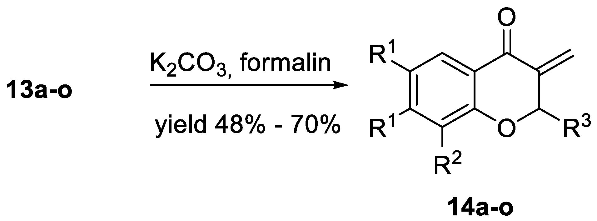
Figure 4.
Effect of 14d on induction of apoptosis in NALM-6 cells. (A) The cytotoxic activity of 14d on NALM-6 cells after 24 h incubation; (B) Quantitative analysis of apoptosis by flow cytometry. The data are presented as mean ± SEM of three independent experiments. Statistical significance was determined using one-way ANOVA and a post-hoc multiple comparison Student–Newman–Keuls test. **** p < 0.0001; *** p < 0.001; ** p < 0.01; ns—not statistically significant. (C) Representative results of cell apoptosis obtained by Annexin V and PI staining using flow cytometry in NALM-6 cells untreated (control) or treated with 14d at IC50 and 2 IC50 concentrations for 24 h.

Figure 5.
Activity of caspase 3, 8 and 9 in HL-60 cells after 6 h treatment with analog 14d at 1.25 µM (IC50) and 2.5 µM (2IC50) concentrations. Results are expressed as mean ± SEM of triplicate experiment. Statistical significance was assessed using one-way ANOVA and a post-hoc multiple comparison Student–Newman–Keuls test; *** p < 0.001; ** p < 0.01; * p < 0.05.


Table 1.
Yields of phosphonates 10a-c and 3-diethoxyphosphorylchromen-4-ones 12a–c.
| Compound | R1,R1 | R2 | 10 Yield [%] 1 | 12 Yield [%] 1 |
|---|---|---|---|---|
| a | H,H | H | 80 | 92 |
| b | H,H | Me | 75 | 89 |
| c | CH=CH-CH=CH | H | 80 | 78 |
1 Yield of pure, isolated product, based on 8 or 10, respectively.
Table 2.
Adducts 13a-o and methylidenechroman-4-ones 14a–o obtained.
| Compound | R1,R1 | R2 | R3 | 13 | 14 Yield [%] 2 | |
|---|---|---|---|---|---|---|
| trans/cis/enol 1 | Yield [%] 2 | |||||
| a | H,H | H | Me | 67/30/3 | 84 | 68 |
| b | H,H | H | Et | 71/25/4 | 68 | 71 |
| c | H,H | H | n-Bu | 76/20/4 | 85 | 59 |
| d | H,H | H | iPr | 71/26/3 | 66 | 53 |
| e | H,H | H | Ph | 81/10/9 | 64 | 84 |
| f | H,H | Me | Me | 73/24/3 | 74 | 64 |
| g | H,H | Me | Et | 75/22/3 | 76 | 59 |
| h | H,H | Me | n-Bu | 72/25/3 | 83 | 52 |
| i | H,H | Me | iPr | 72/26/2 | 70 | 53 |
| j | H,H | Me | Ph | 83/11/6 | 84 | 59 |
| k | CH=CH-CH=CH | H | Me | 79/14/7 | 81 | 66 |
| l | CH=CH-CH=CH | H | Et | 89/4/7 | 64 | 67 |
| m | CH=CH-CH=CH | H | n-Bu | 73/16/11 | 53 | 60 |
| n | CH=CH-CH=CH | H | iPr | 81/15/4 | 66 | 48 |
| o | CH=CH-CH=CH | H | Ph | 65/6/29 | 76 | 70 |
1 Ratios taken from 31P NMR spectra of the crude mixtures. 2 Yield of pure, isolated product, based on 12 or 13, respectively.
Table 3.
Tumor cell growth inhibitory activity of 14a–o on three cancer cell lines.
![Molecules 24 01868 g006]()

| 14 | R1, R1 | R2 | R3 | IC50 [µM] 1 | ||
|---|---|---|---|---|---|---|
| HL-60 | NALM-6 | MCF-7 | ||||
| a | H, H | H | Me | 5.91 ± 0.32 | 2.13 ± 0.04 | 9.70 ± 0.80 |
| b | H, H | H | Et | 5.24 ± 0.52 | 0.60 ± 0.02 | 12.50 ± 0.71 |
| c | H, H | H | n-Bu | 8.79 ± 0.81 | 4.23 ± 0.46 | 16.00 ± 0.20 |
| d | H, H | H | iPr | 1.46 ± 0.16 | 0.50 ± 0.05 | 19.40 ± 0.80 |
| e | H, H | H | Ph | 23.86 ± 2.30 | 5,76 ± 0.23 | 17,10 ± 0.30 |
| f | H, H | Me | Me | 7.52 ± 0.81 | 3.28 ± 0.35 | 10.50 ± 0.34 |
| g | H, H | Me | Et | 20.87 ± 1.73 | 4.83 ± 0.45 | 11.60 ± 0.07 |
| h | H, H | Me | n-Bu | 32.16 ± 3.17 | 6.36 ± 0.36 | 13.50 ± 0.60 |
| i | H, H | Me | iPr | 3.45 ± 0.34 | 0.58 ± 0.05 | 8.48 ± 0.93 |
| j | H, H | Me | Ph | 30.51 ± 3.62 | 6.09 ± 0.61 | 30.00 ± 1.90 |
| k | CH=CH-CH=CH | H | Me | 6.96 ± 0.42 | 4.31 ± 0.36 | 13.50 ± 1.00 |
| l | CH=CH-CH=CH | H | Et | 47.00 ± 4.11 | 10.91 ± 2.8 | 34.60 ± 4.50 |
| m | CH=CH-CH=CH | H | n-Bu | 8.64 ± 0.53 | 6.00 ± 0.23 | 17.40 ± 2.50 |
| n | CH=CH-CH=CH | H | iPr | 10.47 ± 2.04 | 5.52 ± 0.25 | 15.70 ± 0.70 |
| o | CH=CH-CH=CH | H | Ph | 49.96 ± 3.89 | 6.59 ± 0.41 | 71.0 ± 7.50 |
| Carboplatin | 2.9 ± 0.1 | 0.7 ± 0.3 | 3.8 ± 0.45 | |||
1 Compound concentration required to inhibit metabolic activity by 50%. The cells were incubated with the analogs for 48 h. Values are expressed as mean ± SEM from the concentration-response curves of at least three experiments using a nonlinear estimation (quasi-Newton algorithm) method.
© 2019 by the authors. Licensee MDPI, Basel, Switzerland. This article is an open access article distributed under the terms and conditions of the Creative Commons Attribution (CC BY) license (http://creativecommons.org/licenses/by/4.0/).
Share and Cite
MDPI and ACS Style
Kędzia, J.; Bartosik, T.; Drogosz, J.; Janecka, A.; Krajewska, U.; Janecki, T. Synthesis and Cytotoxic Evaluation of 3-Methylidenechroman-4-ones. Molecules 2019, 24, 1868. https://doi.org/10.3390/molecules24101868
AMA Style
Kędzia J, Bartosik T, Drogosz J, Janecka A, Krajewska U, Janecki T. Synthesis and Cytotoxic Evaluation of 3-Methylidenechroman-4-ones. Molecules. 2019; 24(10):1868. https://doi.org/10.3390/molecules24101868
Chicago/Turabian StyleKędzia, Jacek, Tomasz Bartosik, Joanna Drogosz, Anna Janecka, Urszula Krajewska, and Tomasz Janecki. 2019. "Synthesis and Cytotoxic Evaluation of 3-Methylidenechroman-4-ones" Molecules 24, no. 10: 1868. https://doi.org/10.3390/molecules24101868