Article

A Study of the Bisphosphonic Derivatives from the Pudovik Reaction of Dialkyl α-Oxophosphonates and >P(O)H Reagents: X-ray Structure and Bioactivity

by
Zsuzsanna Szalai
1,
Boldizsár Tóth
1,
Rita Oláhné Szabó
2,3,
Szilvia Bősze
2,
Konstantin Karaghiosoff
4,
Mátyás Czugler
1,
László Drahos
5 and
György Keglevich
1,*
1
Department of Organic Chemistry and Technology, Faculty of Chemical Technology and Biotechnology, Budapest University of Technology and Economics, 1521 Budapest, Hungary
2
Eötvös Loránd Research Network (ELKH), Research Group of Peptide Chemistry, Eötvös Loránd University, 1117 Budapest, Hungary
3
Department of Genetics, Cell and Immunobiology, Semmelweis University, Nagyvárad tér 4, 1089 Budapest, Hungary
4
Department Chemie, Ludwig-Maximilians-Universitat München, Butenandtstr. 5-13, D-81377 München, Germany
5
MS Proteomics Research Group, Research Centre for Natural Sciences, 1117 Budapest, Hungary
*
Author to whom correspondence should be addressed.
Molecules 2023, 28(16), 6037; https://doi.org/10.3390/molecules28166037
Submission received: 19 July 2023 / Revised: 7 August 2023 / Accepted: 10 August 2023 / Published: 12 August 2023
(This article belongs to the Special Issue Recent Development of Organophosphorus Chemistry)

Abstract

:
New hydroxy-methylenebisphosphonic derivatives were prepared with different P-functions. The outcome of the reaction of α-oxophosphonates (YC(O)P(O)(OR)2) and dialkyl phosphites or diarylphosphine oxides depended on the Y substituent of the oxo-compound, the nature of the P-reagent and the amount of the diethylamine catalyst. Starting from dimethyl α-oxoethylphosphonate, in the presence of 5% of diethylamine, the corresponding Pudovik adduct was the single product. While using 40% of the catalyst, the rearranged species with the >P(O)–O–CH–P(O)< skeleton was the exclusive component. A similar reaction of α-oxobenzylphosphonate followed the rearrangement protocol. X-ray crystallography revealed not only the spatial structures of the three products, but also an intricate pattern evolving from the interplay of slight chemical differences, solvent inclusion and disorder as well as H-bridge patterns, which invite further investigation. In vitro activity of the compounds was assessed on different tumor cell cultures using end-point-type cell tetrazolium-based measurements. These structure–activity studies revealed a cytostatic effect for four rearranged derivatives containing aromatic units. One of them had a pronounced effect on MDA-MB 231 and Ebc-1 cells, showing IC50 = 37.8 and 25.9 µM, respectively.

Graphical Abstract

1. Introduction

Tetraalkyl methylenebisphosphonates and related derivatives are important intermediates, e.g., they may be modified by substitution on the central carbon atom. A number of methods were elaborated for alkylation [1,2,3,4,5,6,7,8] and acylation [9]. On the other hand, substituted hydroxy-methylenebisphosphonic derivatives form a prominent group called dronates that are used for bone diseases such as osteoporosis and cancer [10,11,12]. Dronic acid derivatives may be synthesized by the reaction of substituted acetic acids with phosphorus trichloride or phosphorous acid in solvents like methanesulfonic acid or sulfolane [13,14,15]. The senior author of this paper with colleagues was the one who elaborated the optimized synthesis of alendronate [16,17], ibandronate [16,17], risedronate [16,18] and zoledronate [16,18]. According to this, if methanesulfonic acid is the solvent, 3.2 equivalents of phosphorus trichloride should be used as the reagent; if sulfolane serves as the medium, phosphorus trichloride and phosphorous acid should be applied in a ratio of 2:2. It was a noteworthy observation that ionic liquid additives promoted the efficiency of the reactions [19,20,21,22]. The other possibility for the preparation of dronic acid derivatives involved the addition of dialkyl phosphites to α-oxophosphonates [23,24,25,26,27,28,29], which is called the Pudovik reaction. In this article, we aimed at the synthesis of methylenebisphosphonic derivatives with mixed P-functions involving a phosphine oxide moiety. We also explored the rearrangement side-reaction, which afforded products with a >P(O)–O–CH–P(O)< moiety.

2. Results and Discussion

2.1. Synthesis

In the first series of experiments, dimethyl α-oxoethylphosphonate (1) was reacted with dimethyl phosphite at 0 °C in diethyl ether for 8 h. The outcome depended on the quantity of the diethylamine (DEA) catalyst applied: using 5%, the Pudovik reaction took place selectively to afford α-hydroxy-methylenebisphosphonate 2a (Table 1/Entry 1); however, in the presence of 40% of the catalyst, tetramethyl phosphonate-phosphate 3a, formed by a rearrangement of the primary hydroxy-methylenebisphosphonate 2a, was the exclusive product (Table 1/Entry 2). The addition of diethyl phosphite and dibutyl phosphite to oxophosphonate 1 applying 5% DEA also selectively provided the adducts 2b and 2c, respectively (Table 1/Entries 3 and 6). At the same time, after stirring the mixture in the presence of 40% of the catalyst at 0 °C for 8 h, the reaction mixtures comprised comparable portions of the adduct (2) and the rearranged product 3 (Table 1/Entries 4 and 7). In these cases, stirring at 26 °C for 3 days was necessary to achieve complete rearrangement (Table 1/Entries 5 and 8). It is noteworthy that the rearrangement of the phosphonate–phosphate compounds led to both possible isomers 3b-1/3b-2 and 3c-1/3c-2.
In the next round, the secondary phosphine oxides diphenylphosphine oxide, bis(4-methylphenyl)phosphine oxide and bis(3,5-dimethylphenyl)phosphine oxide were added to the carbonyl group of dimethyl α-oxoethylphosphonate (1). After adding 40% of the catalyst, the reactions were completed after stirring at 0 °C for 8 h. In these cases, no rearranged products were formed (Table 1/Entries 9–11).
In summary, adducts 2a-c, 2d-f, along with rearranged products 3a, 3b and 3c were obtained in 62–87% yields after column chromatography or recrystallization. All compounds were characterized by 31P, 13C and 1H NMR, as well as HRMS. Tetramethyl bisphosphonate 2a and the rearranged version 3a were described earlier [24,30].
Finally, diethyl α-oxobenzylphosphonate (4) was reacted with the three diarylphosphine oxides also used above. The results are summarized in Table 2. It was not possible to stop at the adduct stage as there was an increased inclination for the rearrangement. Carrying out the reaction in diethyl ether in the presence of 40% DEA at 0 °C for 8 h, only isomers 5-1 and 5-2 of the rearranged products 5(d-f)-1 and 5(d-f)-2 were formed. Compound 5d, comprising isomers 5d-1 and 5d-2 in a comparable 6:4 proportion, was obtained as a mixture of isomers. However, the major isomers 5-1 of products 5e and 5f were prepared in a pure form by column chromatography. Compounds 5e-f were fully characterized new species. Previously, it was also found that the adducts derived from α-oxobenzylphosphonates are less stable than those obtained from α-oxoethylphosphonates [29].

2.2. X-ray Structure of the Three Adducts

Two Pudovik adducts, dimethyl phosphonate–phosphine oxide derivatives 2d and 2e together the earlier described diethyl phosphonate–phosphine oxide 6 [29], were subjected to single crystal X-ray analysis. The results are presented in Figure 1, Figure 2, Figure 3, Figure 4, Figure 5 and Figure 6. The stereostructures of 2d, 2e and 6 are shown in Figure 1, Figure 3 and Figure 5, respectively, while selected geometries were included in Table 3. Connection of the molecules in the crystal structure can be seen in Figure 2, Figure 4 and Figure 6. It is clear, that 2d is present as an H-bonded chain, while 2e and 6 are H-bonded dimers.
Molecules 28 06037 i003
Bond distances in the P1 atom vicinity show nearly identical dimensions (Table 3). At first glance, this statement does not seem to apply to the P2 atom environment, inclining one to suppose that an eventual charge imbalance may be visible through slightly alternating bond lengths of phosphonates 2d, 2e and 6. Such an assumption may eventually lead to the disparity of the H-bonding pattern difference between 2d (continuous chain) and 2e (discrete dimers). Nevertheless, this supposition may not really be supportable as the disorder in the methoxy groups of species 2e obviates interpretations of bond differences in the immediate region of the P2 atom. One can only speculate that the disorder is a reason or a consequence of the dimer formation in the case of compound 2e. It seems probable that a dimer-type of H-bridge buildup may be the consequence of the effect of the p-methyl substitution in the benzene ring, the solvent inclusion, and the disorder of the methoxy groups at P2. Hydroxy-methylenebisphosphonate 6 also forms H-bonded dimers in its crystal. As a very simple tool of assessing packing tightness in respect of 2d, 2e.0.5 C3H6O and 6, it is instructive to compare their calculated densities in their crystals (See Experimental). The chain-forming 2d had the highest value in this series, while 2e.0.5 C3H6O had the lowest, even with the aid of a solvent molecule. It is also worth noting that the real assembly in the crystal had a perfect twofold symmetry rotor image with acetone solvent sitting on the symmetry axis. Thus, the real 2:1 stoichiometry is 2 × 2e.C3H6O. The density of derivative 6 took on an intermediate value between the two others, thus suggesting that dimer formation tends to yield to looser packing.

2.3. Bioactivity of the Compounds Prepared

Hydroxy-methylenebisphosphonic derivatives 2a–f and 6 as well as related rearranged species 3a–c, 5d, 5e-1, 5f-1 and 7 were subjected to bioactivity study. Phosphonate–phosphate 7 was described by us earlier [29]. From among the compounds investigated, the ones listed in Figure 7 (2b, 3b, 5d, 5e-1, 5f-1 and 7) showed significant activity.
In vitro cytostatic activity of the compounds was determined after an overnight treatment using 3-(4,5-dimethylthiazol-2-yl)-2,5-diphenyltetrazolium bromide (MTT) assay. (For the details see Experimental.) Cytostasis (%) and 50% inhibitory concentration (IC50) were determined for each compound on MDA-MB 231 human breast adenocarcinoma, A431 human epidermoid carcinoma, PC-3 human prostate adenocarcinoma, and Ebc-1 human lung squamous cell carcinoma cell lines.
Results indicated that several compounds elicited a cytostatic effect on the human tumor cell lines. Considering the percentage of cytostasis values at c = 50 µM, we concluded that compounds 3b, 5f-1 and 7 had a moderate cytostatic effect on the cells. However, three compounds were more effective. Adduct 2b was efficient on A431 human epidermoid carcinoma cells and showed a cytostasis = 49.9%. Phosphine oxide–phosphate 5d induced a higher cytostasis on MDA-MB 231 human breast adenocarcinoma and Ebc-1 human lung carcinoma cells, showing a cytostasis of 48.9 and 45.3%, respectively, whereas 5e-1 elicited an outstanding effect on these cell lines: cytostasis = 69.9 and 72.4%, respectively). Overall, the MDA-MB 231 human breast adenocarcinoma cell line proved to be the most sensitive for all effective compounds. Cytostasis values at c = 50 µM are shown in Table 4.
The above tendency was also confirmed when the effect was determined on the basis of the calculated IC50 values. While the effect of compounds 2a–f and 3a–c were not very efficient, a higher cytostatic effect was observed for compounds 5d, 5e-1, 5f-1, and 7. Compounds 5f-1 and 7 had a lower cytostatic effect (IC50 = 99.5–115.8 µM), whereas 5e-1 induced a more pronounced cytostasis on A431 cells (IC50 = 40.4 µM). The most effective on MDA–MB 231 and Ebc-1 cells (IC50 = 37.8 and 25.9 µM, respectively) proved to be 5e-1. The IC50 values of the compounds are summarized in Table 5. Daunomycin, an often-used reference compound, was also tested on the cell lines we applied. Its efficiency was much better [32,33] than that of our compounds. For details, see footnote “a” in Table 5.

3. Experimental

3.1. General

The 31P, 13C, 1H-NMR spectra were taken on a Bruker DRX-500 or Bruker Avance-300 spectrometer operating at 202.4, 125.7, and 500 MHz or 121.5, 75.4 and 300 MHz respectively. The couplings were given in Hz. LC–MS measurements were performed with an Agilent 1200 liquid chromatography system, coupled with a 6130 quadrupole mass spectrometer equipped with an ESI ion source (Agilent Technologies, Palo Alto, CA, USA). High-resolution mass spectrometric measurements were performed using a Thermo Velos Pro Orbitrap Elite hybrid mass spectrometer in positive electrospray mode.

3.2. General Procedure for the Synthesis of Tetraalkyl α-Hydroxy-ethylidenebisphosphonates

2.2 mmol (0.33 g) of dimethyl α-oxoethylphosphonate was added dropwise to a mixture of 2.2 mmol dialkyl phosphite (dimethyl phosphite: 0.20 mL, diethyl phosphite: 0.30 mL, dibutyl phosphite: 0.43 mL) and 0.11 mmol (0.010 mL) of diethylamine in diethyl ether (13 mL) at 0 °C on stirring. After an 8 h reaction time, the solvent was evaporated, and the crude product obtained was purified by column chromatography (using DCM–MeOH 97:3 as the eluent on silica gel).

3.2.1. Tetramethyl α-Hydroxy-ethylidenebisphosphonates (2a)

Yield: 0.40 g (68%), 31P NMR (CDCl3) δ 22.3 Ref [24] δP 22.0; 13C NMR (CDCl3) δ 20.0 (t, J = 2.2 Hz, CCH3), 54.2 and 54.3 (t, J = 3.5 Hz, 4 OCH3), 71.7 (t, J = 156.8 Hz, CCH3); 1H NMR (CDCl3) δ 1.70 (t, J = 16.2 Hz, 3H, CCH3), 3.88–3.93 (m, 12H, OCH3); [M + H]+ = 263; [M + Na]+found = 285.0272; C6H16O7P2Na required 285.0269.

3.2.2. Diethyl–Dimethyl α-Hydroxy-ethylidenebisphosphonate (2b)

Yield: 0.51 g (80%), 31P NMR (CDCl3) δP1 19.9 and δP2 22.7 (d, 2JPP = 40.1 Hz), Ref [25] δP1 20.6 and δP2 23.4 (2JPP = 39.3 Hz); 13C NMR (CDCl3) δ 16.4 (d, J = 5.6 Hz, 2 CH2CH3), 20.4 (s, CCH3), 54.2 and 54.3 (d, J = 7.1 Hz, 2 OCH3), 63.7 and 63.8 (d, J = 4.9 Hz, 2 OCH2), 71.6 (t, J = 156.4 Hz, CCH3); 1H NMR (CDCl3) δ 1.37 (t, J = 7.0 Hz, 6H, CH2CH3), 1.68 (t, J = 16.1 Hz, 3H, CCH3), 3.88 (d, J = 10.5 Hz, 6H, OCH3), 4.21–4.32 (m, 4H, OCH2); [M + H]+ = 291; [M + Na]+found = 313.0573; C8H20O7P2Na required 313.0582.

3.2.3. Dibutyl-Dimethyl α-Hydroxy-ethylidenebisphosphonate (2c)

Yield: 0.50 g (66%) 31P NMR (CDCl3) δP1 19.8 and δP2 22.8 (d, 2JPP = 39.3 Hz), Ref [30] δP1 20.4 and δP2 23.3 (2JPP = 40.1 Hz); 13C NMR (CDCl3) δ 13.6 (s, 2 CH2CH3), 18.6 (s, 2 CH2CH3), 20.1 (s, CCH3), 32.5 (d, J = 5.5 Hz, 2 OCH2CH2), 54.0–54.1 and 54.2–54.3 (m, 2 OCH3), 67.4 (m, 2 OCH2), 71.7 (t, J = 156.1 Hz, CCH3); 1H NMR (CDCl3) δ 0.96 (t, J = 7.4 Hz, 6H, 2 CH2CH3), 1.38–1.50 (m, 4H, CH2CH3), 1.63–1.78 (m, 3 + 4H, CCH3 + OCH2CH2), 3.89 (dd, J1 = 10.5 Hz, J2 = 1.6 Hz, 6H, OCH3), 4.16–4.23 (m, 4H, OCH2); [M + H]+ = 347; [M + Na]+found = 369.1209; C12H28O7P2Na required 369.1208.

3.3. General Procedure for the Synthesis of Dimethyl 1-Diarylphosphinoyl-1-hydroxy-ethylphosphonate

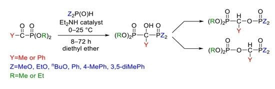
2.2 mmol (0.33 g) of dimethyl α-oxoethylphosphonate was added dropwise to a mixture of 2.2 mmol diarylphosphine oxide (diphenylphosphine oxide: 0.44 g, bis(4-methylphenyl)phosphine oxide: 0.50 g, bis(3,5-dimethylphenyl)phosphine oxide: 0.56 g) and 0.88 mmol (0.090 mL) of diethylamine in diethyl ether (13 mL) at 0 °C on stirring. After an 8 h reaction time, the precipitated material was removed by filtration, washed with diethyl ether, and the residue recrystallized from acetone. The products were white crystalline compounds.

3.3.1. Dimethyl 1-Diphenylphosphinoyl-1-hydroxy-ethylphosphonate (2d)

Yield: 0.50 g (64%), mp: 131–132 °C; 31P NMR (CDCl3) δP1 23.9 and δP2 29.0 (d, 2JPP = 25.4 Hz); 13C NMR (CDCl3) δ 20.3 (s, CCH3), 53.9 and 54.0 (d, J = 7.4 Hz, 2 OCH3), 74.8 (dd, J1 = 154.6 Hz, J2 = 79.0 Hz, CCH3), 127.8 and 128.2 (d, J = 11.7 Hz, 2 Cγ), 130.4 (dd, J1 = 96.9 Hz, J2 = 5.5 Hz, Cα), 130.8 (d, J = 98.1 Hz, Cα), 131.6 and 131.8 (d, J = 2.8 Hz, 2 Cδ), 132.4 and 132.7 (d, J = 8.6 Hz, 2 Cβ); 1H NMR (CDCl3) δ 1.65 (t, J = 15.6 Hz, 3H, CCH3), 3.44 and 3.70 (d, J = 10.6 Hz, 6H, OCH3), 7.40–7.61 (m, 6H, ArH), 8.09 and 8.18 (dd, J1 = 11.1 Hz, J2 = 6.9 Hz, 4H, ArHβ); [M + H]+ = 355; [M + Na]+found = 377.0681; C16H20O5P2Na required 377.0684.

3.3.2. Dimethyl 1-Bis(4-methylphenyl)phosphinoyl-1-hydroxy-ethylphosphonate (2e)

Yield: 0.52 g (62%), mp: 153–154 °C; 31P NMR (CDCl3) δP1 24.0 and δP2 30.2 (d, 2JPP = 29.0 Hz); 13C NMR (CDCl3) δ 20.6 (s, CCH3), 21.5 (s, 2 ArCH3), 53.9 and 54.0 (d, J = 7.4 Hz, 2 OCH3), 74.6 (dd, J1 = 153.7 Hz, J2 = 76.3 Hz, CCH3), 127.1 (dd, J1 = 99.4 Hz, J2 = 4.9 Hz, Cα), 127.2 (d, J = 100.9 Hz, Cα), 128.7 and 128.9 (d, J = 12.1 Hz, 2 Cγ), 132.4 and 132.7 (d, J = 9.1 Hz, 2 Cβ), 142.1 and 142.3 (d, J = 2.9 Hz, 2 Cδ); 1H NMR (CDCl3) δ 1.61 (t, J = 15.4 Hz, 3H, CCH3), 2.39 (s, 6H, ArCH3), 3.55 and 3.67 (d, J = 10.6 Hz, 6H, OCH3), 7.27–7.29 (m, 4H, ArH), 7.92 and 8.03 (dd, J1 = 11.0 Hz, J2 = 8.0 Hz, 4H, ArHβ); [M + H]+ = 383; [M + Na]+found = 405.1003; C18H24O5P2Na required 405.0997.

3.3.3. Dimethyl 1-Bis(3,5-dimethylphenyl)phosphinoyl-1-hydroxy-ethylphosphonate (2f)

Yield: 0.62 g (69%), mp: 161–162 °C; 31P NMR (CDCl3) δP1 24.3 and δP2 30.0 (d, 2JPP = 29.0 Hz); 13C NMR (CDCl3) δ 20.7 (s, CCH3), 21.3 (s, 4 ArCH3), 53.8 and 54.0 (d, J = 7.4 Hz, 2 OCH3), 74.7 (dd, J1 = 153.6 Hz, J2 = 76.3 Hz, CCH3), 129.9 and 130.1 (d, J = 8.7 Hz, 2 Cβ) 130.2 (dd, J1 = 95.7 Hz, J2 = 5.1 Hz, Cα), 130.5 (d, J = 96.0 Hz, Cα), 133.4 and 133.5 (d, J = 3.0 Hz, 2 Cδ), 137.4 and 137.7 (d, J = 12.4 Hz, 2 Cγ); 1H NMR (CDCl3) δ 1.65 (t, J = 14.7 Hz, 3H, CCH3), 2.36 (d, J = 5.5 Hz, 12H, ArCH3), 3.52 and 3.70 (d, J = 10.6 Hz, 6H, OCH3), 7.14 (s, 2H, ArHδ), 7.67 and 7.77 (d, J = 11.3 Hz, 4H, ArHβ); [M + H]+ = 411; [M + Na]+found = 433.1312; C20H28O5P2Na required 433.1310.

3.4. General Procedure for the Synthesis of Dialkyl 1-(Dialkylphosphonoylethyl)phosphate

2.2 mmol (0.33 g) of dimethyl α-oxoethylphosphonate was added dropwise to a mixture of 2.2 mmol dialkyl phosphite (dimethyl phosphite: 0.20 mL, diethyl phosphite: 0.30 mL, dibutyl phosphite: 0.43 mL) and 0.88 mmol (0.090 mL) of diethylamine in diethyl ether (13 mL) at 0 °C on stirring. After 8–72 h reaction time, the solvent was evaporated and the crude product obtained was purified by column chromatography (using DCM–MeOH 97:3 as the eluent on silica gel).

3.4.1. Dimethyl 1-(Dimethylphosphonoylethyl)phosphate (3a)

Yield: 0.43 g (75%), 31P NMR (CDCl3) δP1 1.1 and δP2 22.5 (d, 3JPP = 30.1 Hz), Ref [34] δP1 0.4 and δP2 21.9 (d, 3JPP = 29.3 Hz); 13C NMR (CDCl3) δ 16.6 (s, CCH3), 53.4 and 53.6 (dd, J1 = 6.8 Hz, J2 = 3.8 Hz, 2 OCH3); 54.4 and 54.6 (dd, J1 = 6.3 Hz, J2 = 3.6 Hz, 2 OCH3), 69.1 (dd, J1 = 174.1 Hz, J2 = 6.9 Hz, CH); 1H NMR (CDCl3) δ 1.61 (dd, J1 = 16.7 Hz, J2 = 7.1 Hz, 3H, CCH3), 3.77–3.84 (m, 12H, OCH3), 4.63–4.91 (m, 1H, CH); [M + H]+ = 263; [M + Na]+found = 285.0268; C6H16O7P2Na required 285.0269.

3.4.2. Dimethyl 1-(Diethylphosphonoylethyl)phosphate (3b-1) and Diethyl 1-(Dimethylphosphonoylethyl)phosphate (3b-2)

Yield: 0.56 g (87%), major (83%): 31P NMR (CDCl3) δP1 1.0 and δP2 20.0 (3JPP = 31.3 Hz); 13C NMR (CDCl3) δ 16.38 and 16.43 (d, J = 5.5 Hz, 2 CH2CH3), 16.6 (s, CCH3), 54.4 and 54.5 (d, J = 6.2 Hz, 2 OCH3), 63.0 and 63.1 (d, J = 6.5 Hz, 2 OCH2), 69.4 (dd, J1 = 174.5 Hz, J2 = 6.8 Hz, CH); 1H NMR (CDCl3) δ 1.32 (t, J = 7.0 Hz, 6H, CH2CH3), 1.54 (dd, J1 = 16.7 Hz, J2 = 7.0 Hz, 3H, CCH3), 3.75 and 3.77 (d, J = 11.5 Hz, 6H, OCH3), 4.13–4.20 (m, 4H, CH2CH3); 4.62–4.72 (m, 1H, CH); minor (17%): δP1 −1.3 and δP2 22.6 (3JPP = 31.0 Hz); 13C NMR (CDCl3) δ 16.0 (d, J = 6.8 Hz, 2 CH2CH3), 16.6 (s, CCH3), 53.4 and 53.6 (d, J = 6.5 Hz, 2 OCH3), 64.1 and 64.2 (d, J = 6.1 Hz, 2 OCH2), 68.8 (dd, J1 = 174.1 Hz, J2 = 7.0 Hz, CH); 1H NMR (CDCl3) δ 3.80 and 3.81 (d, J = 10.7 Hz, 6H, OCH3). The other signals were common with those of the major isomer; [M + H]+ = 291; [M + Na]+found = 313.0581; C8H20O7P2Na required 313.0582.

3.4.3. Dimethyl 1-(Dibutylphosphonoylethyl)phosphate (3c-1) and Dibutyl 1-(Dimethylphosphonoylethyl)phosphate (3c-2)

Yield: 0.53 g (70%), major (81%) 31P NMR (CDCl3) δP1 1.1 and δP2 20.0 (3JPP = 31.6 Hz); 13C NMR (CDCl3) δ 13.5 (s, 2 CH2CH3), 16.7 (s, CCH3), 18.6 (s, 2 CH2CH3), 32.5 and 32.6 (d, J = 3.2 Hz, 2 OCH2CH2), 54.3–54.4 and 54.5–54.6 (m, 2 OCH3), 66.4–66.9 (m, 2 OCH2), 69.5 (dd, J1 = 174.6 Hz, J2 = 6.9 Hz, CH); 1H NMR (CDCl3) δ 0.96 (t, J = 7.9 Hz, 6H, CH2CH3), 1.40–1.47 (m, 4H, CH2CH3), 1.60 (dd, J1 = 16.5 Hz, J2 = 7.1 Hz, 3H, CCH3), 1.67–1.72 (m, 4H, OCH2CH2), 3.80 and 3.83 (d, J = 11.5 Hz, 6H, OCH3), 4.04–4.20 (m, 4H, OCH2), 4.66–4.78 (m, 1H, CH); minor (19%) 31P NMR (CDCl3) δP1 –0.9 and δP2 22.8 (3JPP = 31.4 Hz); 13C NMR (CDCl3) δ 68.8 (dd, J1 = 174.1 Hz, J2 = 7.1 Hz, CH). The other signals are common with those of the major isomer; 1H NMR (CDCl3) δ 3.85 and 3.86 (d, J = 10.5 Hz, 6H, OCH3). The other signals are common with those of the major isomer; [M + H]+ = 347; [M + Na]+found = 369.1201; C12H28O7P2Na required 369.1208.

3.5. General Procedure for Diethyl (Diarylphosphinoyloxybenzyl)phosphonate and Diethyl (Diarylphosphinoylbenzyl)phosphate

1.5 mmol (0.36 g) of diethyl α-oxobenzylphosphonate was added slowly to a mixture of 1.5 mmol (bis(4-methylphenyl)phosphine oxide: 0.35 g, bis(3,5-dimethylphenyl)phosphine oxide: 0.40 g) and 0.60 mmol (0.060 mL) of diethylamine in diethyl ether (13 mL) at 0 °C on stirring. After an 8 h reaction time, the solvent was evaporated, and the crude product obtained was purified with column chromatography (using ethyl acetate as the eluent on silica gel).

3.5.1. Diethyl (Diphenylphosphinoylbenzyl)phosphate (5d-1) and Diethyl (Diphenylphosphinoyloxybenzyl)phosphonate (5d-2)

Yield: 0.47 g (70%), major (60%): 31P NMR (CDCl3) δP1 –1.5 and δP2 28.6 (3JPP = 31.3 Hz); 13C NMR (CDCl3) δ 15.6 and 15.8 (d, J = 7.4 Hz, 2 CH2CH3), 63.8 and 63.9 (d, J = 6.0 Hz, 2 OCH2), 77.4 (dd, J1 = 85.7 Hz, J2 = 7.9 Hz, CH). The aromatic range was rather complex between δ 128.0–132.6; 1H NMR (CDCl3) δ 0.90 and 0.96 (t, J = 7.1 Hz, 6H, CH2CH3), 3.41–3.70 (m, 4H, OCH2), 6.06 (dd, J1 = 9.7 Hz, J2 = 4.4 Hz, 1H, CH), aromatic region: 7.15–7.98 (m, 15H, ArH); minor (40%): 31P NMR (CDCl3) δP1 17.2 and δP2 34.7 (3JPP = 26.7 Hz); 13C NMR (CDCl3) δ 16.2 and 16.3 (d, J = 5.8 Hz, 2 CH2CH3), 63.3 and 63.5 (d, J = 6.9 Hz, 2 OCH2), 72.0 (dd, J1 = 172.6 Hz, J2 = 7.0 Hz, CH). The aromatic range was rather complex between δ 128.0–132.6; 1H NMR (CDCl3) δ 1.09 and 1.18 (t, J = 7.1 Hz, 6H, CH2CH3), 3.78–4.15 (m, 4H, OCH2), 5.63 (dd, J1 = 13.5 Hz, J2 = 11.2 Hz, 1H, CH), aromatic region: 7.15–7.98 (m, 15H, ArH); [M + H]+ = 445; [M + Na]+found = 467.1154; C23H26O5P2Na required 467.1153.

3.5.2. Diethyl 1-Bis((4-methylphenyl)phosphinoylbenzyl)phosphate (5e-1)

Yield: 0.40 g (65%), 31P NMR (CDCl3) δP1 –1.3 and δP2 29.0 (3JPP = 31.4 Hz); 13C NMR (CDCl3) δ 15.6 and 15.7 (d, J = 7.4 Hz, 2 CH2CH3), 21.6 (d, J = 9.8 Hz, 2 ArCH3), 63.8 and 63.9 (d, J = 5.9 Hz, 2 OCH2), 77.6 (dd, J1 = 85.3 Hz, J2 = 8.0 Hz, CH). The aromatic range was rather complex between δ 124.6–142.9; 1H NMR (CDCl3) δ 0.94 and 1.00 (t, J = 7.3 Hz, 6H, CH2CH3), 2.35 and 2.42 (s, 6H, ArCH3), 3.46–3.74 (m, OCH2), 6.03 (dd, J1 = 9.8 Hz, J2 = 4.5 Hz, 1H, CH), aromatic region: 7.18–7.33 (m, 9H, ArH), 7.55 and 7.83 (dd, J1 = 11.1 Hz J2 = 8.1 Hz, 4H, ArHβ); [M + H]+ = 473; [M + Na]+found = 495.1467; C25H30O5P2Na required 495.1466.

3.5.3. Diethyl 1-Bis((3,5-dimethylphenyl)phosphinoylbenzyl)phosphate (5f-1)

Yield: 0.42 g (72%), 31P NMR (CDCl3) δP1 −1.2 and δP2 29.1 (3JPP = 30.9 Hz); 13C NMR (CDCl3) δ 15.6 and 15.8 (d, J = 7.5 Hz, 2 CH2CH3), 21.2 (d, J = 13.3 Hz, 4 ArCH3), 63.6 and 63.8 (d, J = 5.9 Hz, 2 OCH2), 77.4 (dd, J1 = 84.8 Hz, J2 = 8.0 Hz, CH). The aromatic range was rather complex between δ 128.0–138.2; 1H NMR (CDCl3) δ 0.95 and 1.04 (t, J = 7.4 Hz, 6H, CH2CH3), 2.27 and 2.40 (s, 12H, ArCH3), 3.48–3.76 (m, 4H, OCH2), 6.06 (dd, J1 = 9.7 Hz, J2 = 3.1 Hz, 1H, CH), aromatic region: 7.22–7.34 (m, 9H, ArH), 7.61 (d, J = 11.7 Hz, 2H, ArHβ); [M + H]+ = 501; [M + Na]+found = 523.1771; C27H34O5P2Na required: 523.1779.
For the 31P, 13C and 1H NMR spectra of the compounds prepared see Supplementary Materials.

3.6. Single Crystal X-ray Diffraction Studies

Single crystals of compound 2d, 2e.0.5 C3H6O and 6 suitable for X-ray diffraction were obtained by slow evaporation of the respective acetone solution. The crystals were introduced into perfluorinated oil and a suitable single crystal was carefully mounted on the top of a thin glass wire. Data collection was performed with an Oxford Xcalibur 3 diffractometer equipped with a Spellman generator (50 kV, 40 mA) and a Kappa CCD detector, operating with Mo-Kα radiation (λ = 0.71071 Ǻ).
Data collection and reduction were performed using CrysAlisPro software [35]. Absorption correction using the multiscan method [35] was applied. The structures were solved with SHELXS-97 [36], refined with SHELXL-97 [37] and finally checked using PLATON [38]. Details of the data collection and structure refinement are summarized in Table 6.
CCDC-2281416, CCDC-2281417 and CCDC-2281418 contain supplementary crystallographic data for compounds 2d, 2e·0.5C3H6O and 6, respectively. These data can be obtained free of charge from The Cambridge Crystallographic Data Centre via www.ccdc.cam.ac.uk/data_request/cif (accessed on 13 July 2023).

3.7. In Vitro Cytotstasis Assays

Cell Lines and Culture Conditions

The in vitro cytostatic effect of the compounds was studied on MDA-MB 231 human breast adenocarcinoma [39], A431 human epidermoid carcinoma [40], PC-3 human prostate adenocarcinoma [41], and Ebc-1 human lung squamous cell carcinoma [42] cell lines. Cells were cultured in a DMEM medium supplemented with 10% FBS, 2 mM L-glutamine, penicillin–streptomycin antibiotic mixture (50 IU/mL and 50 μg/mL, respectively), 1 mM sodium pyruvate and 1% non-essential amino acid mixture. The cell cultures were maintained at 37 °C in a humidified atmosphere with 5% CO2. The cells were grown to confluent state and then they were harvested by trypsinization and divided into 96-well tissue culture plates (initial cell number was of 5.0 × 10³ cells/well). Cells were allowed to attach for 24 h at 37 °C when the culturing medium was removed and they were treated with the compounds in 2, 10, 50, and 250 μM concentration in a serum-free medium. (The treating solutions contained 1.0 v/v% DMSO). Control cells were treated only with serum-free medium or with DMSO (c = 1.0 v/v%) under the same conditions. After overnight incubation, cells were washed twice with a serum-free medium, and then cultured for another 72 h in 10% serum-containing medium at 37 °C. An MTT-solution (at c = 0.37 mg/mL final concentration) was added to each well and incubated for 3 h. The cells were centrifuged for 5 min at 900 g, and then the supernatant was removed. The obtained formazan crystals were dissolved in DMSO (100 µL) and the optical density (OD) of the samples was measured with an ELISA Reader (iEMS Reader, Labsystems, Vantaa, Finland) at detecting wavelength = 540 and reference wavelength = 620 nm. OD620 values were subtracted from the OD540 values, and then cytostasis % was calculated from this corrected OD value by the following equation:
Cytostatic effect (%) = [1 − (ODtreated/ODcontrol)] × 100
where ODtreated and ODcontrol correspond to the optical densities of the treated and control wells, respectively. In each case, two independent experiments were carried out with 4 parallel measurements. Statistical data analysis was performed using Student’s t-test at a 95% confidence level. A 50% inhibitory concentration (IC50, expressed in micromolar units) was determined from the dose–response curves: cytostasis was plotted as a function of concentration on which a sigmoidal curve was fitted using Microcal™ Origin 2018 software [43,44].

4. Conclusions

The outcome of the reaction of α-oxophosphonates (ZC(O)P(O)(OR)2) and Y2P(O)H reagents depended on the nature of the Z substituent of the oxo-compound, the Y substituent of the P-reagent, and the amount of the diethylamine catalyst. In case of Z = Me, new hydroxy-methylenebisphosphonic derivatives with different P-functions were synthesized. Performing the reactions in the presence of an increased amount (40%) of the catalyst, or starting from an α-oxobenzylphosphnate, rearranged species comprising the >P(O)–O–CH–P(O)< motif were the products. The molecular dimensions mostly conformed to those expected for this kind of P-compound. The intermolecular connection pattern may be realized in centrosymmetric H-bridge dimers, but in one case a catameric chain structure was experienced. Solvent inclusion as well as the presence of disorder were also present in one of the crystals hampering deeper insight into the solid-state relations. Part of the compounds we synthesized showed significant in vitro cytotoxic activity on human tumor cell cultures of different tissue origin. The rearranged derivatives with aromatic units possessed considerable antiproliferative activity characterized by low IC50 values.

Supplementary Materials

The following supporting information can be downloaded at: https://www.mdpi.com/article/10.3390/molecules28166037/s1, X-ray data for compounds 2d, 2e and 6; 31P, 13C and 1H NMR spectra of the compounds prepared.

Author Contributions

Conceptualization, G.K. and S.B.; methodology, Z.S., R.O.S. and S.B.; investigation, Z.S., B.T., L.D., K.K., R.O.S. and S.B.; resources, G.K., K.K. and S.B.; data curation, R.O.S., S.B., L.D. and K.K.; writing—original draft preparation, G.K., Z.S., R.O.S., S.B., K.K. and M.C.; writing—review and editing, G.K. and S.B.; supervision, G.K. and S.B.; project administration, G.K.; funding acquisition, G.K. All authors have read and agreed to the published version of the manuscript.

Funding

This project was supported by the National Research, Development and Innovation Office (K134318). The publication, as well as the scientific results presented in its context, were made with the support of the Gedeon Richter Talentum Foundation established by Gedeon Richter Plc. (1103 Budapest, Gyömrői út 19-21.) with the support of the Gedeon Richter Excellence PhD Scholarship.

Institutional Review Board Statement

Not applicable.

Informed Consent Statement

Not applicable.

Data Availability Statement

The data presented in this study are available on request from the corresponding authors.

Conflicts of Interest

The authors declare no conflict of interest.

Sample Availability

Not applicable.

References

  1. Grün, A.; Bálint, E.; Keglevich, G. Solid–liquid phase C-alkylation of active methylene containing compounds under microwave conditions. Catalysts 2015, 5, 634–652. [Google Scholar] [CrossRef] [Green Version]
  2. Hays, H.R.; Logan, T.J. gem-Diphosphinoalkanes. Preparation and characterization. J. Org. Chem. 1966, 31, 3391–3394. [Google Scholar] [CrossRef]
  3. Nguyen, L.M.; Niesor, E.; Bentzen, C.L. gem-Diphosphonate and gem-phosphonate-phosphate compounds with specific high density lipoprotein inducing activity. J. Med. Chem. 1987, 80, 1426–1433. [Google Scholar] [CrossRef]
  4. Roth, A.G.; Drescher, D.; Reamer, S.; Arenz, C.; Yang, Y.; Uhlig, S. Potent and selective inhibition of acid sphingomyelinase by bisphosphonates. Angew. Chem. Int. Ed. 2009, 48, 7560–7563. [Google Scholar] [CrossRef]
  5. Kosolopoff, G.M. The chemistry of aliphatic phosphonic acids. I. Alkylation of methanediphosphonic acid. J. Am. Chem. Soc. 1953, 75, 1500–1501. [Google Scholar] [CrossRef]
  6. Goebel, R.; Richte, F.; Weichmann, H. Synthesis and reactivity of methylene bridged diphosphoryl compounds. Phosphorus Sulfur Silicon Relat. Elem. 1992, 73, 67–80. [Google Scholar] [CrossRef]
  7. Cotton, F.A.; Schunn, R.A. Metal salts and complexes of dialkoxyphosphonylacetylmethanide ions. J. Am. Chem. Soc. 1963, 85, 2394–2402. [Google Scholar] [CrossRef]
  8. Greiner, I.; Grün, A.; Ludányi, K.; Keglevich, G. Solid–liquid two-phase alkylation of tetraethyl methylenebisphosphonate under microwave irradiation. Heteroat. Chem. 2011, 22, 11–14. [Google Scholar] [CrossRef]
  9. Gulyás, K.V.; Keglevich, G. The surprising diacylation of diethyl (ethoxycarbonylmethyl)phosphonate. Mendeleev Commun. 2021, 31, 244–245. [Google Scholar] [CrossRef]
  10. Breuer, E. The development of bisphosphonates as drugs. In Analogue-Based Drug Discovery; Fischer, J., Ganellin, C.R., Eds.; Wiley-VCH: Weinheim, Germany, 2006; Chapter 15. [Google Scholar]
  11. Mucha, A.; Kafarski, P.; Berlicki, L. Remarkable potential of the α-aminophosphonate/phosphinate structural motif in medicinal chemistry. J. Med. Chem. 2011, 54, 5955–5980. [Google Scholar] [CrossRef]
  12. Grembecka, J.; Mucha, A.; Cierpicki, T.; Kafarski, P. The most potent organophosphorus inhibitors of leucine aminopeptidase. Structure-based design, chemistry, and activity. J. Med. Chem. 2003, 46, 2641–2655. [Google Scholar] [CrossRef] [PubMed]
  13. Hudson, H.R.; Wardle, N.J.; Bligh, S.W.A.; Greiner, I.; Grün, A.; Keglevich, G. N-Heterocyclic Dronic acids; applications and synthesis. Mini-Rev. Med. Chem. 2012, 12, 313–325. [Google Scholar] [CrossRef] [PubMed]
  14. Nagy, D.I.; Grün, A.; Greiner, I.; Keglevich, G. The role of phosphorus trichloride and phoshorous acid in the formation of α-hydroxymethylenebisphosphonic acids from the corresponding carboxylic acids—A mechanistic overview. Curr. Org. Chem. 2017, 21, 1567–1578. [Google Scholar] [CrossRef]
  15. Nagy, D.I.; Grün, A.; Garadnay, S.; Greiner, I.; Keglevich, G. Synthesis of hydroxymethylenebisphosphonic acid derivatives in different solvents. Molecules 2016, 21, 1046. [Google Scholar] [CrossRef] [PubMed] [Green Version]
  16. Kovács, R.; Grün, A.; Garadnay, S.; Greiner, I.; Keglevich, G. “Greener” synthesis of bisphosphonic/dronic acid derivatives. Green Process. Synth. 2014, 3, 111–116. [Google Scholar] [CrossRef]
  17. Keglevich, G.; Grün, A.; Kovács, R.; Garadnay, S.; Greiner, I. Rational synthesis of Ibandronate and Alendronate. Curr. Org. Synth. 2013, 10, 640–644. [Google Scholar] [CrossRef]
  18. Keglevich, G.; Grün, A.; Aradi, K.; Garadnay, S.; Greiner, I. Optimized synthesis of N-heterocyclic dronic acids; closing a black-box era. Tetrahedron Lett. 2011, 52, 2744–2746. [Google Scholar] [CrossRef]
  19. Nagy, D.I.; Grün, A.; Garadnay, S.; Greiner, I.; Keglevich, G. Investigation of the effect of medium in the preparation of alendronate: Till now the best synthesis in the presence of an ionic liquid additive. Heteroat. Chem. 2017, 28, e21370. [Google Scholar] [CrossRef]
  20. Nagy, D.I.; Grün, A.; Pavela, O.; Garadnay, S.; Greiner, I.; Keglevich, G. Efficient synthesis of ibandronate in the presence of an ionic liquid. Lett. Drug Des. Discov. 2018, 15, 713–720. [Google Scholar] [CrossRef]
  21. Nagy, D.I.; Grün, A.; Sinkovicz, J.; Garadnay, S.; Greiner, I.; Keglevich, G. A study on the synthesis of risedronic acid; The role of ionic liquid additive. Lett. Drug Des. Discov. 2019, 16, 238–244. [Google Scholar] [CrossRef]
  22. Nagy, D.I.; Grün, A.; Lévay, K.; Garadnay, S.; Greiner, I.; Keglevich, G. Efficient syntheses of zoledronic acid as an active ingredient of a drug against osteoporosis. Synth. Commun. 2018, 48, 663–671. [Google Scholar] [CrossRef]
  23. Fitch, S.J.; Moedritzer, K. NMR study of the P-C(OH)-P to P-CO-P rearrangement: Tetraethyl 1-hydroxyalkylidenediphosphonates. J. Am. Chem. Soc. 1962, 84, 1876–1880. [Google Scholar] [CrossRef]
  24. Nicholson, D.A.; Vaughn, H. A general method of preparation of tetramethyl alkyl-1-hydroxy-1,1-diphosphonates. J. Org. Chem. 1971, 36, 3843–3845. [Google Scholar] [CrossRef]
  25. Turhanen, P.A.; Ahlgren, M.J.; Jarvinen, T.; Vepsalainen, J.J. Bisphosphonate prodrugs. Synthesis and identification of (1-hydroxyetrylidene)-1,1-bisphosphonic acid tetraesters by mass spectrometry, NMR spectroscopy and X-ray crystallography. Phosphorus Sulfur Silicon Relat. Elem. 2001, 170, 115–133. [Google Scholar] [CrossRef]
  26. Turhanen, P.A.; Ahlgren, M.J.; Jarvinen, T.; Vepsalainen, J.J. Bisphosphonate prodrugs, selective synthesis of (1-hydroxyethylidene)-1,1-bisphosphonate partial esters. Synthesis 2001, 2001, 633–637. [Google Scholar] [CrossRef]
  27. Grün, A.; Molnár, I.G.; Bertók, B.; Greiner, I.; Keglevich, G. Synthesis of α-hydroxy-methylenebisphosphonates by the microwave-assisted reaction of α-oxophosphonates and dialkyl phosphites under solventless conditions. Heteroat. Chem. 2009, 20, 350–354. [Google Scholar] [CrossRef]
  28. Keglevich, G.; Grün, A.; Molnár, I.G.; Greiner, I. Phenyl-, benzyl- and unsymmetrical hydroxy-methylenebisphosphonates as dronic acid ester analogues from α-oxophosphonates by microwave-assisted synthesis. Heteroat. Chem. 2011, 22, 640–648. [Google Scholar] [CrossRef]
  29. Szalai, Z.; Keglevich, G. Tetraalkyl hydroxymethylenebisphosphonate and dialkyl 1-diphenylphosphinoyl-1-hydroxy-ethylphosphonate derivatives by the Pudovik reaction and their rearranged products. Molecules 2021, 26, 7575. [Google Scholar] [CrossRef]
  30. Nikander, H.; Heikkila-Hoikka, M.; Pohjala, E.; Hanhijarvi, H.; Lauren, L. New Methylenebisphosphonic Acid Derivatives. U.S. Patent WO9211267, 1992. [Google Scholar]
  31. DIAMOND, version 3.2i; Crystal Impact GbR: Bonn, Germany, 2014.
  32. Berek-Nagy, P.J.; Tóth, G.; Bősze, S.; Horváth, L.B.; Darcsi, A.; Csíkos, S.; Knapp, D.G.; Kovács, G.M.; Boldizsár, I. The grass root endophytic fungus Flavomyces fulophazii: An abundant source of tetramic acid and chlorinated azaphilone derivatives. Phytochemistry 2021, 190, 112851. [Google Scholar] [CrossRef]
  33. Anaya-Eugenio, G.D.; Rebollar-Ramos, D.; González, M.C.; Raja, H.; Mata, R.; Blanco, E.J.C. Apoptotic activity of xanthoquinodin JBIR-99, from Parengyodontium album MEXU 30054, in PC-3 human prostate cancer cells. Chemio-Biol. Int. 2019, 311, 108798. [Google Scholar] [CrossRef]
  34. Tromelin, A.; Elmanouni, D.; Burgada, R. α-Ketophosphonates and cyclic esters of hydroxymethylenediphosphonates. Synthesis, structure, and hydrolysis. Phosphorus Sulfur Relat. Elem. 1986, 27, 301–312. [Google Scholar] [CrossRef]
  35. CrysAlisPro 1.171.40.82a; Program package; Rigaku OD: Tokyo, Japan, 2020.
  36. Sheldrick, G.M. SHELXS-97: Program for Crystal Structure Solution; University of Göttingen: Göttingen, Germany, 1997. [Google Scholar]
  37. Sheldrick, G.M. SHELXL-97: Program for the Refinement of Crystal Structures; University of Göttingen: Göttingen, Germany, 1997. [Google Scholar]
  38. Spek, A.L. PLATON: A Multipurpose Crystallographic Tool; Utrecht University: Utrecht, The Netherlands, 1999. [Google Scholar]
  39. Cailleau, R.; Olivé, M.; Cruciger, Q.V. Long-term human breast carcinoma cell lines of metastatic origin: Preliminary characterization. In Vitro 1978, 14, 911–915. [Google Scholar] [CrossRef]
  40. Haigler, H.; Ash, J.F.; Singer, S.J.; Cohen, S. Visualization by fluorescence of the binding and internalization of epidermal growth factor in human carcinoma cells A-431. Proc. Natl. Acad. Sci. USA 1978, 75, 3317–3321. [Google Scholar] [CrossRef] [PubMed]
  41. Kaighn, M.E.; Narayan, K.S.; Ohnuki, Y.; Lechner, J.F.; Jones, L.W. Establishment and characterization of a human prostatic carcinoma cell line (PC-3). Investig. Urol. 1979, 17, 16–23. [Google Scholar]
  42. Fujita, T.; Kiyama, M.; Tomizawa, Y.; Kohno, T.; Yokota, J. Comprehensive analysis of p53 gene mutation characteristics in lung carcinoma with special reference to histological subtypes. Int. J. Oncol. 1999, 15, 927–961. [Google Scholar] [CrossRef] [PubMed]
  43. Mosmann, T. Rapid colorimetric assay for cellular growth and survival: Application to proliferation and cytotoxicity assays. J. Immunol. Methods 1983, 65, 55–63. [Google Scholar] [CrossRef]
  44. Denizot, F.; Lang, R.J. Rapid colorimetric assay for cell growth and survival. Modifications to the tetrazolium dye procedure giving improved sensitivity and reliability. J. Immunol. Methods 1986, 89, 271–277. [Google Scholar] [CrossRef]
Figure 1. Molecular structure of hydroxymethylenephosphonate–phosphine oxide 2d in the crystal. DIAMOND [31] representation; thermal ellipsoids are drawn at 50% probability level.
Molecules 28 06037 g001
Figure 2. View of the hydrogen-bonded chain of molecules of 2d in the crystal. DIAMOND [31] representation; thermal ellipsoids are drawn at 50% probability level. Symmetry code for the unlabeled molecules: x, 0.5 − y, 0.5 + z (left) and x, 0.5 − y, −0.5 + z (right).
Molecules 28 06037 g002
Figure 3. Molecular structure of phosphonate-phosphine oxide 2e.0.5C3H6O in the crystal. The methoxy groups at P2 are disordered each over two positions. Only the major position is shown. The crystal structure contains one acetone molecule for every two molecules of 2e (in a special position). The solvent molecule was omitted for clarity. DIAMOND [31] representation; thermal ellipsoids are drawn at 50% probability level.
Molecules 28 06037 g003
Figure 4. View of the hydrogen-bonded dimers of 2e.0.5C3H6O in the crystal. Only major disorder methyl positions are shown while the acetone solvate is omitted from the drawing. DIAMOND [31] representation; thermal ellipsoids are drawn at 50% probability level. Symmetry code for the non-labeled molecule: 1.5 − x, 0.5 − y, −z.
Molecules 28 06037 g004
Figure 5. Molecular structure of hydroxymethylenephosphonate–phosphine oxide 6 in the crystal. DIAMOND [31] representation; thermal ellipsoids are drawn at 50% probability level.
Molecules 28 06037 g005
Figure 6. View of the hydrogen-bonded dimers around a symmetry center of 6 in the crystal. DIAMOND [31] representation; thermal ellipsoids are drawn at 50% probability level.
Molecules 28 06037 g006
Figure 7. Members of the compound library showing significant cytotoxic activity.
Molecules 28 06037 g007
Table 1. The reaction of dimethyl α-oxoethylphosphonate (1) with dialkyl phosphites or secondary phosphine oxides under different conditions.
Molecules 28 06037 i001
EntryYCatalyst (%)T (°C)tProduct Composition (%) [a][b]Yield (%)
23-13-2
1MeO508 h10068 (2a)
2MeO4008 h10075 (3a)
3EtO508 h10080 (2b)
4EtO4008 h60328
5EtO40263 days831787 (3b)
6BuO508 h10066 (2c)
7BuO4008 h503416
8BuO40263 days811970 (3c)
9Ph4008 h10064 (2d)
104-MePh4008 h10062 (2e)
113,5-diMePh4008 h10069 (2f)
[a] On the basis of relative 31P NMR intensities. [b] The data set shaded in yellow refer to the best experiments.
Table 2. The reaction of diethyl α-oxobenzylphosphonate (4) with secondary phosphine oxides.
Molecules 28 06037 i002
EntryYProduct Composition (%) [a][b]Yield (%)
5-15-2
1Ph604070 (5d-1 + 5d-2)
24-MePh881265 (5e-1)
33,5-diMePh772372 (5f-1)
[a] On the basis of relative 31P NMR intensities. [b] The data set shaded in yellow refer to the best experiments.
Table 3. Selected bond lengths (Å) of compound 2d, 2e.0.5C3H6O and 6.
2d2e6
P1–O1P1–O11.485 (1)1.493 (1)P1–O11.488 (1)
P1–C7P1–C81.809 (2)1.801 (2)P1–C71.807 (1)
P1–C1P1–C11.809 (2)1.811 (2)P1–C11.809 (1)
P1–C13P1–C151.862 (2)1.863 (2)P1–C131.860 (1)
P2–O5P2–O31.466 (1)1.463 (1)P2–O51.476 (1)
P2–O3P2–O4A1.524 (2)1.572 (1)P2–O31.568 (1)
P2–O4P2–O5A1.642 (3)1.574 (1)P2–O41.570 (1)
P2–C13P2–C151.834 (2)1.831 (2)P2–C131.841 (1)
O3–C15O4A–C17A1.425 (5)1.440 (2)O3–C151.457 (1)
O4–C16O5A–C18A1.415 (7)1.438 (2)O4–C171.464 (1)
C7–C8C1–C21.393 (2)1.398 (2)C1–C21.394 (1)
C7–C12C1–C61.396 (2)1.400 (2)C1–C61.398 (1)
O2–C13O2–C151.424 (2)1.424 (2)O2–C131.427 (1)
Table 4. The cytostatic effect of the P-compounds studied on human tumor cell cultures.
CompoundCytostasis [%] at c = 50 µM
Cell Line
MDA-MB 231PC-3Ebc-1A431
2bMolecules 28 06037 i004
3b
5d
5e-1
5f-1
7
Table 5. 50% inhibitory concentration (IC50) values of the P-compounds studied on human tumor cell cultures.
CompoundIC50 (µM) a,c
Cell Line
MDA_MB-231PC-3Ebc-1A431
2a>250>250>250>250
2b>250>250>250>250
2c>250>250>250>250
2d>250>250>250>250
2e>250>250>250>250
2f>250>250>250>250
3a>250>250>250>250
3b>250>250>250>250
3c>250>250>250>250
5d76.7>25099.540.4
5e-137.8149.525.9>250
5f-1100.7115.894.1110.7
6 bn.d.
7115.0>250>250>250
a For comparison purposes for the above cell lines, reference compound Daunomycin had an IC50 value of 0.20, 4.0, 1.2, and 0.7 µM, respectively [32,33]. b Compound 6 precipitated in aqueous media. c The data shaded in yellow refer to the most efficient compounds.
Table 6. Details for X-ray data collection and structure refinement for compounds 2d, 2e.0.5 C3H6O and 6.
2d2e.0.5C3H6O6
Empirical formulaC16H20O5P2C18H24O5P2.0.5C3H6OC18H24O5P2
Formula mass354.26411.35382.31
T [K]123 (2)123 (2)123 (2)
Crystal size [mm]0.20 × 0.02 × 0.020.35 × 0.20 × 0.100.25 × 0.20 × 0.15
Crystal descriptioncolorless rodcolorless blockcolorless block
Crystal systemmonoclinicmonoclinictriclinic
Space groupP21/cC2/cP21/n
a [Å]9.1252 (3)13.8562 (3)8.6609 (2)
b [Å]18.1309 (6)10.4172 (2)9.8169 (2)
c [Å]10.1680 (4)28.5479 (7)22.1229 (5)
α [°]90.090.090.0
β [°]94.892 (3)96.649 (2)96.193 (2)
γ [°]90.090.090.0
V [Å3]1676.15 (10)4092.97 (16)1869.98 (7)
Z484
ρcalcd. [g cm−3]1.4041.3351.358
μ [mm−1]0.2810.2420.258
F (000)7441744808
Θ range [°]2.24–25.242.45–25.242.27–25.24
Index ranges−12 ≤ h ≤ 12−17 ≤ h ≤ 17−12 ≤ h ≤ 12
Disclaimer/Publisher’s Note: The statements, opinions and data contained in all publications are solely those of the individual author(s) and contributor(s) and not of MDPI and/or the editor(s). MDPI and/or the editor(s) disclaim responsibility for any injury to people or property resulting from any ideas, methods, instructions or products referred to in the content.

