Article

Syntheses and Self-assembling Behaviors of Pentagonal Conjugates of Tryptophane Zipper-Forming Peptide

1
Department of Chemistry and Biochemistry, Graduate School of Engineering, Kyushu University, Moto-oka 744, Nishi-ku, Fukuoka 819-0395, Japan
2
International Research Center for Molecular Systems, Kyushu University, Moto-oka 744, Nishi-ku, Fukuoka 819-0395, Japan
*
Author to whom correspondence should be addressed.
Int. J. Mol. Sci. 2011, 12(8), 5187-5199; https://doi.org/10.3390/ijms12085187
Submission received: 13 June 2011 / Revised: 30 July 2011 / Accepted: 10 August 2011 / Published: 15 August 2011
(This article belongs to the Special Issue Molecular Self-Assembly 2011)

Abstract

:
Pentagonal conjugates of tryptophane zipper-forming peptide (CKTWTWTE) with a pentaazacyclopentadecane core (Pentagonal-Gly-Trpzip and Pentagonal-Ala-Trpzip) were synthesized and their self-assembling behaviors were investigated in water. Pentagonal-Gly-Trpzip self-assembled into nanofibers with the width of about 5 nm in neutral water (pH 7) via formation of tryptophane zipper, which irreversibly converted to nanoribbons by heating. In contrast, Pentagonal-Ala-Trpzip formed irregular aggregates in water.

Graphical Abstract

1. Introduction

Multivalent ligand-receptor interactions play pivotal roles in biological systems [14]. To date, many artificial multivalent bioconjugates have been developed as inhibitors, receptors, artificial enzymes, signaling molecules, and drug delivery materials. For example, inhibition of Shiga-like or cholera toxins by pentavalent conjugates of oligosaccharides has been reported [5,6]. The concept of “template-assembled synthetic proteins (TASP)” provided a chemical approach to design artificial proteins [7]. Peptide dendrimers have also been developed as multifunctional biomaterials [8,9].
Many artificial peptide nano-assemblies have been designed by self-assembly of coiled-coil α-helix peptides and β-sheet-forming peptides [1021]. Application of multivalent molecular design to peptide assembly can promote self-assembly and lead to unique morphologies. For example, Ghosh et al. reported that PAMAM dendrimer containing four leucine zipper-forming peptides self-assembled into nanofibers [22]. We have designed trigonal peptide conjugates containing β-sheet forming peptides [2325] and glutathione [2628], and demonstrated that they are useful components for the spontaneous construction of peptide nanoarchitectures in water. Tryptophane zipper has been attracting much attention as a secondary structure motif, which forms stable twisted β-hairpin structure due to the interaction between tryptophane residues [2934]. Recently, we have reported that a novel trigonal peptide conjugate bearing tryptophane zipper-forming peptides showed pH-responding self-assembly into nanospheres and nanofibers (Figure 1) [35].
Most spherical virus capsids are self-assembled from some multiple of 60 chemically identical protein subunits and have an icosahedral symmetry which possesses three- and five-fold rotation axes [36,37]. Molecular design of C5-symmetric self-assembling molecules would provide chemical strategy for artificial virus capsid, since three-dimensional tiling of pentagon affords dodecahedron. Olson et al. demonstrated by molecular dynamics that C5-symmetric corannulene-based molecules have the potential to self-assemble into dodecahedral nanocapsule [38]. In this paper, we designed a pentagonal peptide conjugate bearing tryptophane zipper-forming peptides (Figure 1), and envisioned that the pentagonal peptide conjugate self-assembles into nanocapsules by formation of intermolecular tryptophane zipper structures. 1,4,7,10,13-Pentaazacyclopentadecane scaffold [6,39] was adopted as a pentagonal core, since the syntheses of pentagonal derivatives are easy to design.

2. Results and Discussion

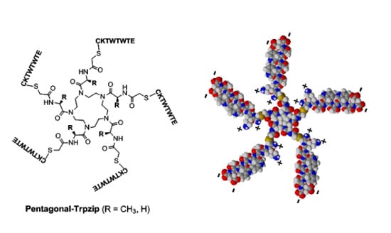
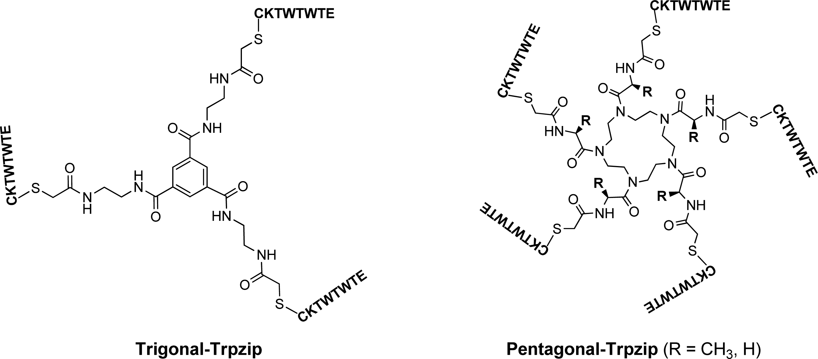
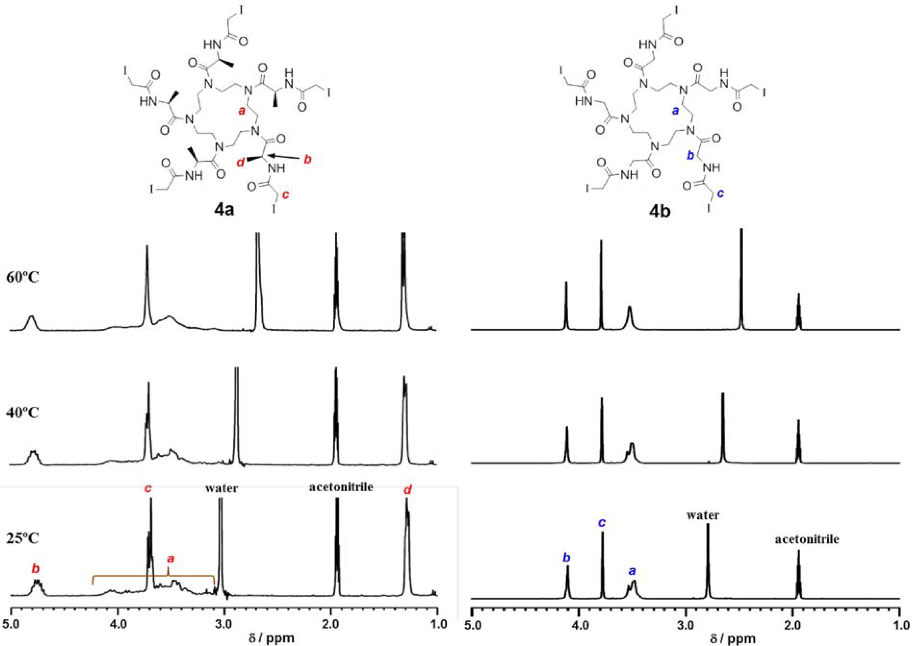
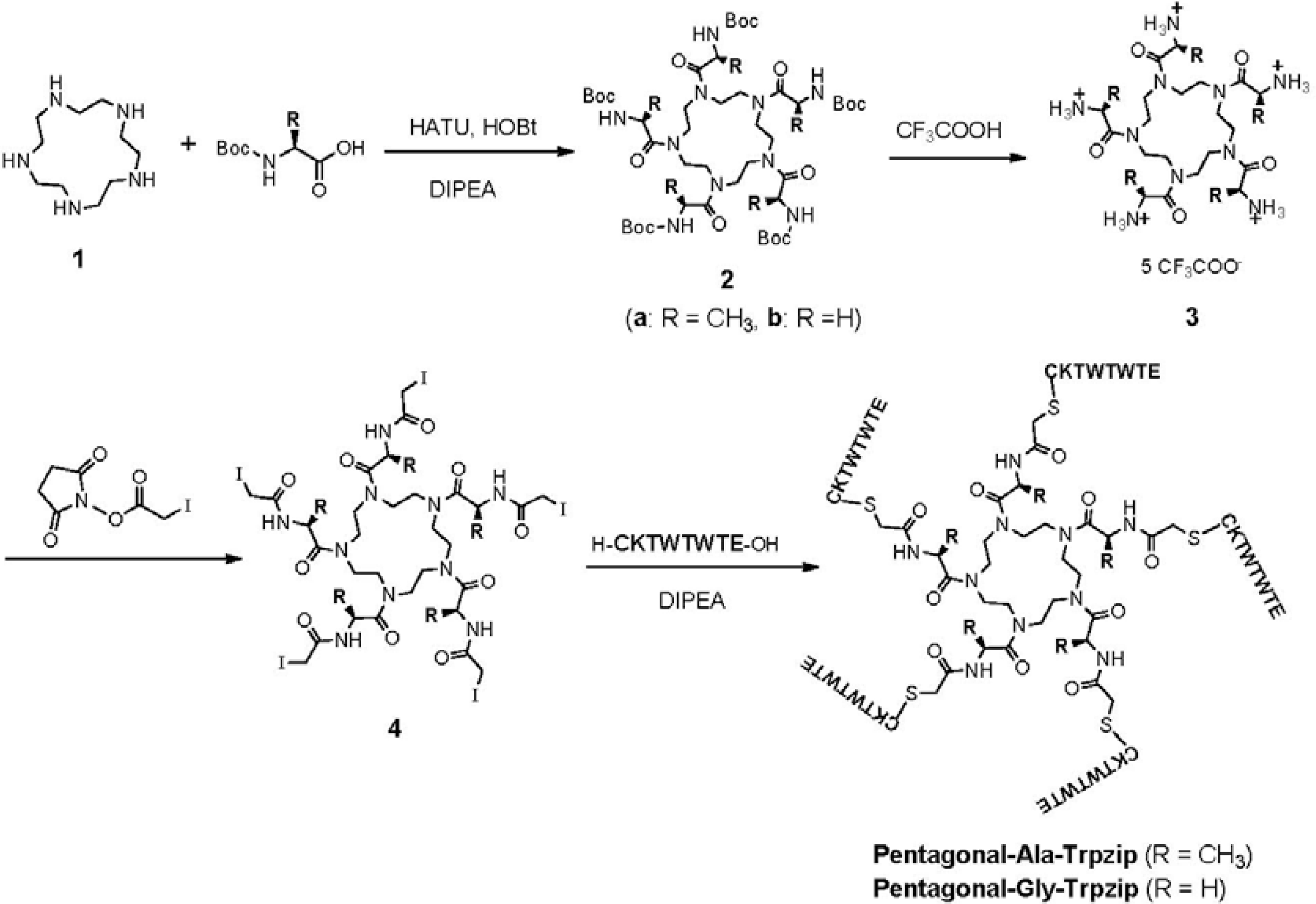
2.1. Synthesis of Pentagonal Conjugates of Tryptophane Zipper-Forming Peptide

1,4,7,10,13-Pentaazacyclopentadecane 1 was synthesized from triethylenetetramine according to the reported procedure [39]. 1 was amidated with Boc-Ala or Boc-Gly, followed by deprotection and iodoacetylation to provide pentagonal core 4 (Scheme 1). 1H NMR spectrum of Ala-containing pentagonal core 4a in mixture of CD3CN/D2O = 15/1 showed notable broad peaks at the range of δ 4.2–3.0 ppm assigned to pentaazacyclopentadecane (Ha) and multiple peaks at δ 4.7 (Hb), 3.7 (Hc) and 1.2 ppm (Hd) (Figure 2). The multiple peaks of Hc were unified by elevating temperature, whereas the peaks assigned to Ha were broad even at 60 °C. This indicates that Ala-containing pentagonal core 4a possesses various irregular conformations of which interconversion are very slow even at 60 °C. In contrast, 1H NMR spectrum of Gly-containing pentagonal core 4b showed relatively sharp peaks (Figure 2), suggesting that 4b possesses flexible conformation. It is probable that steric hindrance of methyl group of Ala to pentaazacyclopentadecane ring of 4a prevent flexible interconversion of conformers, whereas steric hindrance in 4b is smaller than that of 4a.
The 8-mer peptide h-CKTWTWTE-oh, which was designed based on the intermolecular tryptophane zipper (β-hairpin) forming peptide reported by Cohhan et al. [29], was synthesized by a standard Fmoc-protected solid-phase method. Pentagonal-Ala-Trpzip and Pentagonal-Gly-Trpzip were prepared by coupling the 8-mer peptides with pentagonal core 4 in the presence of diisopropylethylamine (Scheme 1). These pentagonal conjugates were purified by reverse-phase HPLC and confirmed by MALDI-TOF-MS (m/z of Pentagonal-Ala-Trpzip = 6038.96 [M + H]+, m/z of Pentagonal-Gly-Trpzip = 5970.58 [M + H]+). They were soluble in water at a whole range of pH.

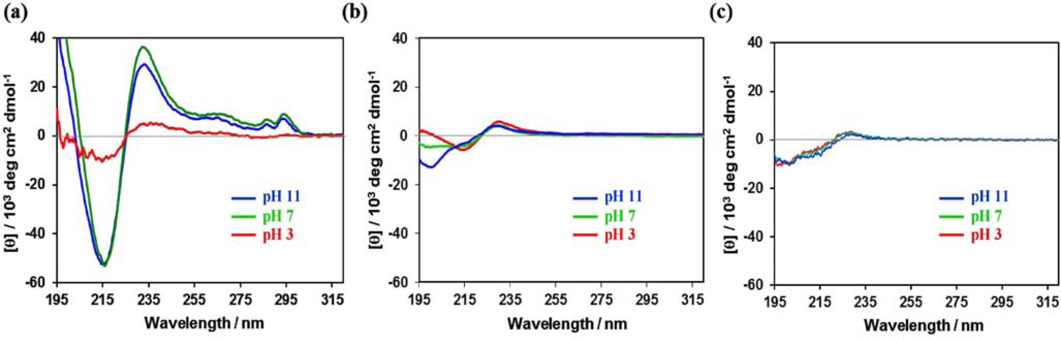
2.2. Secondary Structure of Pentagonal Peptide Conjugates

After the solutions of pentagonal peptide conjugates were incubated for 48 h at 25 °C in 20 mM citrate buffer (pH 3), in 20 mM phosphate buffer (pH 7), and in aqueous NaOH solution (pH 11), circular dichroism (CD) spectra were measured [40]. It has been reported that tryptophane zipper-forming β-hairpin peptide showed exciton-coupling type CD spectrum (positive peak at 230 nm and negative peak at 215 nm) along with small peaks at 285–295 nm based on the interaction between Trp residues [33]. The CD spectra of aqueous solution of Pentagonal-Gly-Trpzip showed negative peak at 216 nm and positive peak at 233 nm together with weak peaks at 280–300 nm in phosphate buffer (pH 7) and aqueous NaOH solution (pH 11) respectively. They indicate the formation of tryptophane zipper and normal β-sheet structure, whereas the CD intensity at 216 and 233 nm considerably decreased in citrate buffer (pH 3) (Figure 3(a)). In contrast, the CD spectra of Pentagonal-Ala-Trpzip showed a weak CD pattern at pH 7 and pH 3 which can be ascribed to tryptophane zipper structure, and the content of random coil increased at pH 11 (Figure 3(b)). The difference in secondary structure between these pentagonal Trpzip conjugates might arise from different flexibility of the pentagonal core, as shown by the 1H NMR spectra (Figure 2).
The CD spectrum of the precursor peptide CKTWTWTE revealed that the peptide adopted random-coil structure at the pH range of 3–11 (Figure 3(c)). We have previously reported that Trigonal-Trpzip also adopts mixed secondary structures of tryptophane zipper and normal β-sheet at pH 7, but the molar elipticity at 216 and 233 nm ([θ]216 = −55,000 and [θ]233 = 21,000 deg cm2 dmol−1) was lower than that of Pentagonal-Gly-Trpzip under the same conditions [35]. These results indicate that the formation of tryptophane zipper structure from the peptide CKTWTWTE is promoted by the pentagonal preorganization, which is more effective than the trigonal preorganization.

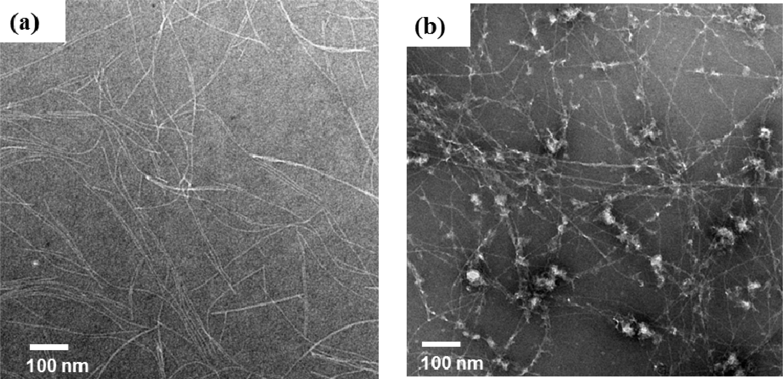
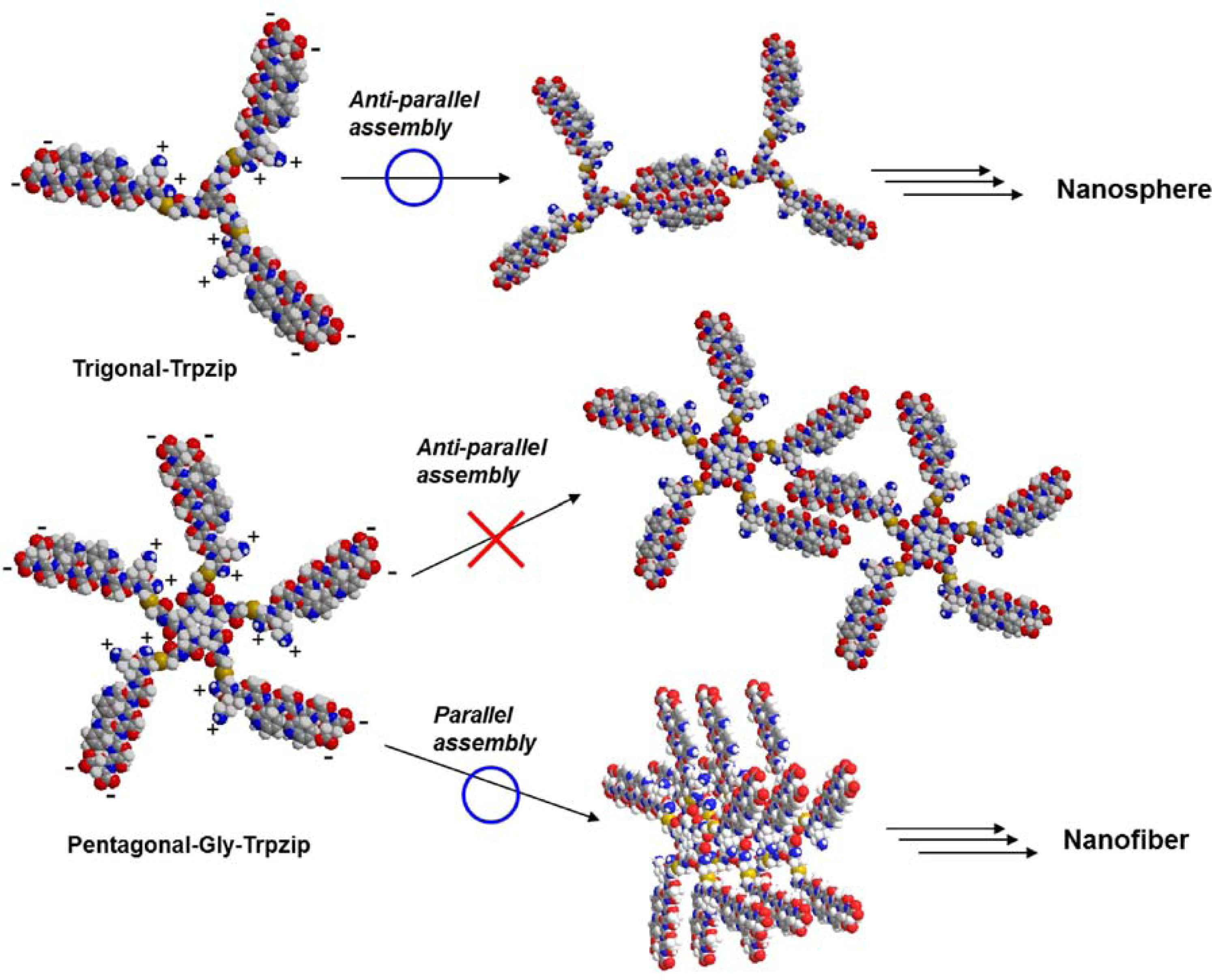
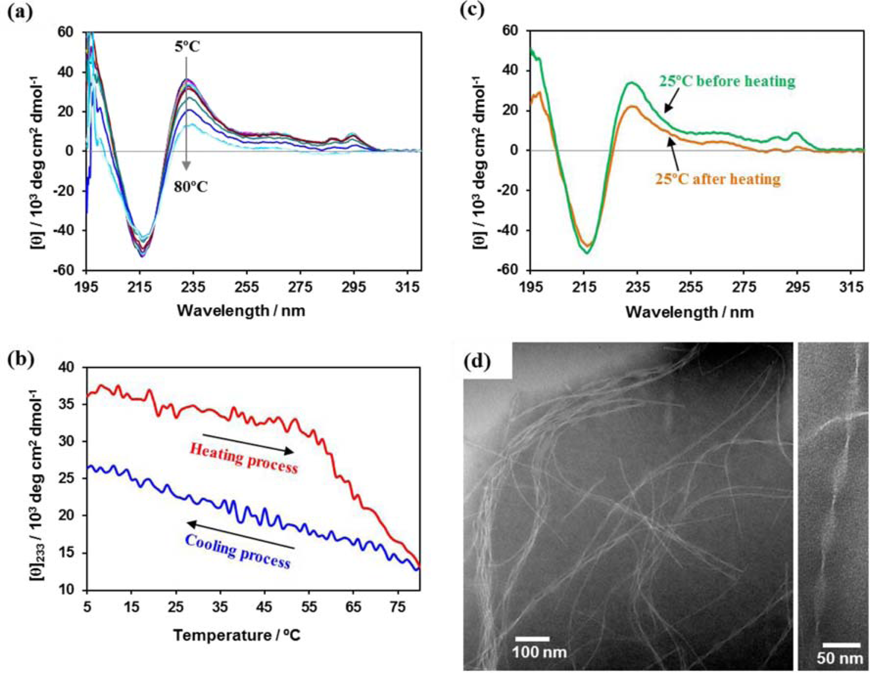
2.3. Self-Assembly of Pentagonal Peptide Conjugates in Water

A transmission electron microscopy (TEM) image revealed that Pentagonal-Gly-Trpzip formed only nanofibers of several micrometers length with uniform width of 4–5 nm in phosphate buffer (pH 7, Figure 4(a)), although we envisioned that the conjugate self-assembles into nanocapsules [40]. It is probable that the nanofibers formed by face-to-face assembly of Pentagonal-Gly-Trpzip via parallel tryptophane zipper and β-sheet structure. On the other hand, in the TEM image of Pentagonal-Ala-Trpzip, irregular aggregates were observed together with nanofibers (Figure 4(b)). This might reflect less formation of tryptophane zipper structure based on rigid irregular core conformations.
We have previously reported that Trigonal-Trpzip selectively self-assembled into nanospheres with the size of 20–30 nm at pH 7 [35]. Since the peptide CKTWTWTE might possess zwitterionic structure at pH7, it is reasonable that Trigonal-Trpzip formed nanospheres by forming intermolecular antiparallel β-sheet-like structures. In contrast, Pentagonal-Gly-Trpzip selectively formed nanofibers probably due to the formation of parallel β-sheet-like structures despite ionic repulsion between peptides (Figure 5). To form nanospheres, it is desirable that the assembly units take concave conformations. The difference in morphology between trignal- and pentagonal-tryptophanezipper conjugtates might arise from difference in the peripheral density of peptide chains and the conformation of scaffold. It seems that steric hindrance among peptide chains in Pentagonal-Gly-Trpzip prevents the formation of anti-parallel β-sheet-like structures and concave conformations, in contrast to the case of Trigonal-Trpzip.
Figure 6(a) shows temperature dependence of CD spectrum for aqueous solution of Pentagonal-Gly-Trpzip (10 μM) at pH 7. The positive peaks at 233 and 285–295 nm were gradually decreased by heating process, but the CD intensity at 233 nm hardly recovered by cooling process (Figure 6(b)). CD spectrum of Pentagonal-Gly-Trpzip at 25 °C after the heating process showed formation of normal β-sheet-rich structure (Figure 6(c)). These results indicate the irreversible structural change of tryptophane zipper to normal β-sheet-rich structure. A TEM image revealed that Pentagonal-Gly-Trpzip formed left-handed helical ribbon structures with the width of 20–30 nm and pitch of 50–100 nm after the heating process (Figure 6(d)). It is probable that kinetically self-assembled nanofibers consisting of tryptphane zipper transformed to thermodynamically stable helical ribbons consisting of normal β-sheet by the heating process.

3. Experimental Section

3.1. General

Reagents were obtained from commercial source and used without further purification. Deionized water of high resistivity (>18 MΩ cm) purified with a Millipore Purification System (Milli-Q water) was used as a solvent of peptide conjugates. 1H-NMR spectra were recorded on Bruker AV300M spectrometer. Reversed-phase HPLC was performed at ambient temperature with a Simadzu LC-6AD liquid chromatograph equipped with a UV/Vis detector (220 nm, Shimadzu SPD-10AVvp) using Inertsil ODS-3 (GL Science) or COSMOSIL Protein-R (Nakarai Tesque) columns (250 × 4.6 mm or 250 × 20 mm). MALDI-TOF mass spectra were obtained on Autoflex III (Bruker Daltonics) under the linear/positive mode with α-cyano-4-hydroxy cinnamic acid (α-CHCA) as matrix.

3.2. Synthesis of Peptide Conjugates

1,4,7,10,13-Pentaazacyclopentadecane (1): 1 was synthesized from triethylenetetramine according to the reported procedure [39].
Tryptophene zipper-forming peptide (H-CKTWTWTE-OH): Peptide H-Cys(Trt)-Lys(Boc)-Thr(tBu)-Trp(Boc)-Thr(tBu)-Trp(Boc)-Thr(tBu)-Glu(OtBu)-Alko resin was synthesized on α-p-alkoxybenzyl alcohol resin (Alko resin, Watanabe Chemical Industries, Ltd., Hiroshima, Japan, 0.69 mmol/g) using standard Fmoc-based FastMoc coupling chemistry (3 eq. Fmoc-amino acids) with an ABI 433A synthesizer (Applied Biosystems, Carlsbad, CA, USA). DMF solution of 2-(1H-benzotriazole-1-yl)-1,1,3,3-tetramethyluronium hexafluorophosphate (HBTU, 0.5 M) and 1-hydroxybenzotriazole hydrate (HOBt·H2O, 0.5 M) was used as a coupling reagent. 2.0 M diisopropylethylamine (DIPEA) in NMP and 20% piperidine in NMP were used for neutralization and for Fmoc deprotection, respectively. The peptidyl-resin was washed with NMP, dichloromethane and methanol then dried under vacuum. The peptide was deprotected and cleaved from the resin by treatment with a cocktail of TFA/1,2-ethanedithiol/water/triisopropylsilane = 94/2.5/2.5/1 in volume at room temperature for 2 h. The reaction mixture was filtered to remove the resin and the filtrate was concentrated under vacuum. The peptide was precipitated by adding ice-cooled methyl-tert-butyl ether to the residue and the supernatent was decanted. After repeating the methyl-tert-butyl ether washing 6 times, the precipitated peptide was dried under vacuum. The crude product was purified by reversed-phase HPLC (column: Inertsil ODS-3) eluting with a liner gradient of CH3CN/water (18/82 to 30/70 over 60 min) containing 0.1% TFA. The elution fraction containing the desired peptide was lyophilized to give a flocculent solid. The isolated yield was 23%. MALDI-TOF-MS (matrix: α-CHCA): m/z = 1054.47 [M + H]+. 1H-NMR (D2O, δ/ppm): 7.37–7.45 (2 H, m), 7.23–7.28 (2H, m), 6.90–7.08 (6H, m), 4.51 (1H, t, J = 7.2 Hz), 4.45 (1H, t, J = 7.2 Hz), 4.20 (1H, t, J = 7.2 Hz), 4.15 (1H, d, J = 5.1 Hz), 4.04–4.10 (3H, m), 3.85–4.01 (4H, m), 3.10 (2H, t, J = 6.3 Hz), 2.89–2.97 (4H, m), 2.67 (2H, t, J = 7.8 Hz), 2.22 (2H, t, J = 7.8 Hz), 1.84–1.99 (1H, m), 1.68–1.84 (1H, m), 1.48–1.58 (2H, m), 1.37–1.48 (2H, m), 1.07–1.20 (2H, m), 0.98 (3H, d, J = 6.3 Hz), 0.91 (3H, d, J = 6.3 Hz), 0.88 (3H, d, J = 6.3 Hz).
N,N″,N″,N″″,N″″-Pentakis(N-tert-butoxycarbonyl-l-alanyl)-1,4,7,10,13-pentaazacyclopentadecane (2a): Boc-Ala-OH (265 mg, 1.40 mmol) and O-(7-azabenzotriazolyl)-tetramethyluronium hexafluorophosphate (HATU, 558 mg, 1.43 mmol) were dissolved in dry DMF (2.0 mL). DIPEA (244 μL, 1.40 mmol) was added to the mixture by microsyringe, and then the mixture was stirred for 45 min at room temperature. Then a solution of 1,4,7,10,13-pentaazacyclopentadecane 1 (30 mg, 0.14 mmol) in dry DMF (1.0 mL) was added to the mixture. After the mixture was stirred for 43 h at room temperature, the solvent was evaporated under reduced pressure. The residue was dissolved in chloroform and washed with 5% aqueous NaHCO3 and deionized water. The organic layer was dried over anhydrous Na2SO4 and evaporated to a sticky solid. The sticky solid was dissolved in 20% aqueous acetonitrile and was lyophilized to provide a yellowish powder (142 mg). The crude yield was 93%. The product was not purified further and was used in the next reaction. MALDI-TOF-MS (matrix: α-CHCA): m/z = 1092.64 [M + Na]+. 1H-NMR (CD3OD, δ/ppm): 3.4–4.9 (25H, br), 1.48 (45H, br), 1.37 (15H, br).
N,N″,N″,N″″,N″″-Pentakis(N-tert-butoxycarbonylglycyl)-1,4,7,10,13-pentaazacyclopentadecane (2b): Compound 2b was prepared by the almost same procedure described above. The crude yield was 96% (135 mg). The product was not purified further and was used in the next reaction. MALDI-TOF-MS (matrix: α-CHCA): m/z = 1022.94 [M + Na]+. 1H-NMR (CD3OD, δ/ppm): 3.98 (10H, s), 3.5–3.7 (20H, br), 1.340 (45H, br).
N,N″,N″,N″″,N″″-Penta-l-alanyl-1,4,7,10,13-pentaazacyclopentadecane trifluoroacetic acid salt (3a): Trifluoroacetic acid (1.0 mL, 13 mmol) and deionized water were added to a solution of compound 2a (142 mg, 0.132 mmol) in dichloromethane (2.0 mL). After the mixture was stirred for 1 h at room temperature, excess trifluoroacetic acid and dichloromethane were evaporated under reduced pressure. The residue was washed with methyl-tert-butyl ether, and the supernatant was decanted. The residue was dried with argon gas to provide a colorless powder (109 mg). The crude yield was 72%. The product was not purified further and was used in the next reaction. MALDI-TOF-MS (matrix: α-CHCA): m/z = 571.40 [M + H]+.
N,N″,N″,N″″,N″″-Pentaglycyl-1,4,7,10,13-pentaazacyclopentadecane trifluoroacetic acid salt (3b): Compound 3b was prepared by the almost same procedure described above. The crude yield was 100% (123 mg). The product was not purified further and was used in the next reaction. MALDI-TOF-MS (matrix: α-CHCA): m/z = 501.33 [M + H]+.
N,N″,N″,N″″,N″″-Pentakis(N-iodoacetyl-l-alanyl)-1,4,7,10,13-pentaazacyclopentadecane (4a): A solution of compound 3a (50 mg, 0.044 mmol) and NaHCO3 (37 mg, 0.44 mmol) in deionized water (2.0 mL) was added to a solution of iodoacetic acid N-hydroxy succinimide ester (248 mg, 0.876 mmol) in acetone (2.0 mL). The mixture became turbid immediately and was stirred in the dark at room temperature. After 48 h, the reaction mixture indicated acidic pH. The mixture was neutralized with NaHCO3 (84 mg, 1.0 mmol) and added iodoacetic acid N-hydroxy succinimide ester (50 mg, 0.18 mmol) in acetone (1.0 mL). After the mixture was stirred for 2 h in the dark, the resulted precipitate was filtered. The residue was washed with deionized water and acetone to provide a colorless powder. The crude product was purified by reversed-phase HPLC (column: Inertsil ODS-3) columns eluting with a linear gradient of CH3CN/water (30/70 to 50/50 over 20 min). The isolated yield was 12%. MALDI-TOF-MS (matrix: α-CHCA): m/z = 1409.88 [M + H]+. 1H-NMR (CD3CN + D2O, δ/ppm): 4.65–4.80 (5H, m), 2.9–4.2 (30H, br m), 1.2–1.3 (15H, m).
N,N″,N″,N″″,N″″-Pentakis(N-iodoacetylglycyl)-1,4,7,10,13-pentaazacyclopentadecane (4b): A solution of compound 3b (47 mg, 0.044 mmol) and NaHCO3 (28 mg, 0.33 mmol) in deionized water (1.0 mL) was added to a solution of iodoacetic acid N-hydroxy succinimide ester (96 mg, 0.34 mmol) in acetone (1.0 mL). The mixture was stirred for 1 h in the dark at room temperature. After the solvent was evaporated under reduced pressure, the residue was purified by reversed-phase HPLC (column: Inertsil ODS-3) eluting with a linear gradient of CH3CN/water (25/75 to 50/50 over 25 min). The isolated yield was 13.4 mg (23%). MALDI-TOF-MS (matrix: α-CHCA): m/z = 1340.79 [M + H]+. 1H-NMR (CD3CN + D2O, δ/ppm): 7.5–7.6 (2H, br), 4.10 (10H, s), 3.76 (10H, s), 3.4–3.6 (20H, br m).
Pentagonal-Ala-Trpzip: Peptide H-CKTWTWTE-OH (13.8 mg, 10.8 μmol) was dissolved in degassed DMF (4.0 mL) under nitrogen at −20 °C. To the mixture were added a solution of compound 4a (2.17 mg, 1.54 μmol) in degassed DMF (4 mL) and then a solution of DIPEA (9.4 μL, 54 μmol) in degassed DMF (0.1 mL) at the same temperature in the dark. The mixture was stirred for 4 h under the same conditions. After removal of DMF under reduced pressure, the residue was purified by reversed-phase HPLC (column: COSMOSIL Protein-R) eluting with a linear gradient of CH3CN/water (23/77 to 28/72 over 40 min) containing 0.1% TFA. The elution fraction containing the desired conjugate was lyophilized to give a flocculent solid. The isolated yield was 1.8 mg (18%). MALDI-TOF-MS (matrix: α-CHCA): m/z = 6038.96 [M + H]+ and 3018 [M + 2H]2+.
Pentagonal-Gly-Trpzip: Pentagonal-Gly-Trpzip was prepared by the almost same procedure described above and was purified by reversed-phase HPLC (column: COSMOSIL Protein-R) eluting with a linear gradient of CH3CN/water (28/72 to 32/68 over 40 min) containing 0.1% TFA. The isolated yield was 1.9 mg (25%). MALDI-TOF-MS (matrix: α-CHCA): m/z = 5970.58 [M + H]+.

3.3. CD Spectrum Measurements

The stock solutions of pentagonal-peptide conjugates (70 μM, the concentration was determined by absorbance at 280 nm) were prepared by dissolving in aqueous NaOH (pH 11) solution, and then diluted in water, 20 mM phosphate buffer (pH 7), and 20 mM citrate buffer (pH 3), respectively. After the solutions of pentagonal peptide conjugates were incubated for 48 h at 25 °C, CD spectra were taken in a 1.0 mm quartz cell with a JASCO J-820 spectrophotometer equipped with a Peltier-type thermostatic cell holder. Temperature dependence of CD spectrum was recorded at 5, 15, 25, 30, 40, 50, 60, 70, and 80 °C. The heating and cooling rates were 1 °C/min. Before the measurement of the CD spectrum, the solution was pre-incubated for 1 min at each temperature.

3.4. Transmission Electron Microscopy (TEM)

A carbon-coated Cu-grid (Oken Co., Ltd., Tokyo, Japan) hydrophilized for 30 sec by hydrophilizing treatment apparatus (JEOL HDT-400). The sample solutions for CD spectra were also used for TEM observation. An aliquot (10 μL) of the solutions was applied to a hydrophilized carbon-coated Cu-grid, left for 60 s, and then removed. Subsequentry, a drop of 2 wt% aqueous phosphotungsic acid was placed on the grids and dried in vacuo (post-staining method). The Cu meshes were subjected to TEM observation (JEOL JEM-2010) with acceleration voltage of 120 kV at 25 °C. All of the measurements were repeated for at least two samples to ensure data reproducibility.

4. Conclusions

We have developed pentagonal conjugates of tryptophane zipper-forming peptide with a pentaazacyclopentadecane core (Pentagonal-Gly-Trpzip and Pentagonal-Ala-Trpzip). CD spectra revealed that Pentagonal-Gly-Trpzip formed tryptophane zipper structure at pH7, but Pentagonal-Ala-Trpzip and the precursor peptide CKTWTWTE showed the formation of weak tryptophane zipper or random coil structure. Pentagonal-Gly-Trpzip self-assembled into nanofibers with uniform width at pH 7, whereas Pentagonal-Ala-Trpzip fromed irregular aggregates which reflect less formation of tryptophane zipper structure. The nanofibers from Pentagonal-Gly-Trpzip showed irreversible transformation to helical ribbon accompanying the secondary structural change by heating process. The present peptide nanostructures would be applied as biodegradable nanomaterials and platforms for nano-biotechnology. The present pentagonal molecular design would extend the feasibility of multivalent peptide conjugates.

Acknowledgments

The present work is supported by PRESTO, Japan Science and Technology Agency, by a Grant-in-Aid for Scientific Research (B) (No. 22350075) from JSPS, and by a Grant-in-Aid for the Global COE Program, “Science for Future Molecular Systems” from MEXT.

References

  1. Mammen, M; Choi, S; Whitesides, GM. Polyvalent interactions in biological systems: Implications for design and use of multivalent ligands and inhibitors. Angew. Chem. Int. Ed 1998, 37, 2754–2794. [Google Scholar]
  2. Kiessling, LL; Gestwicki, JE; Strong, LE. Synthetic multivalent ligands as probes of signal transduction. Angew. Chem. Int. Ed 2006, 45, 2348–2368. [Google Scholar]
  3. Dam, TK; Brewer, CF. Effects of clustered epitopes in multivalent ligand-receptor interactions. Biochemistry 2008, 47, 8470–8476. [Google Scholar]
  4. Mulder, A; Huskens, J; Reinhoudt, DN. Multivalency in supramolecular chemistry and nanofabrication. Organ. Biomol. Chem 2004, 2, 3409–3424. [Google Scholar]
  5. Kitov, PI; Sadowska, JM; Mulvey, G; Armstrong, GD; Ling, H; Pannu, NS; Read, RJ; Bundle, DR. Shiga-like toxins are neutralized by tailored multivalent carbohydrate ligands. Nature 2000, 403, 669–672. [Google Scholar]
  6. Zhang, Z; Merritt, EA; Ahn, M; Roach, C; Hou, Z; Verlinde, CLMJ; Hol, WGJ; Fan, E. Solution and crystallographic studies of branched multivalent ligands that inhibit the receptor-binding of cholera toxin. J. Am. Chem. Soc 2002, 124, 12991–12998. [Google Scholar]
  7. Mutter, M; Vuilleumier, S. A Chemical approach to protein design. Template-assembled synthetic proteins (TASP). Angew. Chem. Int. Ed 1989, 28, 535–676. [Google Scholar]
  8. Crespo, L; Sanclimens, G; Pons, M; Giralt, E; Royo, M; Albericio, F. Peptide and amide bond-containing dendrimers. Chem. Rev 2005, 105, 1663–1681. [Google Scholar]
  9. Darbre, T; Reymond, J-L. Peptide dendrimers as artificial enzymes, receptors, and drug-delivery agents. Acc. Chem. Res 2006, 39, 925–934. [Google Scholar]
  10. Zhang, S. Fabrication of novel biomaterials through molecular self-assembly. Nat. Biotech 2003, 21, 1171–1178. [Google Scholar]
  11. Gao, X; Matsui, H. Peptide-based nanotubes and their application in bionanotechnology. Adv. Mater 2005, 17, 2037–2050. [Google Scholar]
  12. Gazit, E. Self-assembled peptide nanostructures: The design of molecular building blocks and their technological utilization. Chem. Soc. Rev 2007, 36, 1263–1269. [Google Scholar]
  13. Ryadnov, MG; Woolfson, DN. Introducing branches into a self-assembling peptide fiber. Angew. Chem. Int. Ed 2003, 42, 3021–3023. [Google Scholar]
  14. Ryadnov, MG; Woolfson, DN. Engineering the morphology of a self-assembling protein fibre. Nat. Mater 2003, 2, 329–332. [Google Scholar]
  15. Smith, AM; Acquah, SFA; Bone, N; Kroto, HW; Ryadnov, MG; Stevens, MSP; Walton, DRM; Woolfson, DN. Polar assembly in a designed protein fiber. Angew. Chem. Int. Ed 2005, 44, 325–328. [Google Scholar]
  16. Boato, F; Thomas, RM; Ghasparian, A; Freund-Renard, A; Moehle, K; Robinson, JA. Synthetic virus-like particles from self-assembling coiled-coil lipopeptides and their use in antigen display to the immune system. Angew. Chem. Int. Ed 2007, 46, 9015–9018. [Google Scholar]
  17. Marini, DM; Hwang, W; Lauffenburger, A; Zhang, S; Kamm, RD. Left-handed helical ribbon intermediates in the self-assembly of a β-sheet peptide. Nano Lett 2002, 2, 295–299. [Google Scholar]
  18. Mihara, H; Matsumura, S; Takahashi, T. Construction and control of self-assembly of amyloid and fibrous peptides. Bull. Chem. Soc. Jpn 2005, 78, 572–590. [Google Scholar]
  19. Lim, Y-B; Lee, E; Lee, M. Cell-penetrating-peptide-coated nanoribbons for intracellular nanocarriers. Angew. Chem. Int. Ed 2007, 46, 3475–3478. [Google Scholar]
  20. Lim, Y-B; Park, S; Lee, E; Jeong, H; Ryu, J-H; Lee, MS; Lee, M. Glycoconjugate nanoribbons from the self-assembly of carbohydrate-peptide block molecules for controllable bacterial cell cluster formation. Biomacromolecules 2007, 8, 1404–1408. [Google Scholar]
  21. Kwon, S; Jeon, A; Yoo, SH; Chung, IS; Lee, H-S. Unprecedented molecular architectures by the controlled self-assembly of a β-peptide foldamer. Angew. Chem. Int. Ed 2010, 49, 8232–8236. [Google Scholar]
  22. Zhou, M; Bentley, D; Ghosh, I. Helical supramolecules and fibers utilizing leucine zipper-displaying dendrimers. J. Am. Chem. Soc 2004, 126, 734–735. [Google Scholar]
  23. Matsuura, K; Murasato, K; Kimizuka, N. Artificial peptide-nanospheres self-assembled from three-way junctions of β-sheet-forming peptides. J. Am. Chem. Soc 2005, 127, 10148–10149. [Google Scholar]
  24. Murasato, K; Matsuura, K; Kimizuka, N. Self-assembly of nanofiber with uniform width from wheel-type trigonal-β-sheet forming peptide. Biomacromolecules 2008, 9, 913–918. [Google Scholar]
  25. Matsuura, K; Watanabe, K; Sakurai, K; Matsuzaki, T; Kimizuka, N. Self-assembled synthetic viral capsids from a 24-mer viral peptide fragment. Angew. Chem. Int. Ed 2010, 49, 9662–9665. [Google Scholar]
  26. Matsuura, K; Matsuyama, H; Fukuda, T; Teramoto, T; Watanabe, K; Murasato, K; Kimizuka, N. Spontaneous self-assembly of nano-spheres from trigonal conjugate of glutathione in water. Soft Matter 2009, 5, 2463–2470. [Google Scholar]
  27. Matsuura, K; Fujino, K; Teramoto, T; Murasato, K; Kimizuka, N. Glutathione nanospheres: Self-assembly of conformation-regulated trigonal-glutathiones in water. Bull Chem Soc Jpn 2010, 83, 880–886. [Google Scholar]
  28. Matsuura, K; Tochio, K; Watanabe, K; Kimizuka, N. Controlled release of guest molecules from spherical assembly of trigonal-gultathione by a disulfide recombination. Chem. Lett 2011, 40, 711–713. [Google Scholar]
  29. Cochran, AG; Skelton, NJ; Starovasnik, MA. Tryptophan zippers: Stable, monomeric β-hairpins. Proc. Natl. Acad. Sci. USA 2001, 98, 5578–5583. [Google Scholar]
  30. Richardson, JS; Richardson, DC. Natural β-sheet proteins use negative design to avoid edge-to-edge aggregation. Proc. Natl. Acad. Sci. USA 2002, 99, 2754–2759. [Google Scholar]
  31. Dempsey, CE; Piggot, TJ; Mason, PE. Dissecting contributions to the denaturant sensitivities of proteins. Biochemistry 2005, 44, 775–781. [Google Scholar]
  32. Yang, WY; Pitera, JW; Swope, WC; Gruebele, M. Heterogeneous folding of the Trpzip hairpin: Full atom simulation and experiment. J. Mol. Biol 2004, 336, 241–251. [Google Scholar]
  33. Streicher, WW; Makhatadze, GI. Calorimetric evidence for a two-state unfolding of the β-hairpin peptide Trpzip4. J. Am. Chem. Soc 2006, 128, 30–31. [Google Scholar]
  34. Chetal, P; Chauhan, VS; Sahal, D. A meccano set approach of joining trpzip a water soluble β-hairpin peptide with a didehydrophenylalanine containing hydrophobic helical peptide. J. Peptide Res 2005, 65, 475–484. [Google Scholar]
  35. Matsuura, K; Hayashi, H; Murasato, K; Kimizuka, N. Trigonal tryptophane-zipper as a novel building block for pH-responding peptide nano-assemblies. Chem. Commun 2011, 47, 265–267. [Google Scholar]
  36. Harrison, SC. Multiple modes of subunit association in the structures of simple spherical viruses. Trends Biochem. Sci 1984, 9, 345–351. [Google Scholar]
  37. Harrison, SC. The familiar and the unexpected in structures of icosahedral viruses. Curr. Opin. Struct. Biol 2001, 11, 195–199. [Google Scholar]
  38. Olson, AJ; Hu, YHE; Kelnan, E. Chemical mimicry of viral capsid self-assembly. Proc. Natl. Acad. Sci. USA 2007, 104, 20731–20736. [Google Scholar]
  39. Bencini, A; Fabbrizzi, L; Poggi, A. Formation of nickel(III) complexes with n-dentate amine macrocycles (n = 4, 5, 6). ESR and electrochemical studies. Inorg. Chem 1981, 20, 2544–2549. [Google Scholar]
  40. Extension of the incubation time (96 h) minimally affected to the results of CD spectra and TEM images.
  41. The measurement was repeated for three times to confirm data reproducibility. The standard deviation was about 22%.
Figure 1. Structures of trigonal and pentagonal conjugates of tryptophane zipper peptide.
Ijms 12 05187f1
Figure 2. 1H-NMR spectra of pentagonal iodoacetoamidated core molecules (4a and 4b) at 2.8 mM in mixture of CD3CN/D2O = 15/1.
Ijms 12 05187f2
Figure 3. Circular dichroism (CD) spectra of aqueous solution of (a) Pentagonal-Gly-Trpzip (10 μM); (b) Pentagonal-Ala-Trpzip (10 μM); and (c) tryptophane zipper peptide CKTWTWTE (50 μM) at 25 °C in 20 mM citrate buffer (pH 3, red line), in 20 mM phosphate buffer (pH 7, green line), and in aqueous NaOH solution (pH 11, blue line).
Ijms 12 05187f3
Figure 4. Transmission electron microscopy (TEM) images of pentagonal conjugates of tryptophane zipper peptide (10 μM) in 20 mM phosphate buffer (pH 7) at 25 °C: (a) Pentagonal-Gly-Trpzip, and (b) Pentagonal-Ala-Trpzip. TEM samples were stained with phosphotungstic acid.
Ijms 12 05187f4
Figure 5. Schematic illustration of the self-assembly of trigonal and pentagonal conjugates of tryptophane zipper peptide.
Ijms 12 05187f5
Figure 6. Structural change of Pentagonal-Gly-Trpzip by heating process. (a) Temperature dependence of CD spectrum for aqueous solution of Pentagonal-Gly-Trpzip (10 μM) at pH 7; (b) Temperature dependence of [θ] at 233 nm for the aqueous solution. The heating and cooling rates were 1 °C/min [41]; (c) CD spectra of Pentagonal-Gly-Trpzip at 25 °C before (green line) and after (orange line) the heating process; and (d) TEM image of Pentagonal-Gly-Trpzip at 25 °C after the heating process.
Ijms 12 05187f6
Scheme 1. Synthesis of pentagonal conjugates of tryptophane zipper peptide.
Ijms 12 05187f7

