Article

Correlation among Antioxidant, Antimicrobial, Hemolytic, and Antiproliferative Properties of Leiothrix spiralis Leaves Extract

1
Biological Sciences Department, Faculty of Pharmaceutical Sciences, São Paulo State University—UNESP, Araraquara, SP 14801-902, Brazil
2
Organic Chemistry Department, Chemistry Institute, São Paulo State University—UNESP, c.p. 355, Araraquara, SP 14800-900, Brazil
3
Clinical Analysis Department, Faculty of Pharmaceutical Sciences, São Paulo State University—UNESP, Araraquara, SP 14801-902, Brazil
4
Department of Clinical Biochemistry, CIBICI-CONICET, Faculty of Chemical Sciences, National University of Cordoba, Haya de la Torre y Medina Allende, Córdoba 5000, Argentina
*
Author to whom correspondence should be addressed.
Int. J. Mol. Sci. 2012, 13(7), 9260-9277; https://doi.org/10.3390/ijms13079260
Submission received: 20 April 2012 / Revised: 15 July 2012 / Accepted: 20 July 2012 / Published: 24 July 2012

Abstract

:
The biological activities of a plant extract depend on a complex sum of individual properties including the antioxidant activity. Several biological activities protect against the harmful action of reactive oxygen species (ROS), and here we focused our attention on the relationship between the biological activities tested and the antioxidant properties. In this study, the total flavonoid content as well as the antioxidant, antimicrobial, hemolytic and cytotoxicity activities of the methanolic extract of Leitothrix spiralis leaves were evaluated. The extract showed a total flavonoid content of 19.26% and the chemical characterization by HPLC-PAD confirmed the presence of flavonoids as the major secondary metabolite compounds. Significant antioxidant activity (IC50 = 1.743 μg/mL ± 0.063) was demonstrated and was effective against Gram-negative organisms and all Candida strains tested, and showed an ability to inhibit hyphal formation. Non-hemolytic and antiproliferative activity could be demonstrated.

1. Introduction

Reactive oxygen species (ROS) and reactive nitrogen species (RNS), including free radicals such as superoxide radical anion, hydroxyl radicals, singlet oxygen, hydrogen peroxide and nitric oxide are continuously produced in human cells [1,2]. In this sense, natural products have been attracting scientific interest due to their antioxidant and chemopreventive properties. It is well-known that one of the main characteristics responsible for the antioxidant activity of a plant extract is its high content of phenolic compound and its ability to scavenge free radicals, which can play a part in the protection against the harmful action of ROS. Phenolic compounds also exhibit a wide range of other biological effects, such as anti-viral, antibacterial, and anti-tumor [37].
Oxidative stress is now recognized as a major factor associated with the development of chronic diseases, including cancer and cardiovascular disease. This has led to the hypothesis that the beneficial effects of natural products could be largely explained by their high content of antioxidants [8,9]. Alterations in the regulation of a number of key pathways in controlling cell proliferation are necessary for the establishment of all tumors. Several antioxidants in plants have been suggested to contribute to the anticarcinogenic effect, and others such as flavanols have also been able to inhibit cancer cell proliferation in vitro [10].
Leiothrix, a Eriocaulaceae genus, is exclusively found in South America of which 37 species are mainly restricted to Brazil. Some species of this family are of great economic importance since they are exported as ornamental plants to various countries, mainly Germany and Japan [11]. Chemical studies of the contents of soluble phenolic compounds of Leiothrix species were performed and the presence of xanthones and flavones, including nepetin-7-O-β-d-glucopyranoside, nepetin-7-O-β-d-arabinopyranoside and luteolin O- and C-glucopyranoside, rutin, 6-methoxyapigenin-7-O-d-glucopyranoside and 6-methoxyapigenin, were identified [5,1214]. Antimicrobial activity of phenolic compounds from Leiothrix spiralis was investigated, showing activity on different strains of bacteria and yeasts [15]. Interest in natural antioxidant sources prompted us to investigate the biological activity of L. spiralis.
Although many studies concerning the antioxidant activity of plant extracts have been performed, only a few have tried to correlate this activity with antimicrobial and antiproliferative properties. Thus, the aim of the present study was to chemically characterize and quantify total flavonoids in the methanolic extract of L. spiralis leaves, as well as to evaluate its antioxidant, antimicrobial, hemolytic and human cancer cell antiproliferative activities.

2. Results and Discussion

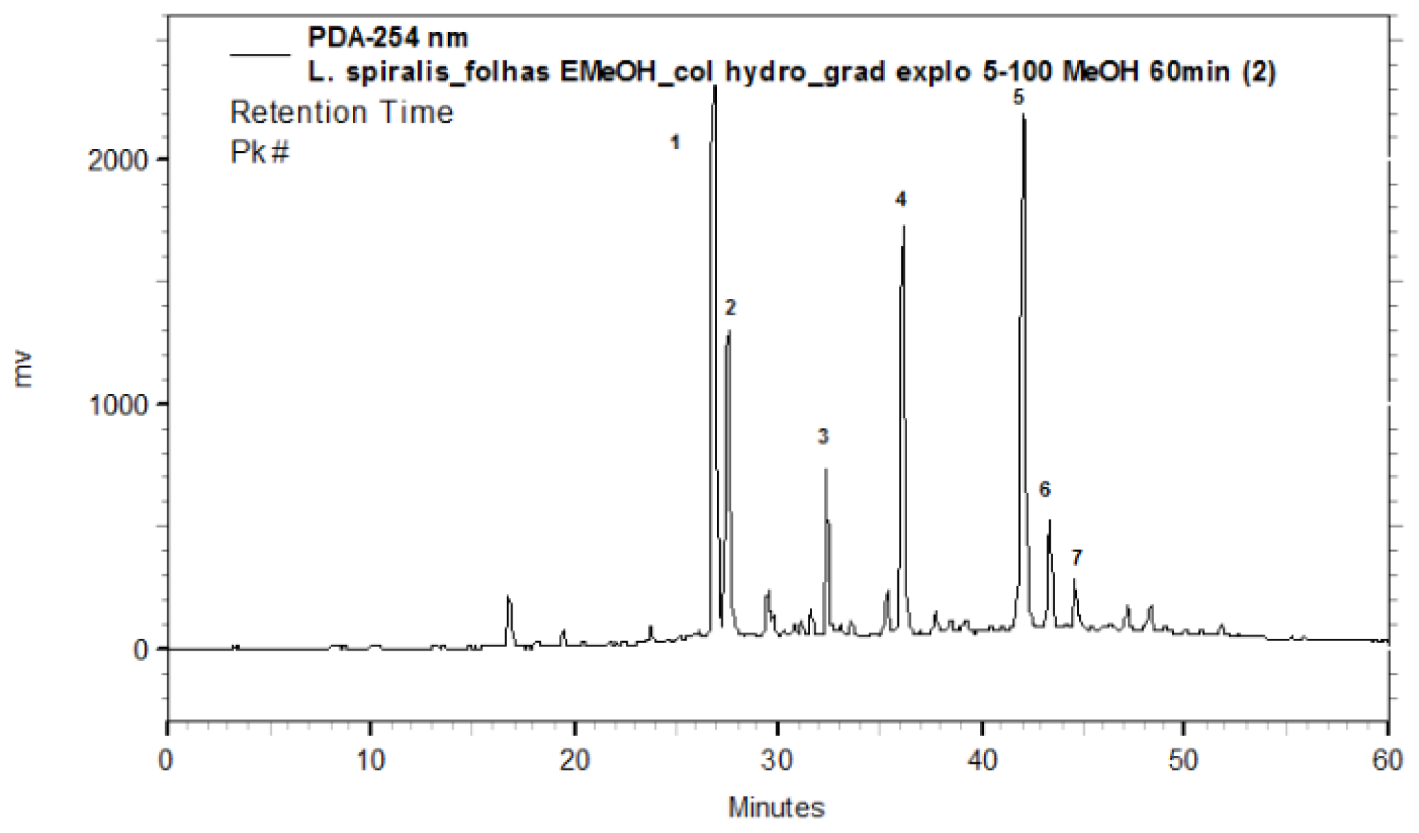
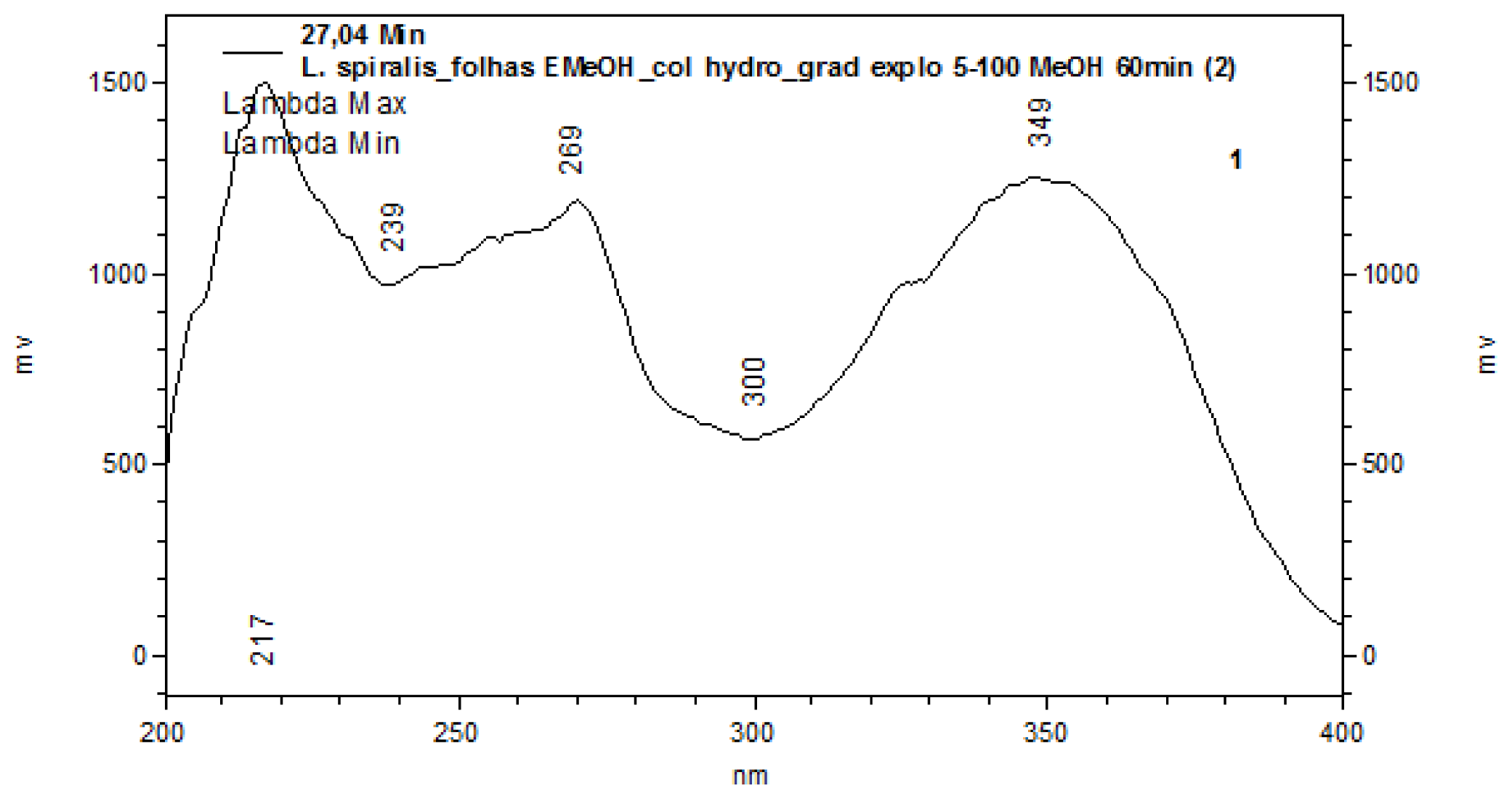
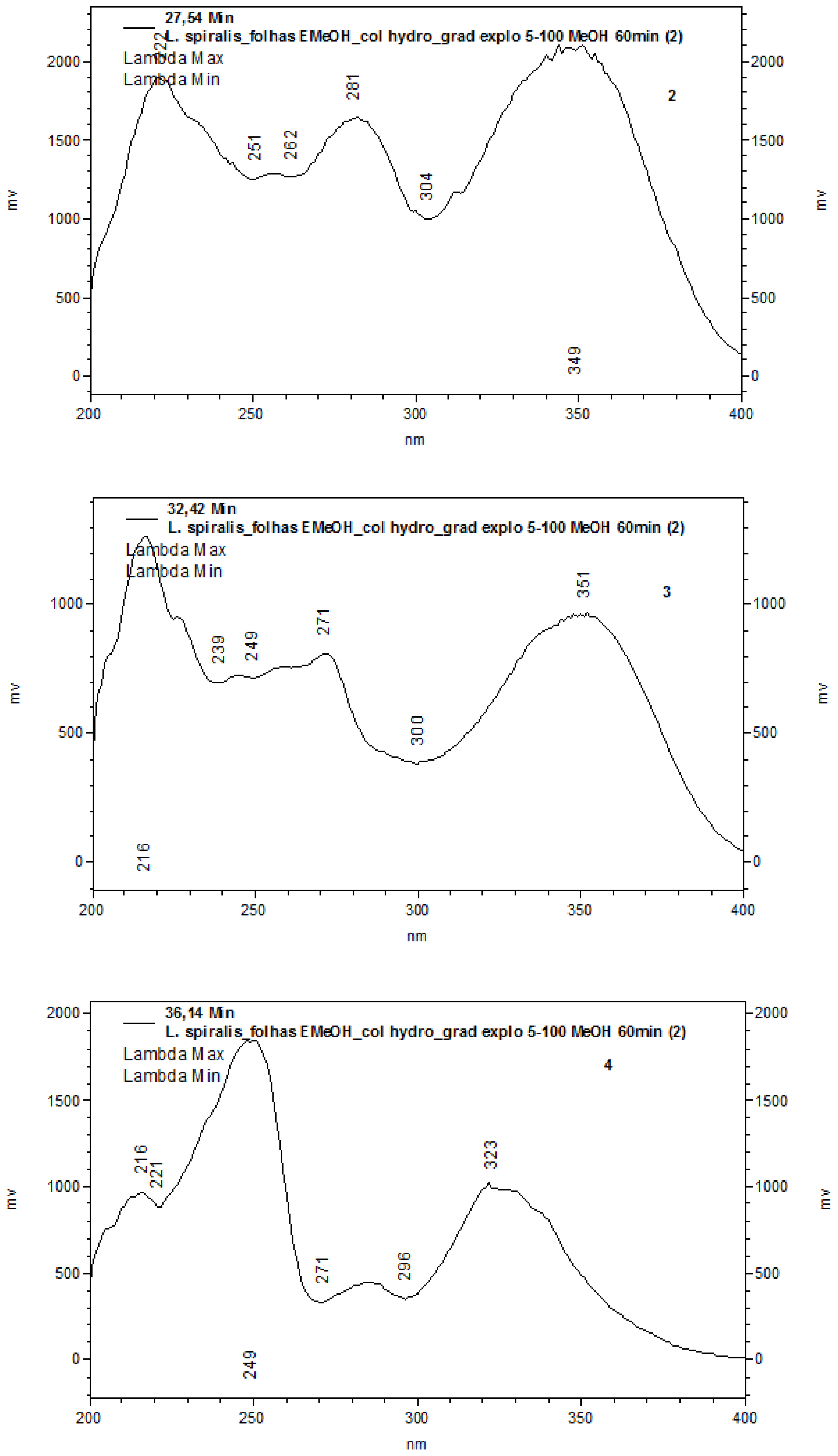
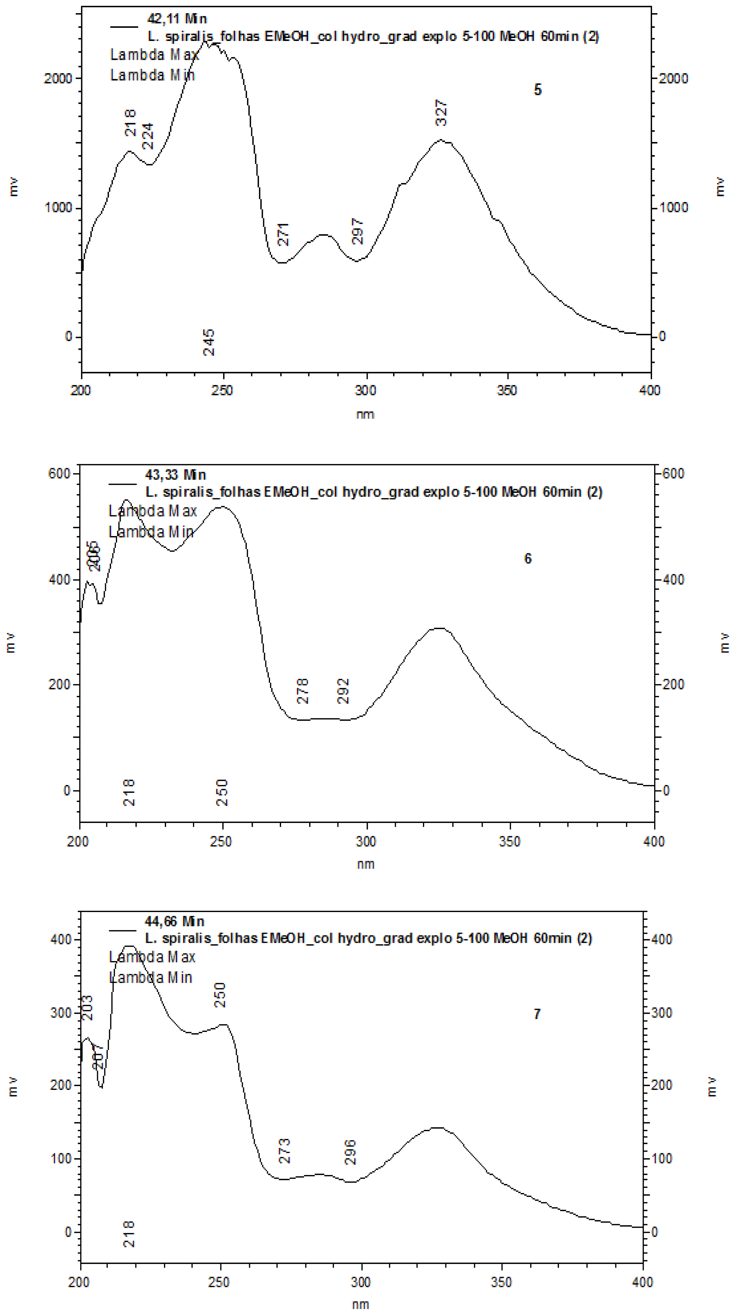
2.1. Chemical Characterization

The characterization of analytes was based on the comparison of the UV spectra of the peaks (Figure 1). With PAD (Photodiodo Array Detector) scanning from 200–600 nm, it was possible to obtain the UV spectra of each peak, which permitted the selection of a suitable wavelength in order to maximize the detection of the constituents. According to their corresponding UV absorbance, a wavelength of 254 nm was determined to be the most appropriate to ensure maximum detection for the simultaneous analyses of all compounds present. The UV spectra of the peaks corroborated the presence of flavone derivatives (peaks 1 to 7 with bands at 239–270 nm and 327–349 nm) [16] (Figure 2).

2.2. Total Flavonoids

The total flavonoid content was calculated based on the luteolin analytical curve and the percentage composition of flavonoid in the extract was 19.26 ± 0.04%. These results indicate the presence of significant amounts of that class of phenols.

2.3. ABTS Radical Cation Scavenging Activity

Table 1 shows the concentrations of the extract needed to decrease the initial ABTS (2,2′-azinobis-(3-ethylbenzothiazoline-6-sulfonic acid)) concentration by 50% (50% Inhibitory Concentration, IC50). The antioxidant activity of the extracts was expressed in quercetin and luteolin equivalents by comparing IC50 of the extracts with IC50 of standards. The IC50 value of the extract showed significant antioxidant activity when compared to standard quercetin and luteolin.

2.4. Antibacterial and Antifungal Susceptibility

The results of minimal inhibitory concentration (MIC) determinations of the antimicrobial activity (Table 2) showed noticeable MIC values for the methanolic extract of leaves from L. spiralis, against Candida species and Gram-positive bacteria. Luteolin showed activity against all microorganisms tested. The results of minimal fungicidal concentration (MFC) determination indicated that the fungicidal effect of the extract on the studied microorganisms could be expected. A close look at MFC and MIC values revealed that most MFC values correspond to MIC values.

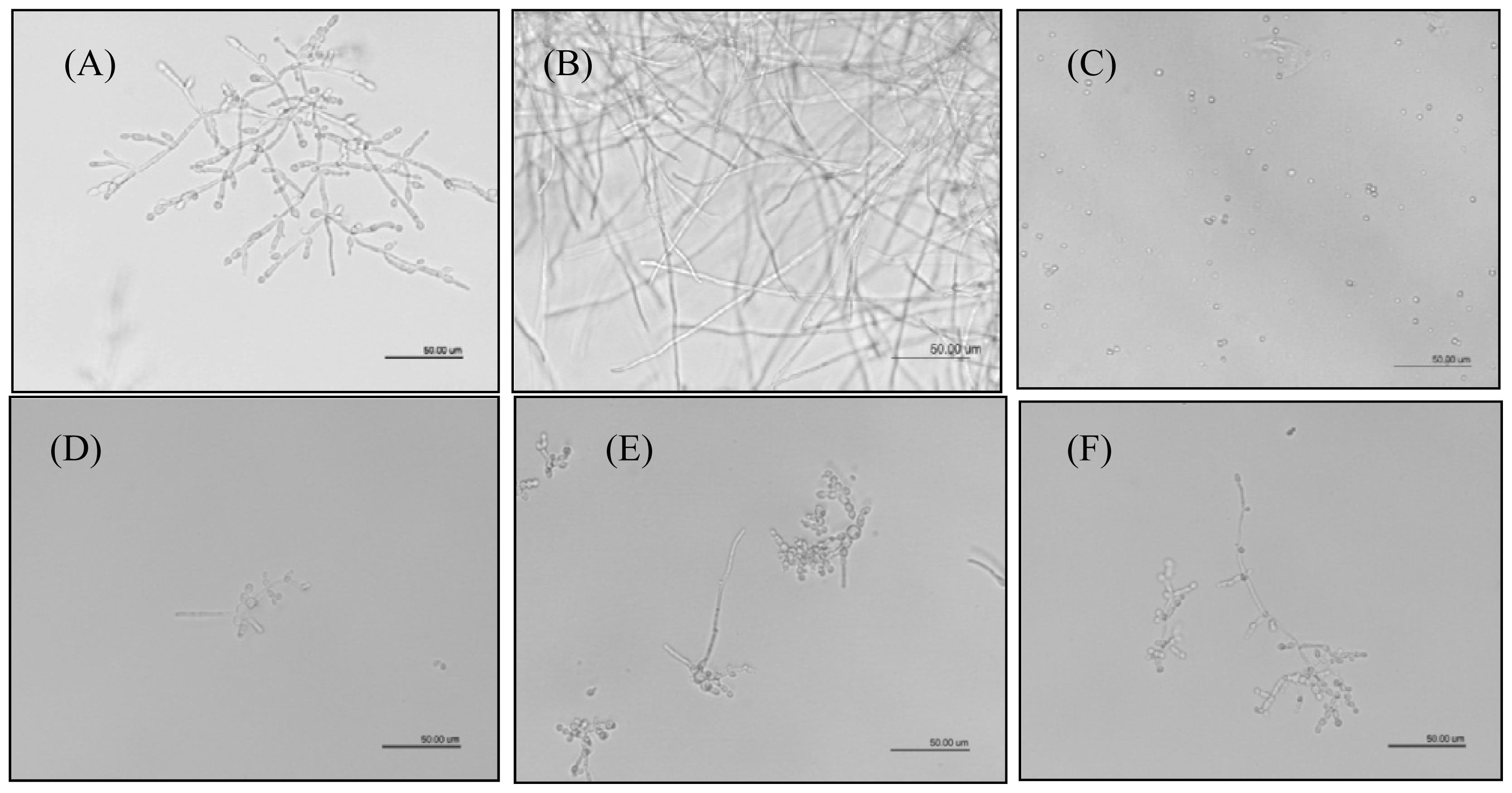
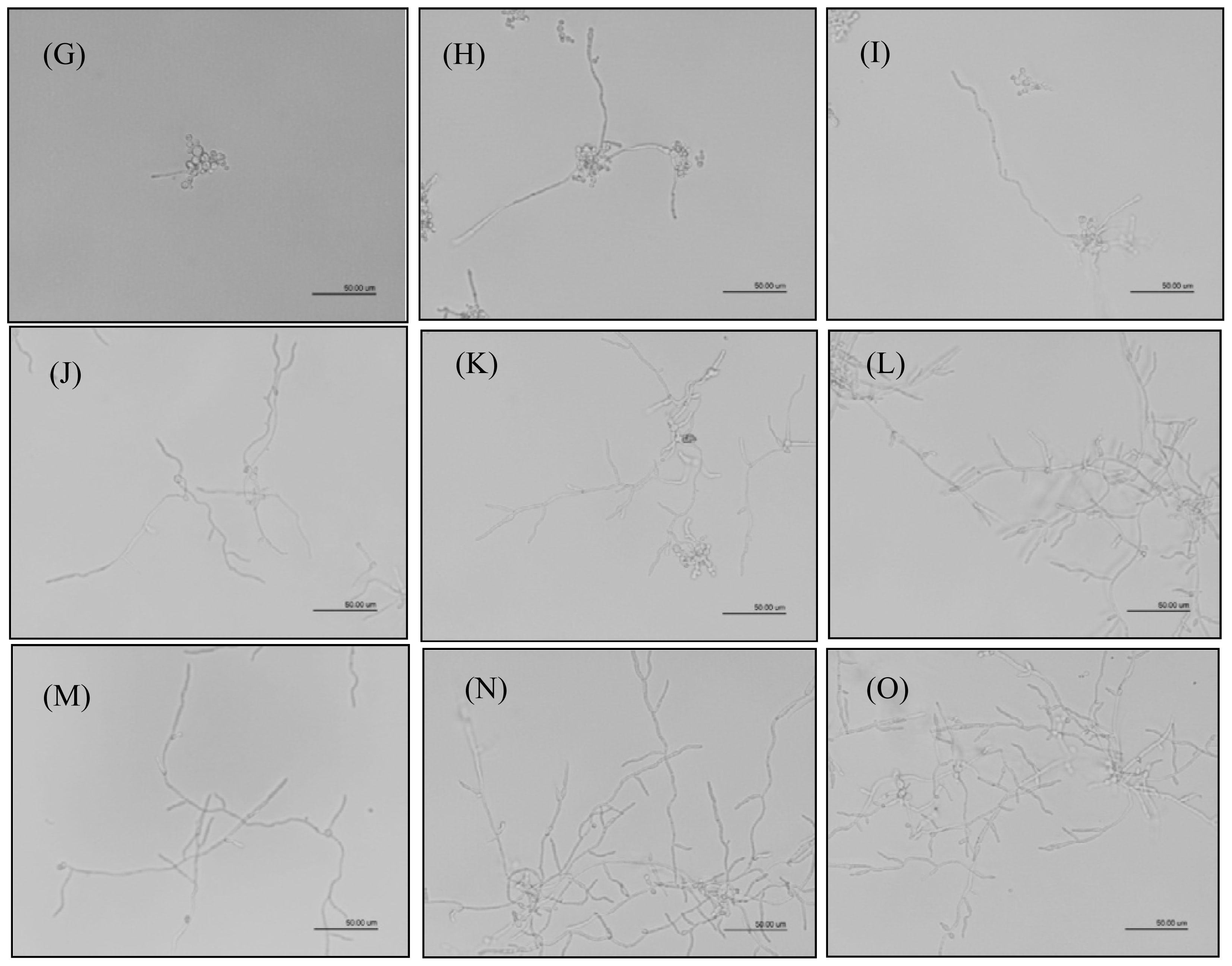
2.5. Inhibition of Hyphal Formation

C. albicans cells were incubated for 12 and 24 h in the presence of three concentrations of extract based on MIC values to C. albicans, and anphotericin B (ANF) at 5 mg/L as positive control, and then observed under an inverted light microscope (Figure 3). In the absence of the drugs, hyphal formation was observed, while in the presence of the highest concentration of extract tested, hyphal formation of C. albicans was significantly inhibited. The different concentrations of luteolin were not able to inhibit hyphal formation.

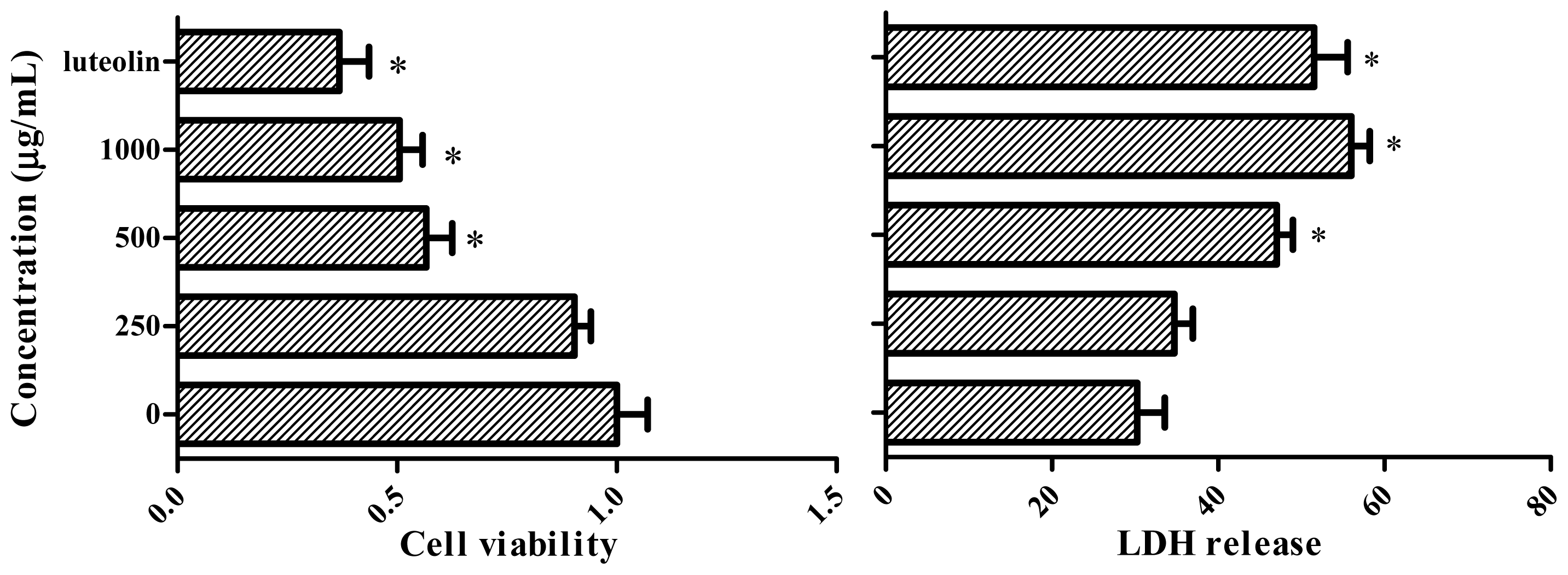
2.6. MTT and LDH Cell Viability

Figure 4 shows the effect of the extract on the cell viability of human cervical adenocarcinoma cells line (HeLa cells) after a 24 h incubation period. Luteolin significantly inhibited cell growth at the tested concentration (p < 0.05), and cell viability was affected by the extract treatment at 500 and 1000 mg/mL when compared with untreated cells. Cell injury was quantitatively assessed by the measurement of lactate dehydrogenase (LDH) release. After a 24 h incubation period, luteolin and the treatment with different extract concentrations significantly increased cell LDH release (p < 0.05), when compared with the untreated control.

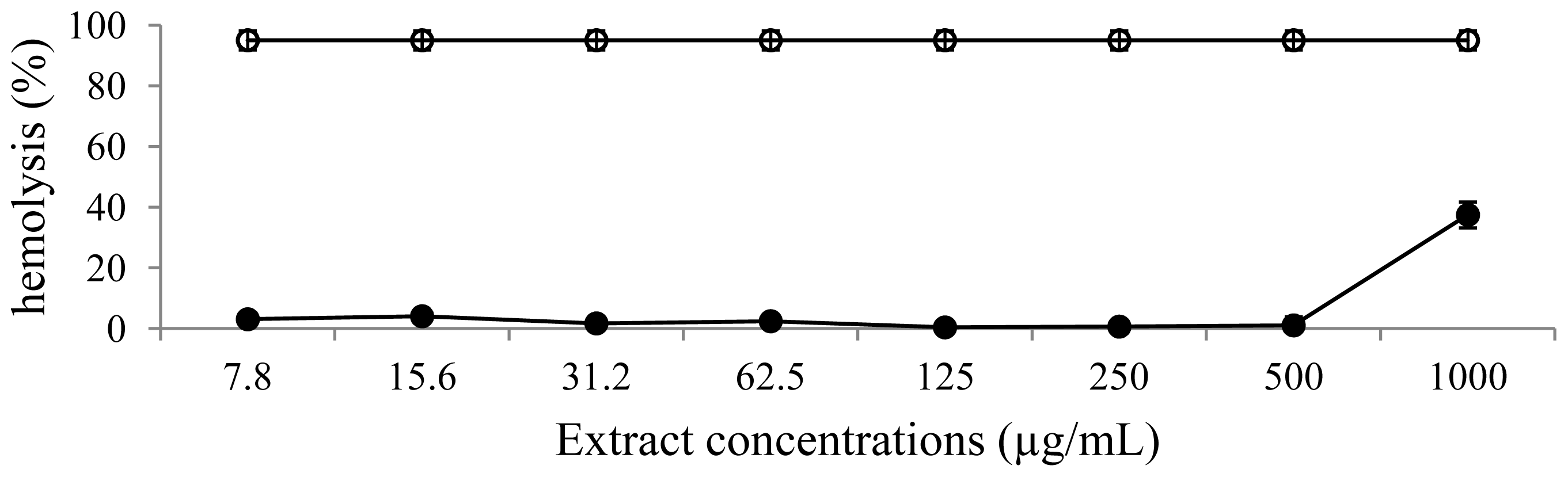
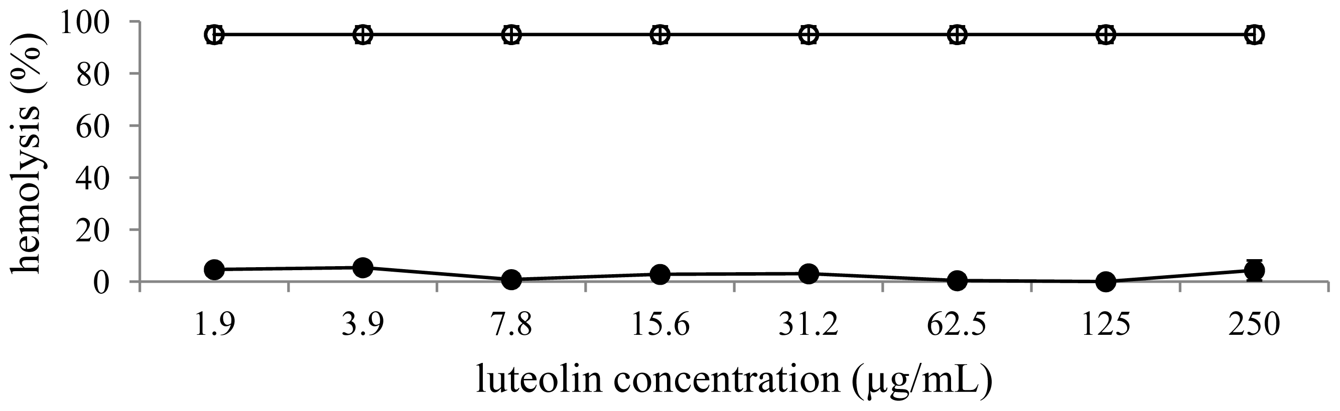
2.7. Hemolytic Assay

Figures 5,6 show the hemolytic activity of the extract investigated by measuring the lysis of a 10% (v/v) human red blood cells suspension in a spectrophotometric assay. In this experiment, Triton X-100 1% (v/v) was used as a positive control and induced 95.0 ± 3.1% of red blood cell lysis. The extract and luteolin showed no significant effect on red blood cells lysis.
ROS and free radicals such as superoxide anion, hydrogen peroxide and hydroxyl radicals are considered to be implicated in degenerative processes related to aging, cancer and atherosclerosis, mainly because they can induce the oxidative damage of cell membranes, DNA, and proteins [17]. Thus, blocking the generation of ROS and free radicals by supplementation of antioxidants might have a beneficial role in preventing these free radical-related diseases. Several studies have shown that phenolics are the bioactive compounds that offer more benefits to human health and many authors have reported a direct relationship between total phenolic content and antioxidant activity in numerous seeds, fruits and vegetables [1820]. As expected, methanolic extract obtained from leaves of L. spiralis presented high flavonoid content. This fact correlates with the remarkable differences in the polarity of the extraction solvents used and the solubility of phenolic compounds in them. In fact, methanol (polar solvent) is considered one of the best solvents for phenolic extraction. The chemical analysis of the extract by HPLC-PAD confirmed the presence of flavonoids as the major secondary metabolite compounds in leaves of L. spiralis. The features of the UV spectra with bands of maximum of absorption at 239–250 (Band II) nm and 327–349 (Band I) nm suggested the presence of flavones [16]. Some authors studied the contents of soluble phenolic compounds of the capitula of 21 species of Leiothrix. The presence of luteolin O- and C-glucopyranoside was determined in Leiothrix species [13] and another study verified the presence of xanthones and flavone with antioxidant activity in capitula of L. flavescens and L. curvifolia [14]. In agreement, in this study the IC50 values of methanolic extract obtained from the ABTS assay were significantly correlated with the total flavonoid standards, suggesting that flavonoids in these extracts constitute an important portion of the antioxidant activity.
It was suggested that antioxidant capacity of L. spiralis was mainly due to the presence of luteolin derivatives. Multiple mechanisms may underlie the antioxidant effect of luteolin. Luteolin functions as a ROS scavenger through its own oxidation and possesses the essential structures to antioxidant activity of flavonoids: 3′,4′-hydroxylation, the presence of a double bond between carbons 2 and 3 and a carbonyl group on carbon 4. The hydrogen atom from an aromatic hydroxyl group can be donated to free radicals. As an aromatic compound, luteolin can support unpaired electrons around the electron system. Moreover, luteolin inhibits ROS generating oxidases and may directly inhibit enzymes that catalyze the oxidation of cellular components [21].
Flavonoids exhibit inhibitory effects against multiple microorganisms. The antimicrobial actions result from an interaction between these compounds and the cell membrane of the target microorganisms, probably due to their ability to bind with extracellular and soluble proteins and also with cell walls [22]. In this study, the extract showed antimicrobial activities against Gram-positive organisms. These differences may be attributed to the fact that the cell wall in Gram-positive bacteria is of a single layer, whereas the Gram negative cell wall is a multilayered structure. This tendency could be explained by the fact that Gram negative bacteria possess an outer membrane surrounding the cell wall, which restricts diffusion of bioactive compounds due to the presence of lipopolysaccharide [23]. However, phenolic compounds maybe not only affect bacterial membranes but also mammalian cells such as erythrocytes [24]. They can interact with membrane lipids and proteins of erythrocytes and result in membrane damage [25]. The in vitro toxicity on the red blood cell membrane of various plant extracts has been studied and correlated with their constituents. The extract and luteolin showed very low hemolytic activity against human blood cells. On the other hand, flavonoids have been revealed to protect biological membranes against free radical-induced oxidative damage. They scavenge reactive oxygen species and, in some cases, their interaction with cellular proteins has been suggested, especially with heme proteins, which exert their physiological functions by the oxidation and reduction of heme iron [2628].
In a previous work, chemical fractionation of the methanolic extract of leaves of L. spiralis Ruhland afforded the flavonoids luteolin-derivatives and xanthones with antimicrobial activity [15]. The methanolic extract of leaves from L. spiralis as well as the flavonoid luteolin, showed activity against all Candida species tested. C. albicans is a dimorphic yeast and its ability to switch from yeast cells to hyphae is considered to be important for the interactions of C. albicans with its host. Hyphae are an important factor of fungal virulence. It is through hyphae that C. albicans invades human tissues [29]. The microscopy observations showed that extract at a concentration of 1000 μg/mL clearly inhibited hyphal formation during the hyphal induction of C. albicans. This result is important to characterize the action of the extract on the yeast tested. The antifungal activity of the phenolic compounds has been attributed to their lipophilic properties, which determine their ability to penetrate into the plasma membrane and induce changes in the physicochemical properties of the cell wall, cell membrane, and cellular organelles [30]. Antioxidant activity may be important in the antifungal mode of action of phenolic compounds. A hypothesis is that during stress, fungi respond by controlling secondary metabolite production and authors have also linked the antioxidant activity of phenolic compounds to their activity on metabolites biosynthesis [3132].
The human body possesses multiple endogenous systems to protect cellular molecules against the oxygen radical-induced damage. These defense mechanisms include antioxidative enzymes, such as superoxide dismutase (SOD); catalase; and glutathione peroxidase [33]. Mammalian cell culture and cell-free studies have begun to elucidate the underlying mechanisms of the potential protective effects of plant constituents against carcinogenesis, including antimutagenic effects of extracts and natural compounds against cancer inducing agents. Cells are very susceptible to mild oxidation, for example, with dilute solutions of hydrogen peroxide. These changes can be protected by additions of various antioxidant compounds such as flavonoids. Therefore, it is possible to speculate that flavonoids should offer cancer protection via their antioxidant properties [34].
Antioxidant activity is involved in cancer prevention at the initiation stage while antiproliferative activity is targeting cancer cells at the promotion and progression stages. However, it should be noted that by using the MTT-assay, it is not possible to differentiate between cell growth inhibition and an increase in cell death [35]. In the meantime, the LDH leakage assay revealed toxicity after exposure at the same concentrations, therefore, the extract has promoted rupture of the cell membrane in HeLa cells. In this regard, data from studies focusing on the elucidation of the molecular basis of the putative anticancer activity of polyphenols indicate that both, the inhibition of cell growth and the induction of cell death, play a role in the antitumor activity of polyphenols [3537]. Furthermore, data from other cell culture studies strongly suggest that the mechanism whereby phenolic compounds modulate cell proliferation is remarkably dose-dependent [2138]. Interestingly, the arrest of HeLa cell growth was evident after a 24 h treatment, with the highest concentration used. This indicates that the polyphenols in the extract promptly initiated a series of cellular events leading to the inhibition of cell proliferation and/or the induction of cell death.

3. Experimental Section

3.1. Plant Material

Leaves of L. spiralis (Bong.) Ruhl. were collected in May 2006 in Diamantina, Minas Gerais State, Brazil, and authenticated by Dr. Paulo Takeo Sano from the Institute of Biosciences of the University of São Paulo (IB-USP), São Paulo. A voucher specimen (SANO n° 4798) was deposited at the Herbarium of the IB-USP. Leaves of L. spiralis were oven dried at 45 °C for one week and powdered separately. The dried leaves of L. spiralis (2.00 g) were powdered and extracted with hexane, methylene chloride, and methanol, successively. The methanol extract (1.2 g) was used for this work. Luteolin was obtained by Sigma®.

3.2. Chemical Characterization of the Extract

The chromatographic profile of the methanolic extract of the leaves from L. spiralis was obtained by using a Jasco (Tokyo, Japan) liquid chromatograph equipped with a PU-2089 Plus pump, a MD-2010 Plus Photodiodo Array detector (PAD) and a Rheodyne 7725 sample injector with a 20 μL sample loop. The analytical column was a Phenomenex Synergi Hydro RP18 (250 × 4.6 mm i.d.; 4 μm) equipped with a Phenomenex security guard column (4.0 × 2.0 mm i.d.). The mobile phase composition was methanol. The gradient program was as follows: 5%–100% methanol in 60 min. The flow rate was 1.0 mL/min and the total run time was 60 min. The software EZChrom Elite 3.17 was used for control analytical system, data collection and processing.

3.3. Total Flavonoids

Total flavonoids were estimated according to Georgetti S.R. et al. [39]. To 0.5 mL of sample, 0.5 mL of 2% AlCl3 ethanol solution was added. After 60 min at room temperature, the absorbance was measured at 351 nm. Total flavonoid contents were calculated as luteolin equivalent from an analytical curve.

3.4. ABTS Radical Cation Scavenging Activity

The free radical scavenging activity of the extract was determined by ABTS radical cation decolorization assay [40]. It involved the generation of ABTS•+ chromophore by the oxidation of ABTS with potassium persulfate. The ABTS•+ radical cation was generated by reacting ABTS and potassium persulfate after incubation at room temperature in the dark for 12–16 h. The solution was then diluted by mixing 1 mL ABTS•+ solution with methanol, to obtain an absorbance of about 0.7 at 734 nm using the spectrophotometer. The reactive mixture (extract in different concentrations and ABTS•+) was allowed to stand at room temperature for 15 min and the absorbance was immediately recorded at 734 nm. Quercetin and luteolin standard solutions were prepared and analyzed under the same conditions. The results were expressed as 50% inhibitory concentration or IC50.

3.5. Antimicrobial Activity

The MIC was determined by the broth microdilution method, according to the standard reference method [41,42]. The antifungal activity was evaluated against C. albicans (ATCC 18804 and NCPF 3153), C. krusei (ATCC 6258), C. parapsilosis (ATCC 22019) and C. tropicalis (ATCC 750) and the antibacterial activity was evaluated against Staphylococcus aureus (ATCC 25923), Bacilus subtillis (ATCC 19659), Enterococcus faecalis (ATCC 29212), Escherichia coli (ATCC 25922), Pseudomonas aeruginosa (ATCC 27853) and Salmonella setubal (ATCC 19196). The extract and luteolin were dissolved in 25% methanol and water to initial concentration of extract of 1000 μg/mL. Then, a two-fold serial dilution was made in order to obtain concentration ranges of 7.8–1000 μg/mL. 100 μL of each concentration were added to 96-well microplates containing 80 μL of RPMI 1640 for yeast and Muller-Hinton broth for bacteria. The inocula of bacteria and yeast were standardized at 1.0 × 10⁷ and 2.5 × 10³ CFU/mL, respectively. Ciprofloxacin, fluconazole and 25% methanol and water were used as positive and negative control. The plates were incubated at 37 °C for 24 h for bacteria and 48 h for yeast. The assay was repeated three times. The MIC of the samples was detected after the addition (50 μL) of resazurin solution (0.2 mg/mL) for bacteria and 2.0% triphenyl-tetrazolium chloride (TTC) solution for yeast, and incubated at 37 °C for 30 min. Growth of bacteria changes the blue dye resazurin to a pink color. The pink color indicates positive growth, and blue indicates growth inhibition. Yeast growth changes the colorless TTC to a red color. MIC was defined as the lowest sample concentration that prevented this change and exhibited inhibition of microorganism growth.
For the determination of minimal bactericidal concentration (MBC) and MFC, a portion from each well that showed antibacterial activity and antifungal activity was plated on Muller-Hinton and Sabouroud agar, and incubated at 37 °C for 24 h. The lowest concentration that yielded no growth after this sub-culturing was taken as the MBC/MFC [43].

3.6. Inhibition of Hyphal Growth

C. albicans (NCPF 3153) cells from a 48 h stationary phase culture were transferred to microplate with RPMI 1640 medium supplemented with fetal bovine serum (FBS) to a final concentration of 2.5 × 10³ CFU/mL, and extract and luteolin solution were added to the growth medium to final concentrations of 0.5× MIC, 1× MIC and 2× MIC, and the cultures were incubated for 12 and 24 h at 37 °C, 5% CO2 [44]. The hyphal formation was observed under an inverted light microscope (Nikon TE 2000-U Eclipse) with the magnification of 400×. ANF (5 mg/L) was used as a positive control.

3.7. MTT and LDH Cell Viability Assay

Cell viability was determined by using the conversion of MTT to formazan via mitochondrial oxidation [45]. The human cervical adenocarcinoma cells line (HeLa—ATCC: CCL-2; 5000 cells/well) were treated with the extract and luteolin (0, 0.5× MIC, 1× MIC and 2× MIC to C. albicans) for 24 h. Then, MTT solution was added to each well at a concentration of 10 mg/mL and the plates were incubated at 37 °C for another 4 h. After incubation, 0.2 mL DMSO was added to each well to dissolve the formazan and the absorbance was read at 570 nm using a spectrophotometric microplate reader. The viability was determined based on a comparison with untreated cells. Luteolin was used as a positive (cytotoxic) control. The experiments were performed in triplicate and repeated at least three times.
Cell injury was quantitatively assessed by the measurement of LDH, released from damaged or destroyed cells, in the extracellular fluid 24 h after the experiment. An aliquot of bathing media was combined with NADH and pyruvate solutions. LDH activity is proportional to the rate of pyruvate loss. The quantity of LDH released by the cells into the medium was measured by the decrease in the absorbance at 340 nm for NADH disappearance within different times (0, 1, 2 and 3 min). The difference in absorbance per minute was determined and the average multiplied by the factor (10080).
The results were statistically analyzed by comparing the LDH and MTT values obtained at baseline with those obtained in different treatments by analysis of variance (ANOVA) followed by Dunnett test (multiple comparisons with one control) with p < 0.05.

3.8. Hemolytic Assay

Human erythrocytes from healthy individuals were collected in vacuum tubes containing heparin as anti-coagulant. The erythrocytes were harvested by centrifugation for 10 min at 2000 rpm and washed three times in phosphate buffered saline (PBS). To the pellet, PBS was added to yield a 10% (v/v) erythrocytes/PBS suspension. The 10% suspension was then diluted 1:10 in PBS. 0.1 mL of this suspension was added in triplicate to 96-well microplates containing 0.1 mL of extract (7.8–1000 μg/mL) or luteolin (1.9–250 μg/mL) serially diluted in PBS. Total hemolysis was achieved with 1% Triton X-100. The microplate was incubated for 1 h at 37 °C and then centrifuged for 10 min at 2000 rpm. The absorbance of the supernatant was measured spectrophotometrically at 450 nm [46]. The percentage of hemolysis was calculated and statistically analyzed by analysis of variance (ANOVA) followed by Dunnett test (multiple comparisons with one control) with p < 0.05.

4. Conclusions

The biological activity of a plant extract depends on a complex sum of individual properties including its composition, the existing compound structures, affinity for the target site, survival within the biological system, transport properties, and state of the target organism. In this study, we focused our attention on the relationship between some biological activities and antioxidant properties. The methanolic extract of L. spiralis leaves showed a significant antioxidant activity, and an antibacterial, antifungal, non-hemolytic and antiproliferative activity could be demonstrated. A better understanding of how antioxidant plant extracts inhibit microorganism growth and inhibit tumor cell proliferation will allow them to be more efficiently used and perhaps synergistically used with other antimicrobial, antiproliferative agents or their anti-hemolytic activity.

Acknowledgments

The authors gratefully acknowledge the financial support of the CAPES which provided the fellowship to M.G.F.A. and FAPESP Program which provided the fellowship to F.H and the project grant to L.C.S. The authors extend their appreciation to the CIBICI-CONICET, Argentina, for support and collaboration the work through the research group of C.E.S.

References

  1. Waris, G.; Ahsan, H. Reactive oxygen species: role in the development of cancer and various chronic conditions. J. Carcinog 2006, 5, 1–8. [Google Scholar]
  2. Hu, H.; Zhang, Z.; Lei, Z.; Yang, Y.; Sugiura, N. Comparative study of antioxidant activity and antiproliferative effect of hot water and ethanol extracts from the mushroom Inonotus obliquus. J. Biosci. Bioeng 2009, 107, 42–48. [Google Scholar]
  3. Song, J.M.; Lee, K.H.; Seong, B.L. Antiviral effect of catechins in green tea on influenza vírus. Antivir. Res 2005, 68, 66–74. [Google Scholar]
  4. Cushnie, T.P.T.; Lamb, A.J. Antimicrobial activity of flavonoids. Int. J. Antimicrob. Agents 2005, 26, 343–356. [Google Scholar]
  5. Silva, M.A.; Cardoso, C.A.L.; Vilegas, W.; Santos, L.C. High-performance liquid chromatographic quantification of flavonoids in Eriocaulaceae species and their antimicrobial activity. Molecules 2009, 14, 4644–4654. [Google Scholar]
  6. Ferguson, P.J.; Kurowska, E.; Freeman, D.J.; Chambers, A.F.; Koropatnick, D.J. A flavonoid fraction from cranberry extract inhibits proliferation of human tumor cell lines. J. Nutr 2004, 134, 1529–1535. [Google Scholar]
  7. Russo, A.; Cardile, V.; Sanchez, F.; Troncoso, N.; Vanella, A.; Garbarino, J.A. Chilean propolis: antioxidant activity and antiproliferative action in human tumor cell lines. Life Sci 2004, 76, 545–558. [Google Scholar]
  8. Boivin, D.; Lamy, S.; Lord-Dufour, S.; Jackson, J.; Beaulieu, E.; Côté, M.; Moghrabi, A.; Barrette, S.; Gingras, D.; Béliveau, R. Antiproliferative and antioxidant activities of common vegetables: A comparative study. Food Chem 2009, 112, 374–380. [Google Scholar]
  9. Hu, W.; Yu, L.; Wanga, M.H. Antioxidant and antiproliferative properties of water extract from Mahonia bealei (Fort.) Carr. leaves. Food Chem. Toxicol 2010, 49, 799–806. [Google Scholar]
  10. Rocha-Guzmán, N.E.; Gallegos-Infante, J.A.; Gonzalez-Laredo, R.F.; Reynoso-Camacho, R.; Ramos-Gomez, M.; García-Gasca, T.; Rodríguez-Muñoz, E.; Guzmán-Maldonado, S.H.; Medina-Torres, L.; Luján-García, B.A. Antioxidant activity and genotoxic effect on HeLa cells of phytophenolic compounds from infusions of Quercus resinosa leaves. Food Chem 2009, 115, 1320–1325. [Google Scholar]
  11. Sano, P.T. Actinocephalus (Koern.) Sano (Paepalanthus sect. Actinocephalus), a new genus of Eriocaulaceae, and other taxonomic and nomenclatural changes involving Paepalanthus. Mart Taxon 2004, 53, 99–107. [Google Scholar]
  12. Salatino, A.; Salatino, M.L.; Giulietti, A.M. Contents of soluble phenolic compounds of capitula of Eriocaulaceae. Quim. Nova 1990, 13, 289–292. [Google Scholar]
  13. Dokkedal, A.L.; Salatino, A. Flavonoids of brazilian species of Leiothrix (Eriocaulacaeae). Biochem. System. Ecol 1992, 20, 31–32. [Google Scholar]
  14. Santos, L.C.; Piacente, S.; Montoro, P.; Pizza, C.; Vilegas, W. Atividade antioxidante de xantonas isoladas de espécies de Leiothix (Eriocaulaceae). Rev. Bras. Farmacogn 2003, 13, 67–74. [Google Scholar]
  15. Araújo, M.G.F.; Hilário, F.; Nogueira, L.G.; Vilegas, W.; Santos, L.C.; Bauab, T.M. Chemical constituents of the methanolic extract of leaves of Leiothrix spiralis ruhland and their antimicrobial activity. Molecules 2011, 16, 10479–10490. [Google Scholar]
  16. Mabry, T.J.; Markham, K.R.; Thomas, M.B. The ultraviolet spectra of flavones and flavonols. In The Systematic Identification of Flavonoids; Mabry, T.J., Markham, K.R., Thomas, M.B., Eds.; Springer-Verlag: New York, USA, 1970; Volume 41–45, pp. 165–166. [Google Scholar]
  17. Cheng, L.X.; Tang, J.J.; Luo, H.; Jin, X.L.; Dai, F.; Yang, J.; Qian, Y.P.; Li, X.Z.; Zhou, B. Antioxidant and antiproliferative activities of hydroxyl-substituted Schiff bases. Bioorg. Med. Chem. Lett 2010, 20, 2417–2420. [Google Scholar]
  18. Parry, J.; Su, L.; Moore, J.; Cheng, Z.; Luther, M.; Rao, J.N.; Wang, J.Y.; Yu, L. Chemical compositions, antioxidant capacities, and antiproliferative activities of selected seed flours. J. Agr. Food Chem. 2006, 54, 3773–3778. [Google Scholar]
  19. Zhang, Y.; Seeram, N.P.; Lee, R.; Feng, L.; Heber, D. Isolation and identification of strawberry phenolics with antioxidant and human cancer cell antiproliferative properties. J. Agr. Food Chem 2008, 56, 670–675. [Google Scholar]
  20. Yang, J.; Liu, R.H.; Halim, L. Antioxidant and antiproliferative activities of common edible nut seeds. Food Sci. Technol 2009, 42, 1–8. [Google Scholar]
  21. Lin, Y.; Shi, R.; Wang, X.; Shen, H-M. Luteolin, a flavonoid with potentials for cancer prevention and therapy. Curr. Cancer Drug Targets. 2008, 8, 634–646. [Google Scholar]
  22. Cowan, N.M. Plant products as antimicrobial agents. Clin. Microbiol. Rev 1999, 12, 564–582. [Google Scholar]
  23. Burt, S. Essential oils: Their antibacterial properties and potential applications in foods—A review. Int. J. Food Microbiol. 2004, 94, 223–253. [Google Scholar]
  24. Tamaki, M.; Imazeki, Y.; Shirane, A.; Fujinuma, K.; Shindo, M.; Kimura, M.; Uchida, Y. Novel gratisin derivatives with high antimicrobial activity and low hemolytic activity. Bioorg. Med. Chem. Lett 2011, 21, 440–443. [Google Scholar]
  25. Singh, R.P.; Kaur, G. Hemolytic activity of aqueous extract of Livistona chinensis fruits. Food Chem. Toxicol 2008, 46, 553–556. [Google Scholar]
  26. Kitagawa, S.; Sakamoto, H.; Tano, H. Inhibitory Effects of Flavonoids on Free Radical-Induced Hemolysis and Their Oxidative Effects on Hemoglobin. Chem Pharm Bull 2004, 52, 999–1001. [Google Scholar]
  27. Asgary, S.; Naderi, G.H.; Askari, N. Protective effect of flavonoids against red blood cell hemolysis by free radicals. Exp. Clin. Cardiol 2005, 10, 88–90. [Google Scholar]
  28. Blasa, M.; Candiracci, M.; Accorsi, A.; Piacentini, M.P.; Piatti, E. Honey flavonoids as protection agents against oxidative damage to human red blood cells. Food Chem 2007, 104, 1635–1640. [Google Scholar]
  29. Yang, Y.L. Virulence factors of Candida species. J. Microbiol. Immunol. Infect 2003, 36, 223–228. [Google Scholar]
  30. Knobloch, E.; Pauli, A.; Iberl, B.; Wies, N.; Weigand, H. Mode of action of essential oil components on whole cells of bacteria-and fungi in plate tests. In Bioflavour: Analyses, Biochemistry, Biotechnology; Schreier, P., Ed.; Walter de Gruyter: Berlin, Germany, 1988; pp. 287–299. [Google Scholar]
  31. Dambolena, J.S.; Zunino, M.P.; López, A.G.; Rubinstein, H.R.; Zygadlo, J.A.; Mwangi, J.W.; Thoithi, G.N.; Kibwage, I.O.; Mwalukumbi, J.M.; Kariuki, S.T. Essential oils composition of Ocimum basilicum L. and Ocimum gratissimum L. from Kenya and their inhibitory effects on growth and fumonisin production by Fusarium verticillioides. Inn. Food. Sci. Emerging. Technol 2010, 11, 410–414. [Google Scholar]
  32. Dambolena, J.S.; Zygadlo, J.A.; Rubinstein, H.R. Antifumonisin activity of natural phenolic compounds. A structure-property-activity relationship study. Int. J. Food Microbiol 2011, 145, 140–146. [Google Scholar]
  33. Lassen, N.; Black, W.J.; Estey, T.; Vasiliou, V. The role of corneal crystallins in the cellular defense mechanisms against oxidative stress. Semin. Cell. Dev. Biol 2008, 19, 100–112. [Google Scholar]
  34. Cutter, H.; Wu, L-Y.; Kim, C.; Morre, D.J.; Morre, D.M. Is the cancer protective effect correlated with growth inhibitions by green tea (–)-epigallocatechin gallate mediated through an antioxidant mechanism? Cancer Lett 2001, 162, 149–154. [Google Scholar]
  35. García-Alonso, J.; Ros, G.; Periago, M.J. Antiproliferative and cytoprotective activities of a phenolic-rich juice in HepG2 cells. Food Res. Int 2006, 39, 982–991. [Google Scholar]
  36. Luo, W.; Zhao, M.; Yang, B.; Ren, J.; Shen, G.; Rao, G. Antioxidant and antiproliferative capacities of phenolics purified from Phyllanthus emblica L. fruit. Food Chem 2011, 126, 277–282. [Google Scholar]
  37. Lazzè, M.C.; Savio, M.; Pizzala, R.; Cazzalini, O.; Perucca, P.; Scovassi, A.I.; Stivala, L.A.; Bianchi, L. Anthocyanins induce cell cycle perturbations and apoptosis in different human cell lines. Carcinogenesis 2004, 25, 1427–1433. [Google Scholar]
  38. Selvendiran, K.; Koga, H.; Ueno, T.; Yoshida, T.; Maeyama, M.; Torimura, T.; Yano, H.; Kojiro, M.; Sata, M. Luteolin Promotes Degradation in Signal Transducer and Activator of Transcription 3 in Human Hepatoma Cells: An Implication for the Antitumor Potential of Flavonoids. Cancer Res 2006, 66, 4826–4834. [Google Scholar]
  39. Georgetti, S.R.; Casagrande, R.; Vicentini, F.T.M.C.; Verri, W.A., Jr; Fonseca, M.J.V. Evaluation of the antioxidant activity of soybean extract by different in vitro methods and investigation of this activity after its incorporation in topical formulations. Eur. J. Pharm. Biopharm 2006, 64, 99–106. [Google Scholar]
  40. Re, R.; Pellegrini, N.; Proteggente, A.; Pannala, A.; Yang, M.; Rice-Evans, C. Antioxidant activity applying an improved ABTS radical cation decolorization assay. Free Radical. Biol. Med 1999, 26, 1231–1237. [Google Scholar]
  41. Rex, J.H.; Alexander, B.D.; Andes, D.; Arthington-Skaggs, B.; Brown, S.D.; Chaturvedi, V.; Ghannoum, M.A.; Espinel-Ingroff, A.; Knapp, C.C.; Ostrosky-Zeichner, L.; et al. Reference Method for Broth Dilution Antifungal Susceptibility Testing of Yeasts: Approved Standard, 3rd ed; CLSI document M27-A3; ISBN 1-56238-666-2. Clinical and Laboratory Standards Institute (CLSI): Wayne, PA, USA, 2002. [Google Scholar]
  42. Wikler, M.A.; Hindler, J.F.; Cockerill, F.R.; Patel, J.B.; Bush, K.; Powell, M.; Dudley, M.N.; Turnidge, J.D.; Elopoulos, G.M.; Weinstein, M.P.; et al. Methods for Dilution Antimicrobial Susceptibility Tests for Bacteria that Grow Aerobically; Approved Standard, 8th ed; CLSI document M07-A8; ISBN ISBN 1-56238-689-1. Clinical and Laboratory Standards Institute: Wayne, PA, USA, 2008. [Google Scholar]
  43. Kuete, V.; Nanab, F.; Ngamenic, B.; Mbaveng, A.T.; Keumedjio, F.; Ngadjuib, B.T. Antimicrobial activity of the crude extract, fractions and compounds from stem bark of Ficus ovata (Moraceae). J. Ethnopharmacol 2009, 124, 556–561. [Google Scholar]
  44. Zhang, J.D.; Xu, Z.; Cao, Y.B.; Chen, H.S.; Yan, L.; An, M.M.; Gao, P.H.; Wang, Y.; Jia, X.M.; Jiang, Y.Y. Antifungal activities and action mechanisms of compounds from Tribulus terrestris L. J. Ethnopharmacol 2006, 103, 76–84. [Google Scholar]
  45. Ding, W.J.; Hasegawa, T.; Peng, D.; Hosaka, H.; Seko, Y. Preliminary investigation on the cytotoxicity of tellurite to cultured HeLa cells. J. Trace. Elem. Med. Biol 2002, 16, 99–102. [Google Scholar]
  46. He, M.; Du, M.; Fan, M.; Bian, Z. In vitro activity of eugenol against Candida albicans biofilms. Mycopathologia 2007, 163, 137–143. [Google Scholar]
Figure 1. HPLC-PAD (Photodiodo Array Detector) chromatogram recorded at 254 nm of the methanolic extract of leaves from L. spiralis. For chromatographic conditions see Experimental section.
Ijms 13 09260f1
Figure 2. UV spectra of the chromatographic (HPLC) data of the methanolic extract of leaves from L. spiralis (peaks 1–7). Conditions: Phenomenex Synergi Hydro RP18 (250 × 4.6 mm i.d.; 4 μm) equipped with a Phenomenex security guard column (4.0 × 2.0 mm i.d.). The mobile phase composition was methanol. The gradient program was as follows: 5%–100% MeOH in 60 min. The flow rate was 1.0 mL/min and the total run time was 60 min.
Ijms 13 09260f2aIjms 13 09260f2bIjms 13 09260f2c
Figure 3. Hyphal formation of C. albicans NCPF 3153 cells. (A) 12 h normal growth; (B) 24 h normal growth; (C) C. albicans cells treated with 0.005 mg/mL anphotericin B (ANF) as the positive control; Hyphal formation of C. albicans cells was obviously inhibited by the extract at (D) 1 mg/mL; (E) 0.5 mg/mL; (F) 0.25 mg/mL at 12 h; and (G) 1 mg/mL; (H) 0.5 mg/mL; (I) 0.25 mg/mL at 24 h. Luteolin did not inhibit the hyphal formation (J) 0.25 mg/mL; (K) 0.125 mg/mL; (L) 0.062 mg/mL at 12 h; (M) 0.25 mg/mL; (N) 0.125 mg/mL; (O) 0.062 mg/mL at 24 h, compared with (A) and (B). The black bar represents a length of 50 μm.
Ijms 13 09260f3aIjms 13 09260f3b
Figure 4. Effect of extract on cell viability and LDH release in cultured HeLa cells. Cells were treated with different concentrations of extract, and luteolin (31 mg/L) was used as positive control.* Significantly different from the basal conditions.
Ijms 13 09260f4
Figure 5. Hemolytic activity of extract of leaves of L. spiralis. Extract (filled circle) and positive control Triton X-100 (open circle).
Ijms 13 09260f5
Figure 6. Hemolytic activity of luteolin. Luteolin (filled circle) and positive control Triton X-100 (open circle).
Ijms 13 09260f6
Table 1. 50% Inhibitory Concentration (IC50) values of extract and standards obtained in the ABTS (2,2′-azinobis-(3-ethylbenzothiazoline-6-sulfonic acid)) radical test.
SamplesIC50 a

Means ± SD
L. spiralis leaves1.743 ± 0.063 b
quercetin1.140 ± 0.038 b
luteolin1.215 ± 0.031 b
aValues in μg/mL;
b<0.001; SD: standard deviation.
Table 2. Antibacterial and antifungal activity of the methanolic extract of L. spiralis leaves and luteolin. Minimal inhibitory concentration (MIC); minimal bactericidal concentration (MBC); minimal fungicidal concentration (MFC).
MicroorganismL. spiralisLuteolin

MIC aMBC/MFC aMIC aMBC/MFC a
C. albicans5001000125500
C. krusei5001000250250
C. parapsilosis250250125125
C. tropicalis10001000250250
S. aureus10001000125125
B. subtilis1000100062.5125
E. faecalis500-31.25125
E. coli--250250
P. aeruginosa--62.5250
S. setubal--250-
aValues of minimal inhibitory concentration given as μg/mL; (-) >1000 μg/mL.

