Review

Calcium in Red Blood Cells—A Perilous Balance

1
Institute of Veterinary Physiology, Vetsuisse Faculty and the Zürich, Center for Integrative Human Physiology, University of Zürich, Zürich 8057, Switzerland
2
Institute for Molecular Cell Biology and Research Centre for Molecular Imaging and Screening, Saarland University, Homburg/Saar 66421, Germany
*
Author to whom correspondence should be addressed.
Int. J. Mol. Sci. 2013, 14(5), 9848-9872; https://doi.org/10.3390/ijms14059848
Submission received: 27 February 2013 / Revised: 18 March 2013 / Accepted: 19 March 2013 / Published: 8 May 2013
(This article belongs to the Special Issue Signalling Molecules and Signal Transduction in Cells)

Abstract

:
Ca2+ is a universal signalling molecule involved in regulating cell cycle and fate, metabolism and structural integrity, motility and volume. Like other cells, red blood cells (RBCs) rely on Ca2+ dependent signalling during differentiation from precursor cells. Intracellular Ca2+ levels in the circulating human RBCs take part not only in controlling biophysical properties such as membrane composition, volume and rheological properties, but also physiological parameters such as metabolic activity, redox state and cell clearance. Extremely low basal permeability of the human RBC membrane to Ca2+ and a powerful Ca2+ pump maintains intracellular free Ca2+ levels between 30 and 60 nM, whereas blood plasma Ca2+ is approximately 1.8 mM. Thus, activation of Ca2+ uptake has an impressive impact on multiple processes in the cells rendering Ca2+ a master regulator in RBCs. Malfunction of Ca2+ transporters in human RBCs leads to excessive accumulation of Ca2+ within the cells. This is associated with a number of pathological states including sickle cell disease, thalassemia, phosphofructokinase deficiency and other forms of hereditary anaemia. Continuous progress in unravelling the molecular nature of Ca2+ transport pathways allows harnessing Ca2+ uptake, avoiding premature RBC clearance and thrombotic complications. This review summarizes our current knowledge of Ca2+ signalling in RBCs emphasizing the importance of this inorganic cation in RBC function and survival.

1. Introduction

Ca2+ is a universal and ubiquitous signalling molecule [1,2], regulating cell cycle and fate, metabolism and structural integrity, motility and volume. Most of the Ca2+ in the cytosol is bound and buffered by numerous Ca2+ binding proteins, phospholipids and inorganic phosphate. When bound and buffered Ca2+ are included, total intracellular Ca2+ in red blood cells (RBCs) reaches 5.7 μM [3]. Basal free Ca2+ concentration in RBCs of healthy human beings under physiological conditions is estimated to be in the range of 30 to 60 nM [4]. A tremendous gradient of at least 40,000-fold between the cytosol and blood plasma where free Ca2+ concentration reaches up to 1.8 mM is maintained due to particularly low permeability of membranes to Ca2+ (~50 μmol/(lcells h)) and efficient extrusion of Ca2+ from the cells by the plasma membrane Ca2+ pump (PMCA) [4]. This gradient may be used for signalling purposes as opening of a few hundreds of channels transporting 106 ions per second over several milliseconds may result in >10-fold changes in free Ca2+ levels in the sub-membrane space, causing acute changes in the activity of multiple Ca2+ sensitive proteins involved in structural, signalling, metabolic and transport functions. Such a huge Ca2+ influx seems to exceed the RBC’s Ca2+-buffering abilities. Long-term increases in the Ca2+ permeability result in serious dysregulation of multiple cellular functions. The subsequent onset of proteolysis, oxidation, irreversible shrinkage and phosphatydylserine (PS) exposure to the extracellular membrane leaflet facilitate clearance of Ca2+ overloaded cells in case the latter have not been haemolysed when passing through capillaries.
This review summarizes the current knowledge on the regulation of RBC Ca2+ levels, Ca2+ dependent processes and the potential role that Ca2+ might play in the development of RBC pathologies. For obvious reasons this review makes no claim to be complete, but wants to draw attention to open questions and mechanisms to be resolved in the field of Ca2+ signalling in RBCs.

2. Ca2+ Transport across the RBC Membrane

2.1. Ca2+ Extrusion Pathway: Plasma Membrane Ca2+ Pump

It was realised very early on that the inhibition of the energy supply in RBCs leads to a Ca2+ increase [5]. Although the nature of the Ca2+ influx remained unknown for several decades, the extrusion mechanism was realized to be mediated by a plasma membrane Ca2+ pump (PMCA). For human RBC membrane the presence of the B-splice isoform of the PMCA1 was shown [6]. This P-type ATPase is ubiquitously expressed. It is composed of 1220 amino acids forming ten transmembrane domains, two intracellular loops containing ATP binding and phosphorylation sites and inward-facing N- and C-terminals. The latter contains a Ca2+ calmodulin binding domain, phosphorylation sites and a PDZ-binding domain serving as a docking terminal for a number of proteins [7]. The maximal Ca2+ extrusion rate in RBCs can vary within RBC populations of a single donor between >60 mmol/(lcells h) and <4 mmol/(lcells h) [8]. The maximal turnover rate of the PCMA is significantly higher in RBCs from light fractions compared to that in RBCs within dehydrated RBC fractions following density centrifugation [9]. This reduction in Vmax in cells from the dense fraction was referred to as a hallmark of RBC senescence [10]. The maximal Ca2+ turnover rate of PMCA in the RBC membrane of healthy humans not loaded with Ca2+ is most likely never reached. The apparent Vmax for PMCA measured in such cells is ~50 μmol/(lcells h), the value equal to the passive Ca2+ uptake. The enzyme half-activation constant for Ca2+ was reported to be 4 μM, far above the actual free cytosolic Ca2+ levels in human RBCs [11].
Increases in the intracellular free Ca2+ are sensed by the PMCA and occur in response to the interaction of the Ca2+ calmodulin complex with the C-terminus of the enzyme. In Ca2+-loaded RBCs the limiting factor of the PMCA transport capacity is ATP availability. The pump is fuelled preferentially by a pre-membrane ATP pool [8,12,13] and has a Kd (ATP) of 145 μM [5,14]. Under conditions of permanent Ca2+ leak, activation of PMCA results in rapid ATP depletion.
RBC-derived plasma membrane vesicles contain factors activating PMCA, such as arachidonic acid, ceramide and acidic phospholipids, whereas sphingosine suppresses its function [1517]. The function of the PMCA is extremely temperature-sensitive with ~30-fold reduction in turnover rate for every 10 °C drop [18].

2.2. Ca2+ Influx Pathways

Ca2+ influx through the plasma membrane of healthy human RBCs is extremely slow but increasing up-to 5-fold in cells of patients with sickle cell disease (SCD) and several other forms of hereditary haemolytic anaemia [4]. Several cation channels mediate inward movement of Ca2+. Significant progress in identification of ion channels in control of Ca2+ uptake has recently been reviewed [19].
Voltage-activated non-selective cation channels [2022] were shown to be Ca2+ permeable [23]. These initial observations have all been performed on excised patches, but the presence of the channel was also shown in the whole cell configuration [24] and further flux-based characterisations have been performed [2527]. However, a proof of the molecular identity remains to be provided.
Furthermore, a P-type Ca2+ channel was pharmacologically identified [28] and shown to be a CaV2.1 channel by Western blot analysis [29]. This channel can be inhibited by ω-agatoxin TK [29]. However, in contrast to the initial investigations, in which activation by protein kinase C (PKC) was proposed, recent findings depicted a rather indirect interaction with PKC [30].
A recent report provided evidence for the presence of a transient receptor potential (TRP) channel of subtype C6 in the RBC membrane [31]. However, most of the work done so far was performed on murine RBCs and detailed characterization of this channel in human RBCs is missing.
In addition, the expression of an NMDA receptor channel was initially reported for rat [32] and later in human RBCs using molecular biological and electrophysiological approaches [33]. NMDA receptor agonists include glutamate, N-methyl d-aspartate (NMDA), homocysteine, homocysteic acid, glycine and d-serine [34].
Recently, the protein PIEZO1 was reported as being mutated in RBCs in hereditary xerocytosis [35] without knowing its physiological function. However, PIEZO1 is characterized as a mechano-sensitive cation channel in heterologous expression systems [36,37].
Furthermore there is evidence for an AMPA receptor related channel activity in RBCs [38].
All the channels mentioned above were reported to be present in human RBCs from healthy donors. However, some currents were only shown to be present in cells of patients. An example is an increase in non-selective cation conductance on RBC of SCD patients mediating or contributing to Psickle [39,40], an increased membrane permeability in SCD RBC. It is still not completely clear if this reflects an increased activity of one or more of the above mentioned channels or yet another conductance [40,41]. However, recent investigations provide evidence for the involvement of the NMDA receptor [42].

3. Ca2+-Sensitive Proteins in RBCs

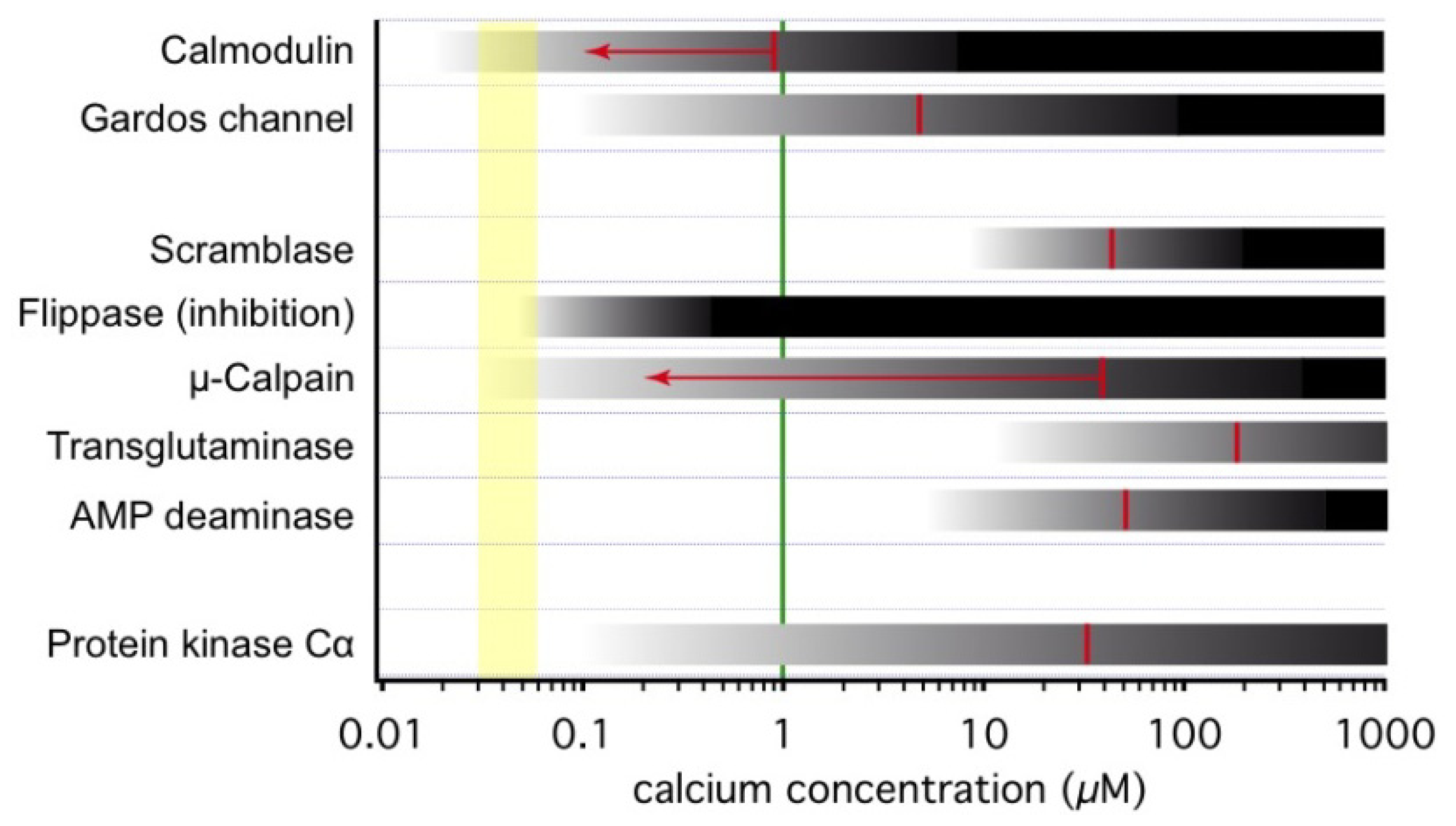
3.1. Onset of Ca2+-Inducible Events and Ca2+ Sensors in RBCs

When in the cell, Ca2+ activates numerous Ca2+ dependent proteins. Each of them has its own activation threshold. Thus, gradual increase in Ca2+ levels is associated with gradual activation of various groups of Ca2+-sensitive proteins involved in physiological and pathophysiological processes in RBCs. In Figure 1 we compiled current knowledge about the activation ranges of some selected proteins. This list of Ca2+ sensitive proteins is by far not comprehensive and can hardly be covered within one review. Despite a large number of such proteins and diversity of their functions, only few of them are “true” Ca2+ sensors interacting directly with calcium ions [43]. One of such ubiquitous sensors highly abundant in RBCs is calmodulin. Calmodulins 1–4 (CaM) are 17 kDa proteins comprising two globular EF hand Ca2+ binding domains enriched with carboxyl and carbonyl groups (Asp, Glu and Thr) interconnected with a flexible linker (for details see, e.g., [44,45]). Upon interaction with Ca2+, CaM wraps around amphipathic regions of the protein compacting into a globular shape and pulling the interacting domains of the target out of lipophilic pockets or out of the membrane lipid bilayer moiety. In RBCs the proteins regulated by interaction with the Ca2+ calmodulin 2 complex (Ca-CaM) include, e.g., elements of the cytoskeletal network, the Na+/H+ exchanger NHE1, PMCA and the endothelial NO synthase (eNOS). Cytoplasmic CaM becomes active when recruited to the plasma membrane where its action is often coupled to that of phosphatidylinositol 4,5-bisphosphate (PIP2) localised at the inner leaflet of the membrane. An example of such coupling is a competitive binding of both co-regulators to the intracellular domain of NHE1 (see Figure 2A).
Another class of “true” Ca2+ sensors (~650 proteins included) contain Ca2+ binding C2 domains interacting with 2–3 Ca2+ [43,46]. In RBCs proteins with C2 domains include phospholipases, PKCα, phosphoinositide 3-kinase (PI3K) and many others. Binding of Ca2+ to the C2 domains, triggers translocation of these proteins to the specific areas within plasma membrane containing their substrates [46].

3.2. Ca2+-Dependent Phosphorylation

Changes in phosphorylation are among the most important modulations of protein activity in RBCs. Among the kinases there is a group of Ca2+ activated protein kinases, the conventional protein kinase C (cPKC) [65]. Among cPKCs, only protein kinase Cα (PKCα) can be found in RBCs [66]. Upon Ca2+ binding, PKCα translocates to the plasma membrane, where it phosphorylates its target proteins. The kinase domain of PKCs lacks specificity [67,68] and therefore numerous proteins can be phosphorylated. Reports include the PMCA [69], cytoskeletal proteins ([70,71] and see below), NADPH oxidase [72] and possibly further proteins [29,30,73] are affected.

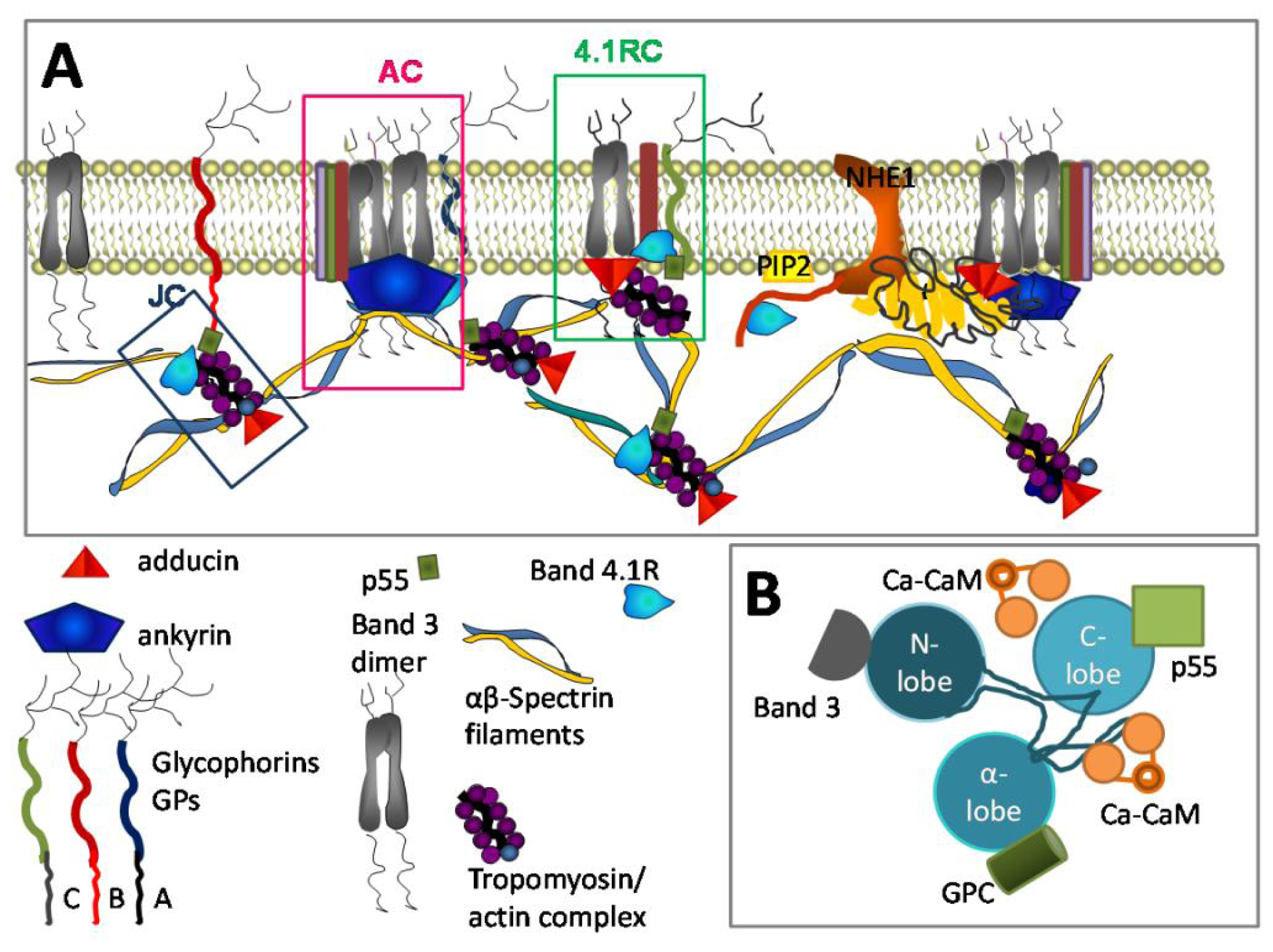
3.3. Ca2+ and RBC Cytoskeleton

Opening of cation channels in response to mechanical stress and the presence of activators, such as amino acids, proinflammatory cytokines and others, result in local transient increase in Ca2+ levels in the vicinity of the plasma membrane. The latter, most likely serves as a signal to mediate rapid reversible changes in cytoskeletal flexibility.
The calcium-calmodulin complex (Ca-CaM) plays a key role in regulation of cytoskeletal stability. Selected elements of the cytoskeletal architecture interacting with Ca-CaM are schematically shown in Figure 2 together with their interacting partners. Those include major components of the cytoskeletal network, protein 4.1R, and adducin. These proteins function as docking stations for spectrin and actin, band 3 protein, glycophorins protein 4.2 and p55 and form a complex known as ankyrin-based complex and junctional complex (Figure 2A) [74,75]. The junctional complex is formed by the three principal components of the skeletal network junctions (spectin, actin, and 4.1R, together with tropomyosin, tropomodulin, adducin, dematin, p55). When associated with transmembrane proteins, GPC, XK, Kell, Duffy, band 3, and Rh junction complex forms a multiprotein 4.1R-based complex [76].
It is shown in Figure 2A, that the band 4.1R protein is a key constituent of all three complexes. Upon interaction with Ca-CaM, affinity of the 4.1R protein to all the interacting partners decreases and the cytoskeletal structure including spectrin-actin-tropomyosin junctions and the spectrin-actomyosin web interaction with the transmembrane protein clusters becomes loose and unstable. Such, Ca-CaM induced dissociation of NHE1 from the ankyrin complexes and band 4.1R protein. This facilitates the interaction of NHE1 with PIP2 resulting in activation of this ion transporter and dysregulation of cell volume and cytosolic alkalosis [62,77].
The Ca2+ concentration required for half-activation of calmodulin is 100 nM [78]–920 nM [49], Figure 1, thus no significant dissociation of the membrane cytoskeleton is expected to occur under physiological concentration of intracellular free Ca2+ [79]. Phosphorylation of calmodulin at Ser-80 and -84 affects its affinity to 4.1R reducing their binding [80].
Ca2+ dependent phosphorylation is a second mode of action of Ca2+ on cytoskeletal proteins. Phosphorylation of the 4.1R protein at serine 312 and serine 331 by PKC was reported [81]. These two phosphorylation sites are localised within a domain flanked by the spectrin- and actin binding domain and a domain containing the interaction sites for transmembrane proteins (Figure 2B). When phosphorylated the 4.1R-β-spectrin interaction appears to be weakened by ~30% [81]. As Ca2+ uptake is known to be triggered by mechanical deformations [82], controlled reversible loosening of cytoskeletal network in cells passing through capillaries is an advantage. Uncontrolled irreversible loss of cytoskeletal stability in RBCs of patients with haemolytic anaemia, in which Ca2+ is permanently upregulated, on the contrary compromises mechanical stability of the RBC membrane [83]. In RBCs of healthy humans the 4.1R protein is not phosphorylated [81]. One more target of PKC is α-adducin which, upon phosphorylation at Ser-726 decreases its affinity to F-actin, as it was observed in SCD RBCs. After the loss of F-actin capping dissociation of spectrin from actin occurs [84], which results in RBC inability to change shape.
In RBCs transaminase 2 responds to Ca2+ entry with a rapid release of its inhibitory GTP and activation resulting in formation of Nɛ (γ-glutaminyl)lysine cross-linking (Figure 1). As a result, in Ca2+ overloaded RBCs formation of numerous polymeric protein complexes such as Glut1-adducin-dematin adducts as well as cross-linked complexes of Band 3-ankyrin-spectrin and glycophorin C-band 4.1-p55 occurs [85]. This Ca2+ induced remodelling of the cytoskeletal structure and concomitant changes in cell shape and membrane plasticity are suggested to contribute to premature RBC clearance.

3.4. Ca2+ and RBC Volume Regulation

RBC volume regulation is a complex process with contributions of numerous molecular players including, e.g., band 3 protein [86]. A marked volume decrease is mediated by the so-called Gardos effect, which was among the first Ca2+ dependent processes recognized in RBCs [87]. The corresponding Gardos channel was the first channel measured by patch-clamp in RBCs [88,89]. This channel is a Ca2+ activated K+ channel and the Gardos effect represents the Ca2+ induced K+ loss of RBCs. The channel is characterized by a single channel conductance of approximately 20 pS [88], a selectivity of K+ to Na+ of about 15:1 [90] and an EC50 (Ca2+) of 4.7 μM, Figure 1 with a Hill-slope of approximately 1 [50]. Later, the molecular identity of the Gardos channel was shown to be the hSK4 channel [91]. Functionally, the opening of the Gardos channel leads to a hyperpolarisation and a loss of K+, Cl and water resulting in cell shrinkage. Although the physiological function of the Gardos channel is not completely elucidated, there are two complementary concepts: (i) Openings of RBC Ca2+ channels by platelet released substances [92,93] initialy trigger the consecutive activation of the Gardos channel. This Gardos channel mediated dehydration of the RBC fosters their contribution in clot formation as outlined below. (ii) Local membrane deformation of RBCs was shown to trigger a transient increase in Ca2+ permeability with secondary activation of the Gardos channels [82]. This was proposed to induce significant dehydration even during a brief deformation event in the microcirculation [82].

3.5. Ca2+ and Lipid Bilayer

Scramblase is a protein responsible for bidirectional transmembrane movement of phospholipids [94] leading to the break-down of the originally asymmetrical distribution of phospholipids between the inner and outer membrane leaflet [95]. It is a passive transport, but Ca2+ activated [51,52]. Its Ca2+ sensitivity is mediated by an EF hand motif [96]. The scramblase activity is complemented by the flippase (aminophospholipid translocase) inhibition [97]. This protein actively builds up phospholipid asymmetry and such can be regarded as the opponent of the scramblase. As shown in Figure 1, flippase activity is almost completely suppressed by 400 nM Ca2+ [53].

3.6. Ca2+ and Metabolism

Numerous reports emphasize the possible role of the Ca-CaM system in regulation of activity of glycolytic enzymes including pyruvate kinase [98,99]. However, even more important is its pivotal role in assembling the glycolytic enzymes at the RBC membrane. Band 3 protein and its cytosolic domain was shown to serve as a docking station for multiple glycolytic enzymes [100]. Ca2+ in turn was suggested to promote band 3 tyrosine phosphorylation [101]. Phosphorylation of the cytosolic domain of band 3 protein (cdb3) at Tyr9 and Tyr21 results in displacement of LDH, PK, GAPDH, PFK and aldolase from RBC membrane in intact cells [102]. A similar effect is induced by interaction of deoxyHb with band 3 protein [103].

3.7. Ca2+ and Redox State Preservation

In RBCs there is a direct link between the intracellular free Ca2+ concentration and the haemoglobin oxygen saturation. In cells of healthy individuals, passive Ca2+ uptake was unaffected by deoxygenation, but the Vmax of the PMCA was reduced by 18%–32% [104]. This is not the case in RBCs of patients with SCD [4]. An increase in free Ca2+ levels was mainly attributed to changes in haemoglobin protonation, increases in protonation of deoxyhaemoglobin and a shift in the intracellular pH towards more alkaline values [104,105]. Along with augmentation of 2,3-diphosphoglycerate and ATP binding to haemoglobin, interaction of deoxyhaemoglobin with protons is associated with a decrease in Ca2+ buffering capacity of haemoglobin. In the cytosol of deoxygenated RBCs release of Ca2+ ions from protein binding sites and lowering of extrusion capacity of the PMCA contribute to both an increase in the ionised Ca2+ fraction by 34%–74% even in the absence of Ca2+ influx from the extracellular medium [104].
Increases in the free Ca2+ were recently linked to a lower oxygen affinity of haemoglobin promoting the release of oxygen [33]. Deoxygenation induced transient release of Ca2+ from intracellular buffers may promote further deoxygenation enhancing O2 dissociation from haemoglobin. Molecular mechanisms of this phenomenon remain to be investigated.
Aside to the control of haemoglobin oxygenation, Ca2+ is also involved in the regulation of the RBC’s redox state. Ca-CaM complexes are co-activators of endothelial NO synthase (eNOS) activity [106,107]. Recently eNOS was shown to be present in circulating RBCs [108,109] and is activated by Ca2+ uptake during shear stress [82,110]. Nitric oxide is a scavenger of superoxide anions, which interacts with them two orders of magnitude faster than superoxide dismutase (SOD) [111]. However, following shortage of tetrahydrobiopterin or l-arginine eNOS itself gets uncoupled and is capable of generating superoxide anions, which is turned into H2O2 in a reaction catalysed by SOD [106]. Pro-oxidative action of uncoupled eNOS was demonstrated in RBCs [112].

3.8. Calpain and Its Targets in RBCs

While the Ca2+ dependent cysteine protease μ-calpain (calpain-1) was mainly detected in human RBCs, m-calpain (calpain-2) was found to be virtually absent [113]. μ-Calpain is highly sensitive to Ca2+ with a half-activation concentration in the range of 3–50 μM [114,115]. The enzyme isolated from human RBCs displayed a value of 40 μM [54]. This is well above the free Ca2+ concentration found in both human RBCs from healthy individuals (30–60 nM) and values of 100–300 nM reported in RBCs from patients with hereditary forms of anaemia [4]. However, a 40 kDa activator protein which makes μ-calpain more Ca2+ sensitive, shifts Ca2+ concentrations required for half-maximal activation from 40–50 μM down to 0.2 μM [55]. In human RBCs a fraction of membrane-associated active μ-calpain was detected [116,117]. Activation mediated recruitment of calpain to the membrane [118] was used to estimate the protease activity revealing that ~7% of the calpain pool is constitutively active in RBCs from healthy individuals [117]. Consequently, the μ-calpain activity in RBCs from patients with hereditary anaemia forms was also increased because their resting Ca2+ levels were raised when compared to those from healthy donors. [4].
Targets of activated calpain are mainly transmembrane or membrane-associated proteins including PMCA, and bands 1, 2, 2.1, 3, 4.1, 4.2 proteins, but also calpain itself [119]. Autolysis occurs at Ca2+ levels beyond physiological concentrations, namely in the range 50–150 μM [114]. Limited digestion of haemoglobin α and β chains by calpain was also reported [120].
Recently, RBCs of a μ-calpain knock-out (KO) mouse displayed an improved deformability [121]. Furthermore it was demonstrated that ankyrin, band 3, band 4.1R, adducin and dematin were degraded in the Ca2+ loaded normal RBCs but not in the KO RBCs [121].
Cleavage of the PMCA by μ-calpain was associated with an activation of the pump, rapid ATP depletion, inactivation of the pump and gradual loss of the transmembrane Ca2+ gradients [122].
Calpastatin, an endogenous inhibitor of μ-calpain, is a natural regulator of the enzyme activity in RBCs. Both major and minor components of calpastatin, calpastatin H and L were detected in human RBCs [54]. Interaction of calpastatin with calpain is also Ca2+ sensitive. Half-maximal activation of calpastatin occurs at 40 μM Ca2+ [114], in bovine skeletal muscle derived enzyme. Activity of calpain and a decrease in calpastatin levels in RBCs was shown to occur in elderly humans [123,124].

3.9. Ca2+ and Inter-Cellular Interactions

Due to the protein activity and lipid remodelling described above one would expect changes in rheological properties of RBC suspensions and indeed in numerous studies provoking increased cytosolic Ca2+ levels, an altered rheology was observed [125127]. These changes were majorly explained by altered RBC deformability [128,129]. However, a second possibility to explain changes in rheological properties of RBC suspensions is RBC aggregation.
A participation of RBC Ca2+ channels in blood clot formation was proposed previously [92,93] following earlier work already suggesting an active participation of RBCs in blood clot formation [130132]. Further support was given by investigations showing Ca2+ mediated aggregation of RBCs [133136] and RBC adhesion to the endothelium [137139]. Further investigations are necessary to specify the required and sufficient conditions for such processes, e.g., the threshold of the intracellular Ca2+ concentration at which aggregation occurs. Additionally the modulations of the aggregation properties under in vivo conditions need to be elucidated.

4. The Physiological Role of Intracellular Ca2+: From RBC Birth to Clearance

4.1. Ca2+ in RBC Haematopoiesis

Ca2+ uptake is of key importance for promoting differentiation and proliferation of erythroid precursors at the stages of burst-forming units erythroid (BFU-E) colony-forming units erythroid (CFU-E) [140,141]. Increase in the intracellular Ca2+ is an integral part of the signalling pathway activated by binding of erythropoietin to its receptor [142,143]. In Ca2+-free medium, Ca2+ uptake is absent and differentiation and survival of erythroid precursors is compromised [140,141]. Inhibition of Ca2+ uptake by erythroid precursor cells cultured from mononuclear cells by the NMDA receptor antagonist MK-801 resulted in 45.5% ± 12.8% mortality of cells at the stage of basophilic and polychromatic erythroblasts suggesting that these receptors are actively contributing to erythropoiesis [33]. Additional evidence indicating that Ca2+ levels in reticulocytes are higher than in mature RBCs comes from secondary Ca2+-dependent processes such as phosphorylation [144]. Protein 4.1R phosphorylation by PKC appeared to be markedly elevated in reticulocytes resulting in weakened interaction between β-spectrin and actin [145].

4.2. Ca2+ in Relation to the Physiological Function of RBCs

For a long time, the physiological function of Ca2+ in mature RBCs was obscure and was believed to be limited to the involvement in RBCs aging and clearance [10,146,147]. However, a prominent part of this report reviews the physiological functions of Ca2+ in RBC regulating a broad range of processes including O2 transport [33], rheology [148], clotting [135,136] and half-life of cells (see Section 4.3). Each of these functions is vital for the organism. Thus, aberrant Ca2+ homeostasis in RBCs results in development of severe life-threatening systemic pathologies.
Very recently, additional evidence in favour of a physiologically important Ca2+ associated mechanism was reported. Here, rises in the intracellular Ca2+ appear to promote the ability of RBCs to deliver oxygen [33].

4.3. Ca2+ in RBC Clearance

At present it is suggested that in senescent RBCs the intracellular Ca2+ levels exceed those in reticulocytes and young RBCs [149]. However, such conclusions on the relationship between cell age and steady-state Ca2+ levels largely depend on the age markers employed. Typical age markers include glycosylated haemoglobin HbA1c, band 4.1a/b ratio, cell density, de-sialation and changes in CD47 abundance at the membrane surface, PS exposure, and several others [149151].
Activity of the PMCA in RBCs was shown to decrease with progressing HbA1c accumulation [152]. However, based on the pump-leak theory, this process will result in Ca2+ accumulation only when coupled to the unchanged or increasing activity in Ca2+-transporting ion channels. However, according to recent findings, this is not necessarily the case. Young rat and human RBCs contain higher number of NMDA receptors, that upon stimulation with plasma glycine and glutamate can cause significant Ca2+ influx [32,33]. Young cells are preferentially removed in subjects with induced or chronic polycytemia, phenomenon known as neocytolysis [112,153155]. Finally, phosphatydylserine (PS) exposure does not always correlate with high Ca2+ levels [156]. Thus, both young and senescent RBCs appear to be prone to Ca2+ overload, which may well trigger RBC clearance, but the relation of this mechanism to other Ca2+ independent clearance mechanisms and to in vivo relevance is still obscure.

5. Ca2+ Dysbalance and Haemolytic Anaemia

Independent of its origin, hereditary haemolytic anaemia is often associated with an increase in the intracellular Ca2+ levels [4]. “Leakiness” of RBC plasma membrane for Ca2+ that could not be compensated for by the activation of PMCA was reported for patients with SCD [157159], beta-thalassemia (although most of the Ca2+ seem to be sequestered in vesicles or bound to cytosolic proteins) [160162], phosphofructokinase deficiency [163]. Most of the information on Ca2+ transport in diseased RBCs was so far obtained for SCD patients. In these cells relatively high rates of Ca2+ uptake in RBCs are partially compensated for by sequestration of Ca2+ into intracellular inside-out vesicles, in which Ca2+ is pumped actively by PMCA [164]. Apparently, this process also exists in RBCs of patients with β-thalalssemia intermedia [162,165]. Furthermore, part of Ca2+ taken into the cells is immobilised by the intracellular proteins. Sickle cell transformation associated with polymerisation of deoxygenated mutated S-haemoglobin is amplified by 20–40-fold by dehydration [166]. Deoxygenation promotes Ca2+ uptake and release of ionised Ca2+ from the intracellular proteins (reduction in buffering capacity) [104,105,167]. In deoxygenated SCD RBC, an acute increase in the intracellular free Ca2+ RBCs causes opening of the Ca2+ sensitive Gardos channel and anion channels [168170].
Downstream events triggered by augmentation of free intracellular Ca2+ comprise activation of μ-calpain [171] and activation of tyrosine phosphorylation [172]. In RBCs from SCD patients, protein 4.1R and p55 appear to be phosphorylated thus contributing to the weakened interaction with beta-spectrin [84].
Cross-linked polymers have been observed in RBCs of patients with SCD suggesting hyperactivation of transglutaminase [85].
Oxidation of membrane proteins and impaired NO production by eNOS [173,174], increase in intercellular and RBC-endothelial adhesion [175] are hallmarks of SCD. Cross-linking of the nature of carrier of sickle cell conductance (Psickle) mediating Ca2+ uptake by RBCs remains unknown (compare Section 2.2).

6. RBC Ca2+ Content and Medicinal Side Effects

6.1. Transfusion Medicine

Ca2+ overload is involved in dramatic reduction of the life span of stored RBCs used for transfusion [176]. Storage of leuco-depleted RBC concentrates is currently performed in Ca2+ free glucose-containing preservation citrate buffers at low temperatures. These storage conditions favour Ca2+ depletion of the cytosol, oxidative stress and ATP deprivation [177,178]. These processes induce deactivation of PMCA, facilitate passive Ca2+ transport and evoke acute Ca2+ overload when re-exposed to the plasma of patients receiving transfusions. The resulting processes include dehydration, rigidity, fragmentation of cytoskeletal proteins and oxidative stress and increased adherence of RBCs to the endothelium and to each other [179,180].

6.2. Therapeutic Side Effects

Additionally, unwanted modulations of the Ca2+ entry into RBC may cause side effects of drugs involved in therapies unrelated to RBCs. An example is photodynamic therapy, where the oxidative stress produced by the photosensitizer leads to the activation of cation channels in the RBC membrane and the consecutive Ca2+ entry triggers the mechanism described above, which is the major cause of an increased formation of blood aggregates as well as haemolysis [181]. Thus, RBC remain to be model cells to develop pharmacological strategies [182] and can even be used in automated safety screens [183].

7. Conclusion and Perspective

The role of Ca2+ in RBCs physiology and pathophysiology cannot be overestimated. Many links between Ca2+ and RBC related diseases still need to be explored [184]. Methodologically single cell based methods will increase in their importance and contribution and complement cell population measurements [185]. This is due to the recent hindsight that intercellular heterogeneity and, in some cases, inhomogeneous distribution of Ca2+ within the cytosol are essential to predict the onset of changes related to the abnormally high Ca2+ levels, which are particularly important in patients with haematological disorders [42,186]. However, the properties of Ca2+ binding entities within the cells will need further attention and research. Following the broad variety of Ca2+ mediated processes mentioned here, monitoring the following parameters may be used to indirectly predict abnormally increased intracellular free Ca2+ levels in RBCs: (i) changes in cell volume and morphology (microcytosis, high MCHC, increase in cell density, echinocytosis or stomatocytosis); (ii) congenital haemolytic anaemia associated with stomatocytosis, reticulocytosis, and shortened RBC survival; (iii) decrease in the intracellular K+ levels, pseudohyperkalemia; (iv) loss of RBC deformability, changes in osmotic resistance, an increase when dehydration has occurred but cytoskeletal stability is still maintained, or a decrease when cytoskeleton is partially disassembled; (v) appearance of calpain-induced band 3 cleavage fragments; (vi) oxidative stress or unusually high NO production (nitrosated Hb, met-Hb); (vii) ATP depletion due to hyperactivation of PMCA; (viii) increase in inter-RBC aggregability; and (ix) increase in PS exposure.
For many years the RBC was the cell of choice for membrane transport investigations. In the age of genomics, interest in RBC research decreased, but numerous signalling cascades—also in respect to the second messenger Ca2+ that occur in other cells and may involve several organelles—have been rediscovered in a modified and/or simplified form in RBCs.

Conflict of Interest

The authors declare no conflict of interest.

References

  1. Berridge, M.J.; Lipp, P.; Bootman, M.D. The versatility and universality of calcium signalling. Nat. Rev. Mol. Cell Biol 2000, 1, 11–21. [Google Scholar]
  2. Bootman, M.D.; Berridge, M.J.; Lipp, P. Cooking with calcium: The recipes for composing global signals from elementary events. Cell 1997, 91, 367–373. [Google Scholar]
  3. Bookchin, R.M.; Lew, V.L. Progressive inhibition of the Ca pump and Ca:Ca exchange in sickle red cells. Nature 1980, 284, 561–563. [Google Scholar]
  4. Tiffert, T.; Bookchin, R.M.; Lew, V.L. Calcium Homeostasis in Normal and Abnormal Human Red Cells. In Red Cell Membrane Transport in Health and Disease; Bernhardt, I., Ellory, C., Eds.; Springer Verlag: Heidelberg, Germany, 2003; pp. 373–405. [Google Scholar]
  5. Wilbrandt, W. A relation between the permeability of the red cell and its metabolism. Trans. Faraday Soc 1937, 33, 956–959. [Google Scholar]
  6. Pasini, E.M.; Kirkegaard, M.; Mortensen, P.; Lutz, H.U.; Thomas, A.W.; Mann, M. In-depth analysis of the membrane and cytosolic proteome of red blood cells. Blood 2006, 108, 791–801. [Google Scholar]
  7. Strehler, E.E.; Caride, A.J.; Filoteo, A.G.; Xiong, Y.; Penniston, J.T.; Enyedi, A. Plasma membrane Ca2+ ATPases as dynamic regulators of cellular calcium handling. Ann. N. Y. Acad. Sci 2007, 1099, 226–236. [Google Scholar]
  8. Tiffert, T.; Lew, V.L. Elevated intracellular Ca2+ reveals a functional membrane nucleotide pool in intact human red blood cells. J. Gen. Physiol 2011, 138, 381–391. [Google Scholar]
  9. Romero, P.J.; Romero, E.A. Differences in Ca2+ pumping activity between sub-populations of human red cells. Cell Calcium 1997, 21, 353–358. [Google Scholar]
  10. Romero, P.J.; Romero, E.A. The role of calcium metabolism in human red blood cell ageing: A proposal. Blood Cells Mol. Dis 1999, 25, 9–19. [Google Scholar]
  11. Schatzmann, H.J. Dependence on calcium concentration and stoichiometry of the calcium pump in human red cells. J. Physiol 1973, 235, 551–569. [Google Scholar]
  12. Chu, H.; Puchulu-Campanella, E.; Galan, J.A.; Taob, W.A.; Low, P.S.; Hoffman, J.F. Identification of cytoskeletal elements enclosing the ATP pools that fuel human red blood cell membrane cation pumps. Proc. Natl. Acad. Sci. USA 2012, 109, 12794–12799. [Google Scholar]
  13. Hoffman, J.F.; Dodson, A.; Proverbio, F. On the functional use of the membrane compartmentalized pool of ATP by the Na+ and Ca++ pumps in human red blood cell ghosts. J. Gen. Physiol 2009, 134, 351–361. [Google Scholar]
  14. Garrahan, P.J.; Rega, A.F. Activation of partial reactions of the Ca2+-ATPase from human red cells by Mg2+ and ATP. Biochim. Biophys. Acta 1978, 513, 59–65. [Google Scholar]
  15. Bredeston, L.M.; Rega, A.F. Phosphatidylcholine makes specific activity of the purified Ca2+-ATPase from plasma membranes independent of enzyme concentration. Biochim. Biophys. Acta 1999, 1420, 57–62. [Google Scholar]
  16. Colina, C.; Cervino, V.; Benaim, G. Ceramide and sphingosine have an antagonistic effect on the plasma-membrane Ca2+-ATPase from human erythrocytes. Biochem. J 2002, 362, 247–251. [Google Scholar]
  17. Oliveira, V.H.; Nascimento, K.S.O.; Freire, M.M.; Moreira, O.C.; Scofano, H.M.; Barrabin, H.; Mignaco, J.A. Mechanism of modulation of the plasma membrane Ca2+-ATPase by arachidonic acid. Prostaglandins Other Lipid Mediat 2008, 87, 47–53. [Google Scholar]
  18. Sarkadi, B.; Szasz, I.; Gerloczy, A.; Gardos, G. Transport parameters and stoichiometry of active calcium-ion extrusion in intact human red-cells. Biochim. Biophys. Acta 1977, 464, 93–107. [Google Scholar]
  19. Kaestner, L. Cation channels in erythrocytes—Historical and future perspective. Open Biol. J 2011, 4, 27–34. [Google Scholar]
  20. Christophersen, P.; Bennekou, P. Evidence for a voltage-gated, non-selective cation channel in the human red cell membrane. Biochim. Biophys. Acta 1991, 1065, 103–106. [Google Scholar]
  21. Bennekou, P. The voltage-gated non-selective cation channel from human red cells is sensitive to acetylcholine. Biochim. Biophys. Acta 1993, 1147, 165–167. [Google Scholar]
  22. Kaestner, L.; Bollensdorff, C.; Bernhardt, I. Non-selective voltage-activated cation channel in the human red blood cell membrane. Biochim. Biophys. Acta 1999, 1417, 9–15. [Google Scholar]
  23. Kaestner, L.; Christophersen, P.; Bernhardt, I.; Bennekou, P. The non-selective voltage-activated cation channel in the human red blood cell membrane: Reconciliation between two conflicting reports and further characterisation. Bioelectrochemistry 2000, 52, 117–125. [Google Scholar]
  24. Rodighiero, S.; de Simoni, A.; Formenti, A. The voltage-dependent nonselective cation current in human red blood cells studied by means of whole-cell and nystatin-perforated patch-clamp techniques. Biochim. Biophys. Acta 2004, 1660, 164–170. [Google Scholar]
  25. Bennekou, P.; Kristensen, B.I.; Christophersen, P. The human red cell voltage-regulated cation channel. The interplay with the chloride conductance, the Ca2+-activated K+ channel and the Ca2+ pump. J. Membr. Biol 2003, 195, 1–8. [Google Scholar]
  26. Bennekou, P.; Barksmann, T.L.; Kristensen, B.I.; Jensen, L.R.; Christophersen, P. Pharmacology of the human red cell voltage-dependent cation channel. Part II: Inactivation and blocking. Blood Cells Mol. Dis 2004, 33, 356–361. [Google Scholar]
  27. Bennekou, P.; Barksmann, T.L.; Christophersen, P.; Kristensen, B.I. The human red cell voltage-dependent cation channel. Part III: Distribution homogeneity and pH dependence. Blood Cells Mol. Dis 2006, 36, 10–14. [Google Scholar]
  28. Yang, L.; Andrews, D.A.; Low, P.S. Lysophosphatidic acid opens a Ca++ channel in human erythrocytes. Blood 2000, 95, 2420–2425. [Google Scholar]
  29. Andrews, D.A.; Yang, L.; Low, P.S. Phorbol ester stimulates a protein kinase C-mediated agatoxin-TK-sensitive calcium permeability pathway in human red blood cells. Blood 2002, 100, 3392–3399. [Google Scholar]
  30. Wagner-Britz, L.; Wang, J.; Kaestner, L.; Bernhardt, I. Protein Kinase Cα and P-Type Ca2+-Channel CaV2.1 in Red Blood Cells cCalcium Signaling. J. Cell. Physiol. Biochem 2013. in revision. [Google Scholar]
  31. Foller, M.; Kasinathan, R.S.; Koka, S.; Lang, C.; Shumilina, E.V.; Birnbaumer, L.; Lang, F.; Huber, S.M. TRPC6 contributes to the Ca2+ leak of human erythrocytes. Cell Physiol. Biochem 2008, 21, 183–192. [Google Scholar]
  32. Makhro, A.; Wang, J.; Vogel, J.; Boldyrev, A.A.; Gassmann, M.; Kaestner, L.; Bogdanova, A.Y. Functional NMDA receptors in rat erythrocytes. Am. J. Physiol. Cell Physiol 2010, 298, C1315–C1325. [Google Scholar]
  33. Makhro, A.; Hanggi, P.; Goede, J.; Wang, J.; Brüggemann, A.; Gassmann, M.; Kaestner, L.; Speer, O.; Bogdanova, A. N-Methyl d-Aspartate (NMDA) Receptors in Erythroid Precursor Cells and in Circulating Human Red Blood Cells Contributes to the Regulation of Intracellular Calcium Levels. Am. J. Physiol 2013. in revision. [Google Scholar]
  34. Madry, C.; Betz, H.; Geiger, J.R.P.; Laube, B. Supralinear potentiation of NR1/NR3A excitatory glycine receptors by Zn2+ and NR1 antagonist. Proc. Natl. Acad. Sci. USA 2008, 105, 12563–12568. [Google Scholar]
  35. Zarychanski, R.; Schulz, V.P.; Houston, B.L.; Maksimova, Y.; Houston, D.S.; Smith, B.; Rinehart, J.; Gallagher, P.G. Mutations in the mechanotransduction protein PIEZO1 are associated with hereditary xerocytosis. Blood 2012, 120, 1908–1915. [Google Scholar]
  36. Gottlieb, P.A.; Sachs, F. Piezo1: Properties of a cation selective mechanical channel. Channels (Austin) 2012, 6, 214–219. [Google Scholar]
  37. Gottlieb, P.A.; Bae, C.; Sachs, F. Gating the mechanical channel Piezo1: A comparison between whole-cell and patch recording. Channels (Austin) 2012, 6, 282–289. [Google Scholar]
  38. Foller, M.; Mahmud, H.; Gu, S.; Kucherenko, Y.; Gehring, E.-M.; Shumilina, E.; Floride, E.; Sprengel, R.; Lang, F. Modulation of suicidal erythrocyte cation channels by an AMPA antagonist. J. Cell. Mol. Med 2009, 13, 3680–3686. [Google Scholar]
  39. Browning, J.A.; Staines, H.M.; Robinson, H.C.; Powell, T.; Ellory, J.C.; Gibson, J.S. The effect of deoxygenation on whole-cell conductance of red blood cells from healthy individuals and patients with sickle cell disease. Blood 2007, 109, 2622–2629. [Google Scholar]
  40. Ma, Y.-L.; Rees, D.C.; Gibson, J.S.; Ellory, J.C. The conductance of red blood cells from sickle cell patients: Ion selectivity and inhibitors. J. Physiol. (Lond.) 2012, 590, 2095–2105. [Google Scholar]
  41. Lew, V.L.; Ortiz, O.E.; Bookchin, R.M. Stochastic nature and red cell population distribution of the sickling-induced Ca2+ permeability. J. Clin. Invest 1997, 99, 2727–2735. [Google Scholar]
  42. Bogdanova, A.; Makhro, A.; Wang, J.; Gassmann, M.; Kaestner, L. Responses of rat erythrocytes to homocysteine and homocysteic acid treatment. Clin. Biochem 2009, 42, 1858–1859. [Google Scholar]
  43. Clapham, D.E. Calcium signaling. Cell 2007, 131, 1047–1058. [Google Scholar]
  44. Meador, W.E.; Means, A.R.; Quiocho, F.A. Target enzyme recognition by calmodulin: 2.4 A structure of a calmodulin-peptide complex. Science 1992, 257, 1251–1255. [Google Scholar]
  45. Meador, W.E.; Means, A.R.; Quiocho, F.A. Modulation of calmodulin plasticity in molecular recognition on the basis of X-ray structures. Science 1993, 262, 1718–1721. [Google Scholar]
  46. Cho, W.; Stahelin, R.V. Membrane-protein interactions in cell signaling and membrane trafficking. Annu. Rev. Biophys. Biomol. Struct 2005, 34, 119–151. [Google Scholar]
  47. Thomas, D.; Tovey, S.C.; Collins, T.J.; Bootman, M.D.; Berridge, M.J.; Lipp, P. A comparison of fluorescent Ca2+ indicator properties and their use in measuring elementary and global Ca2+ signals. Cell Calcium 2000, 28, 213–223. [Google Scholar]
  48. Kaestner, L.; Tabellion, W.; Weiss, E.; Bernhardt, I.; Lipp, P. Calcium imaging of individual erythrocytes: Problems and approaches. Cell Calcium 2006, 39, 13–19. [Google Scholar]
  49. Jarrett, H.W.; Kyte, J. Human erythrocyte calmodulin. Further chemical characterization and the site of its interaction with the membrane. J. Biol. Chem 1979, 254, 8237–8244. [Google Scholar]
  50. Leinders, T.; van Kleef, R.G.; Vijverberg, H.P. Single Ca2+-activated K+ channels in human erythrocytes: Ca2+ dependence of opening frequency but not of open lifetimes. Biochim. Biophys. Acta 1992, 1112, 67–74. [Google Scholar]
  51. Stout, J.G.; Zhou, Q.; Wiedmer, T.; Sims, P.J. Change in conformation of plasma membrane phospholipid scramblase induced by occupancy of its Ca2+ binding site. Biochemistry 1998, 37, 14860–14866. [Google Scholar]
  52. Woon, L.A.; Holland, J.W.; Kable, E.P.; Roufogalis, B.D. Ca2+ sensitivity of phospholipid scrambling in human red cell ghosts. Cell Calcium 1999, 25, 313–320. [Google Scholar]
  53. Bitbol, M.; Fellmann, P.; Zachowski, A.; Devaux, P.F. Ion regulation of phosphatidylserine and phosphatidylethanolamine outside-inside translocation in human erythrocytes. Biochim. Biophys. Acta 1987, 904, 268–282. [Google Scholar]
  54. Murakami, T.; Hatanaka, M.; Murachi, T. The cytosol of human erythrocytes contains a highly Ca2+-sensitive thiol protease (calpain I) and its specific inhibitor protein (calpastatin). J. Biochem 1981, 90, 1809–1816. [Google Scholar]
  55. Salamino, F.; de Tullio, R.; Mengotti, P.; Viotti, P.L.; Melloni, E.; Pontremoli, S. Site-directed activation of calpain is promoted by a membrane-associated natural activator protein. Biochem. J 1993, 290, 191–197. [Google Scholar]
  56. Bergamini, C.M.; Signorini, M. Studies on tissue transglutaminases: Interaction of erythrocyte type-2 transglutaminase with GTP. Biochem. J 1993, 291, 37–39. [Google Scholar]
  57. Almaraz, L.; García-Sancho, J.; Lew, V.L. Calcium-induced conversion of adenine nucleotides to inosine monophosphate in human red cells. J. Physiol. (Lond.) 1988, 407, 557–567. [Google Scholar]
  58. Kohout, S.C.; Corbalán-García, S.; Torrecillas, A.; Goméz-Fernandéz, J.C.; Falke, J.J. C2 domains of protein kinase C isoforms alpha, beta, and gamma: Activation parameters and calcium stoichiometries of the membrane-bound state. Biochemistry 2002, 41, 11411–11424. [Google Scholar]
  59. Kuhlman, P.A.; Hughes, C.A.; Bennett, V.; Fowler, V.M. A new function for adducin. Calcium/calmodulin-regulated capping of the barbed ends of actin filaments. J. Biol. Chem 1996, 271, 7986–7991. [Google Scholar]
  60. Kifor, G.; Toon, M.R.; Janoshazi, A.; Solomon, A.K. Interaction between red cell membrane band 3 and cytosolic carbonic anhydrase. J. Membr. Biol 1993, 134, 169–179. [Google Scholar]
  61. Li, X.; Alvarez, B.; Casey, J.R.; Reithmeier, R.A.F.; Fliegel, L. Carbonic anhydrase II binds to and enhances activity of the Na+/H+ exchanger. J. Biol. Chem 2002, 277, 36085–36091. [Google Scholar]
  62. Nunomura, W.; Denker, S.P.; Barber, D.L.; Takakuwa, Y.; Gascard, P. Characterization of cytoskeletal protein 4.1R interaction with NHE1 (Na+/H+ exchanger isoform 1). Biochem. J 2012, 446, 427–435. [Google Scholar]
  63. An, X.; Zhang, X.; Debnath, G.; Baines, A.J.; Mohandas, N. Phosphatidylinositol-4,5- biphosphate (PIP2) differentially regulates the interaction of human erythrocyte protein 4.1 (4.1R) with membrane proteins. Biochemistry 2006, 45, 5725–5732. [Google Scholar]
  64. Nunomura, W.; Sasakura, D.; Shiba, K.; Nakamura, S.; Kidokoro, S.-I.; Takakuwa, Y. Structural stabilization of protein 4.1R FERM domain upon binding to apo-calmodulin: Novel insights into the biological significance of the calcium-independent binding of calmodulin to protein 4.1R. Biochem. J 2011, 440, 367–374. [Google Scholar]
  65. Lipp, P.; Reither, G. Protein kinase C: The “masters” of calcium and lipid. Cold Spring Harb. Perspect. Biol. 2011, 3. [Google Scholar] [CrossRef]
  66. Govekar, R.B.; Zingde, S.M. Protein kinase C isoforms in human erythrocytes. Ann. Hematol 2001, 80, 531–534. [Google Scholar]
  67. Newton, A.C. Protein kinase C: Structural and spatial regulation by phosphorylation, cofactors, and macromolecular interactions. Chem. Rev 2001, 101, 2353–2364. [Google Scholar]
  68. Steinberg, S.F. Structural basis of protein kinase C isoform function. Physiol. Rev 2008, 88, 1341–1378. [Google Scholar]
  69. Wright, L.C.; Chen, S.; Roufogalis, B.D. Regulation of the activity and phosphorylation of the plasma membrane Ca2+-ATPase by protein kinase C in intact human erythrocytes. Arch. Biochem. Biophys 1993, 306, 277–284. [Google Scholar]
  70. Cohen, C.M.; Gascard, P. Regulation and post-translational modification of erythrocyte membrane and membrane-skeletal proteins. Semin. Hematol 1992, 29, 244–292. [Google Scholar]
  71. De Oliveira, S.; Silva-Herdade, A.S.; Saldanha, C. Modulation of erythrocyte deformability by PKC activity. Clin. Hemorheol. Microcirc 2008, 39, 363–373. [Google Scholar]
  72. George, A.; Pushkaran, S.; Konstantinidis, D.G.; Koochaki, S.; Malik, P.; Mohandas, N.; Zheng, Y.; Joiner, C.H.; Kalfa, T.A. Erythrocyte NADPH oxidase activity modulated by Rac GTPases, PKC, and plasma cytokines contributes to oxidative stress in sickle cell disease. Blood 2013, 121, 2099–2107. [Google Scholar]
  73. Ceolotto, G.; Conlin, P.; Clari, G.; Semplicini, A.; Canessa, M. Protein kinase C and insulin regulation of red blood cell Na+/H+ exchange. Am. J. Physiol 1997, 272, C818–C826. [Google Scholar]
  74. Mohandas, N.; Gallagher, P.G. Red cell membrane: Past, present, and future. Blood 2008, 112, 3939–3948. [Google Scholar]
  75. Bruce, L.J.; Beckmann, R.; Ribeiro, M.L.; Peters, L.L.; Chasis, J.A.; Delaunay, J.; Mohandas, N.; Anstee, D.J.; Tanner, M.J.A. A band 3-based macrocomplex of integral and peripheral proteins in the RBC membrane. Blood 2003, 101, 4180–4188. [Google Scholar]
  76. Salomao, M.; Zhang, X.; Yang, Y.; Lee, S.; Hartwig, J.H.; Chasis, J.A.; Mohandas, N.; An, X. Protein 4.1R-dependent multiprotein complex: New insights into the structural organization of the red blood cell membrane. Proc. Natl. Acad. Sci. USA 2008, 105, 8026–8031. [Google Scholar]
  77. Rivera, A.; de Franceschi, L.; Peters, L.L.; Gascard, P.; Mohandas, N.; Brugnara, C. Effect of complete protein 4.1R deficiency on ion transport properties of murine erythrocytes. Am. J. Physiol. Cell Physiol 2006, 291, C880–C886. [Google Scholar]
  78. Hoeflich, K.P.; Ikura, M. Calmodulin in action: Diversity in target recognition and activation mechanisms. Cell 2002, 108, 739–742. [Google Scholar]
  79. Chin, D.; Means, A.R. Calmodulin: A prototypical calcium sensor. Trends Cell Biol 2000, 10, 322–328. [Google Scholar]
  80. Vetter, S.W.; Leclerc, E. Phosphorylation of serine residues affects the conformation of the calmodulin binding domain of human protein 4.1. Eur. J. Biochem. FEBS 2001, 268, 4292–4299. [Google Scholar]
  81. Gauthier, E.; Guo, X.; Mohandas, N.; An, X. Phosphorylation-dependent perturbations of the 4.1R-associated multiprotein complex of the erythrocyte membrane. Biochemistry 2011, 50, 4561–4567. [Google Scholar]
  82. Dyrda, A.; Cytlak, U.; Ciuraszkiewicz, A.; Lipinska, A.; Cueff, A.; Bouyer, G.; Egée, S.; Bennekou, P.; Lew, V.L.; Thomas, S.L.Y. Local membrane deformations activate Ca2+-dependent K+ and anionic currents in intact human red blood cells. PLoS One 2010, 5, e9447. [Google Scholar]
  83. Manno, S.; Takakuwa, Y.; Mohandas, N. Modulation of erythrocyte membrane mechanical function by protein 4.1 phosphorylation. J. Biol. Chem 2005, 280, 7581–7587. [Google Scholar]
  84. George, A.; Pushkaran, S.; Li, L.; An, X.; Zheng, Y.; Mohandas, N.; Joiner, C.H.; Kalfa, T.A. Altered phosphorylation of cytoskeleton proteins in sickle red blood cells: The role of protein kinase C, Rac GTPases, and reactive oxygen species. Blood Cells Mol. Dis 2010, 45, 41–45. [Google Scholar]
  85. Lorand, L.; Murthy, S.N.P.; Khan, A.A.; Xue, W.; Lockridge, O.; Chishti, A.H. Transglutaminase-mediated remodeling of the human erythrocyte membrane skeleton: Relevance for erythrocyte diseases with shortened cell lifespan. Adv. Enzymol. Relat. Areas Mol. Biol 2011, 78, 385–414. [Google Scholar]
  86. Guizouarn, H.; Gabillat, N.; Motais, R.; Borgese, F. Multiple transport functions of a red blood cell anion exchanger, tAE1: Its role in cell volume regulation. J. Physiol. (Lond.) 2001, 535, 497–506. [Google Scholar]
  87. Gardos, G. The function of calcium in the potassium permeability of human erythrocytes. Biochim. Biophys. Acta 1958, 30, 653–654. [Google Scholar]
  88. Hamill, O.P. Potassium channel currents in human red blood cells. J. Physiol. (Lond.) 1981, 319, 97P–98P. [Google Scholar]
  89. Hamill, O.P. Potassium and Chloride Channels in Red Blood Cells. In Single Channel Recording; Sakmann, B., Neher, E., Eds.; Plenum Press: New York, NY, USA and London, UK, 1983; pp. 451–471. [Google Scholar]
  90. Grygorczyk, R.; Schwarz, W. Properties of the Ca2+-activated K+ conductance of human red cells as revealed by the patch-clamp technique. Cell Calcium 1983, 4, 499–510. [Google Scholar]
  91. Hoffman, J.F.; Joiner, W.; Nehrke, K.; Potapova, O.; Foye, K.; Wickrema, A. The hSK4 (KCNN4) isoform is the Ca2+-activated K+ channel (Gardos channel) in human red blood cells. Proc. Natl. Acad. Sci. USA 2003, 100, 7366–7371. [Google Scholar]
  92. Kaestner, L.; Bernhardt, I. Ion channels in the human red blood cell membrane: Their further investigation and physiological relevance. Bioelectrochemistry 2002, 55, 71–74. [Google Scholar]
  93. Kaestner, L.; Tabellion, W.; Lipp, P.; Bernhardt, I. Prostaglandin E2 activates channel-mediated calcium entry in human erythrocytes: An indication for a blood clot formation supporting process. Thromb. Haemostasis 2004, 92, 1269–1272. [Google Scholar]
  94. Bassé, F.; Stout, J.G.; Sims, P.J.; Wiedmer, T. Isolation of an erythrocyte membrane protein that mediates Ca2+-dependent transbilayer movement of phospholipid. J. Biol. Chem 1996, 271, 17205–17210. [Google Scholar]
  95. Verkleij, A.J.; Zwaal, R.F.; Roelofsen, B.; Comfurius, P.; Kastelijn, D.; van Deenen, L.L. The asymmetric distribution of phospholipids in the human red cell membrane. A combined study using phospholipases and freeze-etch electron microscopy. Biochim. Biophys. Acta 1973, 323, 178–193. [Google Scholar]
  96. Zhou, Q.; Zhao, J.; Stout, J.G.; Luhm, R.A.; Wiedmer, T.; Sims, P.J. Molecular cloning of human plasma membrane phospholipid scramblase. A protein mediating transbilayer movement of plasma membrane phospholipids. J. Biol. Chem 1997, 272, 18240–18244. [Google Scholar]
  97. Morrot, G.; Hervé, P.; Zachowski, A.; Fellmann, P.; Devaux, P.F. Aminophospholipid translocase of human erythrocytes: Phospholipid substrate specificity and effect of cholesterol. Biochemistry 1989, 28, 3456–3462. [Google Scholar]
  98. Beitner, R. Control of glycolytic enzymes through binding to cell structures and by glucose-1,6-bisphosphate under different conditions. The role of Ca2+ and calmodulin. Int. J. Biochem 1993, 25, 297–305. [Google Scholar]
  99. Nakashima, K.; Fujii, S.; Kaku, K.; Kaneko, T. Calcium-calmodulin dependent phosphorylation of erythrocyte pyruvate kinase. Biochem. Biophys. Res. Commun 1982, 104, 285–289. [Google Scholar]
  100. Campanella, M.E.; Chu, H.; Low, P.S. Assembly and regulation of a glycolytic enzyme complex on the human erythrocyte membrane. Proc. Natl. Acad. Sci. USA 2005, 102, 2402–2407. [Google Scholar]
  101. Zipser, Y.; Piade, A.; Barbul, A.; Korenstein, R.; Kosower, N.S. Ca2+ promotes erythrocyte band 3 tyrosine phosphorylation via dissociation of phosphotyrosine phosphatase from band 3. Biochem. J 2002, 368, 137–144. [Google Scholar]
  102. Chu, H.; Low, P.S. Mapping of glycolytic enzyme-binding sites on human erythrocyte band 3. Biochem. J 2006, 400, 143–151. [Google Scholar]
  103. Chu, H.; Breite, A.; Ciraolo, P.; Franco, R.S.; Low, P.S. Characterization of the deoxyhemoglobin binding site on human erythrocyte band 3: Implications for O2 regulation of erythrocyte properties. Blood 2008, 111, 932–938. [Google Scholar]
  104. Tiffert, T.; Etzion, Z.; Bookchin, R.M.; Lew, V.L. Effects of deoxygenation on active and passive Ca2+ transport and cytoplasmic Ca2+ buffering in normal human red cells. J. Physiol. (Lond.) 1993, 464, 529–544. [Google Scholar]
  105. Tiffert, T.; Lew, V.L. Cytoplasmic calcium buffers in intact humanred cells. J. Physiol. (Lond.) 1997, 500, 139–154. [Google Scholar]
  106. Alderton, W.K.; Cooper, C.E.; Knowles, R.G. Nitric oxide synthases: Structure, function and inhibition. Biochem. J 2001, 357, 593–615. [Google Scholar]
  107. Spratt, D.E.; Newman, E.; Mosher, J.; Ghosh, D.K.; Salerno, J.C.; Guillemette, J.G. Binding and activation of nitric oxide synthase isozymes by calmodulin EF hand pairs. FEBS J 2006, 273, 1759–1771. [Google Scholar]
  108. Kleinbongard, P.; Schulz, R.; Rassaf, T.; Lauer, T.; Dejam, A.; Jax, T.; Kumara, I.; Gharini, P.; Kabanova, S.; Ozüyaman, B.; et al. Red blood cells express a functional endothelial nitric oxide synthase. Blood 2006, 107, 2943–2951. [Google Scholar]
  109. Ozüyaman, B.; Grau, M.; Kelm, M.; Merx, M.W.; Kleinbongard, P. RBC NOS: Regulatory mechanisms and therapeutic aspects. Trends Mol. Med 2008, 14, 314–322. [Google Scholar]
  110. Ulker, P.; Yaras, N.; Yalcin, O.; Celik-Ozenci, C.; Johnson, P.C.; Meiselman, H.J.; Baskurt, O.K. Shear stress activation of nitric oxide synthase and increased nitric oxide levels in human red blood cells. Nitric Oxide 2011, 24, 184–191. [Google Scholar]
  111. Cave, A.C.; Brewer, A.C.; Narayanapanicker, A.; Ray, R.; Grieve, D.J.; Walker, S.; Shah, A.M. NADPH oxidases in cardiovascular health and disease. Antioxid. Redox Signal 2006, 8, 691–728. [Google Scholar]
  112. Mihov, D.; Vogel, J.; Gassmann, M.; Bogdanova, A.Y. Erythropoietin activates nitric oxide synthase in murine erythrocytes. Am. J. Physiol. Cell Physiol 2009, 297, C378–C388. [Google Scholar]
  113. Inomata, M.; Nakamura, M.; Imajoh-Ohmi, S.; Kawashima, S. A variety of calpain/calpastatin systems in mammalian erythrocytes. Biochim. Biophys. Acta 1993, 1178, 207–214. [Google Scholar]
  114. Goll, D.E.; Thompson, V.F.; Li, H.; Wei, W.; Cong, J. The calpain system. Physiol. Rev 2003, 83, 731–801. [Google Scholar]
  115. Campbell, R.L.; Davies, P.L. Structure–function relationships in calpains. Biochem. J 2012, 447, 335–351. [Google Scholar]
  116. Hatanaka, M.; Yoshimura, N.; Murakami, T.; Kannagi, R.; Murachi, T. Evidence for membrane-associated calpain I in human erythrocytes. Detection by an immunoelectrophoretic blotting method using monospecific antibody. Biochemistry 1984, 23, 3272–3276. [Google Scholar]
  117. Samis, J.A.; Elce, J.S. Immunogold electron-microscopic localization of calpain I in human erythrocytes. Thromb. Haemost 1989, 61, 250–253. [Google Scholar]
  118. Molinari, M.; Anagli, J.; Carafoli, E. Ca2+-activated neutral protease is active in the erythrocyte membrane in its nonautolyzed 80-kDa form. J. Biol. Chem 1994, 269, 27992–27995. [Google Scholar]
  119. Mortensen, A.M.; Novak, R.F. Dynamic changes in the distribution of the calcium-activated neutral protease in human red blood cells following cellular insult and altered Ca2+ homeostasis. Toxicol. Appl. Pharmacol 1992, 117, 180–188. [Google Scholar]
  120. Melloni, E.; Salamino, F.; Sparatore, B.; Michetti, M.; Pontremoli, S. Ca2+-dependent neutral proteinase from human erythrocytes: Activation by Ca2+ ions and substrate and regulation by the endogenous inhibitor. Biochem. Int 1984, 8, 477–489. [Google Scholar]
  121. Wieschhaus, A.; Khan, A.; Zaidi, A.; Rogalin, H.; Hanada, T.; Liu, F.; de Franceschi, L.; Brugnara, C.; Rivera, A.; Chishti, A.H. Calpain-1 knockout reveals broad effects on erythrocyte deformability and physiology. Biochem. J 2012, 448, 141–152. [Google Scholar]
  122. Molinari, M.; Maki, M.; Carafoli, E. Purification of mu-calpain by a novel affinity chromatography approach. New insights into the mechanism of the interaction of the protease with targets. J. Biol. Chem 1995, 270, 14576–14581. [Google Scholar]
  123. Schwarz-Benmeir, N.; Glaser, T.; Barnoy, S.; Kosower, N.S. Calpastatin in erythrocytes of young and old individuals. Biochem. J 1994, 304, 365–370. [Google Scholar]
  124. Glaser, T.; Schwarz-Benmeir, N.; Barnoy, S.; Barak, S.; Eshhar, Z.; Kosower, N.S. Calpain (Ca2+-dependent thiol protease) in erythrocytes of young and old individuals. Proc. Natl. Acad. Sci. USA 1994, 91, 7879–7883. [Google Scholar]
  125. Rademaker, M.; Thomas, R.H.; Kirby, J.D.; Kovacs, I.B. Calcium influx into red blood cells: The effect of sera from patients with systemic sclerosis. Clin. Exp. Rheumatol 1991, 9, 247–251. [Google Scholar]
  126. Hung, T.C.; Pham, S.; Steed, D.L.; Webster, M.W.; Butter, D.B. Alterations in erythrocyte rheology in patients with severe peripheral vascular disease: 1. Cell volume dependence of erythrocyte rigidity. Angiology 1991, 42, 210–217. [Google Scholar]
  127. Jendryczko, A.; Pardela, M. Abnormal effect of sera from patients with atherosclerosis on calcium influx into normal erythrocytes. Cor. Vasa 1992, 34, 428–433. [Google Scholar]
  128. Piagnerelli, M.; Boudjeltia, K.Z.; Vanhaeverbeek, M.; Vincent, J.-L. Red blood cell rheology in sepsis. Intensive Care Med 2003, 29, 1052–1061. [Google Scholar]
  129. Baskurt, O.; Neu, B.; Meiselman, H.J. Red Blood Cell Aggregation; CRC Press: Boca Raton, FL, USA, 2012. [Google Scholar]
  130. Duke, W.W. The relation of blood platelets to hemorrhagic disease. Description of a method for determining the bleeding time and coagulation time and report of three cases of hemorrhagic disease relieved by transfusion. J. Am. Med. Assoc 1910, 55, 1185–1192. [Google Scholar]
  131. Hellem, A.J.; Borchgrevink, C.F.; Ames, S.B. The role of red cells in haemostasis: The relation between haematocrit, bleeding time and platelet adhesiveness. Br. J. Haematol 1961, 7, 42–50. [Google Scholar]
  132. Andrews, D.A.; Low, P.S. Role of red blood cells in thrombosis. Curr. Opin. Hematol 1999, 6, 76–82. [Google Scholar]
  133. Chung, S.M.; Bae, O.N.; Lim, K.M.; Noh, J.Y.; Lee, M.Y.; Jung, Y.S.; Chung, J.H. Lysophosphatidic acid induces thrombogenic activity through phosphatidylserine exposure and procoagulant microvesicle generation in human erythrocytes. Arterioscl. Thromb. Vasc. Biol 2007, 27, 414–421. [Google Scholar]
  134. Noh, J.-Y.; Lim, K.-M.; Bae, O.-N.; Chung, S.-M.; Lee, S.-W.; Joo, K.-M.; Lee, S.-D.; Chung, J.-H. Procoagulant and prothrombotic activation of human erythrocytes by phosphatidic acid. AJP Heart Circ. Physiol 2010, 299, H347–H355. [Google Scholar]
  135. Steffen, P.; Jung, A.; Nguyen, D.B.; Müller, T.; Bernhardt, I.; Kaestner, L.; Wagner, C. Stimulation of human red blood cells leads to Ca2+-mediated intercellular adhesion. Cell Calcium 2011, 50, 54–61. [Google Scholar]
  136. Kaestner, L.; Steffen, P.; Nguyen, D.B.; Wang, J.; Wagner-Britz, L.; Jung, A.; Wagner, C.; Bernhardt, I. Lysophosphatidic acid induced red blood cell aggregation in vitro. Bioelectrochemistry 2012, 87, 89–95. [Google Scholar]
  137. Wautier, M.-P.; Nemer, El W.; Gane, P.; Rain, J.-D.; Cartron, J.-P.; Colin, Y.; le van Kim, C.; Wautier, J.-L. Increased adhesion to endothelial cells of erythrocytes from patients with polycythemia vera is mediated by laminin alpha5 chain and Lu/BCAM. Blood 2007, 110, 894–901. [Google Scholar]
  138. Yedgar, S.; Kaul, D.K.; Barshtein, G. RBC adhesion to vascular endothelial cells: More potent than RBC aggregation in inducing circulatory disorders. Microcirculation 2008, 15, 581–583. [Google Scholar]
  139. Wautier, M.-P.; Héron, E.; Picot, J.; Colin, Y.; Hermine, O.; Wautier, J.-L. Red blood cell phosphatidylserine exposure is responsible for increased erythrocyte adhesion to endothelium in central retinal vein occlusion. J. Thromb. Haemost 2011, 9, 1049–1055. [Google Scholar]
  140. Miller, B.A.; Cheung, J.Y. Mechanisms of erythropoietin signal transduction: Involvement of calcium channels. Proc. Soc. Exp. Biol. Med 1994, 206, 263–267. [Google Scholar]
  141. Schaefer, A.; Magócsi, M.; Marquardt, H. Signalling mechanisms in erythropoiesis: The enigmatic role of calcium. Cell. Signal 1997, 9, 483–495. [Google Scholar]
  142. Cheung, J.Y.; Zhang, X.Q.; Bokvist, K.; Tillotson, D.L.; Miller, B.A. Modulation of calcium channels in human erythroblasts by erythropoietin. Blood 1997, 89, 92–100. [Google Scholar]
  143. Miller, B.A.; Cheung, J.Y.; Tillotson, D.L.; Hope, S.M.; Scaduto, R.C. Erythropoietin stimulates a rise in intracellular-free calcium concentration in single BFU-E derived erythroblasts at specific stages of differentiation. Blood 1989, 73, 1188–1194. [Google Scholar]
  144. De Haro, C.; de Herreros, A.G.; Ochoa, S. Protein phosphorylation and translational control in reticulocytes: Activation of the heme-controlled translational inhibitor by calcium ions and phospholipid. Curr. Top. Cell. Regul 1985, 27, 63–81. [Google Scholar]
  145. Liu, J.; Guo, X.; Mohandas, N.; Chasis, J.A.; An, X. Membrane remodeling during reticulocyte maturation. Blood 2010, 115, 2021–2027. [Google Scholar]
  146. Bookchin, R.M.; Lew, V.L.; Roth, E.F. Elevated Red Cell Calcium: Innocent Bystander or Kiss of Death? In Cellular and Molecular Aspects of Aging: The Red Cell as a Model; Eaton, J.W., Ed.; John Wiley & Sons: New York, NY, USA, 1985; pp. 369–375. [Google Scholar]
  147. Clark, M.R. Senescence of red blood cells: Progress and problems. Physiol. Rev 1988, 68, 503–554. [Google Scholar]
  148. Friederichs, E.; Meiselman, H.J. Effects of calcium permeabilization on RBC rheologic behavior. Biorheology 1994, 31, 207–215. [Google Scholar]
  149. Bosman, G.J.; Willekens, F.L.; Werre, J.M. Erythrocyte aging: A more than superficial resemblance to apoptosis? Cell Physiol. Biochem 2005, 16, 1–8. [Google Scholar]
  150. Mohandas, N.; Groner, W. Cell membrane and volume changes during red cell development and aging. Ann. N. Y. Acad. Sci 1989, 554, 217–224. [Google Scholar]
  151. Lutz, H.U. Innate immune and non-immune mediators of erythrocyte clearance. Cell. Mol. Biol. (Noisy-le-grand) 2004, 50, 107–116. [Google Scholar]
  152. Lew, V.L.; Daw, N.; Etzion, Z.; Tiffert, T.; Muoma, A.; Vanagas, L.; Bookchin, R.M. Effects of age-dependent membrane transport changes on the homeostasis of senescent human red blood cells. Blood 2007, 110, 1334–1342. [Google Scholar]
  153. Rice, L.; Alfrey, C.P. The negative regulation of red cell mass by neocytolysis: Physiologic and pathophysiologic manifestations. Cell Physiol. Biochem 2005, 15, 245–250. [Google Scholar]
  154. Risso, A.; Turello, M.; Biffoni, F.; Antonutto, G. Red blood cell senescence and neocytolysis in humans after high altitude acclimatization. Blood Cells Mol. Dis 2007, 38, 83–92. [Google Scholar]
  155. Chang, C.-C.; Chen, Y.; Modi, K.; Awar, O.; Alfrey, C.; Rice, L. Changes of red blood cell surface markers in a blood doping model of neocytolysis. J. Investig. Med 2009, 57, 650–654. [Google Scholar]
  156. Nguyen, D.B.; Wagner-Britz, L.; Maia, S.; Steffen, P.; Wagner, C.; Kaestner, L.; Bernhardt, I. Regulation of phosphatidylserine exposure in red blood cells. Cell Physiol. Biochem 2011, 28, 847–856. [Google Scholar]
  157. Eaton, J.W.; Skelton, T.D.; Swofford, H.S.; Kolpin, C.E.; Jacob, H.S. Elevated erythrocyte calcium in sickle cell disease. Nature 1973, 246, 105–106. [Google Scholar]
  158. Etzion, Z.; Tiffert, T.; Bookchin, R.M.; Lew, V.L. Effects of deoxygenation on active and passive Ca2+ transport and on the cytoplasmic Ca2+ levels of sickle cell anemia red cells. J. Clin. Invest 1993, 92, 2489–2498. [Google Scholar]
  159. Joiner, C.H.; Jiang, M.; Franco, R.S. Deoxygenation-induced cation fluxes in sickle cells. IV. Modulation by external calcium. Am. J. Physiol 1995, 269, C403–C409. [Google Scholar]
  160. Wiley, J.S. Increased erythrocyte cation permeability in thalassemia and conditions of marrow stress. J. Clin. Invest 1981, 67, 917–922. [Google Scholar]
  161. Shalev, O.; Mogilner, S.; Shinar, E.; Rachmilewitz, E.A.; Schrier, S.L. Impaired erythrocyte calcium homeostasis in beta-thalassemia. Blood 1984, 64, 564–566. [Google Scholar]
  162. Bookchin, R.M.; Ortiz, O.E.; Shalev, O.; Tsurel, S.; Rachmilewitz, E.A.; Hockaday, A.; Lew, V.L. Calcium transport and ultrastructure of red cells in beta-thalassemia intermedia. Blood 1988, 72, 1602–1607. [Google Scholar]
  163. Sabina, R.L.; Waldenström, A.; Ronquist, G. The contribution of Ca+ calmodulin activation of human erythrocyte AMP deaminase (isoform E) to the erythrocyte metabolic dysregulation of familial phosphofructokinase deficiency. Haematologica 2006, 91, 652–655. [Google Scholar]
  164. Bookchin, R.M.; Ortiz, O.E.; Somlyo, A.V.; Somlyo, A.P.; Sepulveda, M.I.; Hockaday, A.; Lew, V.L. Calcium-accumulating inside-out vesicles in sickle cell anemia red cells. Trans. Assoc. Am. Phys 1985, 98, 10–20. [Google Scholar]
  165. Lew, V.L.; Hockaday, A.; Sepulveda, M.I.; Somlyo, A.P.; Somlyo, A.V.; Ortiz, O.E.; Bookchin, R.M. Compartmentalization of sickle-cell calcium in endocytic inside-out vesicles. Nature 1985, 315, 586–589. [Google Scholar]
  166. Eaton, W.A.; Hofrichter, J. Sickle cell hemoglobin polymerization. Adv. Protein Chem 1990, 40, 263–279. [Google Scholar]
  167. Vandorpe, D.H.; Xu, C.; Shmukler, B.E.; Otterbein, L.E.; Trudel, M.; Sachs, F.; Gottlieb, P.A.; Brugnara, C.; Alper, S.L. Hypoxia activates a Ca2+-permeable cation conductance sensitive to carbon monoxide and to GsMTx-4 in human and mouse sickle erythrocytes. PLoS One 2010, 5, e8732. [Google Scholar]
  168. Ortiz, O.E.; Lew, V.L.; Bookchin, R.M. Calcium accumulated by sickle cell anemia red cells does not affect their potassium (86Rb+) flux components. Blood 1986, 67, 710–715. [Google Scholar]
  169. Rhoda, M.D.; Apovo, M.; Beuzard, Y.; Giraud, F. Ca2+ permeability in deoxygenated sickle cells. Blood 1990, 75, 2453–2458. [Google Scholar]
  170. Clark, M.R.; Rossi, M.E. Permeability characteristics of deoxygenated sickle cells. Blood 1990, 76, 2139–2145. [Google Scholar]
  171. De Franceschi, L.; Franco, R.S.; Bertoldi, M.; Brugnara, C.; Matte, A.; Siciliano, A.; Wieschhaus, A.J.; Chishti, A.H.; Joiner, C.H. Pharmacological inhibition of calpain-1 prevents red cell dehydration and reduces Gardos channel activity in a mouse model of sickle cell disease. FASEB J 2013, 27, 750–759. [Google Scholar]
  172. Siciliano, A.; Turrini, F.; Bertoldi, M.; Matte, A.; Pantaleo, A.; Olivieri, O.; de Franceschi, L. Deoxygenation affects tyrosine phosphoproteome of red cell membrane from patients with sickle cell disease. Blood Cells Mol. Dis 2010, 44, 233–242. [Google Scholar]
  173. Rank, B.H.; Hebbel, R.P.; Carlsson, J. Oxidation of membrane thiols in sickle erythrocytes. Prog. Clin. Biol. Res 1984, 165, 473–477. [Google Scholar]
  174. Wood, K.C.; Hebbel, R.P.; Lefer, D.J.; Granger, D.N. Critical role of endothelial cell-derived nitric oxide synthase in sickle cell disease-induced microvascular dysfunction. Free Radic. Biol. Med 2006, 40, 1443–1453. [Google Scholar]
  175. Hebbel, R.P. Perspectives series: Cell adhesion in vascular biology. Adhesive interactions of sickle erythrocytes with endothelium. J. Clin. Invest 1997, 99, 2561–2564. [Google Scholar]
  176. Antonelou, M.H.; Tzounakas, V.L.; Velentzas, A.D.; Stamoulis, K.E.; Kriebardis, A.G.; Papassideri, I.S. Effects of pre-storage leukoreduction on stored red blood cells signaling: A time-course evaluation from shape to proteome. J. Proteomics 2012, 76, 220–238. [Google Scholar]
  177. Schrier, S.L.; Sohmer, P.R.; Moore, G.L.; Ma, L.; Junga, I. Red blood cell membrane abnormalities during storage: Correlation with in vivo survival. Transfusion 1982, 22, 261–265. [Google Scholar]
  178. Wolfe, L. The red cell membrane and the storage lesion. Clin. Haematol 1985, 14, 259–276. [Google Scholar]
  179. Chin-Yee, I.H.; Gray-Statchuk, L.; Milkovich, S.; Ellis, C.G. Transfusion of stored red blood cells adhere in the rat microvasculature. Transfusion 2009, 49, 2304–2310. [Google Scholar]
  180. Chaudhary, R.; Katharia, R. Oxidative injury as contributory factor for red cells storage lesion during twenty eight days of storage. Blood Transfus 2012, 10, 59–62. [Google Scholar]
  181. Kaestner, L.; Juzeniene, A.; Moan, J. Erythrocytes-the “house elves” of photodynamic therapy. Photochem. Photobiol. Sci 2004, 3, 981–989. [Google Scholar]
  182. Kaestner, L. Evaluation of human erythrocytes as model cells in photodynamic therapy. Gen. Physiol. Biophys 2003, 22, 455–465. [Google Scholar]
  183. Muller, O.; Tian, Q.; Zantl, R.; Kahl, V.; Lipp, P.; Kaestner, L. A system for optical high resolution screening of electrical excitable cells. Cell Calcium 2010, 47, 224–233. [Google Scholar]
  184. Kaestner, L. Calcium Signalling. Approaches and Findings in the Heart and Blood; Springer: Heidelberg, Germany, 2013. [Google Scholar]
  185. Minetti, G.; Egée, S.; Mörsdorf, D.; Steffen, P.; Makhro, A.; Achilli, C.; Ciana, A.; Wang, J.; Bouyer, G.; Bernhardt, I.; et al. Red cell investigations: Art and artefacts. Blood Rev 2013, 27, 91–101. [Google Scholar] [Green Version]
  186. Wang, J.; Wagner-Britz, L.; Bogdanova, A.; Ruppenthal, S.; Wiesen, K.; Kaiser, E.; Tian, Q.; Krause, E.; Bernhardt, I.; Lipp, P.; et al. Morphologically homogeneous red blood cells present a heterogeneous response to hormonal stimulation. PLoS One 2013. submitted for publication. [Google Scholar]
Figure 1. Overview of concentration dependence of Ca2+ activated events in RBCs. The yellow column indicates the estimated range of RBCs’ resting free Ca2+ [4]. The gray/black bars indicate the activation of the proteins with the intensity of darkness related to the activation level (details see below). The red lines depict the half activation concentration. For orientation, the green line provides the in vivo kD for Fluo-4 [47], probably the most appropriate Ca2+ fluorophore to be used in RBCs [48]. The universal intermediate messenger calmodulin has a dissociation constant for Ca2+ of 920 nM [49], which can be shifted down to 100 nM (see main text), indicated by the red arrow. The Gardos channel has an open probability of EC50 of 4.7 μM with a Hill slope factor of approximately 1 [50]. Values were measured in excised patches at a membrane potential of 0 mV. The curve of the opening frequency is almost superimposable (EC50 of 4.3 μM) [50] keeping the values given in the figure valid also for whole cell and hence population based investigations. The values for half maximal activation of the scramblase were determined by different studies with varying methodologies and a slightly different result. Values varied between approximately 30 μM determined in liposomes [51] and 70 μM measured in RBC ghosts [52]. The flippase depicts almost full inhibition already at a Ca2+ concentration of 400 nM [53]. μ-Calpain, a protein that cleaves cytoskeleton and membrane proteins depicts half activation at 40 μM Ca2+ [54] but can be activated and then shifting half-maximal activation down to 200 nM [55]. Transglutaminase mediating polymerisation of RBC membrane proteins in its native form has a dissociation constant for Ca2+ of 190 μM [56]. Adenosine monophosphate (AMP) deaminase is an enzyme that converts AMP into inosin monophosphate and is directly stimulated by Ca2+ at a half maximal concentration of 50 μM free Ca2+ [57]. The binding of Ca2+ to the C2-domain of PKCα was determined in vitro to be 35 μM with a Hill coefficient of 0.9 [58]. Although the Ca2+ dependence of the membrane binding was measured to be an order of magnitude lower [58], the initial Ca2+ binding is the crucial step for PKCα activation and therefore the relevant number in this compilation.
Ijms 14 09848f1
Figure 2. Ca2+ and RBC cytoskeleton. (A) Ca2+ sensitive elements of the cytoskeleton: band 4.1 and adducin interact with the Ca2+-calmodulin (Ca-CaM) complex. Adducin binds to actin blocking elongation of the fast-growing (barbing) ends of actin filaments within junctional complexes (J). When interacting with the band 3 dimers anchoring the spectrin network to the membrane, J become a part of bigger multi-protein complexes known as 4.1R-comples (4.1RC). Interaction with Ca-CaM down-regulates capping activity of adducin regulating thereby actin filament assembly [59]. Furthermore, adducin tetramers participate in docking of carbonic anhydrase II (CAII) to band 3 tetrames. NHE1 is activated as it joins CAII and thereby becomes associated with the ankyrin complex (AC) [60,61]. Band 4.1R is an interacting partner of a number of proteins. Those include spectrin and actin which bind to the 10 kDa domain of the band 4.1R protein; band 3 protein, p55, and GPC docking to the FERM domain of it and NHE1 interacting with its C-terminal 24 kDa domain. Interaction of band 4.1R with Ca-CaM triggers the reduction of the affinity of this protein to all interacting partners. As a result, spectrin network interaction with the integral proteins becomes loose. Decrease in affinity of band 4.1R to the cytosolic domain of NHE1 favours its dissociation from 4.1R and interaction with phoshatidylinositol 4,5-phosphate (PIP2), thus causing NHE activation [62]. PIP2 also modulates interaction of band 4.1R with glycophorin C and band 3 protein [63]; (B) Schematic representation of the FERM (4.1/ezrin/radixin/moesin) domain of band 4.1 protein, indicating docking ports for interacting partners and Ca-CaM binding sites (for details see [64]).
Ijms 14 09848f2

