Review

Noncoding RNA in Oncogenesis: A New Era of Identifying Key Players

1
Department of Cancer Biology and Comprehensive Cancer Center, Wake Forest University School of Medicine, Winston-Salem, NC 27157, USA
2
Hypertension and Vascular Research Center, Wake Forest University School of Medicine, Winston-Salem, NC 27157, USA
*
Author to whom correspondence should be addressed.
Int. J. Mol. Sci. 2013, 14(9), 18319-18349; https://doi.org/10.3390/ijms140918319
Submission received: 8 July 2013 / Revised: 23 August 2013 / Accepted: 30 August 2013 / Published: 5 September 2013
(This article belongs to the Special Issue Regulation by non-coding RNAs 2013)

Abstract

:
New discoveries and accelerating progresses in the field of noncoding RNAs (ncRNAs) continuously challenges our deep-rooted doctrines in biology and sometimes our imagination. A growing body of evidence indicates that ncRNAs are important players in oncogenesis. While a stunning list of ncRNAs has been discovered, only a small portion of them has been examined for their biological activities and very few have been characterized for the molecular mechanisms of their action. To date, ncRNAs have been shown to regulate a wide range of biological processes, including chromatin remodeling, gene transcription, mRNA translation and protein function. Dysregulation of ncRNAs contributes to the pathogenesis of a variety of cancers and aberrant ncRNA expression has a high potential to be prognostic in some cancers. Thus, a new cancer research era has begun to identify novel key players of ncRNAs in oncogenesis. In this review, we will first discuss the function and regulation of miRNAs, especially focusing on the interplay between miRNAs and several key cancer genes, including p53, PTEN and c-Myc. We will then summarize the research of long ncRNAs (lncRNAs) in cancers. In this part, we will discuss the lncRNAs in four categories based on their activities, including regulating gene expression, acting as miRNA decoys, mediating mRNA translation, and modulating protein activities. At the end, we will also discuss recently unraveled activities of circular RNAs (circRNAs).

1. Introduction

The central dogma of biology dictates that genetic information flows in a unidirectional fashion of DNA-mRNA-proteins. Thus, the majority of the early research in molecular and cellular biology was focused on the protein-coding genes and their transcripts, messenger RNAs (mRNAs). However, in the past two decades, noncoding RNAs (ncRNAs) have garnered increased appreciation for their important roles in regulating various biological processes. In the whole human genome, more than 90% of the DNA sequence can be transcribed, but only about 2% of it encodes proteins [15]. Recent studies reveal that most of these excess transcripts are not transcriptional noise, but rather serve as functional ncRNAs regulating chromatin modifications, gene transcription, mRNA translation and protein function [57].
The discovery of the first human oncogene RAS [810] and the first tumor suppressor Retinoblastoma (Rb) in the 1980s invigorated the field of cancer research [11,12]. The fact that a simple point mutation in the RAS gene could transform a normal growth-regulating gene into a cancer-causing factor inspired cancer researchers to seek out additional cancer-regulating genes. In the decades that followed, the search for novel oncogenes and tumor suppressors has dominated the field of cancer research, leading to the discovery of many well-characterized tumor suppressors, such as p53, and oncogenes, such as c-Myc. With the discovery of ncRNAs and their involvement in oncogenesis, a new surge of research in determining the oncogenic and tumor suppressive roles of these noncoding transcripts has taken place. To date, multiple classes of ncRNAs have been demonstrated to regulate different biological or physiological processes. It is possible that some ncRNAs may not have a discernible function; however, the enormous number of the identified noncoding transcripts and their differential expression profiles in cancers indicate that we are still at the beginning of this exciting era to explore the functions and regulatory mechanisms of ncRNAs in cancers.
NcRNAs can be divided into three categories based on length or number of nucleotides (nts) [13]. (1) Short ncRNAs: these RNAs are 17–30 nts in length and include microRNAs (miRNAs), piwi-interacting RNAs (piRNAs), and transcription initiation RNAs (tiRNAs). MiRNAs will be one of the major focuses of this review; (2) Middle-size ncRNAs: these RNAs have variable sizes but are typically between 20 and 200 nts in length. The small nucleolar RNAs (snoRNAs) belong to this category; (3) Long ncRNAs (lncRNAs): these RNAs are over 200 nts. This category includes several well-characterized ncRNA, such as MALAT1 and HOTAIR. Circular RNAs (circRNAs) have recently been discovered and can be grouped here based on their sizes [14]. Many members in this category were identified in the last few years, which will be extensively discussed in this review.
Due to rapid expanding of the ncRNA field, it is difficult to cover all aspects of ncRNA-related studies in a single review article. There are many excellent reviews discussing the activities of ncRNAs from different perspectives. In this review, we will discuss the biological function of miRNAs and lncRNAs in cancers, with particular attention paid to their interplay with p53, c-Myc and PTEN. We will also summarize recent studies of circRNAs that have returned to the arena with a recently unraveled function since their discovery many years ago.

2. MicroRNAs

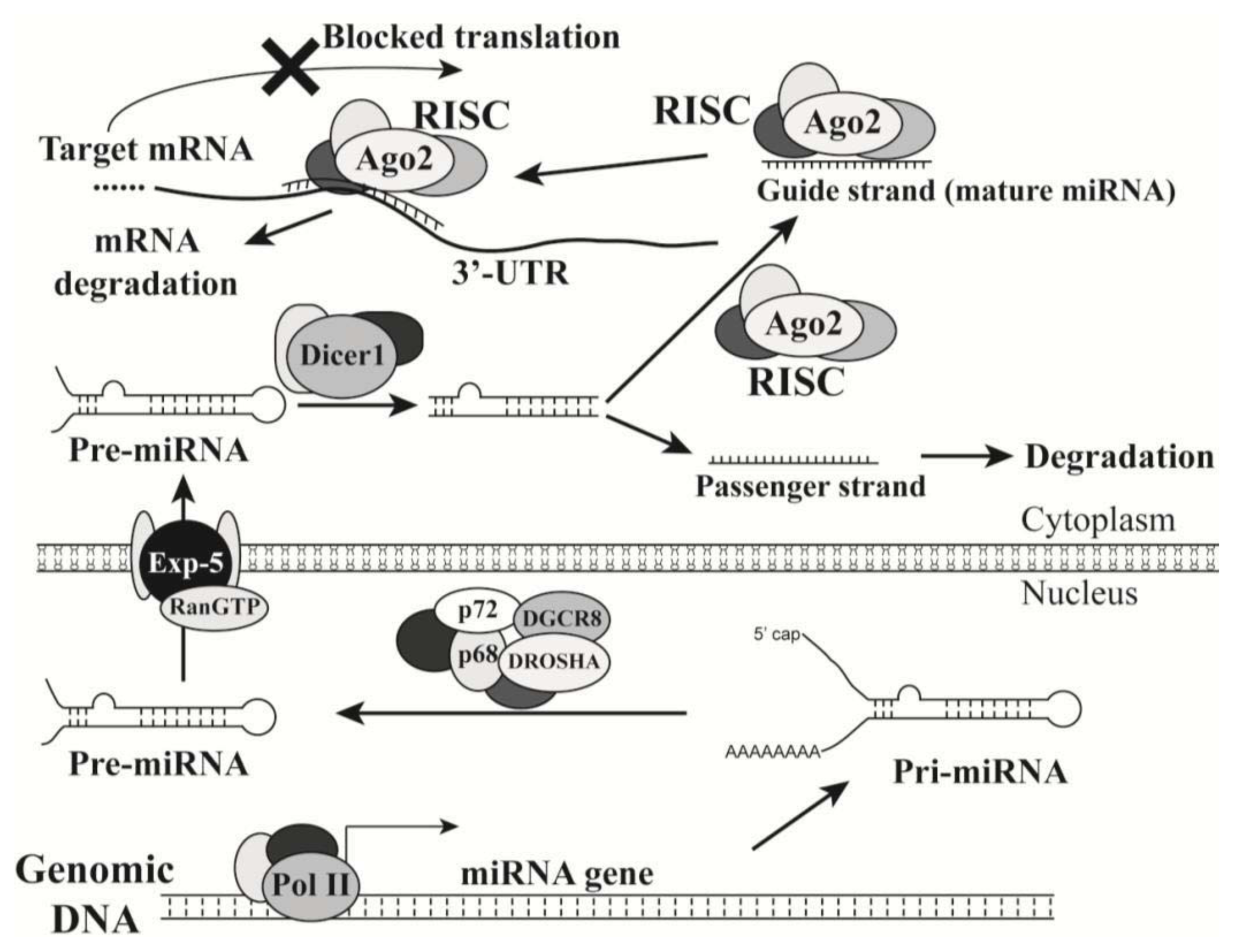
2.1. Introduction of MicroRNAs

MiRNAs are small and evolutionarily conserved noncoding RNAs that are typically 18 to 25 nts in length. The biogenesis of miRNAs begins with the synthesis of the primary transcripts (pri-miRNAs) by RNA polymerase II in the nucleus (Figure 1). Like the transcripts of protein-coding genes, pri-miRNAs contain a 5′ cap structure, a poly(A) tail, and sometimes intron sequences [15,16]. A stem loop structure can be formed by regions of partial complementary sequences in each pri-miRNA. The nuclear ribonuclease DROSHA and its partner DGCR8 recognize this stem-loop structure, and subsequently crop the pri-miRNA to generate the pre-miRNA intermediate. After the pre-miRNA is exported to the cytoplasm by Exportin-5/Ran-GTP, it is further processed by DICER1, another ribonuclease, to generate a double-stranded miRNA molecule. Both strands can act as mature miRNAs; however, if only one strand (guide strand) becomes a functional miRNA, the other strand (passenger strand) will be quickly degraded [17,18].
A mature miRNA can associate with Argonaute proteins to form a RNA-induced silencing complex (RISC), in which the miRNA guides the complex to the 3′-untranslated region (3′-UTR) of the target mRNA to block translational protein synthesis and/or cause its degradation (Figure 1). Each miRNA contains a seed sequence (7 nts; from the nucleotides 2 to 8) at its 5′-end with conserved complementarity to perfectly pair the ‘seed-match’ sequence at the 3′-UTR of its target mRNA [19,20]. Since only a 7-nucleotide match between a target 3′-UTR and a miRNA seed sequence will theoretically make the mRNA a potential target of the miRNA, miRNAs do not have a high specificity for their targets like small interference RNAs (siRNAs) have. MiRNAs can be divided into different families. A miRNA family consists miRNAs that share the same seed sequence and thus may target the same set of genes [19]. MiRNA coding regions, or their genes, can be located in either protein-coding or noncoding regions of transcription units in the human genome. Each of these regions may encode one or a cluster of mRNAs. A miRNA cluster is a set of two or more miRNAs that are transcribed from physically adjacent miRNA genes. Thus, the miRNAs in a cluster are transcribed by the same promoter, in the same direction, and as an unseparated transcription unit. One cluster typically contains two or three miRNAs, but large clusters do exist, such as the miRNA-17-92 cluster consisting of seven miRNAs. Interestingly, some members in one miRNA family are encoded by one miRNA gene cluster, such as miR-15a/miR-16-1 and miR-34b/miR-34c. Different miRNA clusters may belong to the same miRNA family, such as miR-15a/miR-16-1 and miR-497/miR-195.
To date, over 2,500 human microRNAs have been identified based on the microRNA database (miRBase) at the Sanger Institute ( http://www.mirbase.org/). Bioinformatical analyses suggest that miRNAs may regulate over 5,300 human genes, which represents 30% of human genes [19] and each miRNA regulates about 200 genes [21]. Thus, altered expression of microRNAs in cancer can cause significant perturbation of gene expression with a profound effect on malignant transformation and cancer progression [20].
There are much debate and investigation regarding the mechanisms of miRNA-repressed protein expression. Initially, miRNA-mediated gene silencing was portrayed as a different mechanism from that of siRNA-mediated mRNA degradation, based on the observation that the lin-4 miRNA decreased lin-14 protein expression without affecting its mRNA levels in C. elegans [22,23]. This was supported by multiple studies showing that miRNA-mediated gene silencing could be achieved with no or minor change of the target mRNA levels in Drosophila [24], Arabidopsis [25] and mammalian cells [26,27]. However, several later studies suggested that microRNAs can both block translation and trigger target mRNA degradation [28,29]. Notably, a recent report by Djuranovic et al. provided new insights into the kinetics of miRNA-mediated gene silencing in Drosophila cells. The authors demonstrated that miR-9b and miR-279 first repress the translation of the target mRNAs and then cause mRNA deadenylation and degradation [30]. Whether this mechanism is applicable to all miRNAs or other species remains to be determined.
Through the ability to repress the expression of multiple genes, miRNAs play an important role in regulating many cellular activities such as proliferation, differentiation and apoptosis. Calin et al. provided the evidence showing the deletion of the miR-15/miR-16 cluster at 13q14 and its downregulation in patients with B cell chronic lymphocytic leukemia [31]. This was the first study suggesting that noncoding genes correlate with and may even contribute to oncogenesis. The same group further investigated the loci of 186 miRNAs in the human genome to evaluate their potential involvement in cancer pathogenesis [32]. They discovered that over 50% of these miRNA genes are present in the genomic regions with reported alterations in cancers. These are highly instable loci, including fragile sites, minimal heterozygous deletion regions, frequently amplified sections and common breakpoints. However, the instability of these miRNA-coding regions does not generate frequent miRNA somatic mutations within their seed sequences in cancers but rather changes their expression. A more recent study indicated that the genes encoding oncogenic miRNAs are mainly located in the amplified regions in human cancers, whereas the majority of genes for tumor suppressive miRNAs are in the deleted regions [33]. Interestingly, many oncogenes can produce alternative mRNA isoforms with shorter 3′-UTR sequences through a mechanism involved in alternative cleavage and polyadenylation [34]. A short mRNA isoform of an oncogene can avoid miRNA-mediated inhibition, which consequently increases its stability and typically produces ten-fold more protein. Overall, miRNA expression is globally reduced in tumors compared to their matched normal tissues [35]. Thus, numerous studies have demonstrated the potential of using the expression profiles of single or multiple miRNAs as biomarkers to classify tumor origins, stages and clinical outcomes [3639]. Currently, microRNA expression detection is not used clinically in cancer diagnosis and prognosis, but it bears great promise in multiple applications of cancer therapies. Especially, due to the high stability of miRNAs and advance of RNA purification techniques, miRNAs can be extracted from not only tumors samples exposed to variable treatments, including formalin-fixing and paraffin-embedding [40], but also serum and urine [4144].

2.2. MicroRNAs in Oncogenesis

Many miRNAs have been shown to regulate cell survival and proliferation, angiogenesis, and epithelial-mesenchymal transition (EMT). The miRNAs associated with oncogenesis are also known as “oncomirs”. Depending on their major targets, oncomirs can be classified into the oncogenic and tumor suppressive miRNA groups. As the classification implies, tumor suppressive miRNAs repress protein-coding oncogenes whereas oncogenic miRNAs repress protein-coding tumor suppressors. Some miRNAs display both oncogenic and tumor suppressive activities, depending on the tissue and tumor contexts. Wang et al. demonstrated that oncogenic and tumor suppressor miRNAs show clearly different patterns in many aspects, including evolutionary rates, expression patterns, chromosome distribution, molecule sizes and of course targets [33].
An example of miRNAs that have opposing effects is the miRNA-17-92 cluster that has been implicated in regulating cellular survival. The polycistronic miRNA-17-92 cluster (miR-17-92) is located on chromosome 13 open reading frame 25 (c13orf25) in the human genome and contains seven miRNAs (miR-92-1, miR-19a, miR-20a, miR-19b, miR-18a, miR-17-5p and miR-17-3p) [45]. Ota et al. reported that the chromosomal region 13q31-q32, where the miR-17-92 cluster resides, is amplified in malignant lymphoma [46]. He et al. observed that five miRNAs (miR-92-1, miR-19a, miR-20a, miR-19b and miR-17-5p) from this cluster are up-regulated in human B-cell lymphoma and cell lines [47]. Ectopic expression of this miRNA cluster promotes c-Myc-induced lymphomagenesis in mice suggesting the cooperation between mir-17-19b and c-Myc in oncogenesis. In another study, O’Donnell et al. identified an additional miRNA, miR-18a, at the miRNA-17-92 locus and demonstrated that c-Myc transcriptionally activates the expression of these 6 miRNAs (miR-92-1, miR-19a, miR-20a, miR-19b, miR-17-5p and miR-18a) [48]. Interestingly, miR17-5p and miR-20a negatively regulate E2F1 expression, which is also activated by c-Myc. E2F1 is a crucial driver of cell cycle progression from the G1 to S phase, but its expression can induce either pro-metastatic activity or cell apoptosis depending on its molecular contexts [49,50]. Thus, the ectopic miR-17-92 expression in a c-Myc background confers an overall pro-survival trait to the cells through eliminating E2F1-mediated apoptotic potential. This is corroborated by the aforementioned studies of He et al., in which c-Myc and miR-19b overexpression promoted murine lymphomas without causing any indicative sign of apoptosis [47]. The presence of this interactive regulatory network suggests a tight control of c-Myc-mediated proliferation signal during oncogenesis.
Data from several other groups also suggest tumor suppressive activities of the miRNAs in this cluster. Hossain et al. reported that miR-17-5p acts as a tumor suppressor through repressing the expression of AIB1 (or NCOA3) and consequently reducing the proliferation of breast cancer cells [51]. AIB1 was named for its “amplified in breast cancer” and has been suggested to play an oncogenic role in mammary oncogenesis [52,53]. Interestingly, AIB1 functions as a coactivator of E2F1 to promote breast cancer cell proliferation [52]. Thus, through repressing both AIB1 and E2F1, miR-17-5p may “kill two birds with one stone” in reducing breast cancer cell proliferation.
Yu et al. reported a negative feedback regulation between miR-17-5p/miR-20a and cyclin D1 [54], an oncoprotein in breast cancer. Cyclin D1 inversely correlated with miR-17-5p/miR-20a levels in breast cancer samples and cell lines. In a negative regulatory loop, cyclin D1 induces miR-17-5p/miR-20a expression, while these two miRNAs target the cyclin D1 3′-UTR to limit its proliferative activities. The same group also reported that miR-17-5p/miR-20a repress cytokeratin 8 through inhibiting cyclin D1 [55]. Additionally, these two miRNAs target the 3′-UTR of the interleukin-8 mRNA. Thus, miR-17-5p/miR-20a regulates both cellular secretion and tumor microenvironment to block migration and invasion of neighboring cells in breast cancer [55]. The seventh member of this cluster, miR-17-3p, is the “passenger strand” of miR-17-5p that is also processed into a mature miRNA. Recent studies indicate that it represses the expression of vimentin and Mdm2, suggesting a tumor suppressive role in oncogenesis [56,57].
Recent studies revealed a tumor suppressive role of miR-101 in a variety of cancers, including prostate, bladder, breast and gastric cancers [5862]. MiR-101 is frequently downregulated in these cancers compared to their normal adjacent tissues. On the other hand, ectopic expression of miR-101 in various cancer cell lines resulted in decreased cellular proliferation, motility and invasiveness, indicating its antitumoral activities. The tumor suppressive function of miR-101 has been corroborated by its inhibitory effect on EZH2, an oncogene of many solid tumors [63]. Varambally et al. reported the miR-101 loss resulted in increased EZH2 expression [62]. Importantly, genomic loss of the miR-101 locus correlates with EZH2 overexpression in solid tumors. Cao et al. further demonstrated that miR-101 negatively regulates EZH2 expression in prostate cancer cells, while miR-101 expression is modulated by androgen receptor and HIF-1α/HIF-1β [58]. The negative regulation of EZH2 by miR-101 has been confirmed by the studies from several other groups [6466]. Consistent with these observations, a recent report suggested a positive regulation of E-cadherin by miR-101 [67]. E-cadherin is frequently downregulated in aggressive cancers and its loss increases cell dissemination and cancer cell invasiveness. In the study by Qazi et al., ectopic miR-101 restored E-cadherin expression in pancreatic ductal adenocarcinoma cells by reducing EZH2-mediated histone H3-K27 methylation. Overall, these studies suggest that miR-101 primarily targets oncogene EZH2 and its deletion contributes to oncogenesis through aberrant epigenetics caused by EZH2 overexpression.
The let-7 family was one of the first mammalian miRNAs to be discovered and consists of 13 members located on different genomic loci that are often lost in human cancers [68]. Reduced expression of the let-7 members is associated with more dedifferentiated and aggressive cancers [69]. These observations suggested a possible tumor suppressive role of the let-7 family members. Recent studies indicated that let-7 can target oncogenes RAS and c-Myc that have multiple potential binding sites of let-7 miRNAs in their 3′-UTRs. Johnson et al. detected reduced let-7 expression in tumor tissues compared to normal adjacent tissues in lung cancer samples [70]. Interestingly, let-7 miRNA levels inversely correlated with RAS protein expression but not its mRNA levels, suggesting a mechanism of let-7-mediated translational inhibition without mRNA degradation. Overall, let-7 miRNAs may target several key oncogenes, such as RAS and c-Myc, to suppress the proliferative signals from these two oncogenes.
Many other miRNAs have been demonstrated to play a role in cancer development and progression. It is noteworthy that reports from different groups may present paradoxical results. These discrepancies could result from different experimental settings and also reflect the complexity of miRNA-regulated network in cancers. As discussed above, each miRNA potentially target hundreds genes and its ectopic overexpression may perturb multiple cellular processes leading to artificial phenotypic changes. Thus, studies only using miRNA overexpression or reporter assay without any miRNA depletion or tumor sample correlation studies may not truly represent physiological relevance. Nevertheless, currently available literature unequivocally indicates the prognostic potential and biological activities of miRNAs in different cancers.

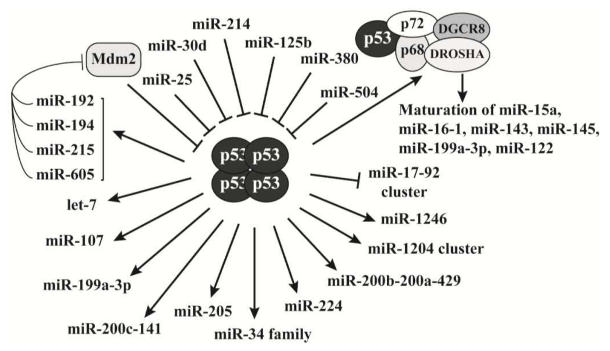
2.3. P53 Is a Key Regulator of MicroRNAs Biogenesis

The tumor suppressor p53 is one of the best-studied proteins in the field of cancer research. Owing its well-characterized regulation, the role of p53 in the miRNA network has been extensively explored in the past decade. As a transcription factor, p53 forms tetramers to bind to its consensus sites on target genes and mediates their transcription. Increasing evidence shows that p53 exerts its antiproliferative activities at least partially through the transcriptional regulation of miRNA expression (Figure 2), in addition to its canonical tumor suppressive role [20,71]. The best characterized p53 target is the miR-34 family [72]. The miR-34 family includes miR-34a, -34b and -34c. MiR-34a is transcribed by chromosome 1, and miR-34b and -34c are transcribed by two proximal loci on chromosome 11 and controlled by the same promoter. These miRNAs exert tumor suppressive activities through repressing proliferative genes, such as c-Myc and BCL2, and their overall function is inducing apoptosis, cell cycle arrest or senescence [7375]. Consistently, they are frequently silenced by promoter methylation in tumors [76]. Thus, the p53-promoted expression of the miR-34 family extended its activated tumor suppressive network.
P53 also stimulates the expression of many other miRNAs that have antiproliferative activities. For example, p53-mediated miRNA expression plays a role in hypoxia. During hypoxia of tumor cells, the upregulated hypoxia inducing factor (HIF)-1α forms a heterodimer with HIF-1β to become a transcription factor HIF-1 that activates pro-angiogenic genes such as vascular endothelial growth factor A (VEGFA), to promote angiogenesis for tumor growth and metastasis [77,78]. Yamakuchi et al. reported that miR-107 reduces hypoxia signaling by inhibiting HIF-1α expression in human colon cancer cells [79]. As a transactivator, p53 promotes the expression of miR-107 to reduce HIF-1α levels, which quenches the hypoxic signal to block tumor angiogenesis. Among other p53-activated miRNAs, miR-145 represses c-Myc expression [80], and miR-200c/miR-141 and miR-200b/miR-200a/miR-429 inhibit EMT through downregulating ZEB1 and ZEB2 [81,82]. As discussed above, miR-17-92 cluster exhibits proliferative activities based on most literature. P53 represses the expression of the miR-17-92 cluster, opposite to its transactivating effects on most other miRNAs (Figure 2), and this regulation likely contributes to the p53-induced apoptosis [83].
In addition to transcriptional regulation, p53 is directly involved in the maturation process of miRNAs. DROSHA is a major component of the complex that processes pri-miRNA into pre-miRNA. P53 associates with DROSHA in a RNA dependent manner and facilitates DROSHA-mediated pri-miRNA processing [84]. Interestingly, although this process is independent of its transcriptional activity, transcriptionally inactive p53 mutants do not show this capability. Actually, these p53 mutants interfere with the assembly of the DROSHA complex and consequently attenuate miRNA processing. Currently, p53 has been reported to regulate the maturation of at least 6 miRNAs (miR-15a, miR-16-1, miR-143, miR-145, miR-199a, and miR-122) [8487]. Whether p53 is involved in the processing of other miRNAs remains to be determined.
Conversely, p53 expression can also be regulated by miRNAs (Figure 2). The functions of these miRNAs are not uniformly oncogenic; thus, whether their role in repressing p53 expression is physiologically significant is unclear. There is a negative feedback loop between p53 and the ubiquitin E3 ligase Mdm2. While Mdm2 stimulates p53 ubiquitination and degradation, p53 activates Mdm2 gene expression. This feedback regulation is important to maintain p53 homeostasis in normal cells, which is disrupted when exposed to genotoxic stresses [88]. Recent studies reveal that some miRNAs can break this negative feedback loop leading to p53 accumulation. Pichiorri et al. reported that p53 activates the expression of miR-192, miR-194 and miR-215 and these miRNAs target the Mdm2 3′-UTR to repress its expression; thus, downregulation of p53-inducible miR-192, miR-194 and miR-215 causes aberrant Mdm2 increase and p53 downregulation in multiple myeloma [89]. Consistently, miR-192, miR-194 and miR-215 are downregulated in multiple myeloma and renal cancers [8991]. Xiao et al. demonstrated that p53 activates miR-605 that also represses Mdm2 expression [92]. Thus, p53 activates these four miRNAs to downregulate its negative regulator Mdm2 leading to p53 accumulation in response to stress.

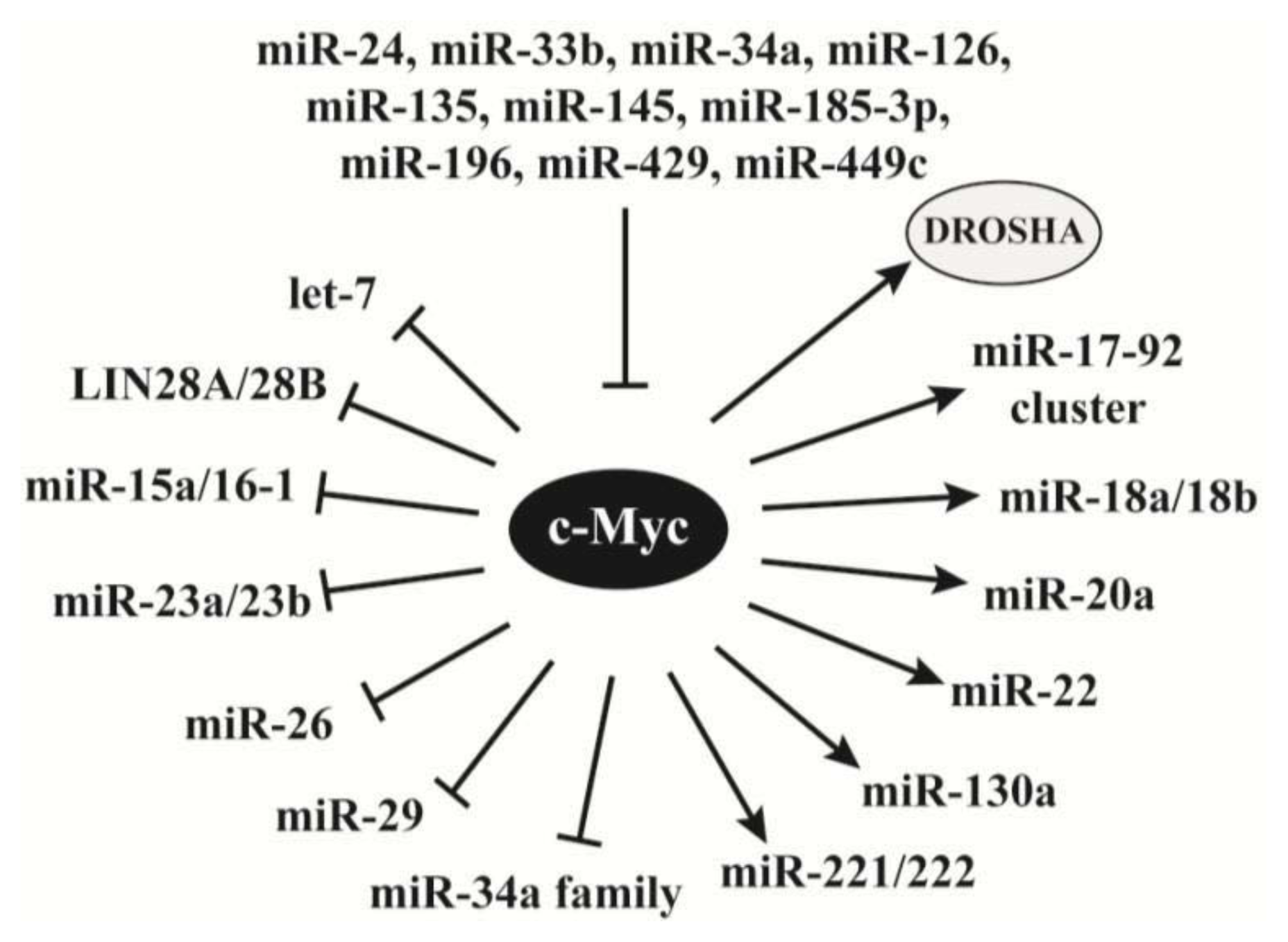
2.4. C-Myc Regulates the Synthesis of miRNAs

The oncogene c-Myc is a transcription factor and mediates target gene expression through recruiting various chromatin modifiers [93]. Recent studies reveal its role in regulating miRNA synthesis. Interestingly, the effects of c-Myc-mediated transcription of many miRNAs are opposite to those regulated by p53.
C-Myc represses the expression of multiple miRNAs (Figure 3). As discussed above, p53 transactivates the miR-34 family that represses c-Myc expression [72,75]. On the other hand, c-Myc represses the expression of the miRNAs in the miR-34a family [94]. C-Myc also recruits HDAC3 to downregulate the expression of miR-15a/miR-16-1 [95] that block the expression of multiple oncogenes, such as BCL2 and cyclin D1 [27,96]. Similarly, c-Myc also recruits HDAC3 and EZH2 to silence the expression of miR-29 [97] that has been defined as a tumor suppressor [98]. Among other c-Myc repressed miRNAs, let-7 inhibits androgen receptor and KRAS [68,99], miR-23a/miR-23b block glutaminase [100], and miR-26 targets EZH2 and cyclin D2 [58]. Thus, c-Myc-mediated repression of these miRNAs can release the expression of multiple oncogenes or proliferative genes to promote oncogenesis.
c-Myc also activates the expression of multiple miRNAs. Similar to its role in antagonizing p53-mediated miR-34 miRNA expression, c-Myc promotes the transcription of miR-17-92 cluster [101] that is downregulated by p53. Kim et al. carried out a combined analysis of mRNA and miRNA expression profiles that revealed multiple c-Myc-induced miRNAs and their downstream targets [102]. Among their discovered miRNAs, miR-221 and miR-222 have been shown to target p27, p57 and PTEN, and exhibit proliferative activities [103106]. For the other c-Myc-activated miRNAs, the authors predicted that miR-20a targets p21, RB1, PTEN and interferon regulatory factor 1 (IRF-1), and miR-130a inhibits tuberous sclerosis 1 (TSC1) and CYLD [102]. While p53 protein direct associates with the miRNA processing machinery, c-Myc activates DROSHA expression through binding to the E-box in its promoter and consequently facilitates miRNA processing [107].
While c-Myc acts as a regulator of miRNAs, its expression has been reported to be modulated by a number of miRNAs (Figure 3). Among them, miR-34a, miR-126 and miR-145 have been documented as tumor suppressors [33].

3. Long Noncoding RNA

Long noncoding RNAs (lncRNAs) were previously defined as RNA molecules longer than 200 nucleotides that are not translated into proteins [6,108]. Recently, Spizzo et al. amended this definition linking to the biological functions and described that lncRNAs are a class of RNA molecules that do not fit into any known class of small and structural RNAs, and possess regulatory roles in their primary or spliced form [109]. The GENCODE Consortium is a part of the ENCODE (ENCyclopedia Of DNA Elements) project and aims to identify all gene features in the human genome. The GENCODE 7 release in 2012 indicates that the human genome contains at least 9,640 long noncoding RNA loci that can potentially encode 15,512 transcripts [110]. LncRNA production is regulated by mechanisms similar to these of protein-coding genes, such as histone modifications and RNA splicing, and their expression shows tissue-specific patterns [111]. Most lncRNAs are localized in nucleus and associated with chromatin, and some of them are preferentially processed into small RNAs [111]. Banfai et al. demonstrated that lncRNAs are rarely translated in two tested human cell lines, suggesting that ribosomes can differentiate the coding and noncoding transcripts for translation [112].
Recent studies continue elucidating novel and unpredicted biological activities of the lncRNAs, which were previously ascribed as protein functions. Based on the regulatory mechanisms, the lncRNAs can be divided into four categories, including the lncRNAs that (1) regulate gene expression, (2) act as miRNA decoys to free target mRNAs, (3) regulate mRNA translation, and (4) regulate protein activities.

3.1. LncRNAs Regulating Gene Expression

The X-inactive-specific transcript (Xist) was one of the first lncRNAs discovered in mammals [113]. Xist is encoded by the inactive X chromosome (Xi) and the genomic locus can transcribe a 17- to 20-kb RNA. This lncRNA binds the Xi in cis to induce chromosome X silencing by recruiting the polycomb repressive complex 2 (PRC2) that induces histone H3-K27 methylation, a hallmark of gene inactivation [114,115]. In this process, the transcription repressor Yin Yang 1 (YY1) confers allele-specific binding of Xist to the Xi with the involvement of two other noncoding RNAs, Jpx [116] and Ftx [117]. There is another lncRNA, Tsix, that is transcribed at the same locus of Xist but in the reverse direction and thus antisense to Xist [118]. Tsix regulates imprinted and random X inactivation in development [119]. An additional Xist-related RNA transcript is Xite that also plays a role in the X chromosome inactivation [120].
The association of epigenetic silencing complexes with Xist to induce transcriptional silencing has been extended to several recently characterized lncRNAs, which also associate with the PRC2 and other chromatin repressive complexes [121,122]. One of these lncRNAs is HOX Antisense Intergenic RNA (HOTAIR) that is a 2.2-kb transcript located at the HOXC gene cluster on chromosome 12 [122]. HOTAIR also modulates gene expression through epigenetic regulation. Unlike Xist that acts in cis, HOTAIR functions in trans to recruit the PRC2 to the HOXD locus on chromosome 2 to induce transcriptional silencing [122]. In addition to associating with PRC2, HOTAIR also interacts with the LSD1/CoREST/REST histone modification complex, leading to both histone H3-K27 methylation and H3-K4 demethylation [123]. Since HOTAIR plays an important role in the epigenetic regulation of its target genes, it is not surprising that its deregulation has been observed in different types of cancers. Recent studies suggest that HOTAIR overexpression is positively associated with increased tumor cell malignancy. Gupta et al. reported that HOTAIR is overexpressed in both primary and metastatic breast cancer tissues, and its levels in the primary tumors could be used as a significant predictor of subsequent tumor metastasis and survival of the patients [124]. Ectopically expressed HOTAIR could confer the breast epithelial cancer cells with invasive and metastatic potential while its depletion in breast cancer cells abrogated these activities [124]. The role of HOTAIR in promoting oncogenesis has also been reported in other cancers. Yang et al. compared HOTAIR levels between tumorigenic and adjacent non-tumorigenic tissues of hepatocellular carcinoma (HCC) samples and found that this lncRNA was expressed at higher levels in malignant tissues [125]. In addition, the survival analysis of a cohort consisting of 60 HCC patients revealed that high HOTAIR expression could serve as an independent prognostic marker for disease recurrence and reduced patient survival [125]. Another HCC-related study suggested that HOTAIR expression is a potential biomarker for lymph node metastasis from the primary tumors [126]. Furthermore, HOTAIR upregulation was also observed in a cohort of patients diagnosed with the stage IV colorectal cancer (CRC) [127]. In this study, Kogo et al. indicated that high HOTAIR expression showed significantly positive correlation with the liver metastasis and poor patient outcome, further supporting that HOTAIR expression is a potential prognostic marker of multiple cancers. In pancreatic cancer, HOTAIR levels were also increased in tumorigenic tissues compared to the non-tumorigenic tissues, and associated with a more aggressive phenotype [128]. The oncogenic role of HOTAIR in pancreatic cancer cell invasion was validated by its siRNA-mediated knockdown and overexpression studies [128], consistent with the observations in the other aforementioned cancers. Interestingly, gene array studies showed only a small overlap of HOTAIR-regulated genes between pancreatic cancer and breast cancer [128], suggesting that HOTAIR may regulate different sets of target genes in a cell type-specific manner.
EZH2 is a core component of the PRC2. EZH2 knockdown followed by chromatin immunoprecipitation demonstrated that HOTAIR-mediated gene repression could be either PRC2-depedent or -independent in pancreatic cancer cells, although the PRC2 is necessary for HOTAIR target gene repression in breast cancer cell [124,128]. This discrepancy between the two cancer types suggests the presence of yet unidentified epigenetic mechanisms regulating HOTAIR-mediated transcriptional silencing.
Wang et al. identified the lincRNA HOTTIP that is transcribed from the 5′ tip of the HOXA locus and modulates the activity of the WDR5-MLL complex, in which the WD40-repeat protein WDR5 binds the MLL complex to activate its histone H3-K4 methyltransferase activity [129]. Chromosomal looping can make HOTTIP stay in the vicinity of its target genes and let it bind WDR5 to promote the WDR5/MLL complexes-mediated histone H3-K4 methylation, which leads to target gene activation. Thus, HOTTIP serves as a key intermediate to transmit information from higher order chromosomal looping into chromatin modifications [129]. Another HOXA-related lncRNA is HOX antisense intergenic RNA myeloid 1 (HOTAIRM1) that is transcribed at a direction antisense to the HOXA gene [130]. The knockdown of HOTAIRM1 reduced the expression of HOXA1 and HOXA4 during the myeloid differentiation in promyelocytic leukemia cells. Whether this lncRNA acts as a miRNA decoy to promote the expression of these HOX genes remains to be determined.
Metastasis-associated lung adenocarcinoma transcript 1 (MALAT1), also known as nuclear-enriched abundant transcript 2 (NEAT2), is one of the first identified cancer-associated lncRNAs [131]. MALAT1 is a highly conserved noncoding transcript of over 8000 nts encoded by a locus on chromosome 11. It was initially recognized as a prognostic marker of increased metastatic risk for the patients of non-small cell lung carcinoma (NSCLC). Subsequent studies revealed that MALAT1 is localized in nuclear structures enriched with splicing and transcription factors, known as nuclear speckles, suggesting that this lncRNA may modulate alternative splicing of target genes [132,133]. Using RNAi-mediated depletion for the components of the nuclear speckles, Miyagawa et al. demonstrated that the pre-mRNA splicing activator RNPS1, the splicing coactivator SRm160 and the spliceosomal intron binding protein IBP160 promote MALAT1 localization to the nuclear speckles [134]. Furthermore, MALAT1 depletion and delocalization from the nuclear speckles resulted in downregulation of two interferon-induced genes (OASL and IFI44) and a potential celiac disease susceptibility gene, SPINK4 [134].
Despite these studies suggesting a role of MALAT1 in RNA splicing, a recent study by Gutschner et al. showed that this lncRNA regulates gene expression but not alternative splicing in lung cancer cells [135]. MALAT1 knockout was achieved by genomic integration of RNA destabilizing elements using zinc finger nucleases, leading to 1000-fold MALAT1 reduction. Using these MALAT1-knockout cells, the authors demonstrated that MALAT1 represses anti-metastatic genes and activates pro-metastatic genes in lung cancer cells, but does not affect genes regulating cell growth. In a xenograft mouse model, the MALAT1-depleted lung cancer cells showed reduced tumor formation compared to the cells with the intact MALAT1. This observation is in contrast to the findings reported by Yang et al. showing that MALAT1 cooperates with Polycomb 2 protein (Pc2) in regulating the activation of the growth-control gene program in 293T cells [136]. The different cell types employed in their experiments might contribute to the discrepancy between the two studies. Consistent with this prediction, MALAT1 is widely expressed in most normal human tissues, such as pancreas and lung, but absent in several other tissues including skin, stomach, bone marrow and uterus [131]. This suggests that this lncRNA may possess tissue specific functions. In addition to its role in regulating lung cancer metastasis, MALAT1 is upregulated in uterine endometrial stromal sarcoma [137], cervical cancer [4] and hepatocellular carcinoma [138], while its expression in the corresponding healthy tissues is undetectable or intermediate [131]. Although the oncogenic role of MALAT1 in different cancers has been demonstrated by correlational and functional studies, the molecular mechanisms underlying its activities in regulating gene expression and RNA splicing remain undetermined. To date, several studies suggested the essential role of the 3′ end sequence and structure to its metastasis-promoting function, nuclear localization and stability [139142].
ANRIL is a large antisense ncRNA of the INK4b/ARF/INK4a locus [143]. Yap et al. demonstrated that ANRIL binds chromobox 7 (CBX7), a component of the polycomb repressive complex 1. This interaction contributes to the role of CBX in promoting EZH2-mediated H3-K27 methylation at the INK4b/ARF/INK4a locus and consequently represses the tumor suppresser INK4a gene. Consistently, both CBX7 and ANRIL are increasingly expressed in prostate cancer [144].
GAS5 (growth arrest-specific transcript 5) is a lncRNA regulating growth arrest of T-cells and lymphocytes [145]. Ectopic GAS5 increases apoptosis and reduces cell cycle progression. Consistently, its downregulation inhibits apoptosis and promotes cell cycle. Kino et al. investigated the mechanism underlying the growth suppressive activities of GAS5 and discovered its role in blocking gene expression mediated by glucocorticoid receptor (GR) [146]. GAS5 binds to the DNA-binding domain of GR and thus prevents its association with the glucocorticoid response element of the GR target genes with anti-apoptotic activities, such as inhibitor of apoptosis 2 (cIAP2). Thus, abundantly expressed GAS5 during starvation can sensitize the cells to apoptosis. The nonsense-mediated mRNA decay (NMD) is a system that controls the quality of gene transcripts and reduces errors in gene expression by eliminating RNAs with premature stop codons [147]. Meanwhile, this mechanism also regulates the abundance of cellular transcripts, including ncRNAs. Recently, Zhang et al. demonstrated a reciprocally negative regulation between GAS5 and miR-21 [148]. While miR-21 represses GAS5 by targeting a sequence encoded by its exon 4, GAS5 inhibits miR-21 expression. Thus, GAS5 antagonizes the oncogenic activity of miR-21 [149] through reducing its cellular levels. Consistently, miR-21 and GAS5 showed negative correlation in breast cancer specimens [148]. The tumor suppressive role of GAS5 is supported by the identification of genetic susceptibility of its genomic locus, 1q25, to several cancers, including melanoma [150], prostate cancer [151], breast cancer [152,153], colorectal cancer [154] and B-cell lymphoma [155]. Additionally, Tani et al. indicated that GAS5 can be stabilized with the depletion of UPF1, an essential component of NMD or during starvation [156] and the GAS5 introns encode multiple snoRNAs [157160].
Several other lncRNAs have also been demonstrated to regulate chromatin remodeling and gene transcription. PTENP1 is the PTEN pseudogene and encodes two antisense RNA (asRNA) transcripts, asRNA α and β [161]. The α asRNA isoform can recruit DNMT3A, EZH2 and G9A to the PTEN promoter and repress its transcription. This will be further discussed below with other regulatory mechanisms of PTENP1. Evf2 is a polyadenylated lncRNA identified in embryonic brain cells [162]. This lncRNA regulates the transcription of homeodomain transcription factors DLX5 and DLX6 through recruiting DLX and MECP2 to the DNA regulatory elements in the intergenic region of these two genes. As a p53 transactivated lncRNA, lincRNA-p21 is a key mediator of p53-dependent gene repression through a mechanism of recruiting heterogeneous nuclear ribonucleoprotein K (HNRNP) to these p53 target genes [163]. Thus, inhibition of lincRNA-p21 affects the expression of a number of p53 repressed genes. Sheik et al. identified an Oct4-activated lncRNA, AK028326, and discovered that this lncRNA regulates pluripotency in mouse embryonic stem cells [164]. Interestingly, AK028326 activates Oct4 expression in a regulatory feedback loop.

3.2. LncRNAs Acting as miRNA Decoys to Free Target mRNAs

Since the regulation of gene expression by miRNAs was revealed, researchers have been using miRNA sponges, RNA molecules containing the target sequence or reverse complementary sequence of a miRNA to be sponged, as a tool to inhibit the function of miRNAs and release their target gene expression [165]. Recent studies suggest that this approach naturally exists in cancers to modulate tumor suppressor and oncogene levels. In some literature, these decoy RNAs are also named as competitive endogenous RNAs (ceRNAs).
PTEN (phosphatase and tensin homolog) is a well characterized tumor suppressor with phosphatase activity. It is encoded at the 10q23.3 locus on chromosome 10 and frequently inactivated through diverse mechanisms in human cancers, highlighting its crucial role in oncogenesis. The 3′-UTR of the PTEN mRNA has 3,329 nts, markedly longer than the average 3′-UTR length (740 nts) of eukaryotic mRNAs [166], implicating its vulnerability as a target of miRNAs. Thus, while PTEN inactivation can be achieved by gene deletion and epigenetic silencing in cancers, its expression is also regulated by multiple miRNAs. For example, the PTEN 3′-UTR contains potential binding sites for over 10 miRNAs overexpressed in glioblastoma multiforme, which is more than 2 times higher than any other tumor suppressor [167]. PTEN expression can be repressed by miR-21, miR-221 and miR-222 [106,168]. Recent studies revealed novel mechanisms regulating PTEN expression through its pseudogene PTENP1 (also called PTH2 or ψPTEN). Pseudogenes are dysfunctional relatives of their cognate genes but have lost the protein-coding ability due to premature stop codons, deletions/insertions or frameshift mutations, and thus cannot be translated into functional proteins [169]. The PTEN pseudogene PTENP1 is highly transcribed in certain tissues and cells, suggesting that this lncRNA may have biological activities [170]. The functional relationship between the PTEN and its pseudogene was first discovered by Poliseno et al. [171]. In their study, the authors demonstrated that PTENP1 modulates endogenous PTEN transcript levels by acting as a molecular sponge for PTEN-targeting miRNAs; thus, the PTENP1 transcript serves as an effective decoy and exerts tumor suppressive functions (Figure 4). This novel regulatory role of the PTENP1 can be extended to KRAS1P, the pseudogene of the oncogene KRAS. In a study by Poliseno et al., the overexpression of KRAS1P led to increased KRAS mRNA levels in prostate cancer DU145 cells through a mechanism of sequestering KRAS-targeting microRNAs, and consequently promoted cell proliferation [171].
As briefly discussed above, a recent study by Johnsson et al. provided evidence of another regulatory mechanism of PTEN by its pseudogene PTENP1 (Figure 4). The authors discovered that the PTENP1 locus can be transcribed from a reverse direction to create two isoforms of an antisense RNA (asRNA), α and β [161]. The α isoform of the asRNA binds the PTEN promoter and recruits DNMT3A and two histone methyltransferases EZH2 and G9A, which mediate the methylation of histone H3-K27 and H3-K9, respectively, two well-characterized markers of gene repression [172]; thus, the PTENP1 α asRNA negatively regulates PTEN gene expression through promoting the epigenetic silencing of its promoter. The β asRNA isoform interacts with the PTENP1 lncRNA through RNA-RNA pairing, which can maintain stable PTENP1 lncRNA levels in cytoplasm, increase its stability and facilitate its role as a microRNA sponge; thus, the PTENP1 β asRNA activates PTEN expression through facilitating the decoy activity of the PTENP1 lncRNA. Overall, the PTENP1 α and β asRNAs exhibit oncogenic and tumor suppressive roles, respectively, based on their effects on PTEN expression (Figure 4). Whether the two asRNA isoforms are differentially expressed in human cancers remains to be determined.
Interestingly, PTEN expression can also be regulated at the translational level by the sponge effect of the transcript from another gene. The full length ZEB2 mRNA has a very long 3′-UTR (over 5,000 nts) and contains multiple potential binding sites of miRNAs that can also target the PTEN 3′-UTR, such as miR-181a and miR-200/miR-141 [173]. Thus, the ZEB2 mRNA serves as a decoy or ceRNA of the PTEN mRNA (Figure 4) and reduced ZEB2 expression activates the PI3K/AKT pathway through downregulating PTEN.
There are several other examples showing that lncRNAs act as decoys to stabilize mRNAs. BACE-AS regulates the expression of β-secretase-1 (BACE1), a crucial enzyme in Alzheimer’s disease pathophysiology [174]. Linc-MD1 is a muscle-specific lncRNA and activates the expression of MAML1 and MEF2C through its decoy role for miR-133 [175].

3.3. LncRNAs Regulating mRNA Translation

As discussed above, MALAT1 was initially demonstrated to possess activities in regulating alternative splicing. MALAT1 associates with serine/arginine (SR) splicing factors at the nuclear speckle domains of nucleus and is involved in the process of alternative splicing [133]. Thus, MALAT1 depletion or ectopic SR protein expression affects the alternative splicing of a similar set of pre-mRNAs. Additionally, MALAT1 alters the phosphorylation of the SR splicing factors, which is essential to their activities in regulating alternative splicing. However, this regulation may only be present in specific cell types or under particular conditions, because MALAT1-knockout mice were viable and fertile, and showed regularly localized nuclear speckle markers [176]. Whether this regulatory mechanism contributes to the activity of MALAT1 in promoting tumor metastasis has not been determined.
A recent study from Zhang et al. demonstrated the lncRNA, lincRNA-RoR, acts as a strong negative regulator of p53 [177]. LincRNA-RoR reduces p53 expression in cells exposed to DNA damage stress, but not in unstressed cells, through directly binding to the heterogeneous nuclear ribonucleoprotein I (hnRNPI) and repressing p53 mRNA translation. As an autoregulatory feedback regulation, p53 transcriptionally induces lincRNA-RoR expression. The functional interplay between lincRNA-RoR and p53 may serve as additional surveillance to mediate cell response to genotoxic stresses.
As discussed above, another p53-transactivated lncRNA, lincRNA-p21, is involved in p53-dependent gene transrepression [163]. A recent study from Yoon et al. demonstrated that lincRNA-p21 also modulates translation [178]. This lncRNA can associate with JunB and β-catenin mRNAs to reduce their translation rates. A RNA-binding protein, HuR, can recruit let-7/AGO2 to lincRNA-p21 to reduce its stability; thus, elevated HuR releases lincRNA-p21-mediated repression of JunB and β-catenin expression. Since lincRNA-p21 is transactivated by p53, the negative regulation of JunB and β-catenin by lincRNA-p21 is consistent to the tumor suppressive role of p53.

3.4. LncRNAs Regulating Protein Activities

Telomeres are the DNA-protein complexes at the end of eukaryotic chromosomes and essential to chromosome stability. As a frequently activated reverse transcriptase, telomerase adds DNA sequence repeats (“TTAGGG” in vertebrates) to the telomere regions of chromosome to maintain the telomere length. Azzalin et al. discovered that telomeres can be transcribed into telomeric repeat-containing RNA (TERRA) [179]. These molecules have different lengths and were predicted to play a role in the maintenance of telomere integrity through an unclear mechanism. A later study from Redon et al. provided a possible mechanism underlying this activity of TERRA [180]. The authors demonstrated that TERRA binds telomerase and thus acts as a potent competitive inhibitor for the telomeric DNA. Consistently, TERRA expression is significantly downregulated in multiple tumor cell lines, which plays a role in telomere maintenance and cell immortalization of cancer cells.
Wang et al. demonstrated an indirect regulation of the histone acetyltransferase activities of CBP and p300 by ncRNAs [181]. The 5′-regulatory region of the cyclin D1 gene encodes at least four ncRNAs. In response to ionizing radiation, these ncRNAs bind TLS (translocated in liposarcoma) protein at the chromatin of the cyclin D1 promoter region and cause an allosteric effect on this protein. This TLS conformational change promotes its activity of inhibiting CBP/p300-mediated histone acetylation and consequently silences cyclin D1 gene. These data indicate that ncRNAs encoded by a promoter can act as selective ligands to modulate the activities of transcription cofactors in response to genotoxic stresses.
LncRNAs can also regulate protein activity through altering their subcellular localization. Nuclear factor of activated T-cells (NFAT) represents a family of transcription factors regulating immune response. Some of NFAT proteins, such as NFAT1 and NFAT5, contribute to tumor metastasis and cell motility [182,183]. The noncoding repressor of NFAT (NRON) is a ncRNA associated with multiple proteins [184]. NRON binds phosphorylated NFAT1 to sequester NFAT in the cytoplasm. The depletion of NRON leads to NFAT dephosphorylation and nuclear import. Thus, ncRNAs can be a part of a scaffold to trap a latent transcription factor [185] and regulate the expression of its target genes.

4. Circular RNAs

Recent advances in high-throughput sequencing coupled with powerful computational analyses of expression data have allowed researchers to identify and characterize new RNA species, one of which is a new class of RNA molecules, circular RNAs (circRNAs), present in humans and animals. CircRNAs were first discovered in plants and considered as viroids due to their predicted role as subviral agents [186]. These covalently linked and single-stranded ncRNAs have molecular weights of over 100,000 Da and are highly thermal stable. Initially, most circRNA molecules were dismissed as transcription noise or by-products generated by cellular splicing machineries. However, this notion has been challenged by emerging studies suggesting that some circRNAs are evolutionary conserved in human and mice [14,187]. The exact mechanism of circRNA biogenesis remains to be elucidated. Salzman et al. indicated that circRNAs are produced by a non-canonical mode of RNA splicing [188]. Several other studies suggested that circRNA may be at least partially contributed by exon-skipping events, which can create an exon-containing lariat and then possibly undergo internal splicing to generate an exon circle [187,189,190].
In 2011, Hansen et al. reported the miRNA decoy function of a noncoding circRNA [191]. In this study, the authors demonstrated that miR-671 directly targets and cleaves a circular antisense transcript of the cerebellar degeneration-related protein 1 (CDR1) in an AGO2-dependent manner. In 2013, thousands of well-expressed and stable circRNAs with tissue- and developmental stage-specific expression were identified [14]. The antisense to the CDR1 transcript (CDR1as) is densely bound by Argonaute proteins and contains 63 conserved binding sites of miR-7, suggesting that this circRNA functions as a miRNA sponge of miR-7 to release its target gene expression [14,190]. Indeed, using a zebrafish model, Memczak et al. demonstrated that introduction of human CDR1as resulted in a phenotype similar to that of miR-7 knockdown, while injection of the miR-7 precursor partially reversed this phenotype, further implicating CDR1as as an antagonist of miR-7 [14]. The results of this study provided evidence that circRNAs may function as regulators of gene expression at the post-transcriptional level. In addition to these studies, Jeck et al. recently discovered over 25,000 circRNAs in human fibroblasts [187]. Due to the lack of exposed 5′ and 3′ ends, circRNAs are predicted to be resistant to degradation by cellular enzymes such as ribonucleases and thus more stable than linear RNAs. The increased stability of circRNAs makes these novel RNA molecules act as efficient miRNA sponges in modulating gene expression.

5. Conclusions

NcRNAs have gained intensive and growing attention for their potential as both regulators and biomarkers of cancers in the past two decades. We are now witnessing the beginning of a new era of identifying key players and determining their underlying mechanisms during oncogenesis. In this review, we are only able to summarize some studies of miRNAs, lncRNAs and circRNAs. We did not intentionally ignore other excellent reports, but just could not include them due to the enormous amounts of ncRNA-related studies and the limitation of this article. Many review papers have summarized the roles of ncRNAs in different diseases including cancers and some of them also discussed several other types of ncRNAs, such as snoRNA and small nuclear RNAs (snRNA).
Currently, many thousands of ncRNAs have been identified and their differential expression profiles in a variety of cancers or between normal and tumorigenic specimens have been demonstrated; however, only a small number of ncRNAs, especially lncRNAs, have been well characterized. Thus, what we have discovered is just a tip of the iceberg in this area. Understanding the functions and regulatory mechanisms of ncRNAs in cancer pathogenesis remains a fertile research field to be explored at least in next decade. Future studies are needed to dissect the upstream mechanisms regulating ncRNA expression and processing, and the downstream proteins or pathways mediated by ncRNAs at different stages of cancer development. These efforts may lead to the discovery of entirely novel regulatory mechanisms or advance our understanding for the currently recognized signaling pathways. The achievement of these ncRNA studies in cancers will definitely provide insights into discovering new biomarkers for cancer diagnosis and prognosis, unraveling novel therapeutic targets, and developing unconventional therapeutic modalities to reduce cancer-related mortality.

Acknowledgements

We thank Steven J Kridel for critical reading of the manuscript. GD is supported by the Randi B. Weiss Cancer Fund. GS is supported by American Cancer Society (116403-RSG-09-082-01-MGO) and NIH R01 (5R01CA106314).

Conflicts of Interest

The authors declare no conflict of interest.

References

  1. Bertone, P.; Stolc, V.; Royce, T.E.; Rozowsky, J.S.; Urban, A.E.; Zhu, X.; Rinn, J.L.; Tongprasit, W.; Samanta, M.; Weissman, S.; et al. Global identification of human transcribed sequences with genome tiling arrays. Science 2004, 306, 2242–2246. [Google Scholar]
  2. Birney, E.; Stamatoyannopoulos, J.A.; Dutta, A.; Guigo, R.; Gingeras, T.R.; Margulies, E.H.; Weng, Z.; Snyder, M.; Dermitzakis, E.T.; Thurman, R.E.; et al. Identification and analysis of functional elements in 1% of the human genome by the ENCODE pilot project. Nature 2007, 447, 799–816. [Google Scholar]
  3. Cheng, J.; Kapranov, P.; Drenkow, J.; Dike, S.; Brubaker, S.; Patel, S.; Long, J.; Stern, D.; Tammana, H.; Helt, G.; et al. Transcriptional maps of 10 human chromosomes at 5-nucleotide resolution. Science 2005, 308, 1149–1154. [Google Scholar]
  4. Kapranov, P.; Cheng, J.; Dike, S.; Nix, D.A.; Duttagupta, R.; Willingham, A.T.; Stadler, P.F.; Hertel, J.; Hackermuller, J.; Hofacker, I.L.; et al. RNA maps reveal new RNA classes and a possible function for pervasive transcription. Science 2007, 316, 1484–1488. [Google Scholar]
  5. Wilusz, J.E.; Sunwoo, H.; Spector, D.L. Long noncoding RNAs: Functional surprises from the RNA world. Genes Dev 2009, 23, 1494–1504. [Google Scholar]
  6. Mercer, T.R.; Dinger, M.E.; Mattick, J.S. Long non-coding RNAs: Insights into functions. Nat. Rev. Genet 2009, 10, 155–159. [Google Scholar]
  7. Wang, K.C.; Chang, H.Y. Molecular mechanisms of long noncoding RNAs. Mol. Cell 2011, 43, 904–914. [Google Scholar]
  8. Cooper, G.M. Cellular transforming genes. Science 1982, 217, 801–806. [Google Scholar]
  9. Santos, E.; Tronick, S.R.; Aaronson, S.A.; Pulciani, S.; Barbacid, M. T24 human bladder carcinoma oncogene is an activated form of the normal human homologue of BALB- and Harvey-MSV transforming genes. Nature 1982, 298, 343–347. [Google Scholar]
  10. Parada, L.F.; Tabin, C.J.; Shih, C.; Weinberg, R.A. Human EJ bladder carcinoma oncogene is homologue of Harvey sarcoma virus ras gene. Nature 1982, 297, 474–478. [Google Scholar]
  11. Godbout, R.; Dryja, T.P.; Squire, J.; Gallie, B.L.; Phillips, R.A. Somatic inactivation of genes on chromosome 13 is a common event in retinoblastoma. Nature 1983, 304, 451–453. [Google Scholar]
  12. Murphree, A.L.; Benedict, W.F. Retinoblastoma: Clues to human oncogenesis. Science 1984, 223, 1028–1033. [Google Scholar]
  13. Esteller, M. Non-coding RNAs in human disease. Nat. Rev. Genet 2011, 12, 861–874. [Google Scholar]
  14. Memczak, S.; Jens, M.; Elefsinioti, A.; Torti, F.; Krueger, J.; Rybak, A.; Maier, L.; Mackowiak, S.D.; Gregersen, L.H.; Munschauer, M.; et al. Circular RNAs are a large class of animal RNAs with regulatory potency. Nature 2013, 495, 333–338. [Google Scholar]
  15. Kim, V.N. MicroRNA biogenesis: Coordinated cropping and dicing. Nat. Rev. Mol. Cell Biol 2005, 6, 376–385. [Google Scholar]
  16. Czech, B.; Hannon, G.J. Small RNA sorting: Matchmaking for Argonautes. Nat. Rev. Genet 2011, 12, 19–31. [Google Scholar]
  17. Bartel, D.P. MicroRNAs: Target recognition and regulatory functions. Cell 2009, 136, 215–233. [Google Scholar]
  18. Guo, L.; Lu, Z. The fate of miRNA* strand through evolutionary analysis: Implication for degradation as merely carrier strand or potential regulatory molecule? PLoS One 2010, 5, e11387. [Google Scholar]
  19. Lewis, B.P.; Burge, C.B.; Bartel, D.P. Conserved seed pairing, often flanked by adenosines, indicates that thousands of human genes are microRNA targets. Cell 2005, 120, 15–20. [Google Scholar]
  20. Lujambio, A.; Lowe, S.W. The microcosmos of cancer. Nature 2012, 482, 347–355. [Google Scholar]
  21. Krek, A.; Grun, D.; Poy, M.N.; Wolf, R.; Rosenberg, L.; Epstein, E.J.; MacMenamin, P.; da Piedade, I.; Gunsalus, K.C.; Stoffel, M.; et al. Combinatorial microRNA target predictions. Nat. Genet 2005, 37, 495–500. [Google Scholar]
  22. Lee, R.C.; Feinbaum, R.L.; Ambros, V. The C. elegans heterochronic gene lin-4 encodes small RNAs with antisense complementarity to lin-14. Cell 1993, 75, 843–854. [Google Scholar]
  23. Wightman, B.; Ha, I.; Ruvkun, G. Posttranscriptional regulation of the heterochronic gene lin-14 by lin-4 mediates temporal pattern formation in C. elegans. Cell 1993, 75, 855–862. [Google Scholar]
  24. Brennecke, J.; Hipfner, D.R.; Stark, A.; Russell, R.B.; Cohen, S.M. Bantam encodes a developmentally regulated microRNA that controls cell proliferation and regulates the proapoptotic gene hid in Drosophila. Cell 2003, 113, 25–36. [Google Scholar]
  25. Chen, X. A microRNA as a translational repressor of APETALA2 in Arabidopsis flower development. Science 2004, 303, 2022–2025. [Google Scholar]
  26. Poy, M.N.; Eliasson, L.; Krutzfeldt, J.; Kuwajima, S.; Ma, X.; Macdonald, P.E.; Pfeffer, S.; Tuschl, T.; Rajewsky, N.; Rorsman, P.; et al. A pancreatic islet-specific microRNA regulates insulin secretion. Nature 2004, 432, 226–230. [Google Scholar]
  27. Cimmino, A.; Calin, G.A.; Fabbri, M.; Iorio, M.V.; Ferracin, M.; Shimizu, M.; Wojcik, S.E.; Aqeilan, R.I.; Zupo, S.; Dono, M.; et al. miR-15 and miR-16 induce apoptosis by targeting BCL2. Proc. Natl. Acad. Sci. USA 2005, 102, 13944–13949. [Google Scholar]
  28. Valencia-Sanchez, M.A.; Liu, J.; Hannon, G.J.; Parker, R. Control of translation and mRNA degradation by miRNAs and siRNAs. Genes Dev 2006, 20, 515–524. [Google Scholar]
  29. Huntzinger, E.; Izaurralde, E. Gene silencing by microRNAs: Contributions of translational repression and mRNA decay. Nat. Rev. Genet 2011, 12, 99–110. [Google Scholar]
  30. Djuranovic, S.; Nahvi, A.; Green, R. miRNA-mediated gene silencing by translational repression followed by mRNA deadenylation and decay. Science 2012, 336, 237–240. [Google Scholar]
  31. Calin, G.A.; Dumitru, C.D.; Shimizu, M.; Bichi, R.; Zupo, S.; Noch, E.; Aldler, H.; Rattan, S.; Keating, M.; Rai, K.; et al. Frequent deletions and down-regulation of micro- RNA genes miR15 and miR16 at 13q14 in chronic lymphocytic leukemia. Proc. Natl. Acad. Sci. USA 2002, 99, 15524–15529. [Google Scholar]
  32. Calin, G.A.; Sevignani, C.; Dumitru, C.D.; Hyslop, T.; Noch, E.; Yendamuri, S.; Shimizu, M.; Rattan, S.; Bullrich, F.; Negrini, M.; et al. Human microRNA genes are frequently located at fragile sites and genomic regions involved in cancers. Proc. Natl. Acad. Sci. USA 2004, 101, 2999–3004. [Google Scholar]
  33. Wang, D.; Qiu, C.; Zhang, H.; Wang, J.; Cui, Q.; Yin, Y. Human microRNA oncogenes and tumor suppressors show significantly different biological patterns: From functions to targets. PLoS One 2010, 5, e13067. [Google Scholar]
  34. Mayr, C.; Bartel, D.P. Widespread shortening of 3′UTRs by alternative cleavage and polyadenylation activates oncogenes in cancer cells. Cell 2009, 138, 673–684. [Google Scholar]
  35. Diederichs, S.; Haber, D.A. Sequence variations of microRNAs in human cancer: Alterations in predicted secondary structure do not affect processing. Cancer Res 2006, 66, 6097–6104. [Google Scholar]
  36. Lu, J.; Getz, G.; Miska, E.A.; Alvarez-Saavedra, E.; Lamb, J.; Peck, D.; Sweet-Cordero, A.; Ebert, B.L.; Mak, R.H.; Ferrando, A.A.; et al. MicroRNA expression profiles classify human cancers. Nature 2005, 435, 834–838. [Google Scholar]
  37. Murakami, Y.; Yasuda, T.; Saigo, K.; Urashima, T.; Toyoda, H.; Okanoue, T.; Shimotohno, K. Comprehensive analysis of microRNA expression patterns in hepatocellular carcinoma and non-tumorous tissues. Oncogene 2006, 25, 2537–2545. [Google Scholar]
  38. Yanaihara, N.; Caplen, N.; Bowman, E.; Seike, M.; Kumamoto, K.; Yi, M.; Stephens, R.M.; Okamoto, A.; Yokota, J.; Tanaka, T.; et al. Unique microRNA molecular profiles in lung cancer diagnosis and prognosis. Cancer Cell 2006, 9, 189–198. [Google Scholar]
  39. Rosenfeld, N.; Aharonov, R.; Meiri, E.; Rosenwald, S.; Spector, Y.; Zepeniuk, M.; Benjamin, H.; Shabes, N.; Tabak, S.; Levy, A.; et al. MicroRNAs accurately identify cancer tissue origin. Nat. Biotechnol 2008, 26, 462–469. [Google Scholar]
  40. Xi, Y.; Nakajima, G.; Gavin, E.; Morris, C.G.; Kudo, K.; Hayashi, K.; Ju, J. Systematic analysis of microRNA expression of RNA extracted from fresh frozen and formalin-fixed paraffin-embedded samples. RNA 2007, 13, 1668–1674. [Google Scholar]
  41. Mitchell, P.S.; Parkin, R.K.; Kroh, E.M.; Fritz, B.R.; Wyman, S.K.; Pogosova-Agadjanyan, E.L.; Peterson, A.; Noteboom, J.; O’Briant, K.C.; Allen, A.; et al. Circulating microRNAs as stable blood-based markers for cancer detection. Proc. Natl. Acad. Sci. USA 2008, 105, 10513–10518. [Google Scholar]
  42. Chan, M.; Liaw, C.S.; Ji, S.M.; Tan, H.H.; Wong, C.Y.; Thike, A.A.; Tan, P.H.; Ho, G.H.; Lee, A.S. Identification of circulating microRNA signatures for breast cancer detection. Clin. Cancer Res 2013, 19, 4477–4487. [Google Scholar]
  43. Chen, J.; Yao, D.; Li, Y.; Chen, H.; He, C.; Ding, N.; Lu, Y.; Ou, T.; Zhao, S.; Li, L.; et al. Serum microRNA expression levels can predict lymph node metastasis in patients with early-stage cervical squamous cell carcinoma. Int. J. Mol. Med 2013, 32, 557–567. [Google Scholar]
  44. Xiao, Y.F.; Yong, X.; Fan, Y.H.; Lu, M.H.; Yang, S.M.; Hu, C.J. microRNA detection in feces, sputum, pleural effusion and urine: Novel tools for cancer screening (Review). Oncol. Rep 2013, 30, 535–544. [Google Scholar]
  45. Xiang, J.; Wu, J. Feud or Friend? The role of the miR-17-92 cluster in tumorigenesis. Curr. Genomics 2010, 11, 129–135. [Google Scholar]
  46. Ota, A.; Tagawa, H.; Karnan, S.; Tsuzuki, S.; Karpas, A.; Kira, S.; Yoshida, Y.; Seto, M. Identification and characterization of a novel gene, C13orf25, as a target for 13q31-q32 amplification in malignant lymphoma. Cancer Res 2004, 64, 3087–3095. [Google Scholar]
  47. He, L.; Thomson, J.M.; Hemann, M.T.; Hernando-Monge, E.; Mu, D.; Goodson, S.; Powers, S.; Cordon-Cardo, C.; Lowe, S.W.; Hannon, G.J.; et al. A microRNA polycistron as a potential human oncogene. Nature 2005, 435, 828–833. [Google Scholar]
  48. O’Donnell, K.A.; Wentzel, E.A.; Zeller, K.I.; Dang, C.V.; Mendell, J.T. c-Myc-regulated microRNAs modulate E2F1 expression. Nature 2005, 435, 839–843. [Google Scholar]
  49. Engelmann, D.; Putzer, B.M. The dark side of E2F1: In transit beyond apoptosis. Cancer Res 2012, 72, 571–575. [Google Scholar]
  50. Biswas, A.K.; Johnson, D.G. Transcriptional and nontranscriptional functions of E2F1 in response to DNA damage. Cancer Res 2012, 72, 13–17. [Google Scholar]
  51. Hossain, A.; Kuo, M.T.; Saunders, G.F. Mir-17-5p regulates breast cancer cell proliferation by inhibiting translation of AIB1 mRNA. Mol. Cell. Biol 2006, 26, 8191–8201. [Google Scholar]
  52. Louie, M.C.; Zou, J.X.; Rabinovich, A.; Chen, H.W. ACTR/AIB1 functions as an E2F1 coactivator to promote breast cancer cell proliferation and antiestrogen resistance. Mol. Cell. Biol 2004, 24, 5157–5171. [Google Scholar]
  53. Qin, L.; Liao, L.; Redmond, A.; Young, L.; Yuan, Y.; Chen, H.; O’Malley, B.W.; Xu, J. The AIB1 oncogene promotes breast cancer metastasis by activation of PEA3-mediated matrix metalloproteinase 2 (MMP2) and MMP9 expression. Mol. Cell. Biol 2008, 28, 5937–5950. [Google Scholar]
  54. Yu, Z.; Wang, C.; Wang, M.; Li, Z.; Casimiro, M.C.; Liu, M.; Wu, K.; Whittle, J.; Ju, X.; Hyslop, T.; et al. A cyclin D1/microRNA 17/20 regulatory feedback loop in control of breast cancer cell proliferation. J. Cell Biol 2008, 182, 509–517. [Google Scholar]
  55. Yu, Z.; Willmarth, N.E.; Zhou, J.; Katiyar, S.; Wang, M.; Liu, Y.; McCue, P.A.; Quong, A.A.; Lisanti, M.P.; Pestell, R.G. microRNA 17/20 inhibits cellular invasion and tumor metastasis in breast cancer by heterotypic signaling. Proc. Natl. Acad. Sci. USA 2010, 107, 8231–8236. [Google Scholar]
  56. Li, H.; Yang, B.B. Stress response of glioblastoma cells mediated by miR-17-5p targeting PTEN and the passenger strand miR-17-3p targeting MDM2. Oncotarget 2012, 3, 1653–1668. [Google Scholar]
  57. Shan, S.W.; Fang, L.; Shatseva, T.; Rutnam, Z.J.; Yang, X.; Du, W.; Lu, W.Y.; Xuan, J.W.; Deng, Z.; Yang, B.B. Mature miR-17-5p and passenger miR-17-3p induce hepatocellular carcinoma by targeting PTEN, GalNT7 and vimentin in different signal pathways. J. Cell Sci 2013, 126, 1517–1530. [Google Scholar]
  58. Cao, P.; Deng, Z.; Wan, M.; Huang, W.; Cramer, S.D.; Xu, J.; Lei, M.; Sui, G. MicroRNA-101 negatively regulates Ezh2 and its expression is modulated by androgen receptor and HIF-1alpha/HIF-1beta. Mol. Cancer 2010, 9. [Google Scholar] [CrossRef] [Green Version]
  59. Wang, H.J.; Ruan, H.J.; He, X.J.; Ma, Y.Y.; Jiang, X.T.; Xia, Y.J.; Ye, Z.Y.; Tao, H.Q. MicroRNA-101 is down-regulated in gastric cancer and involved in cell migration and invasion. Eur. J. Cancer 2010, 46, 2295–2303. [Google Scholar]
  60. Hu, Z.; Lin, Y.; Chen, H.; Mao, Y.; Wu, J.; Zhu, Y.; Xu, X.; Li, S.; Zheng, X.; Xie, L. MicroRNA-101 suppresses motility of bladder cancer cells by targeting c-Met. Biochem. Biophys. Res. Commun 2013, 435, 82–87. [Google Scholar]
  61. Wang, R.; Wang, H.B.; Hao, C.J.; Cui, Y.; Han, X.C.; Hu, Y.; Li, F.F.; Xia, H.F.; Ma, X. MiR-101 is involved in human breast carcinogenesis by targeting Stathmin1. PLoS One 2012, 7, e46173. [Google Scholar]
  62. Varambally, S.; Cao, Q.; Mani, R.S.; Shankar, S.; Wang, X.; Ateeq, B.; Laxman, B.; Cao, X.; Jing, X.; Ramnarayanan, K.; et al. Genomic loss of microRNA-101 leads to overexpression of histone methyltransferase EZH2 in cancer. Science 2008, 322, 1695–1699. [Google Scholar]
  63. Sellers, W.R.; Loda, M. The EZH2 polycomb transcriptional repressor—A marker or mover of metastatic prostate cancer? Cancer Cell 2002, 2, 349–350. [Google Scholar]
  64. Friedman, J.M.; Liang, G.; Liu, C.C.; Wolff, E.M.; Tsai, Y.C.; Ye, W.; Zhou, X.; Jones, P.A. The putative tumor suppressor microRNA-101 modulates the cancer epigenome by repressing the polycomb group protein EZH2. Cancer Res 2009, 69, 2623–2629. [Google Scholar]
  65. Banerjee, R.; Mani, R.S.; Russo, N.; Scanlon, C.S.; Tsodikov, A.; Jing, X.; Cao, Q.; Palanisamy, N.; Metwally, T.; Inglehart, R.C.; et al. The tumor suppressor gene rap1GAP is silenced by miR-101-mediated EZH2 overexpression in invasive squamous cell carcinoma. Oncogene 2011, 30, 4339–4349. [Google Scholar]
  66. Kottakis, F.; Polytarchou, C.; Foltopoulou, P.; Sanidas, I.; Kampranis, S.C.; Tsichlis, P.N. FGF-2 regulates cell proliferation, migration, and angiogenesis through an NDY1/KDM2B-miR-101-EZH2 pathway. Mol. Cell 2011, 43, 285–298. [Google Scholar]
  67. Qazi, A.M.; Gruzdyn, O.; Semaan, A.; Seward, S.; Chamala, S.; Dhulipala, V.; Sethi, S.; Ali-Fehmi, R.; Philip, P.A.; Bouwman, D.L.; et al. Restoration of E-cadherin expression in pancreatic ductal adenocarcinoma treated with microRNA-101. Surgery 2012, 152, 704–711, , discussion 711–713.. [Google Scholar]
  68. Nadiminty, N.; Tummala, R.; Lou, W.; Zhu, Y.; Zhang, J.; Chen, X.; eVere White, R.W.; Kung, H.J.; Evans, C.P.; Gao, A.C. MicroRNA let-7c suppresses androgen receptor expression and activity via regulation of Myc expression in prostate cancer cells. J. Biol. Chem 2012, 287, 1527–1537. [Google Scholar]
  69. Boyerinas, B.; Park, S.M.; Hau, A.; Murmann, A.E.; Peter, M.E. The role of let-7 in cell differentiation and cancer. Endocr. Relat. Cancer 2010, 17, F19–F36. [Google Scholar]
  70. Johnson, S.M.; Grosshans, H.; Shingara, J.; Byrom, M.; Jarvis, R.; Cheng, A.; Labourier, E.; Reinert, K.L.; Brown, D.; Slack, F.J. RAS is regulated by the let-7 microRNA family. Cell 2005, 120, 635–647. [Google Scholar]
  71. Hermeking, H. MicroRNAs in the p53 network: Micromanagement of tumour suppression. Nat. Rev. Cancer 2012, 12, 613–626. [Google Scholar]
  72. Hunten, S.; Siemens, H.; Kaller, M.; Hermeking, H. The p53/microRNA network in cancer: Experimental and bioinformatics approaches. Adv. Exp. Med. Biol 2013, 774, 77–101. [Google Scholar]
  73. Yamamura, S.; Saini, S.; Majid, S.; Hirata, H.; Ueno, K.; Chang, I.; Tanaka, Y.; Gupta, A.; Dahiya, R. MicroRNA-34a suppresses malignant transformation by targeting c-Myc transcriptional complexes in human renal cell carcinoma. Carcinogenesis 2012, 33, 294–300. [Google Scholar]
  74. Yamamura, S.; Saini, S.; Majid, S.; Hirata, H.; Ueno, K.; Deng, G.; Dahiya, R. MicroRNA-34a modulates c-Myc transcriptional complexes to suppress malignancy in human prostate cancer cells. PLoS One 2012, 7, e29722. [Google Scholar]
  75. Cole, K.A.; Attiyeh, E.F.; Mosse, Y.P.; Laquaglia, M.J.; Diskin, S.J.; Brodeur, G.M.; Maris, J.M. A functional screen identifies miR-34a as a candidate neuroblastoma tumor suppressor gene. Mol. Cancer Res 2008, 6, 735–742. [Google Scholar]
  76. Hermeking, H. The miR-34 family in cancer and apoptosis. Cell Death Differ 2010, 17, 193–199. [Google Scholar]
  77. Wong, C.S.; Sceneay, J.; House, C.M.; Halse, H.M.; Liu, M.C.; George, J.; Hunnam, T.C.; Parker, B.S.; Haviv, I.; Ronai, Z.; et al. Vascular normalization by loss of Siah2 results in increased chemotherapeutic efficacy. Cancer Res 2012, 72, 1694–1704. [Google Scholar]
  78. Keith, B.; Johnson, R.S.; Simon, M.C. HIF1alpha and HIF2alpha: Sibling rivalry in hypoxic tumour growth and progression. Nat. Rev. Cancer 2012, 12, 9–22. [Google Scholar]
  79. Yamakuchi, M.; Lotterman, C.D.; Bao, C.; Hruban, R.H.; Karim, B.; Mendell, J.T.; Huso, D.; Lowenstein, C.J. P53-induced microRNA-107 inhibits HIF-1 and tumor angiogenesis. Proc. Natl. Acad. Sci. USA 2010, 107, 6334–6339. [Google Scholar]
  80. Sachdeva, M.; Zhu, S.; Wu, F.; Wu, H.; Walia, V.; Kumar, S.; Elble, R.; Watabe, K.; Mo, Y.Y. p53 represses c-Myc through induction of the tumor suppressor miR-145. Proc. Natl. Acad. Sci. USA 2009, 106, 3207–3212. [Google Scholar]
  81. Chang, C.J.; Chao, C.H.; Xia, W.; Yang, J.Y.; Xiong, Y.; Li, C.W.; Yu, W.H.; Rehman, S.K.; Hsu, J.L.; Lee, H.H.; et al. p53 regulates epithelial-mesenchymal transition and stem cell properties through modulating miRNAs. Nat. Cell Biol 2011, 13, 317–323. [Google Scholar]
  82. Kim, T.; Veronese, A.; Pichiorri, F.; Lee, T.J.; Jeon, Y.J.; Volinia, S.; Pineau, P.; Marchio, A.; Palatini, J.; Suh, S.S.; et al. p53 regulates epithelial-mesenchymal transition through microRNAs targeting ZEB1 and ZEB2. J. Exp. Med 2011, 208, 875–883. [Google Scholar]
  83. Yan, H.L.; Xue, G.; Mei, Q.; Wang, Y.Z.; Ding, F.X.; Liu, M.F.; Lu, M.H.; Tang, Y.; Yu, H.Y.; Sun, S.H. Repression of the miR-17–92 cluster by p53 has an important function in hypoxia-induced apoptosis. EMBO J 2009, 28, 2719–2732. [Google Scholar]
  84. Suzuki, H.I.; Yamagata, K.; Sugimoto, K.; Iwamoto, T.; Kato, S.; Miyazono, K. Modulation of microRNA processing by p53. Nature 2009, 460, 529–533. [Google Scholar]
  85. Kohlstedt, K.; Trouvain, C.; Boettger, T.; Shi, L.; Fisslthaler, B.; Fleming, I. AMP-activated protein kinase regulates endothelial cell angiotensin-converting enzyme expression via p53 and the post-transcriptional regulation of microRNA-143/145. Circ. Res 2013, 112, 1150–1158. [Google Scholar]
  86. Wang, J.; He, Q.; Han, C.; Gu, H.; Jin, L.; Li, Q.; Mei, Y.; Wu, M. p53-facilitated miR-199a-3p regulates somatic cell reprogramming. Stem Cells 2012, 30, 1405–1413. [Google Scholar]
  87. Manfe, V.; Biskup, E.; Rosbjerg, A.; Kamstrup, M.; Skov, A.G.; Lerche, C.M.; Lauenborg, B.T.; Odum, N.; Gniadecki, R. miR-122 regulates p53/Akt signalling and the chemotherapy-induced apoptosis in cutaneous T-cell lymphoma. PLoS One 2012, 7, e29541. [Google Scholar]
  88. Michael, D.; Oren, M. The p53-Mdm2 module and the ubiquitin system. Semin. Cancer Biol 2003, 13, 49–58. [Google Scholar]
  89. Pichiorri, F.; Suh, S.S.; Rocci, A.; De Luca, L.; Taccioli, C.; Santhanam, R.; Zhou, W.; Benson, D.M., Jr; Hofmainster, C.; Alder, H.; et al. Downregulation of p53-inducible microRNAs 192, 194, and 215 impairs the p53/MDM2 autoregulatory loop in multiple myeloma development. Cancer Cell 2010, 18, 367–381. [Google Scholar]
  90. Khella, H.W.; Bakhet, M.; Allo, G.; Jewett, M.A.; Girgis, A.H.; Latif, A.; Girgis, H.; von Both, I.; Bjarnason, G.A.; Yousef, G.M. miR-192, miR-194 and miR-215: A convergent microRNA network suppressing tumor progression in renal cell carcinoma. Carcinogenesis 2013. [Google Scholar] [CrossRef]
  91. Senanayake, U.; Das, S.; Vesely, P.; Alzoughbi, W.; Frohlich, L.F.; Chowdhury, P.; Leuschner, I.; Hoefler, G.; Guertl, B. miR-192, miR-194, miR-215, miR-200c and miR-141 are downregulated and their common target ACVR2B is strongly expressed in renal childhood neoplasms. Carcinogenesis 2012, 33, 1014–1021. [Google Scholar]
  92. Xiao, J.; Lin, H.; Luo, X.; Wang, Z. miR-605 joins p53 network to form a p53:miR-605:Mdm2 positive feedback loop in response to stress. EMBO J 2011, 30, 524–532. [Google Scholar]
  93. Pelengaris, S.; Khan, M.; Evan, G. c-MYC: More than just a matter of life and death. Nat. Rev. Cancer 2002, 2, 764–776. [Google Scholar]
  94. Craig, V.J.; Cogliatti, S.B.; Imig, J.; Renner, C.; Neuenschwander, S.; Rehrauer, H.; Schlapbach, R.; Dirnhofer, S.; Tzankov, A.; Muller, A. Myc-mediated repression of microRNA-34a promotes high-grade transformation of B-cell lymphoma by dysregulation of FoxP1. Blood 2011, 117, 6227–6236. [Google Scholar] [Green Version]
  95. Zhang, X.; Chen, X.; Lin, J.; Lwin, T.; Wright, G.; Moscinski, L.C.; Dalton, W.S.; Seto, E.; Wright, K.; Sotomayor, E.; et al. Myc represses miR-15a/miR-16–1 expression through recruitment of HDAC3 in mantle cell and other non-Hodgkin B-cell lymphomas. Oncogene 2012, 31, 3002–3008. [Google Scholar]
  96. Bonci, D.; Coppola, V.; Musumeci, M.; Addario, A.; Giuffrida, R.; Memeo, L.; D’Urso, L.; Pagliuca, A.; Biffoni, M.; Labbaye, C.; et al. The miR-15a-miR-16-1 cluster controls prostate cancer by targeting multiple oncogenic activities. Nat. Med 2008, 14, 1271–1277. [Google Scholar]
  97. Zhang, X.; Zhao, X.; Fiskus, W.; Lin, J.; Lwin, T.; Rao, R.; Zhang, Y.; Chan, J.C.; Fu, K.; Marquez, V.E.; et al. Coordinated silencing of MYC-mediated miR-29 by HDAC3 and EZH2 as a therapeutic target of histone modification in aggressive B-Cell lymphomas. Cancer Cell 2012, 22, 506–523. [Google Scholar]
  98. Schmitt, M.J.; Margue, C.; Behrmann, I.; Kreis, S. MiRNA-29: A microRNA family with tumor-suppressing and immune-modulating properties. Curr. Mol. Med 2013, 13, 572–585. [Google Scholar]
  99. Chin, L.J.; Ratner, E.; Leng, S.; Zhai, R.; Nallur, S.; Babar, I.; Muller, R.U.; Straka, E.; Su, L.; Burki, E.A.; et al. A SNP in a let-7 microRNA complementary site in the KRAS 3′ untranslated region increases non-small cell lung cancer risk. Cancer Res 2008, 68, 8535–8540. [Google Scholar]
  100. Dang, C.V.; Le, A.; Gao, P. MYC-induced cancer cell energy metabolism and therapeutic opportunities. Clin. Cancer Res 2009, 15, 6479–6483. [Google Scholar]
  101. Jung, Y.J.; Kim, J.W.; Park, S.J.; Min, B.Y.; Jang, E.S.; Kim, N.Y.; Jeong, S.H.; Shin, C.M.; Lee, S.H.; Park, Y.S.; et al. c-Myc-mediated overexpression of miR-17-92 suppresses replication of hepatitis B virus in human hepatoma cells. J. Med. Virol 2013, 85, 969–978. [Google Scholar]
  102. Kim, J.W.; Mori, S.; Nevins, J.R. Myc-induced microRNAs integrate Myc-mediated cell proliferation and cell fate. Cancer Res 2010, 70, 4820–4828. [Google Scholar]
  103. Medina, R.; Zaidi, S.K.; Liu, C.G.; Stein, J.L.; van Wijnen, A.J.; Croce, C.M.; Stein, G.S. MicroRNAs 221 and 222 bypass quiescence and compromise cell survival. Cancer Res 2008, 68, 2773–2780. [Google Scholar]
  104. Visone, R.; Russo, L.; Pallante, P.; de Martino, I.; Ferraro, A.; Leone, V.; Borbone, E.; Petrocca, F.; Alder, H.; Croce, C.M.; et al. MicroRNAs (miR)-221 and miR-222, both overexpressed in human thyroid papillary carcinomas, regulate p27Kip1 protein levels and cell cycle. Endocr. Relat. Cancer 2007, 14, 791–798. [Google Scholar]
  105. Fornari, F.; Gramantieri, L.; Ferracin, M.; Veronese, A.; Sabbioni, S.; Calin, G.A.; Grazi, G.L.; Giovannini, C.; Croce, C.M.; Bolondi, L.; et al. MiR-221 controls CDKN1C/p57 and CDKN1B/p27 expression in human hepatocellular carcinoma. Oncogene 2008, 27, 5651–5661. [Google Scholar]
  106. Garofalo, M.; di Leva, G.; Romano, G.; Nuovo, G.; Suh, S.S.; Ngankeu, A.; Taccioli, C.; Pichiorri, F.; Alder, H.; Secchiero, P.; et al. miR-221&222 regulate TRAIL resistance and enhance tumorigenicity through PTEN and TIMP3 downregulation. Cancer Cell 2009, 16, 498–509. [Google Scholar]
  107. Wang, X.; Zhao, X.; Gao, P.; Wu, M. c-Myc modulates microRNA processing via the transcriptional regulation of Drosha. Sci. Rep. 2013, 3. [Google Scholar] [CrossRef]
  108. Goodrich, J.A.; Kugel, J.F. Non-coding-RNA regulators of RNA polymerase II transcription. Nat. Rev. Mol. Cell Biol 2006, 7, 612–616. [Google Scholar]
  109. Spizzo, R.; Almeida, M.I.; Colombatti, A.; Calin, G.A. Long non-coding RNAs and cancer: A new frontier of translational research? Oncogene 2012, 31, 4577–4587. [Google Scholar]
  110. Harrow, J.; Frankish, A.; Gonzalez, J.M.; Tapanari, E.; Diekhans, M.; Kokocinski, F.; Aken, B.L.; Barrell, D.; Zadissa, A.; Searle, S.; et al. GENCODE: The reference human genome annotation for The ENCODE Project. Genome Res 2012, 22, 1760–1774. [Google Scholar]
  111. Derrien, T.; Johnson, R.; Bussotti, G.; Tanzer, A.; Djebali, S.; Tilgner, H.; Guernec, G.; Martin, D.; Merkel, A.; Knowles, D.G.; et al. The GENCODE v7 catalog of human long noncoding RNAs: Analysis of their gene structure, evolution, and expression. Genome Res 2012, 22, 1775–1789. [Google Scholar]
  112. Banfai, B.; Jia, H.; Khatun, J.; Wood, E.; Risk, B.; Gundling, W.E., Jr; Kundaje, A.; Gunawardena, H.P.; Yu, Y.; Xie, L.; et al. Long noncoding RNAs are rarely translated in two human cell lines. Genome Res 2012, 22, 1646–1657. [Google Scholar]
  113. Brown, C.J.; Hendrich, B.D.; Rupert, J.L.; Lafreniere, R.G.; Xing, Y.; Lawrence, J.; Willard, H.F. The human XIST gene: Analysis of a 17 kb inactive X-specific RNA that contains conserved repeats and is highly localized within the nucleus. Cell 1992, 71, 527–542. [Google Scholar]
  114. Clemson, C.M.; McNeil, J.A.; Willard, H.F.; Lawrence, J.B. XIST RNA paints the inactive X chromosome at interphase: Evidence for a novel RNA involved in nuclear/chromosome structure. J. Cell Biol 1996, 132, 259–275. [Google Scholar]
  115. Zhao, J.; Sun, B.K.; Erwin, J.A.; Song, J.J.; Lee, J.T. Polycomb proteins targeted by a short repeat RNA to the mouse X chromosome. Science 2008, 322, 750–756. [Google Scholar]
  116. Tian, D.; Sun, S.; Lee, J.T. The long noncoding RNA, Jpx, is a molecular switch for X chromosome inactivation. Cell 2010, 143, 390–403. [Google Scholar]
  117. Chureau, C.; Chantalat, S.; Romito, A.; Galvani, A.; Duret, L.; Avner, P.; Rougeulle, C. Ftx is a non-coding RNA which affects Xist expression and chromatin structure within the X-inactivation center region. Hum. Mol. Genet 2011, 20, 705–718. [Google Scholar]
  118. Lee, J.T.; Davidow, L.S.; Warshawsky, D. Tsix, a gene antisense to Xist at the X-inactivation centre. Nat. Genet 1999, 21, 400–404. [Google Scholar]
  119. Lee, J.T. Disruption of imprinted X inactivation by parent-of-origin effects at Tsix. Cell 2000, 103, 17–27. [Google Scholar]
  120. Ogawa, Y.; Lee, J.T. Xite, X-inactivation intergenic transcription elements that regulate the probability of choice. Mol. Cell 2003, 11, 731–743. [Google Scholar]
  121. Khalil, A.M.; Guttman, M.; Huarte, M.; Garber, M.; Raj, A.; Rivea Morales, D.; Thomas, K.; Presser, A.; Bernstein, B.E.; van Oudenaarden, A.; et al. Many human large intergenic noncoding RNAs associate with chromatin-modifying complexes and affect gene expression. Proc. Natl. Acad. Sci. USA 2009, 106, 11667–11672. [Google Scholar]
  122. Rinn, J.L.; Kertesz, M.; Wang, J.K.; Squazzo, S.L.; Xu, X.; Brugmann, S.A.; Goodnough, L.H.; Helms, J.A.; Farnham, P.J.; Segal, E.; et al. Functional demarcation of active and silent chromatin domains in human HOX loci by noncoding RNAs. Cell 2007, 129, 1311–1323. [Google Scholar]
  123. Tsai, M.C.; Manor, O.; Wan, Y.; Mosammaparast, N.; Wang, J.K.; Lan, F.; Shi, Y.; Segal, E.; Chang, H.Y. Long noncoding RNA as modular scaffold of histone modification complexes. Science 2010, 329, 689–693. [Google Scholar]
  124. Gupta, R.A.; Shah, N.; Wang, K.C.; Kim, J.; Horlings, H.M.; Wong, D.J.; Tsai, M.C.; Hung, T.; Argani, P.; Rinn, J.L.; et al. Long non-coding RNA HOTAIR reprograms chromatin state to promote cancer metastasis. Nature 2010, 464, 1071–1076. [Google Scholar]
  125. Yang, Z.; Zhou, L.; Wu, L.M.; Lai, M.C.; Xie, H.Y.; Zhang, F.; Zheng, S.S. Overexpression of long non-coding RNA HOTAIR predicts tumor recurrence in hepatocellular carcinoma patients following liver transplantation. Ann. Surg. Oncol 2011, 18, 1243–1250. [Google Scholar]
  126. Geng, Y.J.; Xie, S.L.; Li, Q.; Ma, J.; Wang, G.Y. Large intervening non-coding RNA HOTAIR is associated with hepatocellular carcinoma progression. J. Int. Med. Res 2011, 39, 2119–2128. [Google Scholar]
  127. Kogo, R.; Shimamura, T.; Mimori, K.; Kawahara, K.; Imoto, S.; Sudo, T.; Tanaka, F.; Shibata, K.; Suzuki, A.; Komune, S.; et al. Long noncoding RNA HOTAIR regulates polycomb-dependent chromatin modification and is associated with poor prognosis in colorectal cancers. Cancer Res 2011, 71, 6320–6326. [Google Scholar]
  128. Kim, K.; Jutooru, I.; Chadalapaka, G.; Johnson, G.; Frank, J.; Burghardt, R.; Kim, S.; Safe, S. HOTAIR is a negative prognostic factor and exhibits pro-oncogenic activity in pancreatic cancer. Oncogene 2013, 32, 1616–1625. [Google Scholar]
  129. Wang, K.C.; Yang, Y.W.; Liu, B.; Sanyal, A.; Corces-Zimmerman, R.; Chen, Y.; Lajoie, B.R.; Protacio, A.; Flynn, R.A.; Gupta, R.A.; et al. A long noncoding RNA maintains active chromatin to coordinate homeotic gene expression. Nature 2011, 472, 120–124. [Google Scholar]
  130. Zhang, X.; Lian, Z.; Padden, C.; Gerstein, M.B.; Rozowsky, J.; Snyder, M.; Gingeras, T.R.; Kapranov, P.; Weissman, S.M.; Newburger, P.E. A myelopoiesis-associated regulatory intergenic noncoding RNA transcript within the human HOXA cluster. Blood 2009, 113, 2526–2534. [Google Scholar]
  131. Ji, P.; Diederichs, S.; Wang, W.; Boing, S.; Metzger, R.; Schneider, P.M.; Tidow, N.; Brandt, B.; Buerger, H.; Bulk, E.; et al. MALAT-1, a novel noncoding RNA, and thymosin beta4 predict metastasis and survival in early-stage non-small cell lung cancer. Oncogene 2003, 22, 8031–8041. [Google Scholar]
  132. Hutchinson, J.N.; Ensminger, A.W.; Clemson, C.M.; Lynch, C.R.; Lawrence, J.B.; Chess, A. A screen for nuclear transcripts identifies two linked noncoding RNAs associated with SC35 splicing domains. BMC Genomics 2007, 8, 39. [Google Scholar]
  133. Tripathi, V.; Ellis, J.D.; Shen, Z.; Song, D.Y.; Pan, Q.; Watt, A.T.; Freier, S.M.; Bennett, C.F.; Sharma, A.; Bubulya, P.A.; et al. The nuclear-retained noncoding RNA MALAT1 regulates alternative splicing by modulating SR splicing factor phosphorylation. Mol. Cell 2010, 39, 925–938. [Google Scholar]
  134. Miyagawa, R.; Tano, K.; Mizuno, R.; Nakamura, Y.; Ijiri, K.; Rakwal, R.; Shibato, J.; Masuo, Y.; Mayeda, A.; Hirose, T.; et al. Identification of cis- and trans-acting factors involved in the localization of MALAT-1 noncoding RNA to nuclear speckles. RNA 2012, 18, 738–751. [Google Scholar]
  135. Gutschner, T.; Hammerle, M.; Eissmann, M.; Hsu, J.; Kim, Y.; Hung, G.; Revenko, A.; Arun, G.; Stentrup, M.; Gross, M.; et al. The noncoding RNA MALAT1 is a critical regulator of the metastasis phenotype of lung cancer cells. Cancer Res 2013, 73, 1180–1189. [Google Scholar]
  136. Yang, L.; Lin, C.; Liu, W.; Zhang, J.; Ohgi, K.A.; Grinstein, J.D.; Dorrestein, P.C.; Rosenfeld, M.G. ncRNA- and Pc2 methylation-dependent gene relocation between nuclear structures mediates gene activation programs. Cell 2011, 147, 773–788. [Google Scholar]
  137. Guo, F.; Li, Y.; Liu, Y.; Wang, J.; Li, G. Inhibition of metastasis-associated lung adenocarcinoma transcript 1 in CaSki human cervical cancer cells suppresses cell proliferation and invasion. Acta Biochim. Biophys. Sin. (Shanghai) 2010, 42, 224–229. [Google Scholar]
  138. Yamada, K.; Kano, J.; Tsunoda, H.; Yoshikawa, H.; Okubo, C.; Ishiyama, T.; Noguchi, M. Phenotypic characterization of endometrial stromal sarcoma of the uterus. Cancer Sci 2006, 97, 106–112. [Google Scholar]
  139. Wilusz, J.E.; Freier, S.M.; Spector, D.L. 3′ end processing of a long nuclear-retained noncoding RNA yields a tRNA-like cytoplasmic RNA. Cell 2008, 135, 919–932. [Google Scholar]
  140. Xu, C.; Yang, M.; Tian, J.; Wang, X.; Li, Z. MALAT-1: A long non-coding RNA and its important 3′ end functional motif in colorectal cancer metastasis. Int. J. Oncol 2011, 39, 169–175. [Google Scholar]
  141. Wilusz, J.E.; JnBaptiste, C.K.; Lu, L.Y.; Kuhn, C.D.; Joshua-Tor, L.; Sharp, P.A. A triple helix stabilizes the 3′ ends of long noncoding RNAs that lack poly(A) tails. Genes Dev 2012, 26, 2392–2407. [Google Scholar]
  142. Brown, J.A.; Valenstein, M.L.; Yario, T.A.; Tycowski, K.T.; Steitz, J.A. Formation of triple-helical structures by the 3′-end sequences of MALAT1 and MENbeta noncoding RNAs. Proc. Natl. Acad. Sci. USA 2012, 109, 19202–19207. [Google Scholar]
  143. Pasmant, E.; Laurendeau, I.; Heron, D.; Vidaud, M.; Vidaud, D.; Bieche, I. Characterization of a germ-line deletion, including the entire INK4/ARF locus, in a melanoma-neural system tumor family: Identification of ANRIL, an antisense noncoding RNA whose expression coclusters with ARF. Cancer Res 2007, 67, 3963–3969. [Google Scholar]
  144. Yap, K.L.; Li, S.; Munoz-Cabello, A.M.; Raguz, S.; Zeng, L.; Mujtaba, S.; Gil, J.; Walsh, M.J.; Zhou, M.M. Molecular interplay of the noncoding RNA ANRIL and methylated histone H3 lysine 27 by polycomb CBX7 in transcriptional silencing of INK4a. Mol. Cell 2010, 38, 662–674. [Google Scholar]
  145. Mourtada-Maarabouni, M.; Hedge, V.L.; Kirkham, L.; Farzaneh, F.; Williams, G.T. Growth arrest in human T-cells is controlled by the non-coding RNA growth-arrest-specific transcript 5 (GAS5). J. Cell Sci 2008, 121, 939–946. [Google Scholar]
  146. Kino, T.; Hurt, D.E.; Ichijo, T.; Nader, N.; Chrousos, G.P. Noncoding RNA gas5 is a growth arrest- and starvation-associated repressor of the glucocorticoid receptor. Sci. Signal 2010, 3. [Google Scholar] [CrossRef]
  147. Conti, E.; Izaurralde, E. Nonsense-mediated mRNA decay: Molecular insights and mechanistic variations across species. Curr. Opin. Cell Biol 2005, 17, 316–325. [Google Scholar]
  148. Zhang, Z.; Zhu, Z.; Watabe, K.; Zhang, X.; Bai, C.; Xu, M.; Wu, F.; Mo, Y.Y. Negative regulation of lncRNA GAS5 by miR-21. Cell Death Differ. 2013. [Google Scholar] [CrossRef]
  149. Pan, X.; Wang, Z.X.; Wang, R. MicroRNA-21: A novel therapeutic target in human cancer. Cancer Biol. Ther 2010, 10, 1224–1232. [Google Scholar]
  150. Smedley, D.; Sidhar, S.; Birdsall, S.; Bennett, D.; Herlyn, M.; Cooper, C.; Shipley, J. Characterization of chromosome 1 abnormalities in malignant melanomas. Genes Chromosomes Cancer 2000, 28, 121–125. [Google Scholar]
  151. Nupponen, N.N.; Carpten, J.D. Prostate cancer susceptibility genes: Many studies, many results, no answers. Cancer Metastasis Rev 2001, 20, 155–164. [Google Scholar]
  152. Stange, D.E.; Radlwimmer, B.; Schubert, F.; Traub, F.; Pich, A.; Toedt, G.; Mendrzyk, F.; Lehmann, U.; Eils, R.; Kreipe, H.; et al. High-resolution genomic profiling reveals association of chromosomal aberrations on 1q and 16p with histologic and genetic subgroups of invasive breast cancer. Clin. Cancer Res 2006, 12, 345–352. [Google Scholar]
  153. Morrison, L.E.; Jewell, S.S.; Usha, L.; Blondin, B.A.; Rao, R.D.; Tabesh, B.; Kemper, M.; Batus, M.; Coon, J.S. Effects of ERBB2 amplicon size and genomic alterations of chromosomes 1, 3, and 10 on patient response to trastuzumab in metastatic breast cancer. Genes Chromosomes Cancer 2007, 46, 397–405. [Google Scholar]
  154. Peters, U.; Jiao, S.; Schumacher, F.R.; Hutter, C.M.; Aragaki, A.K.; Baron, J.A.; Berndt, S.I.; Bezieau, S.; Brenner, H.; Butterbach, K.; et al. Identification of genetic susceptibility loci for colorectal tumors in a genome-wide meta-analysis. Gastroenterology 2013, 144, 799–807. [Google Scholar]
  155. Nakamura, Y.; Takahashi, N.; Kakegawa, E.; Yoshida, K.; Ito, Y.; Kayano, H.; Niitsu, N.; Jinnai, I.; Bessho, M. The GAS5 (growth arrest-specific transcript 5) gene fuses to BCL6 as a result of t(1;3)(q25;q27) in a patient with B-cell lymphoma. Cancer Genet Cytogenet 2008, 182, 144–149. [Google Scholar]
  156. Tani, H.; Torimura, M.; Akimitsu, N. The RNA degradation pathway regulates the function of GAS5 a non-coding RNA in mammalian cells. PLoS One 2013, 8, e55684. [Google Scholar]
  157. Smith, C.M.; Steitz, J.A. Classification of gas5 as a multi-small-nucleolar-RNA (snoRNA) host gene and a member of the 5′-terminal oligopyrimidine gene family reveals common features of snoRNA host genes. Mol. Cell. Biol 1998, 18, 6897–6909. [Google Scholar]
  158. Tanaka, R.; Satoh, H.; Moriyama, M.; Satoh, K.; Morishita, Y.; Yoshida, S.; Watanabe, T.; Nakamura, Y.; Mori, S. Intronic U50 small-nucleolar-RNA (snoRNA) host gene of no protein-coding potential is mapped at the chromosome breakpoint t(3;6)(q27;q15) of human B-cell lymphoma. Genes Cells 2000, 5, 277–287. [Google Scholar]
  159. Raho, G.; Barone, V.; Rossi, D.; Philipson, L.; Sorrentino, V. The gas 5 gene shows four alternative splicing patterns without coding for a protein. Gene 2000, 256, 13–17. [Google Scholar]
  160. Hirose, T.; Steitz, J.A. Position within the host intron is critical for efficient processing of box C/D snoRNAs in mammalian cells. Proc. Natl. Acad. Sci. USA 2001, 98, 12914–12919. [Google Scholar]
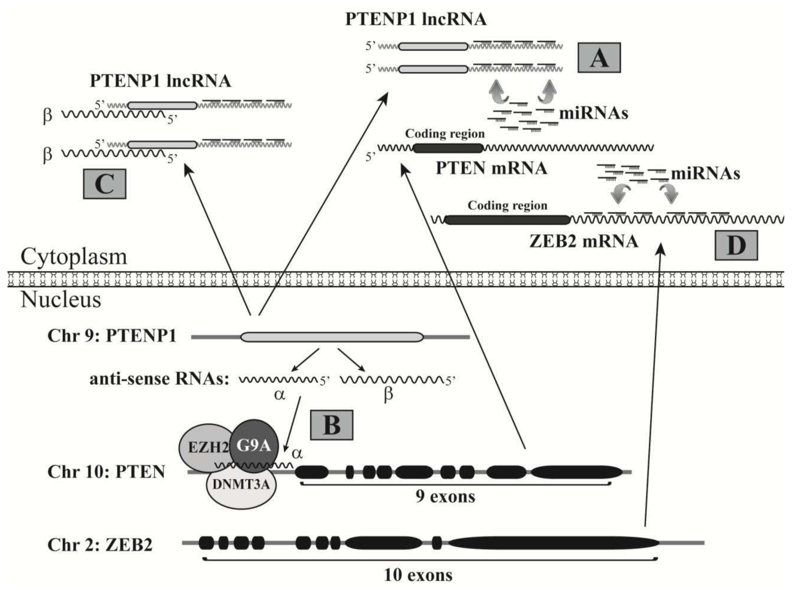
  161. Johnsson, P.; Ackley, A.; Vidarsdottir, L.; Lui, W.O.; Corcoran, M.; Grander, D.; Morris, K.V. A pseudogene long-noncoding-RNA network regulates PTEN transcription and translation in human cells. Nat. Struct. Mol. Biol 2013, 20, 440–446. [Google Scholar]
  162. Bond, A.M.; Vangompel, M.J.; Sametsky, E.A.; Clark, M.F.; Savage, J.C.; Disterhoft, J.F.; Kohtz, J.D. Balanced gene regulation by an embryonic brain ncRNA is critical for adult hippocampal GABA circuitry. Nat. Neurosci 2009, 12, 1020–1027. [Google Scholar]
  163. Huarte, M.; Guttman, M.; Feldser, D.; Garber, M.; Koziol, M.J.; Kenzelmann-Broz, D.; Khalil, A.M.; Zuk, O.; Amit, I.; Rabani, M.; et al. A large intergenic noncoding RNA induced by p53 mediates global gene repression in the p53 response. Cell 2010, 142, 409–419. [Google Scholar]
  164. Sheik Mohamed, J.; Gaughwin, P.M.; Lim, B.; Robson, P.; Lipovich, L. Conserved long noncoding RNAs transcriptionally regulated by Oct4 and Nanog modulate pluripotency in mouse embryonic stem cells. RNA 2010, 16, 324–337. [Google Scholar]
  165. Ebert, M.S.; Neilson, J.R.; Sharp, P.A. MicroRNA sponges: Competitive inhibitors of small RNAs in mammalian cells. Nat. Methods 2007, 4, 721–726. [Google Scholar]
  166. Pesole, G.; Liuni, S.; Grillo, G.; Saccone, C. Structural and compositional features of untranslated regions of eukaryotic mRNAs. Gene 1997, 205, 95–102. [Google Scholar]
  167. Huse, J.T.; Brennan, C.; Hambardzumyan, D.; Wee, B.; Pena, J.; Rouhanifard, S.H.; Sohn-Lee, C.; le Sage, C.; Agami, R.; Tuschl, T.; et al. The PTEN-regulating microRNA miR-26a is amplified in high-grade glioma and facilitates gliomagenesis in vivo. Genes Dev 2009, 23, 1327–1337. [Google Scholar]
  168. Liu, Z.L.; Wang, H.; Liu, J.; Wang, Z.X. MicroRNA-21 (miR-21) expression promotes growth, metastasis, and chemo- or radioresistance in non-small cell lung cancer cells by targeting PTEN. Mol. Cell. Biochem 2013, 372, 35–45. [Google Scholar]
  169. Vanin, E.F. Processed pseudogenes: Characteristics and evolution. Annu. Rev. Genet 1985, 19, 253–272. [Google Scholar]
  170. Fujii, G.H.; Morimoto, A.M.; Berson, A.E.; Bolen, J.B. Transcriptional analysis of the PTEN/MMAC1 pseudogene, psiPTEN. Oncogene 1999, 18, 1765–1769. [Google Scholar]
  171. Poliseno, L.; Salmena, L.; Zhang, J.; Carver, B.; Haveman, W.J.; Pandolfi, P.P. A coding-independent function of gene and pseudogene mRNAs regulates tumour biology. Nature 2010, 465, 1033–1038. [Google Scholar]
  172. Lund, A.H.; van Lohuizen, M. Epigenetics and cancer. Genes Dev 2004, 18, 2315–2335. [Google Scholar]
  173. Karreth, F.A.; Tay, Y.; Perna, D.; Ala, U.; Tan, S.M.; Rust, A.G.; DeNicola, G.; Webster, K.A.; Weiss, D.; Perez-Mancera, P.A.; et al. In vivo identification of tumor- suppressive PTEN ceRNAs in an oncogenic BRAF-induced mouse model of melanoma. Cell 2011, 147, 382–395. [Google Scholar]
  174. Faghihi, M.A.; Modarresi, F.; Khalil, A.M.; Wood, D.E.; Sahagan, B.G.; Morgan, T.E.; Finch, C.E.; St Laurent, G., 3rd; Kenny, P.J.; Wahlestedt, C. Expression of a noncoding RNA is elevated in Alzheimer’s disease and drives rapid feed-forward regulation of beta-secretase. Nat. Med. 2008, 14, 723–730. [Google Scholar]
  175. Cesana, M.; Cacchiarelli, D.; Legnini, I.; Santini, T.; Sthandier, O.; Chinappi, M.; Tramontano, A.; Bozzoni, I. A long noncoding RNA controls muscle differentiation by functioning as a competing endogenous RNA. Cell 2011, 147, 358–369. [Google Scholar]
  176. Nakagawa, S.; Ip, J.Y.; Shioi, G.; Tripathi, V.; Zong, X.; Hirose, T.; Prasanth, K.V. Malat1 is not an essential component of nuclear speckles in mice. RNA 2012, 18, 1487–1499. [Google Scholar]
  177. Zhang, A.; Zhou, N.; Huang, J.; Liu, Q.; Fukuda, K.; Ma, D.; Lu, Z.; Bai, C.; Watabe, K.; Mo, Y.Y. The human long non-coding RNA-RoR is a p53 repressor in response to DNA damage. Cell Res 2013, 23, 340–350. [Google Scholar]
  178. Yoon, J.H.; Abdelmohsen, K.; Srikantan, S.; Yang, X.; Martindale, J.L.; De, S.; Huarte, M.; Zhan, M.; Becker, K.G.; Gorospe, M. LincRNA-p21 suppresses target mRNA translation. Mol. Cell 2012, 47, 648–655. [Google Scholar]
  179. Azzalin, C.M.; Reichenbach, P.; Khoriauli, L.; Giulotto, E.; Lingner, J. Telomeric repeat containing RNA and RNA surveillance factors at mammalian chromosome ends. Science 2007, 318, 798–801. [Google Scholar]
  180. Redon, S.; Reichenbach, P.; Lingner, J. The non-coding RNA TERRA is a natural ligand and direct inhibitor of human telomerase. Nucleic Acids Res 2010, 38, 5797–5806. [Google Scholar]
  181. Wang, X.; Arai, S.; Song, X.; Reichart, D.; Du, K.; Pascual, G.; Tempst, P.; Rosenfeld, M.G.; Glass, C.K.; Kurokawa, R. Induced ncRNAs allosterically modify RNA-binding proteins in cis to inhibit transcription. Nature 2008, 454, 126–130. [Google Scholar]
  182. Jauliac, S.; Lopez-Rodriguez, C.; Shaw, L.M.; Brown, L.F.; Rao, A.; Toker, A. The role of NFAT transcription factors in integrin-mediated carcinoma invasion. Nat. Cell Biol 2002, 4, 540–544. [Google Scholar]
  183. Yoeli-Lerner, M.; Yiu, G.K.; Rabinovitz, I.; Erhardt, P.; Jauliac, S.; Toker, A. Akt blocks breast cancer cell motility and invasion through the transcription factor NFAT. Mol. Cell 2005, 20, 539–550. [Google Scholar]
  184. Willingham, A.T.; Orth, A.P.; Batalov, S.; Peters, E.C.; Wen, B.G.; Aza-Blanc, P.; Hogenesch, J.B.; Schultz, P.G. A strategy for probing the function of noncoding RNAs finds a repressor of NFAT. Science 2005, 309, 1570–1573. [Google Scholar]
  185. Sharma, S.; Findlay, G.M.; Bandukwala, H.S.; Oberdoerffer, S.; Baust, B.; Li, Z.; Schmidt, V.; Hogan, P.G.; Sacks, D.B.; Rao, A. Dephosphorylation of the nuclear factor of activated T cells (NFAT) transcription factor is regulated by an RNA-protein scaffold complex. Proc. Natl. Acad. Sci. USA 2011, 108, 11381–11386. [Google Scholar]
  186. Sanger, H.L.; Klotz, G.; Riesner, D.; Gross, H.J.; Kleinschmidt, A.K. Viroids are single-stranded covalently closed circular RNA molecules existing as highly base-paired rod-like structures. Proc. Natl. Acad. Sci. USA 1976, 73, 3852–3856. [Google Scholar]
  187. Jeck, W.R.; Sorrentino, J.A.; Wang, K.; Slevin, M.K.; Burd, C.E.; Liu, J.; Marzluff, W.F.; Sharpless, N.E. Circular RNAs are abundant, conserved, and associated with ALU repeats. RNA 2013, 19, 141–157. [Google Scholar]
  188. Salzman, J.; Gawad, C.; Wang, P.L.; Lacayo, N.; Brown, P.O. Circular RNAs are the predominant transcript isoform from hundreds of human genes in diverse cell types. PLoS One 2012, 7, e30733. [Google Scholar]
  189. Capel, B.; Swain, A.; Nicolis, S.; Hacker, A.; Walter, M.; Koopman, P.; Goodfellow, P.; Lovell-Badge, R. Circular transcripts of the testis-determining gene Sry in adult mouse testis. Cell 1993, 73, 1019–1030. [Google Scholar]
  190. Hansen, T.B.; Jensen, T.I.; Clausen, B.H.; Bramsen, J.B.; Finsen, B.; Damgaard, C.K.; Kjems, J. Natural RNA circles function as efficient microRNA sponges. Nature 2013, 495, 384–388. [Google Scholar]
  191. Hansen, T.B.; Wiklund, E.D.; Bramsen, J.B.; Villadsen, S.B.; Statham, A.L.; Clark, S.J.; Kjems, J. miRNA-dependent gene silencing involving Ago2-mediated cleavage of a circular antisense RNA. EMBO J 2011, 30, 4414–4422. [Google Scholar]
Figure 1. MicroRNA biogenesis and their role in inhibiting gene expression. A miRNA is transcribed by RNA polymerase II (Pol II) mediated by other transcription factors, such as p53 and c-Myc. The generated pri-miRNA is processed by the DROSHA complex to become a pre-miRNA, which is transported from nucleus to cytoplasm with a complex consisting of Exportin-5 (Exp-5) and Ran-GTP. In cytoplasm, the pre-miRNA is further processed by a Dicer-1 complex to become a duplex that consists of a guide strand (to become a mature miRNA) and a passenger strand. The guide strand associates with the RNA-induced silencing complex (RISC) and guides it to the target site on the 3′-UTR of an mRNA to inhibit the mRNA translation and cause mRNA degradation. For some miRNA-duplexes post Dicer-1 processing, both strands can become mature miRNAs; the one at the 5′-end of pre-miRNA is suffixed by “-5p”, while the 3′-end one is suffixed by “-3”, such as miR-17-5p and miR-17-3p.
Ijms 14 18319f1
Figure 2. The interplay between miRNAs and p53. While p53 regulates the gene expression of many miRNAs, its expression is also inhibited by miRNAs. When cells are exposed to genotoxic stresses, p53 activates four miRNAs that repress Mdm2 expression, which leads to p53 accumulation and cell cycle arrest or apoptosis. P53 protein also associates with the DROSHA complex to directly regulate miRNA maturation.
Ijms 14 18319f2
Figure 3. The interplay between miRNAs and c-Myc. While c-Myc regulates the expression of multiple miRNAs, its expression is inhibited by different miRNAs. C-Myc also activates the gene expression of DROSHA to directly promote miRNA procession.
Ijms 14 18319f3
Figure 4. PTEN expression is regulated by multiple ncRNAs. (A) The PTEN pseudogene, PTENP1, can transcribe into the PTENP1 lncRNA that acts as a decoy to sponge the miRNAs targeting at the 3′-UTR of the PTEN mRNA; (B,C) The locus of the PTEN pseudogene can also transcribe from the reverse direction to make two antisense RNA (asRNA) isoforms, α and β; (B) The α asRNA isoform binds the PTEN promoter and recruits DNMT3A, EZH2 and G9A, which causes epigenetic silencing of the PTEN gene; (C) The β asRNA isoform associates with the PTENP1 lncRNA to increase its stability and miRNA decoy activity; (D) The ZEB2 mRNA has a long 3′-UTR with many binding sites of miRNAs that also potentially target the PTEN 3′-UTR. Thus, the ZEB2 mRNA acts as a decoy to sponge many miRNAs and consequently promotes PTEN expression.
Ijms 14 18319f4

