Cytotoxicity Evaluation of a New Set of 2-Aminobenzo[de]iso-quinoline-1,3-diones


Abstract
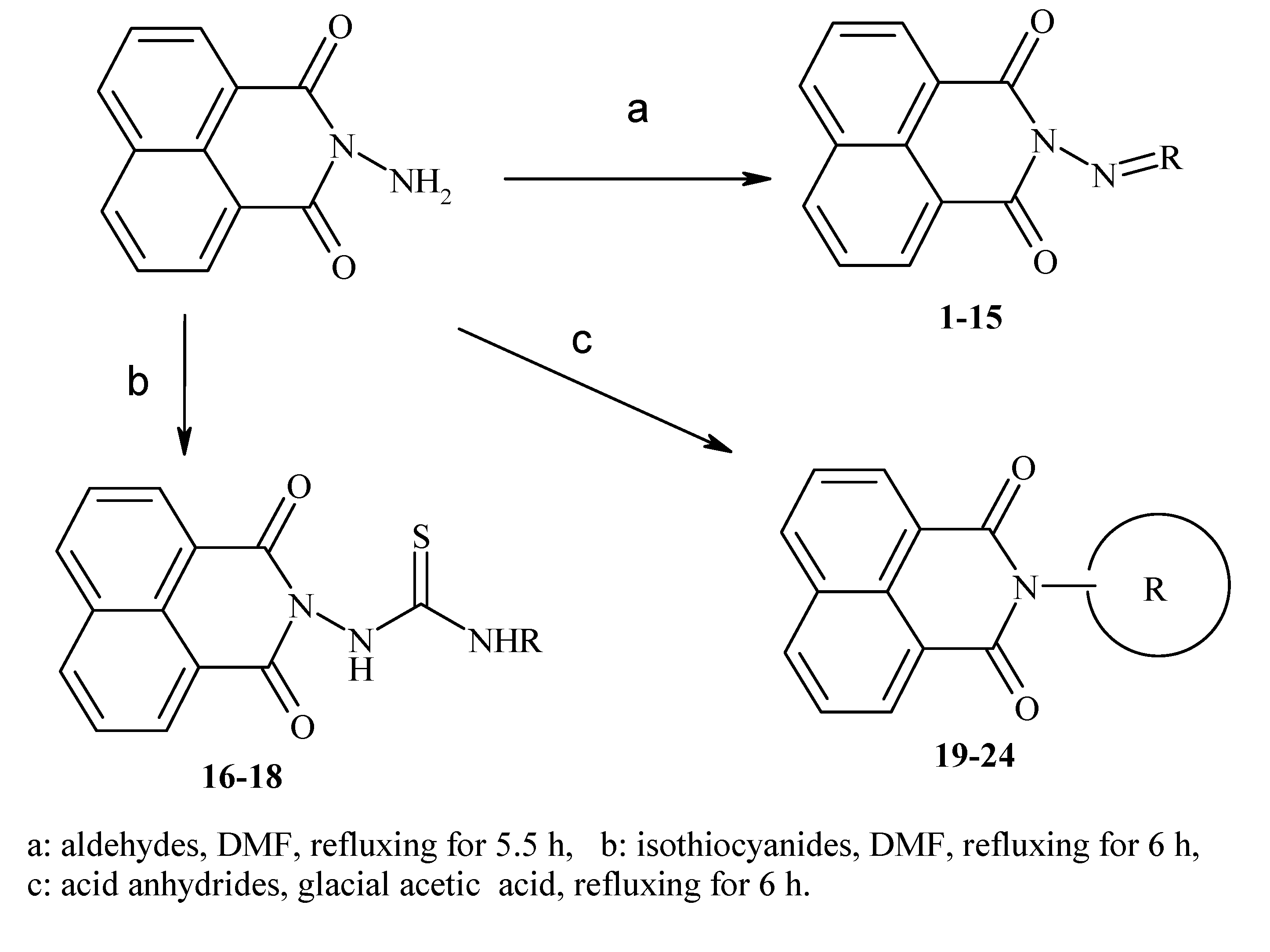
:1. Introduction
2. Results

| Compound Number | R | Compound Number | R |
|---|---|---|---|
| 1 |  | 13 |  |
| 2 |  | 14 |  |
| 3 |  | 15 |  |
| 4 |  | 16 |  |
| 5 |  | 17 |  |
| 6 |  | 18 |  |
| 7 |  | 19 |  |
| 8 |  | 20 |  |
| 9 |  | 21 |  |
| 10 |  | 22 |  |
| 11 |  | 23 |  |
| 12 |  | 24 |  |

3. Discussion
| Compound Number | IC50 (µg/mL) | ||
|---|---|---|---|
| HCT-116 | Hep-G2 | MCF-7 | |
| 1 | 37.3 | 37.0 | 42.1 |
| 2 | 41.9 | 35.4 | 33.8 |
| 3 | 27.9 | 14.8 ** | 30.4 |
| 4 | 16.3 ** | 19.7 * | 17.9 * |
| 5 | 36.1 | 30.9 | 30.1 |
| 6 | 31.2 | 24.4 * | 27.9 |
| 7 | 16.9 ** | 12.2 ** | 25.8 |
| 8 | 45.0 | 47.1 | 43.5 |
| 9 | 20.3 * | 21.2 * | 11.2 ** |
| 10 | 18.2 * | 17.0 * | 19.1 * |
| 11 | 40.2 | 47.6 | 35.0 |
| 12 | 41.1 | 44.1 | 38.2 |
| 13 | 40.0 | >50 | 19.7 * |
| 14 | 3.5 *** | 2.5 *** | 1.3 *** |
| 15 | 3.1 *** | 2.7 *** | 3.7 *** |
| 16 | 6.7 *** | 8.3 *** | 6.1 *** |
| 17 | 16.7 ** | 20.6 | 16.3 ** |
| 18 | 41.2 | 43.6 | 40.4 |
| 19 | 35.9 | 39.4 | 23.7 * |
| 20 | 24.7 * | 19.0 * | 23.7 * |
| 21 | 11.0 ** | 19.8 * | 5.0 *** |
| 22 | 5.3 *** | 4.4 *** | 3.5 *** |
| 23 | 23.3 * | 24.7 | 21.4 * |
| 24 | >50 | 27.9 * | >50 |
| Parent | >50 | >50 | >50 |
| Doxorubicin | 0.469 | 0.892 | 0.426 |
4. Experimental Section
4.1. Cell Lines
4.2. Evaluation of Antitumor Activity
4.3. Statistical Analysis
5. Conclusions
Acknowledgments
Author Contributions
Conflicts of Interest
References
- Xu, Y.; Qu, B.; Qian, X.; Li, Y. Five-member thio-heterocyclic fused naphthalimides with aminoalkyl side chains: Intercalation and photocleavage to DNA. Bioorg. Med. Chem. Lett. 2005, 15, 1139–1142. [Google Scholar]
- Mukherjee, A.; Dutta, S.; Shanmugavel, M.; Mondhe, D.M.; Sharma, P.R.; Singh, S.K.; Saxena, A.K.; Sanyal, U. 6-Nitro-2-(3-hydroxypropyl)-1H-benz[de]isoquinoline-1,3-dione, a potent antitumor agent, induces cell cycle arrest and apoptosis. J. Exp. Clin. Cancer Res. 2010, 29, 175–182. [Google Scholar]
- Mukherjee, A.; Hazra, S.; Dutta, S.; Muthiah, S.; Mondhe, D.M.; Sharma, P.R.; Singh, S.K.; Saxena, A.K.; Qazi, G.N.; Sanyal, U. Antitumor efficacy and apoptotic activity of substituted chloroalkyl 1H-Benzo[de]Isoquinoline-1,3-diones: A new class of potential antineoplastic agents. Investig. New Drugs 2011, 29, 434–442. [Google Scholar]
- Brana, M.F.; Castellano, J.M.; Moran, M.; Pérez de Vega, M.J.; Romerdahl, C.R.; Qian, X.D.; Bousquet, P.; Emling, F.; Schlick, E.; Keilhauer, G. Bis-naphthalimides: A new class of antitumor agents. Anti-Cancer Drug Des. 1993, 8, 257–268. [Google Scholar]
- Aibin, W.; Jide, L.L; Shaoxiong, Q.; Ping, M. Derivatives of 5-nitro-1H benzo[de]isoquinolin-1,3(2H)-dione: Design, synthesis, and biological activity. Monatsh. Chem. 2010, 141, 95–99. [Google Scholar]
- Qazi, G.N.; Saxena, A.K.; Muthiah, S.; Mondhe, D.M.; Sharma, P.R.; Singh, S.K.; Sanyal, U.; Mukherjee, A.; Hazra, S.; Dutta, S. Preparation of benzisoquinolinedione derivatives for us as antitumor agents. 17 July 2008. [Google Scholar]
- Cholody, W.M.; Kosakowska-Cholody, T.; Michejda, C.J. Preparation of 1,8-naphthalimido-linked imidazo[4,5,1-de]acridones as bis-intercalating antitumor agents. 13 September 2001. [Google Scholar]
- Vaisburg, A.; Bernstein, N.; Frechette, S.; Allan, M.; Abou-Khalil, E.; Leit, S.; Moradei, O.; Bouchain, G.; Wang, J.; Woo, S.H.; et al. (2-amino-phenyl)-amides of omega-substituted alkanoic acids as new histone deacetylase inhibitors. Bioorg. Med. Chem. Lett. 2004, 14, 283–287. [Google Scholar]
- Llombart, M.; Poved, A.; Forner, E.; Fernández-Martos, C.; Gaspar, C.; Muñoz, M.; Olmos, T.; Ruiz, A.; Soriano, V.; Benavides, A. Phase I study of mitonafide in solid tumors. Investig. New Drugs 1992, 10, 177–181. [Google Scholar]
- Leaf, A.N.; Neuberg, D.; Schwartz, E.L.; Wadler, S.; Ritch, P.S.; Dutcher, J.P.; Adams, G.L. An ECOG phase II study of amonafide in unresectable or recurrent carcinoma of the head and neck (PB390).Eastern Cooperative Oncology Group. Investig. New Drugs 1997, 15, 165–172. [Google Scholar]
- Kuran, B.; Krawiecka, M.; Kossakowski, J.; Szymanek, K.; Kierzkowska, M.; Mlynarczyk, G. Synthesis and antimicrobial activity of derivatives of 1H-benzo[de]isoquinoline-1,3(2H)-dione. Heterocycl. Commun. 2012, 18, 275–278. [Google Scholar]
- Lacivita, E.; Leopoldo, M.; Masotti, A.C.; Inglese, C.; Berardi, F.; Perrone, R.; Ganguly, S.; Jafurulla, M.; Chattopadhyay, A. Synthesis and characterization of environment-sensitive fluorescent ligands for human 5-HT1A receptors with 1-arylpiperazine structure. J. Med. Chem. 2009, 52, 7892–7896. [Google Scholar]
- Al-Salahi, R.; Marzouk, M. Synthesis of novel 2-aminobenzo[de]isoquinolin-1,3-dione derivatives. Asian J. Chem. 2014, 26, 2166–2172. [Google Scholar]
- Al-Salahi, R.; Geffken, D. Synthesis and reactivity of [1,2,4]triazolo-annelated quinazolines. Molecules 2010, 15, 7016–7034. [Google Scholar]
- Al-Salahi, R.; Marzouk, M.; Ashour, A.; Alswaidan, I. Synthesis and antitumor activity of 1,2,4-triazolo[1,5-a]quinazolines. Asian J. Chem. 2014, 26, 2173–2176. [Google Scholar]
- Mothana, R.A.; Gruenert, R.; Lindequist, U.; Bednarski, P.J. Study of the anticancer potential of Yemeni plants used in folk medicine. Pharmazie 2007, 62, 305–307. [Google Scholar]
- Bajbouj, K.; Schulze-Luehrmann, J.; Diermeier, S.; Amin, A.; Schneider-Stock, R. The anticancer effect of saffron in two p53 isogenic colorectal cancer cell lines. BMC Complement. Altern. Med. 2012, 12, 69. [Google Scholar]
- Ušaj, M.; Trontelj, K.; Hudej, R.; Kandušer, M.; Miklavčič, D. Cell size dynamics and viability of cells exposed to hypotonic treatment and electroporation for electrofusion optimization. Radiol. Oncol. 2009, 43, 108–119. [Google Scholar]
- Bernhardt, G.; Reile, H.; Birnboeck, H.; Spruss, T.; Schoenenberger, H. Standardized kinetic microassay to quantify differential chemosensitivity on the basis of proliferative activity. J. Cancer Res. Clin. Oncol. 1992, 118, 35–43. [Google Scholar]
- Ait Mbarek, L.; Ait Mouse, H.; Elabbadi, N.; Bensalah, M.; Gamouh, A.; Aboufatima, R.; Benharref, A.; Chait, A.; Kamal, M.; Dalal, A.; et al. Anti-tumor properties of blackseed (Nigella sativa L.) extracts. Braz. J. Med. Biol. Res. 2007, 40, 839–847. [Google Scholar]
- Vega-Avila1, E.; Pugsley, M.K. An overview of colorimetric assay methods used to assess survival or proliferation of mammalian cells. Proc. West. Pharmacol. Soc. 2011, 54, 10–14. [Google Scholar]
- Mosmann, T. Rapid colorimetric assay for cellular growth and survival: Application to proliferation and cytotoxicity assays. J. Immunol. Methods 1983, 65, 55–63. [Google Scholar]
- Gangadevi, V.; Muthumary, J. Preliminary studies on cytotoxic effect of fungal taxol on cancer cell lines. Afr. J. Biotechnol. 2007, 6, 1382–1386. [Google Scholar]
- Castilla-Serna, L.; Cravioto, J. Simply Statistic for Health Investigation, 1st ed.; Trillas: Mexico City, Mexico, 1999. [Google Scholar]
© 2014 by the authors; licensee MDPI, Basel, Switzerland. This article is an open access article distributed under the terms and conditions of the Creative Commons Attribution license (http://creativecommons.org/licenses/by/4.0/).
Share and Cite
Al-Salahi, R.; Alswaidan, I.; Marzouk, M. Cytotoxicity Evaluation of a New Set of 2-Aminobenzo[de]iso-quinoline-1,3-diones. Int. J. Mol. Sci. 2014, 15, 22483-22491. https://doi.org/10.3390/ijms151222483
Al-Salahi R, Alswaidan I, Marzouk M. Cytotoxicity Evaluation of a New Set of 2-Aminobenzo[de]iso-quinoline-1,3-diones. International Journal of Molecular Sciences. 2014; 15(12):22483-22491. https://doi.org/10.3390/ijms151222483
Chicago/Turabian StyleAl-Salahi, Rashad, Ibrahim Alswaidan, and Mohamed Marzouk. 2014. "Cytotoxicity Evaluation of a New Set of 2-Aminobenzo[de]iso-quinoline-1,3-diones" International Journal of Molecular Sciences 15, no. 12: 22483-22491. https://doi.org/10.3390/ijms151222483