Article

Fatty Acid Elongation in Non-Alcoholic Steatohepatitis and Hepatocellular Carcinoma

1
Department of Pharmacy, Pharmaceutical Biology, Saarland University, Campus C2 2, 66123 Saarbrücken, Germany
2
Institute of Pathology, Medical University of Graz, 8010 Graz, Austria
3
Department of Pharmacy, Pharmaceutical Biotechnology, Saarland University and Helmholtz Institute for Pharmaceutical Research Saarland (HIPS), Helmholtz Centre for Infection Research (HZI), 66123 Saarbrücken, Germany
4
Systems Toxicology, Leibniz Research Centre for Working Environment and Human Factors (IfADo) at the TU Dortmund, Ardeystr. 67, 44139 Dortmund, Germany
5
Department of Medicine II, Saarland University Medical Center, Kirrberger Str., 66421 Homburg, Germany
6
Institute of Pathology, Saarland University, Kirrberger Str., 66421 Homburg, Germany
7
Center for Bioinformatics, Saarland University, Campus E2 1, 66123 Saarbrücken, Germany
8
Department of Molecular Cell Biology, Faculty of Medicine, Vrije Universiteit, Van der Boechorststraat 7, 1081 BT Amsterdam, The Netherlands
*
Author to whom correspondence should be addressed.
Int. J. Mol. Sci. 2014, 15(4), 5762-5773; https://doi.org/10.3390/ijms15045762
Submission received: 27 February 2014 / Revised: 17 March 2014 / Accepted: 21 March 2014 / Published: 4 April 2014
(This article belongs to the Special Issue Molecular Mechanisms of Human Liver Diseases)

Abstract

:
Non-alcoholic steatohepatitis (NASH) represents a risk factor for the development of hepatocellular carcinoma (HCC) and is characterized by quantitative and qualitative changes in hepatic lipids. Since elongation of fatty acids from C16 to C18 has recently been reported to promote both hepatic lipid accumulation and inflammation we aimed to investigate whether a frequently used mouse NASH model reflects this clinically relevant feature and whether C16 to C18 elongation can be observed in HCC development. Feeding mice a methionine and choline deficient diet to model NASH not only increased total hepatic fatty acids and cholesterol, but also distinctly elevated the C18/C16 ratio, which was not changed in a model of simple steatosis (ob/ob mice). Depletion of Kupffer cells abrogated both quantitative and qualitative methionine-and-choline deficient (MCD)-induced alterations in hepatic lipids. Interestingly, mimicking inflammatory events in early hepatocarcinogenesis by diethylnitrosamine-induced carcinogenesis (48 h) increased hepatic lipids and the C18/C16 ratio. Analyses of human liver samples from patients with NASH or NASH-related HCC showed an elevated expression of the elongase ELOVL6, which is responsible for the elongation of C16 fatty acids. Taken together, our findings suggest a detrimental role of an altered fatty acid pattern in the progression of NASH-related liver disease.

1. Introduction

Non-alcoholic fatty liver disease (NAFLD) is regarded as the most common liver disorder [1]. Although NAFLD is often an asymptomatic disease and therefore difficult to detect, the prevalence appears to be around 20%–35% of the adult population in Western countries [24]. NAFLD/NASH (non-alcoholic steatohepatitis) strongly correlates with characteristics of the metabolic syndrome, such as obesity and diabetes mellitus, and NAFLD/NASH [57]. Liver pathogenesis of NAFLD is widely believed to start with simple steatosis, which is characterized by excessive lipid accumulation [2,8]. The progression from simple steatosis to NASH is mediated by the release of inflammatory cytokines [9] and can result in hepatic cirrhosis and finally in hepatocellular carcinoma (HCC) [10]. Due to this inflammatory environment 4% to 27% of individuals with NASH and cirrhosis [11] develop HCC.
There is increasing evidence that in steatosis besides the total amount of accumulated lipids the composition of lipids has an impact on pathophysiology [12,13]. In fact, human NAFLD is characterized by numerous changes in hepatic lipid composition and free fatty acid ratios [14,15]. Viral hepatitis has also been described to lead to strongly altered hepatic lipid content and composition [1619]. In HCC a decreased stearic acid (C18:0) to oleic acid (C18:1) ratio compared to normal tissue has been reported, suggesting the importance of desaturation in HCC development [20]. Regarding changes in hepatic fatty acid pattern it is important to note that the ELOVL fatty acid elongase 6 (ELOVL6), which catalyzes the elongation of C16 to C18 fatty acids [21], has been shown to promote NASH [22,23]. A role of ELOVL6 in murine NASH-related HCC has recently been suggested, but still remains unclear in human NASH-associated HCC [23].
The aims of our study were to investigate the occurrence of fatty acid elongation in lipid metabolism in different NAFLD mouse models and to elucidate its relevance in human NASH and NASH-associated HCC.

2. Results and Discussion

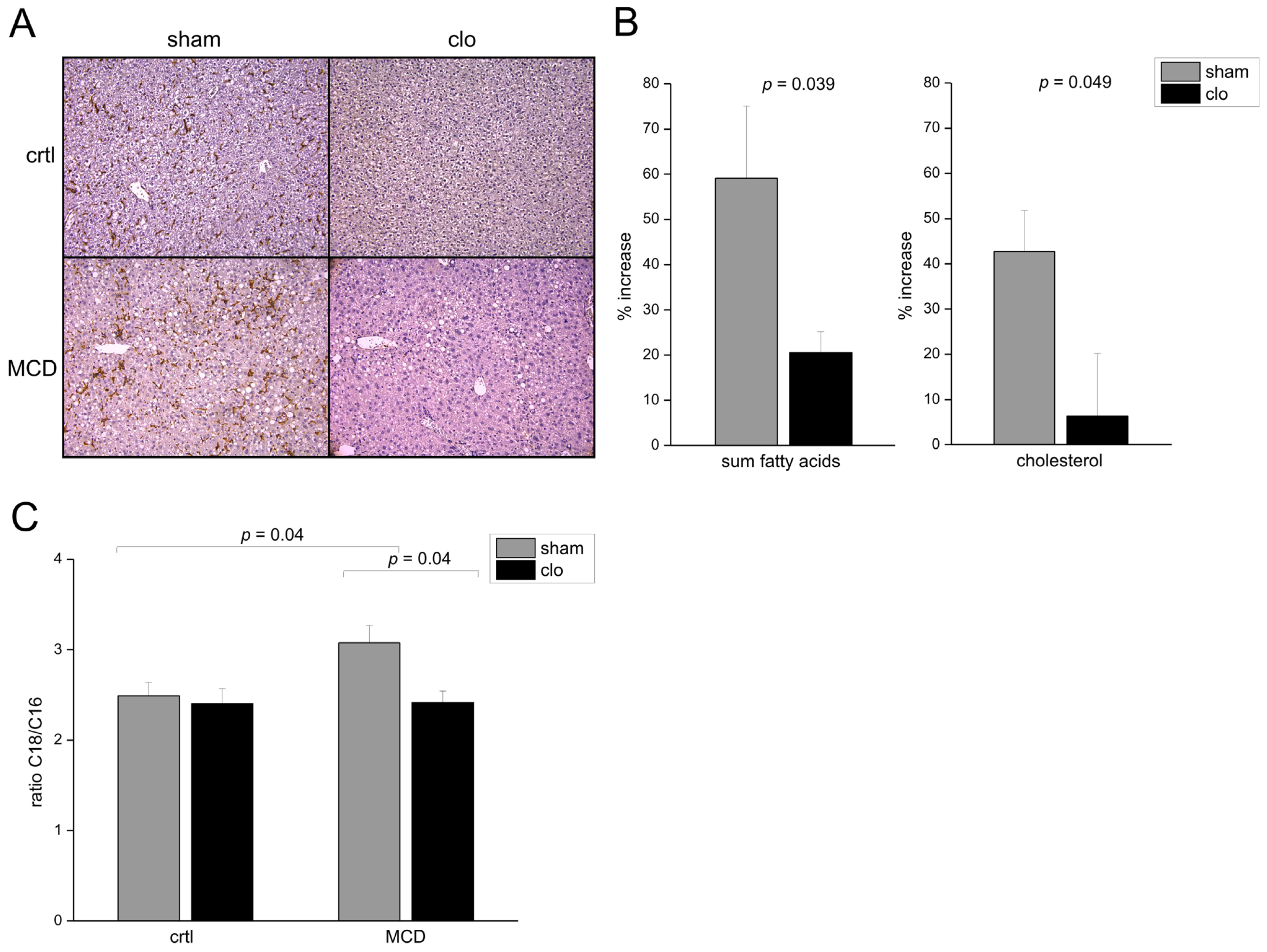
2.1. Fatty Acid Elongation in Murine Non-Alcoholic Steatohepatitis (NASH) Is Kupffer Cell Dependent

Fatty acid elongation plays a role in murine and human NASH development [22,23]. Feeding mice a methionine-choline deficient diet (MCD) led to increased total fatty acid and cholesterol levels and profound inflammation (Figure 1A,B). Having a closer look at the composition of the fatty acids, we observed that the chain length of fatty acids was different in MCD-fed livers compared to tissues from a control diet: NASH livers exhibited an increased ratio of C18 to C16 fatty acids (Figure 1B). Increased ELOVL6 mRNA expression has been observed in NASH animal models, such as low-density lipoprotein receptor knockout animals fed a western type diet [23] or a fructose diet [24]. In order to study whether this increased fatty acid elongation from C16 to C18 could also be observed in simple steatosis we investigated the well-established leptin-deficiency (ob/ob) mouse model [24,25]. As expected, livers of ob/ob mice showed excessive hepatic lipid accumulation compared to lean controls (Figure 1C,D). However, no distinct inflammation was observed and the ratio of C18 to C16 fatty acids was not changed compared to wild-type animals (Figure 1D). Adipose tissue of ob/ob mice is known to show inflammation [26]. Concordantly, adipose tissue macrophages of ob/ob mice exhibit increased ELOVL6 mRNA expression [27]. Another study reported increased hepatic ELOVL6 expression in older ob/ob mice, whereby some of the animals were in a fasted condition [28]. The study does not contain any information on fatty acid composition.
Leroux et al. reported that lipid storage by Kupffer cells correlates with a pro-inflammatory phenotype in NASH [29]. In order to clarify whether the altered fatty acid elongation in murine NASH is due to co-existing inflammation, we depleted Kupffer cells by clodronate liposomes. This intervention is known to attenuate inflammatory and metabolic events in the MCD model [30]. In line with these findings we observed a strong decrease of hepatic lipid accumulation after Kupffer cell depletion (Figure 2A,B). Besides hepatocytes also macrophages are known to express ELOVL6 [27], which was shown to be relevant for lipid storage [31]. After Kupffer cell depletion the MCD-induced changes in C18 to C16 fatty acids and cholesterol were completely abrogated (Figure 2A,B). Kupffer cell depletion was confirmed by immunohistochemical F4/80 staining (Figure 2A).

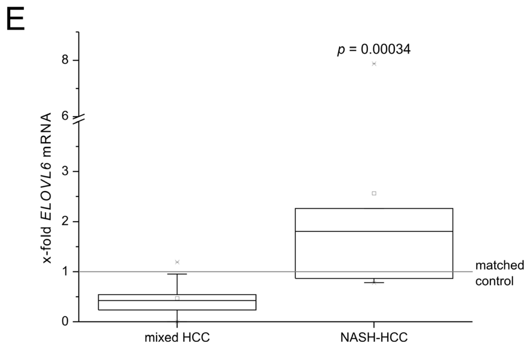
2.2. Role of Fatty Acid Elongation in NASH-Related Hepatocellular Carcinoma (HCC) and Human NASH

To further investigate the role of fatty acid elongation in hepatocarcinogenesis, we used short-term (48 h) treatment with the carcinogen diethylnitrosamine (DEN) to model early inflammatory events associated with hepatocarcinogenesis [32,33]. In fact, we histologically observed inflammatory foci in the livers exposed to DEN (Figure 3A). Little is known about increased lipid accumulation after DEN treatment: Histologically detected hepatic lipid deposition by DEN was reported in fish (Oryzias lapites) [34,35]. Changes in the lipid composition have been reported for cancerous tissues compared to normal tissue in DEN-induced hepatocarcinogenesis [3638], but not in the precancerous short-term protocol. We observed that inflammatory events were paralleled by distinct metabolic alterations similar to the murine NASH model: fatty acids, cholesterol levels, and the C18/C16 ratio were elevated upon 48 h DEN treatment (Figure 3B). This short-term model might therefore resemble NASH-related hepatocarcinogenesis. In later stages of DEN-induced carcinogenesis and during tumor progression we observed that fatty acid elongation was rather repressed [39]. This can be explained by the fact that the long-term DEN model predominantly acts via genotoxic effects of the carcinogen and therefore no NASH-related HCCs are induced.
In order to study the relevance of increased fatty acid elongation in human NASH and human NASH-related HCC, we analyzed the hepatic mRNA expression of the enzyme ELOVL6, which is responsible for the elongation of C16 fatty acids. We observed increased levels of ELOVL6 in NASH versus steatosis samples (GSE48452 [41]) (Figure 3C) as well as in NASH compared to healthy control tissues (GSE37031 [42]) (Figure 3D). Interestingly, expression of ELOVL6 was also altered in NASH-associated HCCs compared to HCC tissues of mixed etiology (Figure 3E): human NASH-related HCCs express increased levels of ELOVL6, indicating fatty acid elongation to play a critical role in this particular HCC subtype.

3. Experimental Section

3.1. Animals

All animal procedures were performed in accordance with the local animal welfare committee (#13/2009, 09/06/2009; #34/2010, 15/11/2010; Landesamt für Soziales, Gesundheit und Verbraucherschutz Saarland). Mice were kept under stable conditions regarding temperature, humidity, food delivery, and 12 h day/night rhythm. At the age of 3 weeks mice (DBA2/Bl6/J background) were fed either a methionine-choline deficient (MCD) or a methionine-choline supplemented control (crtl) diet for 3 weeks. Intraperitoneal clodronate or empty liposome injections [43] were started two days prior to MCD or control diet and repeated every five days to ensure Kupffer cell depletion. Leptin deficient mice ob/ob (Bl6:Cg-Lepob/J) and lean control mice (ob/+) were obtained from Charles River and sacrificed at an age of 10 weeks. DEN treatment of mice (DBA2/Bl6/J background) on regular chow was performed by a single intraperitoneal injection of 100 mg/kg body weight at the age of 2.5 weeks. Mice were sacrificed 48 h after DEN injection. Animals of all experimental groups were sacrificed in a non-fasted state.

3.2. Human Liver Tissue

Paraffin-embedded liver samples from randomly selected pseudonymized HCC patients who underwent liver resection at the Saarland University Medical Center between 2005 and 2010 were obtained as described previously [40]. The study protocol was approved by the local Ethics Committee (#47/07). Samples had a mixed etiology including NASH, alcoholic liver disease, viral hepatitis, hemochromatosis, porphyria, and cryptogenic [40].

3.3. Fatty Acid Measurement by Gas Chromatography-Mass Spectrometry (GC-MS)

Murine liver samples were lyophilized and analyzed according to Bode et al. [44]. In short, lyophilized samples were dissolved in a mixture of 500 μL methanol/toluene/sulfuric acid (50:50:2, v/v/v) and incubated at 55 °C overnight. Subsequently, 400 μL of a 0.5 M NH4CO3, 2 M KCl solution were added and samples were centrifuged. The organic phase was transferred into a new glass vial, derivatized with 25 μL N-methyl-N-(trimethylsilyl)trifluoroacetamide at 37 °C for 1 h. Fatty acid separation was performed on an Agilent 6890N gas chromatograph coupled to an Agilent 5973N mass selective detector and equipped with a non-polar J&WDB-5HT capillary column (Agilent Technologies, Böblingen, Germany). The column temperature was kept at 130 °C for 2.5 min, increased to 240 °C at a rate of 5 °C/min, and then ramped to 300 °C at 30 °C/min, and held at 300 °C for 5 min. Helium was used as the carrier gas at a flow rate of 1 mL/min. The mass selective detector was operated in scan mode, average spectra were acquired in the m/z range of 40–700 m/z and were recorded at a scan speed of 2.24 scans/s. Scan control, data acquisition, and processing were performed by MSD ChemStation (Agilent Technologies, Böblingen, Germany) and AMDIS software based on the fragmentation patterns and retention time, in comparison with the reference standards Supelco 37 Component FAME Mix (Sigma-Aldrich, Taufkirchen, Germany), and NIST 08 library. Methyl-nonadecanoate (74208, Sigma-Aldrich, Taufkirchen, Germany) was used as an internal standard. The method detects both free and bound fatty acids.

3.4. Histochemistry and Immunohistochemistry

Hematoxylin-eosin staining of paraffin-embedded tissues was performed as previously reported [45,46]. Immunohistochemical F4/80 detection was achieved using the Vectastain Peroxidase Elite ABC kit/DAB with anti-F4/80 antibody (AbD Serotec, Puchheim, Germany) 1:1000 overnight at 4 °C. Epitopes were demasked with citrate buffer pH 6.0 for 10 min in a waterbath at 95 °C.

3.5. Analysis of the Public Gene Omnibus (GEO) Datasets

Datasets GSE48452 and GSE37031 [41,42] normalized using log2-RMA and log2-GCRMA respectively, were downloaded from Gene omnibus (GEO) [47]. Dataset GSE48452 with samples from different stages of NAFLD contained 18 NASH and 14 steatosis samples while dataset GSE37031 included 8 NASH and 7 control samples. The statistical significance was determined by Kolmogorov-Smirnov test.

3.6. Statistical Analysis

Results are expressed as means ± SEM. The statistical significance was determined by independent two-sample t-test. Expression data of human tissues were analyzed using Mann-Whitney U tests. The results were considered as statistically significant when p value was less than 0.05.

4. Conclusions

In the present study, we identified that NASH-induced fatty acid elongation is an inflammation-associated pathophysiological step in liver disease. Furthermore, the fatty acid elongase ELOVL6 is elevated in human NASH and NASH-related HCC.

Acknowledgments

The project was funded, in part, by an EASL Dame Sheila Sherlock Fellowship, the research committee of Saarland University (61-cl/Anschub2012) (both to S.M.K.); and the Deutsche Forschungsgemeinschaft (DFG LA 997/7-1) (to F.L.). The results upon which this publication is based were partly funded by the Federal Ministry of Education and Research under the Project Number [O1KU1216F] (to K.G., C.C., J.G.H., and A.K.K.). The responsibility for the content of this publication lies with the author.

Conflicts of Interest

The authors declare no conflict of interest.
  • Author ContributionsS.M.K., Y.S., and A.K.K. designed experiments, analysed data and wrote the manuscript. A.K.K. initiated and directed the study. The others designed experiments and participated in data acquisition.

References

  1. Kanuri, G.; Bergheim, I. In vitro and in vivo models of non-alcoholic fatty liver disease (NAFLD). Int. J. Mol. Sci 2013, 14, 11963–11980. [Google Scholar]
  2. Adams, L.A.; Angulo, P.; Lindor, K.D. Nonalcoholic fatty liver disease. CMAJ 2005, 172, 899–905. [Google Scholar]
  3. Browning, J.D.; Szczepaniak, L.S.; Dobbins, R.; Nuremberg, P.; Horton, J.D.; Cohen, J.C.; Grundy, S.M.; Hobbs, H.H. Prevalence of hepatic steatosis in an urban population in the United States: Impact of ethnicity. Hepatology 2004, 40, 1387–1395. [Google Scholar]
  4. De Alwis, N.M.W.; Day, C.P. Non-alcoholic fatty liver disease: The mist gradually clears. J. Hepatol 2008, 48, S104–S112. [Google Scholar]
  5. Adams, L.A.; Waters, O.R.; Knuiman, M.W.; Elliott, R.R.; Olynyk, J.K. NAFLD as a risk factor for the development of diabetes and the metabolic syndrome: An eleven-year follow-up study. Am. J. Gastroenterol 2009, 104, 861–867. [Google Scholar]
  6. Ascha, M.S.; Hanouneh, I.A.; Lopez, R.; Tamimi, T.A.R.; Feldstein, A.F.; Zein, N.N. The incidence and risk factors of hepatocellular carcinoma in patients with nonalcoholic steatohepatitis. Hepatology 2010, 51, 1972–1978. [Google Scholar]
  7. Fabbrini, E.; Sullivan, S.; Klein, S. Obesity and nonalcoholic fatty liver disease: Biochemical, metabolic, and clinical implications. Hepatology 2010, 51, 679–689. [Google Scholar]
  8. Angulo, P. Medical progress: Nonalcoholic fatty liver disease. N. Engl. J. Med 2002, 346, 1221–1231. [Google Scholar]
  9. Day, C.P. Genetic and environmental susceptibility to non-alcoholic fatty liver disease. Dig. Dis 2006, 28, 255–260. [Google Scholar]
  10. Malhi, H.; Gores, G.J. Molecular mechanisms of lipotoxicity in nonalcoholic fatty liver disease. Semin. Liver Dis 2008, 28, 360–369. [Google Scholar]
  11. Cohen, J.C.; Horton, J.D.; Hobbs, H.H. Human fatty liver disease: Old questions and new insights. Science 2011, 332, 1519–1523. [Google Scholar]
  12. Alkhouri, N.; Dixon, L.J.; Feldstein, A.E. Lipotoxicity in nonalcoholic fatty liver disease: Not all lipids are created equal. Expert Rev. Gastroenterol. Hepatol 2009, 3, 445–451. [Google Scholar]
  13. Takaki, A.; Kawai, D.; Yamamoto, K. Multiple hits, including oxidative stress, as pathogenesis and treatment target in non-alcoholic steatohepatitis (NASH). Int. J. Mol. Sci 2013, 14, 20704–20728. [Google Scholar]
  14. Puri, P.; Baillie, R.A.; Wiest, M.M.; Mirshahi, F.; Choudhury, J.; Cheung, O.; Sargeant, C.; Contos, M.J.; Sanyal, A.J. A lipidomic analysis of nonalcoholic fatty liver disease. Hepatology 2007, 46, 1081–1090. [Google Scholar]
  15. Puri, P.; Wiest, M.M.; Cheung, O.; Mirshahi, F.; Sargeant, C.; Min, H.K.; Contos, M.J.; Sterling, R.K.; Fuchs, M.; Zhou, H.; et al. The plasma lipidomic signature of nonalcoholic steatohepatitis. Hepatology 2009, 50, 1827–1838. [Google Scholar]
  16. Kim, K.H.; Shin, H.; Kim, K.; Choi, H.M.; Rhee, S.H.; Moon, H.; Kim, H.H.; Yang, U.S.; Yu, D.; Cheong, J. Hepatitis B virus X protein induces hepatic steatosis via transcriptional activation of SREBP1 and PPARγ. Gastroenterology 2007, 132, 1955–1967. [Google Scholar]
  17. Miyoshi, H.; Moriya, K.; Tsutsumi, T.; Shinzawa, S.; Fujie, H.; Shintani, Y.; Fujinaga, H.; Goto, K.; Todoroki, T.; Suzuki, T.; et al. Pathogenesis of lipid metabolism disorder in hepatitis C: Polyunsaturated fatty acids counteract lipid alterations induced by the core protein. J. Hepatol 2011, 54, 432–438. [Google Scholar]
  18. Moriya, K.; Todoroki, T.; Tsutsumi, T.; Fujie, H.; Shintani, Y.; Miyoshi, H.; Ishibashi, K.; Takayama, T.; Makuuchi, M.; Watanabe, K.; et al. Increase in the concentration of carbon 18 monounsaturated fatty acids in the liver with hepatitis C: Analysis in transgenic mice and humans. Biochem. Biophys. Res. Commun 2001, 281, 1207–1212. [Google Scholar]
  19. Yamaguchi, A.; Tazuma, S.; Nishioka, T.; Ohishi, W.; Hyogo, H.; Nomura, S.; Chayama, K. Hepatitis C virus core protein modulates fatty acid metabolism and thereby causes lipid accumulation in the liver. Dig. Dis. Sci 2005, 50, 1361–1371. [Google Scholar]
  20. Wood, C.B.; Habib, N.A.; Apostolov, K. Reduction in the stearic to oleic acid ratio in human malignant liver neoplasms. Eur. J. Surg. Oncol 1985, 11, 347–348. [Google Scholar]
  21. Matsuzaka, T.; Shimano, H.; Yahagi, N.; Kato, T.; Atsumi, A.; Yamamoto, T.; Inoue, N.; Ishikawa, M.; Okada, S.; Ishigaki, N. Crucial role of a long-chain fatty acid elongase, ELOVL6, in obesity-induced insulin resistance. Nat. Med 2007, 13, 1193–1202. [Google Scholar]
  22. Matsuzaka, T.; Atsumi, A.; Matsumori, R.; Nie, T.; Shinozaki, H.; Suzuki-Kemuriyama, N.; Kuba, M.; Nakagawa, Y.; Ishii, K.; Shimada, M. Elovl6 promotes nonalcoholic steatohepatitis. Hepatology 2012, 56, 2199–2208. [Google Scholar]
  23. Muir, K.; Hazim, A.; He, Y.; Peyressatre, M.; Kim, D.Y.; Song, X.; Beretta, L. Proteomic and lipidomic signatures of lipid metabolism in NASH-associated hepatocellular carcinoma. Cancer Res 2013, 73, 4722–4731. [Google Scholar]
  24. Imajo, K.; Yoneda, M.; Kessoku, T.; Ogawa, Y.; Maeda, S.; Sumida, Y.; Hyogo, H.; Eguchi, Y.; Wada, K.; Nakajima, A. Rodent models of nonalcoholic fatty liver disease/nonalcoholic steatohepatitis. Int. J. Mol. Sci 2013, 14, 21833–21857. [Google Scholar]
  25. Trak-Smayra, V.; Paradis, V.; Massart, J.; Nasser, S.; Jebara, V.; Fromenty, B. Pathology of the liver in obese and diabetic ob/ob and db/db mice fed a standard or high-calorie diet. Int. J. Exp. Pathol 2011, 92, 413–421. [Google Scholar]
  26. Xu, H.; Barnes, G.T.; Yang, Q.; Tan, G.; Yang, D.; Chou, C.J.; Sole, J.; Nichols, A.; Ross, J.S.; Tartaglia, L.A.; et al. Chronic inflammation in fat plays a crucial role in the development of obesity-related insulin resistance. J. Clin. Investig 2003, 112, 1821–1830. [Google Scholar]
  27. Prieur, X.; Mok, C.Y.L.; Velagapudi, V.R.; Núnez, V.; Fuentes, L.; Montaner, D.; Ishikawa, K.; Camacho, A.; Barbarroja, N.; O’Rahilly, S.; et al. Differential lipid partitioning between adipocytes and tissue macrophages modulates macrophage lipotoxicity and M2/M1 polarization in obese mice. Diabetes 2011, 60, 797–809. [Google Scholar]
  28. Matsuzaka, T.; Shimano, H.; Yahagi, N.; Yoshikawa, T.; Amemiya-Kudo, M.; Hasty, A.H.; Okazaki, H.; Tamura, Y.; Iizuka, Y.; Ohashi, K.; et al. Cloning and characterization of a mammalian fatty acyl-CoA elongase as a lipogenic enzyme regulated by SREBPs. J. Lipid Res 2002, 43, 911–920. [Google Scholar]
  29. Leroux, A.; Ferrere, G.; Godie, V.; Cailleux, F.; Renoud, M.L.; Gaudin, F.; Naveau, S.; Prévot, S.; Makhzami, S.; Perlemuter, G.; et al. Toxic lipids stored by Kupffer cells correlates with their pro-inflammatory phenotype at an early stage of steatohepatitis. J. Hepatol 2012, 57, 141–149. [Google Scholar]
  30. Vonghia, L.; Michielsen, P.; Francque, S. Immunological mechanisms in the pathophysiology of non-alcoholic steatohepatitis. Int. J. Mol. Sci 2013, 14, 19867–19890. [Google Scholar]
  31. Saito, R.; Matsuzaka, T.; Karasawa, T.; Sekiya, M.; Okada, N.; Igarashi, M.; Matsumori, R.; Ishii, K.; Nakagawa, Y.; Iwasaki, H.; et al. Macrophage Elovl6 deficiency ameliorates foam cell formation and reduces atherosclerosis in low-density lipoprotein receptor-deficient mice. Arterioscler. Thromb. Vasc. Biol 2011, 31, 1973–1979. [Google Scholar]
  32. Naugler, W.E.; Sakurai, T.; Kim, S.; Maeda, S.; Kim, K.; Elsharkawy, A.M.; Karin, M. Gender disparity in liver cancer due to sex differences in MyD88-dependent IL-6 production. Science 2007, 317, 121–124. [Google Scholar]
  33. Park, E.J.; Lee, J.H.; Yu, G.Y.; He, G.; Ali, S.R.; Holzer, R.G.; Österreicher, C.H.; Takahashi, H.; Karin, M. Dietary and genetic obesity promote liver inflammation and tumorigenesis by enhancing IL-6 and TNF expression. Cell 2010, 140, 197–208. [Google Scholar]
  34. Braunbeck, T.A.; Teh, S.J.; Lester, S.M.; Hinton, D.E. Ultrastructural alterations in liver of medaka (Oryzias latipes) exposed to diethylnitrosamine. Toxicol. Pathol 1992, 20, 179–196. [Google Scholar]
  35. Laurén, D.J.; Teh, S.J.; Hinton, D.E. Cytotoxicity phase of diethylnitrosamine-induced hepatic neoplasia in medaka. Cancer Res 1990, 50, 5504–5514. [Google Scholar]
  36. Abel, S.; Smuts, C.M.; de Villiers, C.; Gelderblom, W.C.A. Changes in essential fatty acid patterns associated with normal liver regeneration and the progression of hepatocyte nodules in rat hepatocarcinogenesis. Carcinogenesis 2001, 22, 795–804. [Google Scholar]
  37. Canuto, R.A.; Biocca, M.E.; Muzio, G.; Dianzani, M.U. Fatty acid composition of phospholipids in mitochondria and microsomes during diethylnitrosamine carcinogenesis in rat liver. Cell Biochem. Funct 1989, 7, 11–19. [Google Scholar]
  38. Yoshimura, K.; Mandal, M.K.; Hara, M.; Fujii, H.; Chen, L.C.; Tanabe, K.; Hiraoka, K.; Takeda, S. Real-time diagnosis of chemically induced hepatocellular carcinoma using a novel mass spectrometry-based technique. Anal. Biochem 2013, 441, 32–37. [Google Scholar]
  39. Kessler, S.M.; Laggai, S.; Barghash, A.; Helms, V.; Kiemer, A.K. Lipid metabolism signatures in NASH-associated HCC. Cancer Res 2014. [Google Scholar] [CrossRef]
  40. Kessler, S.M.; Pokorny, J.; Zimmer, V.; Laggai, S.; Lammert, F.; Bohle, R.M.; Kiemer, A.K. IGF2 mRNA binding protein p62/IMP2-2 in hepatocellular carcinoma: Antiapoptotic action is independent of IGF2/PI3K signaling. Am. J. Physiol 2013, 304, G328–G336. [Google Scholar]
  41. Ahrens, M.; Ammerpohl, O.; von Schönfels, W.; Kolarova, J.; Bens, S.; Itzel, T.; Teufel, A.; Herrmann, A.; Brosch, M.; Hinrichsen, H.; et al. DNA methylation analysis in nonalcoholic fatty liver disease suggests distinct disease—Specific and remodeling signatures after bariatric surgery. Cell Metab 2013, 18, 296–302. [Google Scholar]
  42. López-Vicario, C.; González-Périz, A.; Rius, B.; Morán-Salvador, E.; García-Alonso, V.; Lozano, J.J.; Bataller, R.; Cofán, M.; Kang, J.X.; Arroyo, V.; et al. Molecular interplay between D5/D6 desaturases and long-chain fatty acids in the pathogenesis of non-alcoholic steatohepatitis. Gut 2014, 63, 344–355. [Google Scholar]
  43. Keller, M.; Gerbes, A.L.; Kulhanek-Heinze, S.; Gerwig, T.; Grützner, U.; van Rooijen, N.; Vollmar, A.M.; Kiemer, A.K. Hepatocyte cytoskeleton during ischemia and reperfusion— Influence of ANP-mediated p38 MAPK activation. World J. Gastroenterol 2005, 11, 7418–7429. [Google Scholar]
  44. Bode, H.B.; Ring, M.W.; Schwär, G.; Kroppenstedt, R.M.; Kaiser, D.; Müller, R. 3-Hydroxy-3- methylglutaryl-coenzyme A (CoA) synthase is involved in biosynthesis of isovaleryl-CoA in the myxobacterium Myxococcus xanthus during fruiting body formation. J. Bacteriol 2006, 188, 6524–6528. [Google Scholar]
  45. Simon, Y.; Kessler, S.M.; Bohle, R.M.; Haybaeck, J.; Kiemer, A.K. The insulin-like growth factor 2 (IGF2) mRNA-binding protein p62/IGF2BP2-2 as a promoter of NAFLD and HCC? Gut 2013, in press. [Google Scholar] [CrossRef]
  46. Tybl, E.; Shi, F.-D.; Kessler, S.M.; Tierling, S.; Walter, J.; Bohle, R.M.; Wieland, S.; Zhang, J.; Tan, E.M.; Kiemer, A.K. Overexpression of the IGF2-mRNA binding protein p62 in transgenic mice induces a steatotic phenotype. J. Hepatol 2011, 54, 994–1001. [Google Scholar]
  47. Edgar, R.; Domrachev, M.; Lash, A.E. Gene expression omnibus: NCBI gene expression and hybridization array data repository. Nucleic Acids Res 2002, 30, 207–210. [Google Scholar]
Figure 1. Non-alcoholic steatohepatitis (NASH), but not non-alcoholic fatty liver disease (NAFLD), is accompanied by elevation of C18 over C16. (A) Representative liver sections stained with hematoxylin-eosin (HE) from animals fed with either a methionine-choline deficient (MCD) or a control (crtl) diet for 3 weeks (original magnification 200×). Arrows denote inflammatory foci; (B) Sum of all hepatic fatty acids, hepatic cholesterol, and ratio of hepatic C18/C16 fatty acids of MCD fed animals compared to ctrl were analyzed by GC-MS (gas chromatography-mass spectrometry) (n = 9–10); and (C,D) Representative HE-stained liver sections (C), hepatic fatty acids as well as hepatic cholesterol, and ratio of hepatic C18/C16 fatty acids (D) of ob/+ and ob/ob mice (n = 8).
Ijms 15 05762f1
Figure 2. Kupffer cell depletion abrogated elevation of C18 over C16. (A) Representative liver sections immunohistologically stained against F4/80 as Kupffer cell marker from animals fed with the respective diet for 3 weeks with simultaneous administration of clodronate (clo) or empty (sham) liposomes (original magnification 200×); and (B,C) Increase of the sum of all hepatic fatty acids, hepatic cholesterol (B), and ratio of hepatic C18/C16 fatty acids (C) of MCD fed animals treated with clodronate (clo) or empty (sham) liposomes compared to ctrl analyzed by GC-MS (n = 9–10).
Ijms 15 05762f2
Figure 3. (A) Representative liver sections stained with HE from animals treated with DEN (DEN) compared to untreated control (crtl) (original magnification 200×). Arrows denote inflammatory foci; (B) Sum of all fatty acids, hepatic cholesterol, and ratio of C18/C16 fatty acids of DEN treated animals compared to untreated control (ctrl) are displayed (n = 6–15); (C,D) Expression of ELOVL6 in human NASH (n = 18) compared to steatosis (n = 14) (GSE48452) (n.s. = not statistically significant) (C) as well as healthy control samples (n = 8 for NASH; n = 7 for control; GSE37031) (D); and (E) ELOVL6 mRNA expression in human NASH-related HCC samples (NASH-HCC) (n = 6) compared to HCC with mixed etiology (mixed HCC) (n = 26) [40]. Expression of tumor tissues was normalized to matched normal liver tissue (matched control).
Ijms 15 05762f3aIjms 15 05762f3b

