Article

Effects of Nitric Oxide on Voltage-Gated K+ Currents in Human Cardiac Fibroblasts through the Protein Kinase G and Protein Kinase A Pathways but Not through S-Nitrosylation

1
Department of Physiology, College of Medicine, Chung-Ang University, 84 Heukseok-ro, Seoul 06974, Korea
2
Department of Family Medicine, College of Medicine, Chung-Ang University Hospital, 102 Heukseok-ro, Seoul 06973, Korea
3
Department of Internal Medicine, College of Medicine, Chung-Ang University Hospital, 102 Heukseok-ro, Seoul 06973, Korea
*
Authors to whom correspondence should be addressed.
Int. J. Mol. Sci. 2018, 19(3), 814; https://doi.org/10.3390/ijms19030814
Submission received: 12 February 2018 / Revised: 2 March 2018 / Accepted: 6 March 2018 / Published: 12 March 2018
(This article belongs to the Special Issue Ion Channel and Ion-Related Signaling)

Abstract

:
This study investigated the expression of voltage-gated K+ (KV) channels in human cardiac fibroblasts (HCFs), and the effect of nitric oxide (NO) on the KV currents, and the underlying phosphorylation mechanisms. In reverse transcription polymerase chain reaction, two types of KV channels were detected in HCFs: delayed rectifier K+ channel and transient outward K+ channel. In whole-cell patch-clamp technique, delayed rectifier K+ current (IK) exhibited fast activation and slow inactivation, while transient outward K+ current (Ito) showed fast activation and inactivation kinetics. Both currents were blocked by 4-aminopyridine. An NO donor, S-nitroso-N-acetylpenicillamine (SNAP), increased the amplitude of IK in a concentration-dependent manner with an EC50 value of 26.4 µM, but did not affect Ito. The stimulating effect of SNAP on IK was blocked by pretreatment with 1H-(1,2,4)oxadiazolo[4,3-a]quinoxalin-1-one (ODQ) or by KT5823. 8-bromo-cyclic GMP stimulated the IK. The stimulating effect of SNAP on IK was also blocked by pretreatment with KT5720 or by SQ22536. Forskolin and 8-bromo-cyclic AMP each stimulated IK. On the other hand, the stimulating effect of SNAP on IK was not blocked by pretreatment of N-ethylmaleimide or by DL-dithiothreitol. Our data suggest that NO enhances IK, but not Ito, among KV currents of HCFs, and the stimulating effect of NO on IK is through the PKG and PKA pathways, not through S-nitrosylation.

Graphical Abstract

1. Introduction

Human cardiac fibroblasts (HCFs) are the highest cell population in the myocardium, accounting for approximately two-thirds of the cells [1], and play a role in cardiac development, myocardial structuring, cell signaling, and electro-mechanical function in healthy and diseased myocardium [2]. Although HCFs are not electrically excitable, they express a plethora of ion channels that modulate cardiac electrical function. The distribution and properties of their ion channels are distinct from those of cardiomyocytes [3]. The electrical coupling between fibroblasts/myofibroblasts and ventricular cardiomyocytes has been demonstrated not only in co-culture conditions [1,4,5], but also in the whole heart [6]. In fact, the two types of cells can directly couple to each other via connexin-based gap junction [7]. High rate of electrical activation from tachy-paced atrial cardiomyocytes induce them to secret factors into the culture medium that in turn activate fibroblasts [8]. Conversely, cardiac fibroblasts paracrine factors alter impulse conduction and ion channel expression of cardiomyocytes [9]. In addition, these interactions are enhanced in response to cardiac injury [10].
Voltage-gated K+ (KV) currents are activated on membrane depolarization, regulation of resting membrane potential, influence the amplitudes and durations of myocardial action potentials, and the electrical coupling between the cells and cardiac fibroblasts [11,12]. Mainly, two classes of KV currents have been distinguished based primarily on differences in time- and voltage-dependent properties: slowly inactivating delayed rectifier K+ current, referred to as IK and rapidly inactivating transient outward K+ current, Ito, which can be differentiated by electrophysiological and pharmacological studies [3,13,14,15]. The two types of KV currents are also present in HCFs [3] but the molecular determinants underlying IK and Ito have not been extensively studied.
Nitric oxide (NO), a ubiquitous cellular messenger, is synthesized by essentially all cardiac cell types and plays a key role in the regulation of cardiac functions [16,17], including the direct modulation of myocardial contractility [18], myocardial regeneration [19], hypertrophic remodeling [20], and apoptosis [21]. These effects of NO are related to the modulation of the activity of cardiac ion channels implicated in the generation of cardiac action potential [22]. Previous reports demonstrated that NO inhibits IK in mouse ventricular myocytes [23], but increases this current in guinea-pig ventricular myocytes [24]. In addition, NO inhibits Ito in human atrial and ventricular myocytes [25].
NO exerts its biological effects through cyclic GMP (cGMP)-dependent and cGMP-independent signaling pathways. Specificity of cGMP signals is achieved through cGMP synthesis by soluble guanylate cyclase (sGC). In cardiomyocytes, the physiological effects of cGMP are exerted through the activation of protein kinase G (PKG). Increasing evidence suggests that cGMP-dependent signaling pathways play an important role in inhibiting cardiac remodeling and represent a promising therapeutic target for treatment of cardiovascular diseases [26].
Although sGC was the first identified receptor for NO, NO exerts a ubiquitous influence in a cGMP-independent manner. NO can modulate the cAMP/protein kinase A (PKA) signaling pathway; low levels of NO increase cAMP, by activation of adenylate cyclase (AC) in rat ventricular myocytes [27]. In addition, many effects of NO are mediated by S-nitrosylation, the covalent modification of a protein cysteine thiol by an NO group to generate an S-nitrosothiol that implicates in all major functions of NO in the cardiovascular system [28,29,30].
However, the effects of NO on KV channels of HCFs and the underlying cyclic nucleotide mechanisms remain unclear. Therefore, this study aimed to analyze the effects of NO on KV channels in cultured human ventricular fibroblasts, as well as the intracellular signaling pathway responsible for these effects.

2. Results

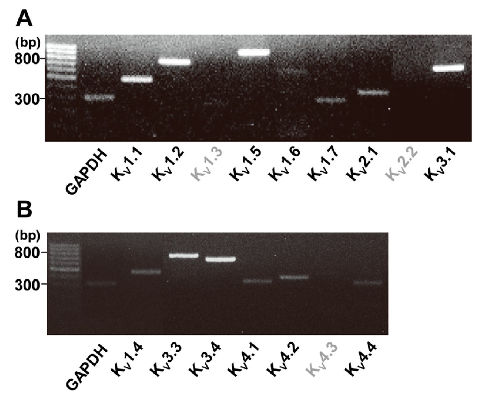
2.1. Identification of Two Types of Voltage-Gated K+ Channels in Human Cardiac Fibroblasts by RT-PCR

In the present study, we used RT-PCR targeting human genes for KV channel pore-forming α subunit in HCFs. The expressions of important genes for KV currents in cardiac myocytes, the pore-forming α subunits of KV1 (Shaker) family, KV2 (Shab) family, KV3 (Shaw) faimly, and KV4 (Shal) faimly were tested [14]. Strong gene expression was observed with KV1.1, KV1.2, KV1.5, and KV3.1, while weak gene expression was exhibited with KV1.6, KV1.7, and KV2.1 (Figure 1A). All of the products of these genes are parts of IK channel. For the Ito channel, strong gene expression of KV3.3 and KV3.4 and weak expression of KV1.4, KV4.1, KV4.2, and KV4.4 were seen in the HCFs (Figure 1B). Gene expression of KV1.3, KV 2.2 (for IK channel) and KV4.3 (for Ito channel) were not observed in the HCFs.

2.2. Identification of Two Types of Voltage-Gated K+ Channels using Electrophysiological Methods

The two types of KV currents with typical behavior were recorded (IK and Ito) with the whole-cell patch clamp technique. The stimulation voltage protocol consisted of depolarizing steps repeated every 10 s with a 10-mV increment for 400 milliseconds (−80 mV of holding potential). To exclude the influence of large-conductance Ca2+-activated K+ current (IBKCa), which is another major K+ current, iberiotoxin (100 nM) was added to the bath solution and EGTA (10 mM) into the pipette solutions. These currents could be distinguished based on their activation and inactivation kinetics. IK exhibited fast activation and slow or partial inactivation (Figure 2A) and Ito demonstrated fast activation and inactivation kinetics (Figure 2B).
IK and Ito were detected in 82.5% (n = 312 of 378) and 17.5% (n = 66 of 378) of the cells, respectively. These currents could also be distinguished based on the effects of K+ current blockers. Both KV currents were sensitive to 4-aminopyridine (4-AP). IK was sensitive to a high concentration of 4-AP (10 mM, −39.6 ± 6.4% of the control, in steady state current at +30 mV, n = 4, p < 0.05, Figure 2A) and Ito was sensitive to a lower concentration of 4-AP (1 mM, −30.7 ± 8.6% of the control, n = 4, p < 0.05; 10 mM, −62.3 ± 7.7% of the control, in peak current at +30 mV, n = 4, p < 0.01, Figure 2B).
On the other hand, when assessing the effects of tetraethylammonium chloride (TEA), another K+ channel blocker, IK was inhibited at a high concentration of TEA (10 mM, −42.1 ± 9.6% of the control, n = 4, p < 0.05, Figure 3A) but not at a low concentration of TEA (1 mM). However, Ito was not inhibited by 10 mM TEA (−4.5 ± 8.2% of the control, n = 4, Figure 3B).

2.3. Effect of NO on Two Types of Voltage-Gated K+ Currents

To determine the effect of NO on KV currents in HCFs, SNAP (an NO donor, 100 µM) was added to the bath solution. The amplitude of IK was significantly increased by SNAP (+46.0 ± 10.9% of the control, n = 6, p < 0.05, Figure 4A). On the other hand, Ito was not activated by SNAP (+1.6 ± 9.8% of the control, n = 6, Figure 4B). After the addition of SNAP, the current density of IK at +30 mV increased from 2.67 ± 0.70 pA/pF to 3.89 ± 0.76 pA/pF. On the other hand, the current density of Ito in the peak state was not altered by SNAP (2.56 ± 0.96 pA/pF to 2.60 ± 0.94 pA/pF).
Various concentrations of SNAP (0.1 to 300 µM) were applied and the IK was then examined to explore whether SNAP-induced stimulation was concentration dependent (Figure 4C). IK was elicited by one-step depolarizing pulses of +30 mV. The IK was increased by increasing concentrations of SNAP, but the low level of NO observed in the physiologic condition did not affect the IK of HCFs. Steady-state currents normalized by control data were fitted with the Hill equation, producing an EC50 value of 26.4 µM and a Hill coefficient of 0.96 (n = 7).

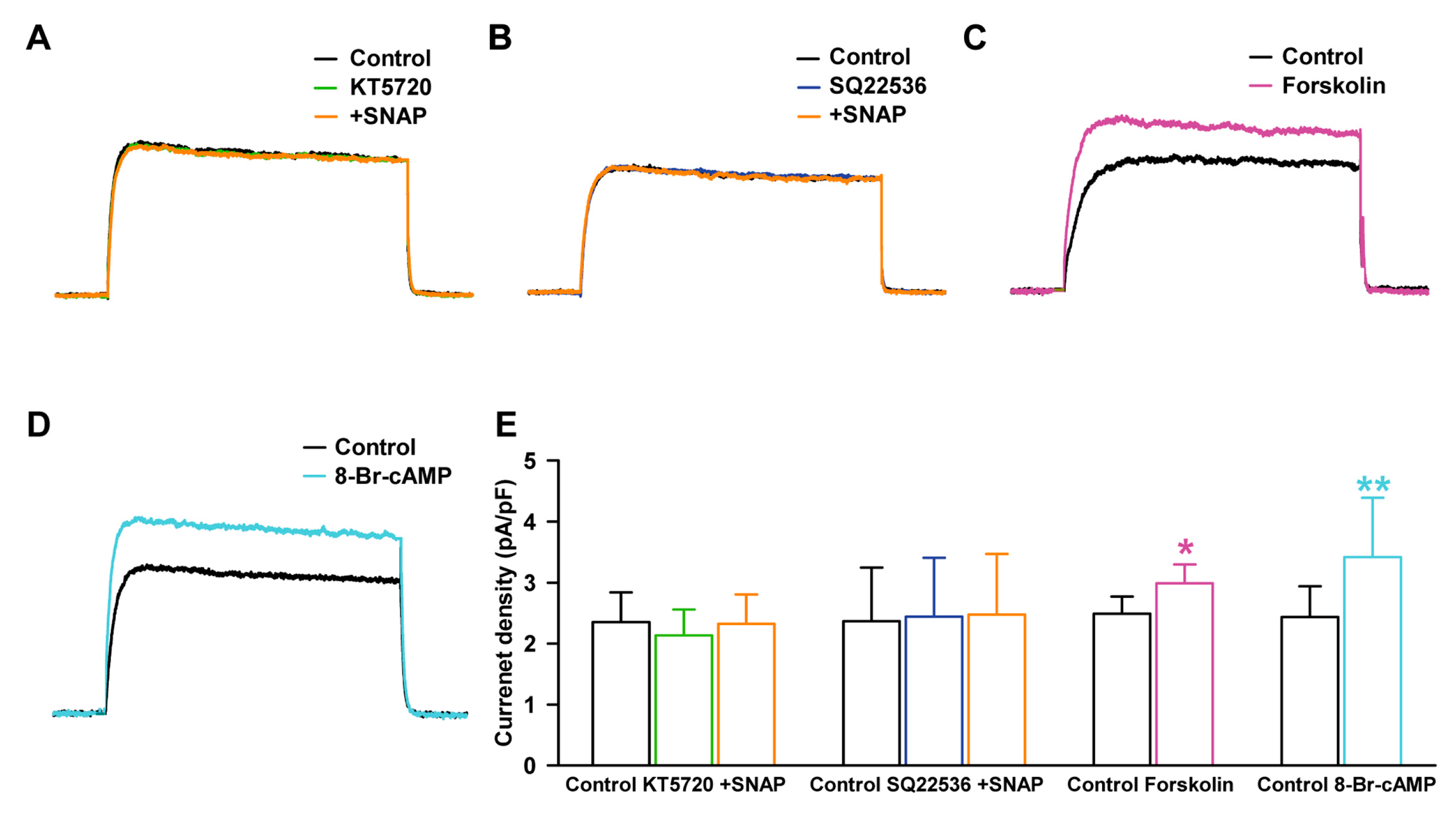
2.4. Effect of NO on Delayed Rectifier K+ Current through PKG Signaling Pathway

NO activates sGC and produces cGMP, which activates PKG. We examined the contribution of cGMP to SNAP-induced IK enhancement, using 1H-(1,2,4)oxadiazolo[4,3-a]quinoxalin-1-one (ODQ, a sGC blocker). When the cells were pretreated with ODQ (10 µM), IK did not increase in the presence of 100 µM SNAP (–2.6 ± 9.3% of the control, n = 7, Figure 5A). To further confirm the contribution of the cGMP signaling pathway, KT5823 (a PKG blocker, 1 µM) was added to the bath, and SNAP then failed to increase IK (+2.3 ± 6.2% of the control, n = 7, Figure 5B).
We also assessed the effect of cGMP, which is generated from NO binding to sGC. 8-Bromo-cyclic GMP (8-Br-cGMP, an activator of PKG, 300 µM) increased IK to +84.0 ± 14.5% of the control (n = 4, p < 0.05, Figure 5C). The current densities at +30 mV with the addition of SNAP were not altered after ODQ pretreatment (2.58 ± 0.81 pA/pF at control, 2.49 ± 0.77 pA/pF at ODQ, 2.51 ± 0.75 pA/pF at SNAP, Figure 5D) or after KT5823 pretreatment (2.39 ± 0.85 pA/pF at control, 2.47 ± 0.42 pA/pF at KT5823, 2.44 ± 0.53 pA/pF at SNAP). On the other hand, the current density with 8-Br-cGMP treatment increased significantly (from 2.40 ± 0.58 pA/pF to 4.42 ± 0.84 pA/pF).

2.5. Effect of NO on Delayed Rectifier K+ Current through PKA Signaling Pathway

We examined the contribution of cAMP to SNAP-induced IK enhancement, using KT5720 (a PKA blocker). When the cells were pretreated with KT5720 (1 µM) in the bath solution for 20 min, 100 µM SNAP did not increase the IK (−1.1 ± 9.8% of the control, n = 7, Figure 6A).
To further confirm the contribution of the cAMP signaling pathway, the cells were pretreated with SQ22536 (an AC blocker, 100 µM), 100 µM SNAP then failed to increase IK significantly (+4.7 ± 11.3% of the control, n = 7, Figure 6B). On the other hand, forskolin (a stimulator of AC and an activator of PKA, 10 µM) increased the amplitude of IK (+20.0 ± 11.0% of the control, n = 6, p < 0.05, Figure 6C). 8-Bromo-cyclic AMP (8-Br-cAMP, a cell-permeable cAMP, and an activator of PKA, 300 µM) also increased the activity of IK (+40.4 ± 19.2% of the control, n = 5, p < 0.01, Figure 6D). The current densities at +30 mV with SNAP were not altered after KT5720 pretreatment (2.35 ± 0.49 pA/pF with control, 2.13 ± 0.42 pA/pF at KT5720, 2.33 ± 0.48 pA/pF with SNAP, Figure 6E) or after SQ22536 pretreatment (2.36 ± 0.88 pA/pF with control, 2.44 ± 0.97 pA/pF with SQ22536, 2.48 ± 0.99 pA/pF with SNAP). On the other hand, the current densities with the forskolin or 8-Br-cAMP effects on IK increased significantly (2.49 ± 0.28 pA/pF with control, 2.99 ± 0.31 pA/pF with forskolin; from 2.44 ± 0.51 pA/pF to 3.42 ± 0.97 pA/pF with 8-Br-cAMP, Figure 6E).

2.6. Effect of NO on Delayed Rectifier K+ Current through the S-Nitrosylation Pathway

NO affects ubiquitous signaling pathways via posttranslational modification of cysteine residues, a reaction termed S-nitrosylation [28]. To examine the involvement of S-nitrosylation in SNAP-induced IK enhancement, we pretreated the cells with N-ethylmaleimide (NEM, a thiol-alkylating reagent, 0.5 mM) and then applied SNAP (100 µM). In the presence of NEM, SNAP increased IK significantly (+42.3 ± 10.1% of the control, n = 5, p < 0.05, Figure 7A), which suggests that the thiol residue was not the ultimate target of NO.
When DL-dithiothreitol (DTT, a reducing agent, 5 mM) was applied after IK had been enhanced by SNAP (100 µM), it could not reverse the SNAP-induced enhancement of IK (+45.5 ± 12.2% of the control with SNAP, n = 6, p < 0.05; +45.7 ± 6.9% of the control with DTT, n = 6, p < 0.05 for the control, Figure 7B). These findings suggest that S-nitrosylation is not the main mechanism for the NO stimulation of IK in HCFs. Figure 7C shows the current densities with the SNAP effects on IK after NEM pretreatment (2.38 ± 0.1 pA/pF with control, 2.53 ± 0.11 pA/pF with NEM, 3.39 ± 0.09 pA/pF with SNAP) and the current densities after DTT treatment following SNAP (2.38 ± 0.13 pA/pF with control, 3.40 ± 0.16 pA/pF with SNAP, 3.47 ± 0.09 pA/pF with DTT).

3. Discussion

In this study, we characterized the voltage-gated K+ (KV) channels in human cardiac ventricular fibroblasts and the effects of NO on the channels. We demonstrated the functional expression of two types of KV channels: delayed rectifier K+ channel and transient outward K+ channel. We also observed that NO stimulated delayed rectifier K+ current (IK), but not transient outward K+ current (Ito), through the sGC/cGMP/PKG pathway and AC/cAMP/PKA pathway, but not through S-nitrosylation.

3.1. Identification of Two Types of Kv Channels in Human Cardiac Fibroblasts by Molecular Methods

The electrophysiological properties of cardiac cells are determined by the composition of ion channels and by their absolute abundance and proportional ratio. In healthy human hearts, the significant expression levels of KV1.2, KV1.5, KV1.7, KV2.1, and KV3.1 (for the IK) and KV1.4, KV3.3, KV3.4, KV4.1, and KV4.3 (for the Ito) are found [31], which might indicate a functional role of these ion channel subunits in the formation of action potential in the human atrium and ventricle. KV1.5 is thought to be the major contributor to the IK in human heart [23,32] and KV4.3 is responsible for the Ito [25,33,34]. However, very little is known about the expression of K+ channel subunits in human cardiac fibroblasts.
In our RT-PCR analysis of mRNA expression of KV channel genes in HCFs, there was a strong mRNA expression of KV1.1, KV1.2, KV1.5, and KV3.1 and a weak expression of KV1.6, KV1.7, and KV2.1 (for the IK). We also found that a strong mRNA expression of KV3.3 and KV3.4, and a weak expression of KV1.4, KV4.1, KV4.2, and KV4.4 (for the Ito).
These results differed from those of a previous report on KV channels in HCFs [3]. In that report, the investigators found RT-PCR products corresponding to significant gene expression of KV1.5 and KV1.6 (for the IK) and KV4.2 and KV4.3 (for the Ito), but found none for KV2.1 and KV3.1 (for the IK) or for KV1.4 (for the Ito). The reason for the discrepancy in our results is unclear.
The KV4.3 represents the predominant K+ channel subunit underlying Ito in human cardiomyocytes, as the most abundant K+ channel mRNAs were KV4.3 (80.7%) [35], but in our HCFs experiments, KV4.3 mRNA was not detected. To confirm our RT-PCR results for the KV channels in HCFs, we repeated the RT-PCR several times to measure the gene expression of KV4.3 for the Ito, but we found no gene expression.
Changes in the expression of K+ channels explain the regional variations in morphology and duration of cardiac action potential among different cardiac regions and are influenced by heart rate, intracellular signaling pathways, drug and cardiovascular disorders [36]. Further investigations using molecular and electrophysiological approaches are needed to reveal the basis of ionic currents in cardiac fibroblasts in different heart regions because the extensive networks exist between cardiac fibroblasts and cardiomyocytes, that make the heterocellular electrical coupling [2,4,37,38,39].

3.2. Identification of Voltage-Gated K+ Channels in Human Cardiac Fibroblasts via Electrophysiological Methods

In our results, among the two types of KV currents that could be distinguished by their activation and inactivation kinetics, IK was the main subtype of KV currents in HCFs, as IK was recorded in 82.5% of HCFs with the whole-cell patch-clamp technique. This result was consistent with previous reports [11,40,41], but was somewhat discrepant with the results found by Li et al. [3].
According to their report on K+ currents in HCFs, IBKCa was present in most HCFs (88%), and IK and Ito were equally present but in smaller populations (15% and 14%, respectively).
The two types of KV currents were 4-AP sensitive in HCFs and these results are consistent with others [15,42,43,44]. Ito was more sensitive than IK to 4-AP, and similar results were found with human atrial Ito [35]. However, for TEA, another K+ channel blocker, the KV currents showed different responses: a high concentration of TEA (10 mM) inhibited IK (−42%) but not Ito. In a study of human ventricular myocytes, IK was not very sensitive to TEA: that is, the reduction was <20% with 10 mM external TEA [44]. On the other hand, 1 mM TEA could not inhibit either KV current, consistent with previous reports: that is, low concentrations of TEA (≤1 mM) predominantly blocked Ca2+-activated K+ (KCa) currents [45,46,47].

3.3. Effect of NO on Two Types of Voltage-Gated K+ Currents

We demonstrated that NO produced a concentration-dependent stimulation of IK in HCFs, and these results were consistent with other reports in guinea-pig cardiomyocytes [24,48]. While a low concentration of NO (0.1–1 µM SNAP) did not affect the IK of HCFs, a high concentration of NO (100 µM SNAP) increased the amplitude of IK with an EC50 of approximately 26.4 µM in our experiment. In previous study, NO inhibited the hKv1.5 channel current, which generates IK in transfected Chinese hamster ovary (CHO) cells and mouse ventricular myocytes [23].
This discrepancy could be explained by the concentration difference, because the effect of NO on cell function is determined by its concentration. Lower levels of NO (0.1–1 µM SNAP), which are observed under physiologic conditions, led to concentration-dependent inhibition of hKv1.5 current in cardiac myocytes [27], with the IC50 being approximately 340 nM [23]. Higher levels of NO (100 µM SNAP), which are observed in pathologic states, increased IK [49,50].
We also demonstrated that NO could not affect Ito in HCFs; however, in previous study, NO inhibited KV4.3 in transfected CHO cells and Ito in human atrial and mouse ventricular myocytes in a concentration and voltage-dependent manner with an IC50 of 375 nM [25]. These differences could be explained by differences in the cell type and expressed K+ channel mRNA [36].

3.4. Effect of NO on Delayed Rectifier K+ Current through Protein Kinases Signaling Pathways and S-Nitrosylation

In our results, the stimulating effect of NO on IK was suppressed in the presence of sGC inhibitor (ODQ) or PKG inhibitor (KT5823), and IK was increased by 8-Br-cGMP, a cell-permeable analogue of cGMP. These results suggest that the stimulatory effects of NO are dependent on the sGC/cGMP/PKG pathway in HCFs. In previous reports, the cGMP-dependent pathway was suggested to play a principal role in NO action on hKv1.5-induced IK in guinea-pig cardiomyocytes [24] and sinoatrial nodal cells [48].
We also investigated the effects of the AC/cAMP/PKA pathway on KV currents in HCFs, because the PKA pathway is also responsible for NO-mediated cardioprotection [51], NO can directly activate AC and thereby increase cAMP levels [27], and AC activators or membrane-permeable cAMP analogs can increase NO production [52]. We found that specific PKA blockers (KT5720 or SQ22536) inhibited the stimulating effect of NO for IK, and forskolin (an AC activator), and 8-Br-cAMP (a membrane-permeable cAMP) increased the currents in HCFs.
These results indicate that NO (100 µM SNAP) increased IK of HCFs through AC/cAMP/PKA signaling mechanisms. It has been reported that NO also modulates cardiac IBKCa channels, which are also important K+ channels in HCFs [13], and Na+ channels in guinea pig and mouse ventricular myocytes through the cAMP and cGMP pathways [53].
We also examined whether NO increased IK through a cGMP-independent pathway, a direct S-nitrosylation of the thiol residue of target proteins, which is a well-known alternative pathway for the biological effects of NO [23,24,25,29]. S-nitrosylation regulates many cardiac ion channels and modulates the major currents involved in the generation of the action potential in cardiomyocytes [28]. Inwardly rectifying K+ current that determine resting membrane potential is also expressed in rat ventricular fibroblasts [3] and human ventricular fibroblasts [3]. NO increases the current by nitrosylation in cardiomyocytes and transfected CHO cells with Kir2.1 channel (the major isoform of inwardly rectifying K+ channel) [54].
It has been reported that S-nitrosylation increases IK in guinea pig cardiomyocytes [55], but inhibits these currents in human atria [23]. However, in our results, the stimulating effect of NO on IK was not blocked by NEM pretreatment in HCFs. In addition, the reducing reagent DTT did not reverse NO-induced IK enhancement. These results suggest that S-nitrosylation is not involved in NO effects on IK in HCFs.
In summary, we provide evidence to demonstrate that NO modulates cardiac IK but not Ito via a second messenger pathway through activation of PKG and PKA. This modulation occurs at a relatively high concentration of NO that was observed in pathological states. Therefore, it is reasonable to speculate that the modulation of IK by NO may play a significant functional role in the pathological states.

4. Materials and Methods

4.1. Cell Preparation and Culture

Commercially available primary adult human ventricular cardiac fibroblasts (HCF-av, Catalogue #6310 from ScienCell, Carisbad, CA, USA) were used. These cells were used for many bimolecular and electrophysiological experiments [3,13,40,56,57,58] and confirmed as fibroblasts by discoidin domain receptor 2 staining [58]. The cells were cultured in Dulbecco’s modified eagle’s medium (DMEM; Welgene, Gyeongsan, Gyeongsangbuk-do, Korea) that was supplemented with fetal bovine serum (10%, Welgene) and a penicillin-streptomycin solution (100×; GenDEPOT, Barker, TX, USA) in a humidified atmosphere of 5% CO2 and 95% air at 37 °C. Confluent fibroblasts were detached by incubation with trypsin (0.25%, Welgene) and ethylene diamine tetraacetic acid (0.02%) in DMEM for several minutes. The detached cells were pelleted by centrifugation, the supernatant was removed, and the pellet was suspended in 1 mL of bath solution. The cells used in this study were from early passages (3 to 7) to limit possible variation due to culture.

4.2. Reverse Transcription Polymerase Chain Reaction (RT-PCR)

mRNA was generated using the RT-PCR technique with the primers indicated in the Table 1 and Table 2. Total RNA was extracted from HCFs using the Total RNA Isolation PureLink RNA Mini Kit (Ambion, Carlsbad, CA, USA). First-strand cDNA was prepared with the SuperScript III Cells Direct cDNA Synthesis Kit (Invitrogen, Tokyo, Japan). Reverse transcription was performed in a S1000 Thermal Cycler (Bio-Rad, Hercules, CA, USA) according to the manufacturer’s instructions. RT-PCR reaction products (cDNA) were resolved by 1.2% agarose gel electrophoresis and stained with ethidium bromide for visualization under ultraviolet light.

4.3. Electrophysiological Recordings

Recordings were obtained, using a whole-cell patch clamp with an Axopatch 200B Patch Clamp Amplifier (Axon Instruments, Union City, CA, USA) at room temperature. The pCLAMP 9.0 software (Axon Instruments) was used for data acquisition and analysis of whole-cell currents. Activated currents were filtered at 2 kHz and digitized at 10 kHz. Recording patch pipettes were prepared from filament-containing borosilicate tubes (TW150F-4; World Precision Instruments, Sarasota, FL, USA), using a two-stage microelectrode puller (PC-10; Narishige, Tokyo, Japan), and were then fire polished on a microforge (MF-830; Narishige). When filled with pipette solution, the pipettes exhibited a resistance of 2–3 MΩ.
The bath solution to record KV currents contained (in mM): 150 NaCl, 5.4 KCl, 1 CaCl2, 1 MgCl2, 10 glucose, and 5 HEPES (pH adjusted to 7.35 with NaOH). The pipette solution contained (in mM): 130 KCl, 1 CaCl2, 2 MgCl2, 10 HEPES, 10 EGTA, and 2 Mg-ATP (pH adjusted to 7.3 with KOH). TEA, 4-AP, SNAP, KT5823, ODQ, 8-Br-cGMP, KT5720, SQ22536, 8-Br-cAMP, and all other chemicals were purchased from Sigma-Aldrich (St. Louis, MO, USA).

4.4. Statistical Analysis

Statistical analysis was performed using SPSS version 22.0 (SPSS Inc., Chicago, IL, USA). Results were presented as mean ± standard error of the mean (SEM). The paired Student’s t-test was used when appropriate to evaluate the statistical significance of differences between two group means, while one-way analysis of variance (ANOVA) was used for multiple groups. p values < 0.05 were considered statistically significant.

Acknowledgments

This research was supported by the Basic Science Research Program through the National Research Foundation of Korea (NRF) funded by the Ministry of Education (NRF-2016R1D1A1B03933569).

Author Contributions

Inja Lim and Taeho Kim conceived and designed the experiments and wrote the paper; Hyemi Bae performed the experiments, analyzed the results, and made figures and tables; Jeongyoon Choi, Young-Won Kim, and Donghee Lee performed the experiments; Jung-Ha Kim, Jae-Hong Ko, and Hyoweon Bang contributed to scientific discussions.

Conflicts of Interest

The authors declare no conflict of interest.

References

  1. Camelliti, P.; Borg, T.K.; Kohl, P. Structural and functional characterisation of cardiac fibroblasts. Cardiovasc. Res. 2005, 65, 40–51. [Google Scholar] [CrossRef] [PubMed]
  2. Kohl, P. Heterogeneous cell coupling in the heart: An electrophysiological role for fibroblasts. Circ. Res. 2003, 93, 381–383. [Google Scholar] [CrossRef] [PubMed]
  3. Li, G.R.; Sun, H.Y.; Chen, J.B.; Zhou, Y.; Tse, H.F.; Lau, C.P. Characterization of multiple ion channels in cultured human cardiac fibroblasts. PLoS ONE 2009, 4, e7307. [Google Scholar] [CrossRef] [PubMed] [Green Version]
  4. Gaudesius, G.; Miragoli, M.; Thomas, S.P.; Rohr, S. Coupling of cardiac electrical activity over extended distances by fibroblasts of cardiac origin. Circ. Res. 2003, 93, 421–428. [Google Scholar] [CrossRef] [PubMed]
  5. Kohl, P.; Camelliti, P.; Burton, F.L.; Smith, G.L. Electrical coupling of fibroblasts and myocytes: Relevance for cardiac propagation. J. Electrocardiol. 2005, 38, 45–50. [Google Scholar] [CrossRef] [PubMed]
  6. Quinn, T.A.; Camelliti, P.; Rog-Zielinska, E.A.; Siedlecka, U.; Poggioli, T.; O’Toole, E.T.; Knopfel, T.; Kohl, P. Electrotonic coupling of excitable and nonexcitable cells in the heart revealed by optogenetics. Proc. Natl. Acad. Sci. USA 2016, 113, 14852–14857. [Google Scholar] [CrossRef] [PubMed]
  7. Pellman, J.; Zhang, J.; Sheikh, F. Myocyte-fibroblast communication in cardiac fibrosis and arrhythmias: Mechanisms and model systems. J. Mol. Cell. Cardiol. 2016, 94, 22–31. [Google Scholar] [CrossRef] [PubMed]
  8. Burstein, B.; Oi, X.Y.; Yeh, Y.H.; Calderone, A.; Nattel, S. Atrial cardiomyocyte tachycardia alters cardiac fibroblast function: A novel consideration in atrial remodeling. Circulation 2007, 116, 442–452. [Google Scholar] [CrossRef] [PubMed]
  9. Pedrotty, D.M.; Klinger, R.Y.; Kirkton, R.D.; Bursac, N. Cardiac fibroblast paracrine factors alter impulse conduction and ion channel expression of neonatal rat cardiomyocytes. Cardiovasc. Res. 2009, 83, 688–697. [Google Scholar] [CrossRef] [PubMed]
  10. Vasquez, C.; Mohandas, P.; Louie, K.L.; Benamer, N.; Bapat, A.C.; Morley, G.E. Enhanced fibroblast-myocyte interactions in response to cardiac injury. Circ. Res. 2010, 107, 1011–1120. [Google Scholar] [CrossRef] [PubMed]
  11. Chilton, L.; Ohya, S.; Freed, D.; George, E.; Drobic, V.; Shibukawa, Y.; Maccannell, K.A.; Imaizumi, Y.; Clark, R.B.; Dixon, I.M.; et al. K+ currents regulate the resting membrane potential, proliferation, and contractile responses in ventricular fibroblasts and myofibroblasts. Am. J. Physiol. Heart Circ. Physiol. 2005, 288, H2931–H2939. [Google Scholar] [CrossRef] [PubMed]
  12. Wang, Y.J.; Sung, R.J.; Lin, M.W.; Wu, S.N. Contribution of bk(ca)-channel activity in human cardiac fibroblasts to electrical coupling of cardiomyocytes-fibroblasts. J. Membr. Biol. 2006, 213, 175–185. [Google Scholar] [CrossRef] [PubMed]
  13. Bae, H.; Lim, I. Effects of nitric oxide on large-conductance Ca2+ -activated K+ currents in human cardiac fibroblasts through pka and pkg-related pathways. Clin. Exp. Pharmacol. Physiol. 2017, 44, 1116–1124. [Google Scholar] [CrossRef] [PubMed]
  14. Nerbonne, J.M. Molecular basis of functional voltage-gated K+ channel diversity in the mammalian myocardium. J. Physiol. (Lond.) 2000, 525, 285–298. [Google Scholar] [CrossRef]
  15. Nerbonne, J.M. Molecular basis of functional myocardial potassium channel diversity. Card. Electrophysiol. Clin. 2016, 8, 257–273. [Google Scholar] [CrossRef] [PubMed]
  16. Seddon, M.; Shah, A.M.; Casadei, B. Cardiomyocytes as effectors of nitric oxide signalling. Cardiovasc. Res. 2007, 75, 315–326. [Google Scholar] [CrossRef] [PubMed]
  17. Ziolo, M.T.; Kohr, M.J.; Wang, H. Nitric oxide signaling and the regulation of myocardial function. J. Mol. Cell. Cardiol. 2008, 45, 625–632. [Google Scholar] [CrossRef] [PubMed]
  18. Massion, P.B.; Pelat, M.; Belge, C.; Balligand, J.L. Regulation of the mammalian heart function by nitric oxide. Comp. Biochem. Physiol. A Mol. Integr. Physiol. 2005, 142, 144–150. [Google Scholar] [CrossRef] [PubMed]
  19. Kanno, S.; Kim, P.K.; Sallam, K.; Lei, J.; Billiar, T.R.; Shears, L.L., 2nd. Nitric oxide facilitates cardiomyogenesis in mouse embryonic stem cells. Proc. Natl. Acad. Sci. USA 2004, 101, 12277–12281. [Google Scholar] [CrossRef] [PubMed]
  20. Takimoto, E.; Champion, H.C.; Li, M.; Ren, S.; Rodriguez, E.R.; Tavazzi, B.; Lazzarino, G.; Paolocci, N.; Gabrielson, K.L.; Wang, Y.; et al. Oxidant stress from nitric oxide synthase–3 uncoupling stimulates cardiac pathologic remodeling from chronic pressure load. J. Clin. Investig. 2005, 115, 1221–1231. [Google Scholar] [CrossRef] [PubMed]
  21. Andreka, P.N.Z.; Muzes, G.; Szantho, G.; Vandor, L.; Konya, L.; Turner, M.S.; Tulassay, Z.; Bishopric, N.H. Possible therapeutic targets in cardiac myocyte apoptosis. Curr. Pharm. Des. 2004, 10, 2445–2461. [Google Scholar] [CrossRef] [PubMed]
  22. Tamargo, J.; Caballero, R.; Gomez, R.; Delpon, E. Cardiac electrophysiological effects of nitric oxide. Cardiovasc. Res. 2010, 87, 593–600. [Google Scholar] [CrossRef] [PubMed]
  23. Nunez, L.; Vaquero, M.; Gomez, R.; Caballero, R.; Mateos-Caceres, P.; Macaya, C.; Iriepa, I.; Galvez, E.; Lopez-Farre, A.; Tamargo, J.; et al. Nitric oxide blocks hKv1.5 channels by s-nitrosylation and by a cyclic gmp-dependent mechanism. Cardiovasc. Res. 2006, 72, 80–89. [Google Scholar] [CrossRef] [PubMed]
  24. Bai, C.X.; Takahashi, K.; Masumiya, H.; Sawanobori, T.; Furukawa, T. Nitric oxide-dependent modulation of the delayed rectifier k+ current and the l-type ca2+ current by ginsenoside re, an ingredient of panax ginseng, in guinea-pig cardiomyocytes. Br. J. Pharmacol. 2004, 142, 567–575. [Google Scholar] [CrossRef] [PubMed]
  25. Gomez, R.; Nunez, L.; Vaquero, M.; Amoros, I.; Barana, A.; de Prada, T.; Macaya, C.; Maroto, L.; Rodriguez, E.; Caballero, R.; et al. Nitric oxide inhibits kv4.3 and human cardiac transient outward potassium current (ito1). Cardiovasc. Res. 2008, 80, 375–384. [Google Scholar] [CrossRef] [PubMed]
  26. Hammond, J.; Balligand, J.L. Nitric oxide synthase and cyclic gmp signaling in cardiac myocytes: From contractility to remodeling. J. Mol. Cell. Cardiol. 2012, 52, 330–340. [Google Scholar] [CrossRef] [PubMed]
  27. Vila-Petroff, M.G.; Younes, A.; Egan, J.; Lakatta, E.G.; Sollott, S.J. Activation of distinct camp-dependent and cgmp-dependent pathways by nitric oxide in cardiac myocytes. Circ. Res. 1999, 84, 1020–1031. [Google Scholar] [CrossRef] [PubMed]
  28. Gonzalez, D.R.; Treuer, A.; Sun, Q.A.; Stamler, J.S.; Hare, J.M. S-nitrosylation of cardiac ion channels. J. Cardiovasc. Pharmacol. 2009, 54, 188–195. [Google Scholar] [CrossRef] [PubMed]
  29. Lima, B.; Forrester, M.T.; Hess, D.T.; Stamler, J.S. S-nitrosylation in cardiovascular signaling. Circ. Res. 2010, 106, 633–646. [Google Scholar] [CrossRef] [PubMed]
  30. Martinez-Ruiz, A.; Lamas, S. S-nitrosylation: A potential new paradigm in signal transduction. Cardiovasc. Res. 2004, 62, 43–52. [Google Scholar] [CrossRef] [PubMed]
  31. Ordog, B.; Brutyo, E.; Puskas, L.G.; Papp, J.G.; Varro, A.; Szabad, J.; Boldogkoi, Z. Gene expression profiling of human cardiac potassium and sodium channels. Int. J. Cardiol. 2006, 111, 386–393. [Google Scholar] [CrossRef] [PubMed]
  32. Feng, J.L.; Wible, B.; Li, G.R.; Wang, Z.G.; Nattel, S. Antisense oligodeoxynucleotides directed against kv1.5 mRNA specifically inhibit ultrarapid delayed rectifier k+ current in cultured adult human atrial myocytes. Circ. Res. 1997, 80, 572–579. [Google Scholar] [CrossRef] [PubMed]
  33. Brahmajothi, M.V.; Campbell, D.L.; Rasmusson, R.L.; Morales, M.J.; Trimmer, J.S.; Nerbonne, J.M.; Strauss, H.C. Distinct transient outward potassium current (ito) phenotypes and distribution of fast-inactivating potassium channel alpha subunits in ferret left ventricular myocytes. J. Gen. Physiol. 1999, 113, 581–600. [Google Scholar] [CrossRef] [PubMed]
  34. Isbrandt, D.; Leicher, T.; Waldschutz, R.; Zhu, X.; Luhmann, U.; Michel, U.; Sauter, K.; Pongs, O. Gene structures and expression profiles of three human kcnd (kv4) potassium channels mediating a-type currents i(to) and i(sa). Genomics 2000, 64, 144–154. [Google Scholar] [CrossRef] [PubMed]
  35. Bertaso, F.; Sharpe, C.C.; Hendry, B.M.; James, A.F. Expression of voltage-gated k+ channels in human atrium. Basic Res. Cardiol. 2002, 97, 424–433. [Google Scholar] [CrossRef] [PubMed]
  36. Tamargo, J.; Caballero, R.; Gomez, R.; Valenzuela, C.; Delpon, E. Pharmacology of cardiac potassium channels. Cardiovasc. Res. 2004, 62, 9–33. [Google Scholar] [CrossRef] [PubMed]
  37. Mahoney, V.M.; Mezzano, V.; Morley, G.E. A review of the literature on cardiac electrical activity between fibroblasts and myocytes. Prog. Biophys. Mol. Biol. 2016, 120, 128–133. [Google Scholar] [CrossRef] [PubMed]
  38. Wang, Y.J.; Wu, S.N.; Lin, M.W.; Sung, R.J. Characterization of Ca2+-activated K+ current in human cardiac fibroblasts: Its contribution to electrical coupling of cardiomyocyte-fibroblast. FASEB J. 2007, 21. [Google Scholar] [CrossRef]
  39. Zlochiver, S.; Munoz, V.; Vikstrom, K.L.; Taffet, S.M.; Berenfeld, O.; Jalife, J. Electrotonic myofibroblast-to-myocyte coupling increases propensity to reentrant arrhythmias in two-dimensional cardiac monolayers. Biophys. J. 2008, 95, 4469–4480. [Google Scholar] [CrossRef] [PubMed]
  40. Bae, H.; Lee, D.; Kim, Y.W.; Choi, J.; Lee, H.J.; Kim, S.W.; Kim, T.; Noh, Y.H.; Ko, J.H.; Bang, H.; et al. Effects of hydrogen peroxide on voltage-dependent K(+) currents in human cardiac fibroblasts through protein kinase pathways. Korean J. Physiol. Pharmacol. 2016, 20, 315–324. [Google Scholar] [CrossRef] [PubMed]
  41. He, M.L.; Liu, W.J.; Sun, H.Y.; Wu, W.; Liu, J.; Tse, H.F.; Lau, C.P.; Li, G.R. Effects of ion channels on proliferation in cultured human cardiac fibroblasts. J. Mol. Cell. Cardiol. 2011, 51, 198–206. [Google Scholar] [CrossRef] [PubMed]
  42. Cole, W.C.; Clement-Chomienne, O.; Aiello, E.A. Regulation of 4-aminopyridine-sensitive, delayed rectifier k+ channels in vascular smooth muscle by phosphorylation. Biochem. Cell Biol. 1996, 74, 439–447. [Google Scholar] [CrossRef] [PubMed]
  43. Rogers, P.A.; Chilian, W.M.; Bratz, I.N.; Bryan, R.M., Jr.; Dick, G.M. H2O2 activates redox- and 4-aminopyridine-sensitive kv channels in coronary vascular smooth muscle. Am. J. Physiol. Heart Circ. Physiol. 2007, 292, H1404–H1411. [Google Scholar] [CrossRef] [PubMed]
  44. Snyders, D.J.; Tamkun, M.M.; Bennett, P.B. A rapidly activating and slowly inactivating potassium channel cloned from human heart. Functional analysis after stable mammalian cell culture expression. J. Gen. Physiol. 1993, 101, 513–543. [Google Scholar] [CrossRef] [PubMed]
  45. Beech, D.J.; Bolton, T.B. Two components of potassium current activated by depolarization of single smooth muscle cells from the rabbit portal vein. J. Physiol. 1989, 418, 293–309. [Google Scholar] [CrossRef] [PubMed]
  46. Krick, S.; Platoshyn, O.; Sweeney, M.; McDaniel, S.S.; Zhang, S.; Rubin, L.J.; Yuan, J.X.J. Nitric oxide induces apoptosis by activating K+ channels in pulmonary vascular smooth muscle cells. Am. J. Physiol. Heart Circ. Physiol. 2002, 282, H184–H193. [Google Scholar] [CrossRef] [PubMed]
  47. Nelson, M.T.; Quayle, J.M. Physiological roles and properties of potassium channels in arterial smooth muscle. Am. J. Physiol. 1995, 268, C799–C822. [Google Scholar] [CrossRef] [PubMed]
  48. Shimizu, K.; Shintani, Y.; Ding, W.G.; Matsuura, H.; Bamba, T. Potentiation of slow component of delayed rectifier k(+) current by cgmp via two distinct mechanisms: Inhibition of phosphodiesterase 3 and activation of protein kinase g. Br. J. Pharmacol. 2002, 137, 127–137. [Google Scholar] [CrossRef] [PubMed]
  49. Brady, A.J.B.; Warren, J.B.; Poolewilson, P.A.; Williams, T.J.; Harding, S.E. Nitric-oxide attenuates cardiac myocyte contraction. Am. J. Physiol. 1993, 265, H176–H182. [Google Scholar] [CrossRef] [PubMed]
  50. Hare, J.M.; Loh, E.; Creager, M.A.; Colucci, W.S. Nitric-oxide inhibits the positive inotropic response to beta-adrenergic stimulation in humans with left-ventricular dysfunction. Circulation 1995, 92, 2198–2203. [Google Scholar] [CrossRef] [PubMed]
  51. Kamga Pride, C.; Mo, L.; Quesnelle, K.; Dagda, R.K.; Murillo, D.; Geary, L.; Corey, C.; Portella, R.; Zharikov, S.; St Croix, C.; et al. Nitrite activates protein kinase a in normoxia to mediate mitochondrial fusion and tolerance to ischaemia/reperfusion. Cardiovasc. Res. 2014, 101, 57–68. [Google Scholar] [CrossRef] [PubMed]
  52. Liu, R.; Flammer, J.; Haefliger, I.O. Isoproterenol, forskolin, and camp-induced nitric oxide production in pig ciliary processes. Investig. Ophthalmol. Vis. Sci. 1999, 40, 1833–1837. [Google Scholar]
  53. Ahmmed, G.U.; Xu, Y.; Hong Dong, P.; Zhang, Z.; Eiserich, J.; Chiamvimonvat, N. Nitric oxide modulates cardiac na(+) channel via protein kinase a and protein kinase g. Circ. Res. 2001, 89, 1005–1013. [Google Scholar] [CrossRef] [PubMed]
  54. Gomez, R.; Caballero, R.; Barana, A.; Amoros, I.; Calvo, E.; Lopez, J.A.; Klein, H.; Vaquero, M.; Osuna, L.; Atienza, F.; et al. Nitric oxide increases cardiac ik1 by nitrosylation of cysteine 76 of kir2.1 channels. Circ. Res. 2009, 105, 383–392. [Google Scholar] [CrossRef] [PubMed]
  55. Bai, C.X.; Namekata, I.; Kurokawa, J.; Tanaka, H.; Shigenobu, K.; Furukawa, T. Role of nitric oxide in Ca2+ sensitivity of the slowly activating delayed rectifier K+ current in cardiac myocytes. Circ. Res. 2005, 96, 64–72. [Google Scholar] [CrossRef] [PubMed]
  56. Bunda, S.; Liu, P.; Wang, Y.; Liu, K.; Hinek, A. Aldosterone induces elastin production in cardiac fibroblasts through activation of insulin-like growth factor-i receptors in a mineralocorticoid receptor-independent manner. Am. J. Pathol. 2007, 171, 809–819. [Google Scholar] [CrossRef] [PubMed]
  57. Chen, J.B.; Tao, R.; Sun, H.Y.; Tse, H.F.; Lau, C.P.; Li, G.R. Multiple Ca2+ signaling pathways regulate intracellular Ca2+ activity in human cardiac fibroblasts. J. Cell. Physiol. 2010, 223, 68–75. [Google Scholar] [CrossRef] [PubMed] [Green Version]
  58. Meredith, A.; Boroomand, S.; Carthy, J.; Luo, Z.S.; McManus, B. 1,25 dihydroxyvitamin d-3 inhibits tgf beta(1)-mediated primary human cardiac myofibroblast activation. PLoS ONE 2015, 10. [Google Scholar] [CrossRef] [PubMed]
Figure 1. Identification of two types of voltage-gated K+ (KV) channels in human cardiac fibroblasts by reverse transcription polymerase chain reaction (RT-PCR). (A) Strong gene expression was shown for KV1.1, KV1.2 KV1.5, and KV3.1 for delayed rectifier K+ (IK) channels and (B) KV3.3 and KV3.4 for transient outward K+ (Ito) channels. Weak expression was observed for KV1.6, KV1.7 and KV2.1 (for IK) and KV1.4, KV4.1, KV4.2, and KV4.4 (for Ito). Glyceraldehyde 3-phosphate dehydrogenase (GAPDH) was used as a positive control.
Ijms 19 00814 g001
Figure 2. Effect of 4-aminopyridine (4-AP) on KV currents in HCFs. (A) The representative rapid activated and slow inactivated, IK recorded in a single HCF in control conditions and after application of 4-AP (1 or 10 mM). The current-voltage (I-V) relationship of steady-state currents with IK changed in the presence of 4-AP, and the concentration-response bar graphs for the 4-AP effect on IK are shown. (B) Typical rapid-activated and inactivated Ito in control and after 4-AP (1 or 10 mM) addition. The I-V relationship of peak currents changed by the presence of 4-AP on Ito and concentration-response bar graphs are shown. * p < 0.05, ** p < 0.01 versus the control.
Ijms 19 00814 g002
Figure 3. Effect of tetraethylammonium chloride (TEA) on KV currents in HCFs. (A) The representative IK recorded in a single HCF in control conditions and after application of TEA (1 or 10 mM) are shown. The I-V relationship of the steady-state current change of IK by TEA and bar graphs for concentration change are also shown. (B) The typical Ito was not changed by TEA. The I-V relationship of the peak current change for the TEA of Ito and bar graph for concentration response are shown. * p < 0.05 versus the control.
Ijms 19 00814 g003
Figure 4. Effect of nitric oxide (NO) on two subtypes of KV currents. Raw data and the I-V relationship of steady-state currents changed by the presence of SNAP on (A) delayed rectifier K+ current (IK) and (B) transient outward K+ current (Ito). (C) The concentration-response curve of S-nitroso-N-acetylpenicillamine (SNAP) on IK. The continuous line represents the fit to the Hill equation. The normalized currents (percent inhibition) were calculated from the IK in the absence of SNAP and plotted against various concentrations of SNAP. * p < 0.05 versus the control.
Ijms 19 00814 g004
Figure 5. Effects of the PKG pathway on the stimulating effect of NO on IK. Representative currents showing the effect of SNAP on IK after pretreatment with (A) 1H-(1,2,4)oxadiazolo[4,3-a]quinoxalin-1-one (ODQ) or (B) KT5823. (C) Effect of 8-Br-cyclic GMP on IK. (D) Bar graph summarizing current density changes for the effects of SNAP on IK after pre-incubation with ODQ or KT5823, and the effect of 8-Br-cyclic GMP on IK current. * p < 0.05 versus the control.
Ijms 19 00814 g005
Figure 6. Effects of the protein kinase A (PKA) pathway on the stimulating effect of NO on IK current. Representative currents showing the effect of SNAP on IK after pretreatment with (A) KT5720 or (B) SQ22536. Effect of (C) forskolin or (D) 8-Br-cyclic AMP on IK. (E) Bar graph summarizing current density changes for the effects of SNAP on IK after pre-incubation with KT5720 or SQ22536, and the effect of forskolin or 8-Br-cAMP on IK. * p < 0.05, ** p < 0.01 versus the control.
Ijms 19 00814 g006
Figure 7. Influence of S-nitrosylation on the stimulating effect of NO on IK. (A) Representative currents showing the effect of SNAP on IK after pretreatment with N-ethylmaleimide (NEM). (B) Effect of DL-dithiothreitol (DTT) on SNAP-stimulating IK. (C) Bar graph summarizing current density changes for SNAP effects on IK after pretreatment with NEM and DTT. * p < 0.05 versus the control.
Ijms 19 00814 g007
Table 1. Primers of delayed rectifier potassium channels used for RT-PCR.
GeneForward PrimerReverse PrimerSize
GAPDH5′-AGCCACATCGCTCAGACACC-3′5′-GTACTCAGCGGCCAGCATCG-3302
KV1.15′-CCATCATTCCTTATTTCATCAC-35′-CTCTTCCCCCTCAGTTTCTC-3′488
KV1.25′-TCCGGGATGAGAATGAAGAC-3′5′-TTGGACAGCTTGTCACTTGC-3′747
KV1.35′-TCTGCCTATGCCCTTGTTTT-3′5′-TTCCTCCCAGGATGTACTGC-3′259
KV1.55′-TGCGTCATCTGGTTCACCTTCG-3′5′-TGTTCAGCAAGCCTCCCATTCC-3′906
KV1.65′-TCAACAGGATGGAAACCAGCCC-3′5′-CTGCCATCTGCAACACGATTCC-3′608
KV1.75′-CTTCCAGGGGCATGTTATTT-3′5′-CTCAATGGAACTCAATTCAG-3′300
KV2.15′-ACAGAGCAAACCAAAGGAAGAAC-3′5′-CACCCTCCATGAAGTTGACTTTA-3′383
KV2.25′-AACGAACTGAGGCGAGAG -3′5′-ACTCCGCCTAAGGGTGAAAC-3′546
KV3.15′-AACCCCATCGTGAACAAGACGG-3′5′-TCATGGTGACCACGGCCCA-3′550
Table 2. Primers of transient outward potassium channels used for RT-PCR.
GeneForward PrimerReverse PrimerSize
GAPDH5′-AGCCACATCGCTCAGACACC-3′5′-ATCATTCAACAACCCACCAT-3′302
KV1.45′-TGGCGGCTACAGTTCAGTCC-3′5′-TGTTGACAATGACGGGCACAGG-3′571
KV3.35′-TTCTGCCTGGAAACCCATGAGG-3′5′-TGCCAAATCCCAAGGTCTGAGG-3′694
KV3.45′-TTCAAGCTCACACGCCACTTCG-3′5′-TTCTTTCGGTCCCGATAC-3′656
KV4.15′-ATCTCGAGGAGATGAGGTTC-3′5′-GATCCGCACGGCACTGTTTC-3′318
KV4.25′-ATCTTCCGCCACATCCTGAA-3′5′-GATCCGCACGGCACTGTTTC-3′362
KV4.35′-GATGAGCAGATGTTTGAGCAG-3′5′-AGCAGGTGGTAGTGAGGCC-3′106
KV4.45′-AGCCAAGAAGAACAAGCTG-3′5′-AGGAAGTTTAGGACATGCC-3′315

