Article

The Effect of Ca2+, Lobe-Specificity, and CaMKII on CaM Binding to NaV1.1

1
Department of Pharmaceutical Toxicology, School of Pharmacy, China Medical University, Shenyang 110122, China
2
Department of Neurobiology, Harvard Medical School, Boston, MA 02115, USA
3
Department of Physiology, Graduate School of Medical and Dental Sciences, Kagoshima University, Kagoshima 890-8544, Japan
4
Department of Ophthalmology, Stanford University School of Medicine, Palo Alto, CA 94305, USA
5
Department of Clinical Pharmacy, School of Life Science and Biopharmaceutics, Shenyang Pharmaceutical University, Shenyang 110016, China
6
Department of Cellular and Molecular Biology, University of Texas Health Science Center at Tyler, Tyler, TX 75708, USA
*
Authors to whom correspondence should be addressed.
Int. J. Mol. Sci. 2018, 19(9), 2495; https://doi.org/10.3390/ijms19092495
Submission received: 4 July 2018 / Revised: 6 August 2018 / Accepted: 13 August 2018 / Published: 23 August 2018
(This article belongs to the Special Issue Calcium Binding Proteins)

Abstract

:
Calmodulin (CaM) is well known as an activator of calcium/calmodulin-dependent protein kinase II (CaMKII). Voltage-gated sodium channels (VGSCs) are basic signaling molecules in excitable cells and are crucial molecular targets for nervous system agents. However, the way in which Ca2+/CaM/CaMKII cascade modulates NaV1.1 IQ (isoleucine and glutamine) domain of VGSCs remains obscure. In this study, the binding of CaM, its mutants at calcium binding sites (CaM12, CaM34, and CaM1234), and truncated proteins (N-lobe and C-lobe) to NaV1.1 IQ domain were detected by pull-down assay. Our data showed that the binding of Ca2+/CaM to the NaV1.1 IQ was concentration-dependent. ApoCaM (Ca2+-free form of calmodulin) bound to NaV1.1 IQ domain preferentially more than Ca2+/CaM. Additionally, the C-lobe of CaM was the predominant domain involved in apoCaM binding to NaV1.1 IQ domain. By contrast, the N-lobe of CaM was predominant in the binding of Ca2+/CaM to NaV1.1 IQ domain. Moreover, CaMKII-mediated phosphorylation increased the binding of Ca2+/CaM to NaV1.1 IQ domain due to one or several phosphorylation sites in T1909, S1918, and T1934 of NaV1.1 IQ domain. This study provides novel mechanisms for the modulation of NaV1.1 by the Ca2+/CaM/CaMKII axis. For the first time, we uncover the effect of Ca2+, lobe-specificity and CaMKII on CaM binding to NaV1.1.

Graphical Abstract

1. Introduction

Voltage-gated sodium channels (VGSCs) are basic signaling molecules in excitable cells and are molecular targets for local anesthetic agents and antiepileptic agents [1,2]. So far, ten isoform members have been identified—NaV1.1-NaV1.9 and NaX—forming the VGSCs superfamily [3,4] in which NaV1.1 is widely expressed in cell bodies and axon initial segments of neurons in the brains [5,6,7,8,9,10,11,12,13]. A sequence within C-terminal of NaV1.1 contains a classical calmodulin (CaM)-binding IQ (isoleucine and glutamine) domain [14,15,16,17], which is involved in Ca2+ signal transduction and alters the activity in response to changes in free Ca2+ concentration ([Ca2+]). All ten isoforms of VGSCs contain a unique IQ domain [14,15].
It has previously been demonstrated that Ca2+ plays a crucial role in the physiology of mammalians and that it is involved in the regulation of many intracellular processes ranging from gene transcription to neurotransmitter release [18,19,20]. The intracellular free [Ca2+] increases from ~10−7 M during resting state and up to ~10−3 M during active state [21,22]. It has been found that Ca2+/CaM modulates VGSCs activity [12,23,24], but the molecular mechanism of how Ca2+/CaM binds to NaV1.1 is still unclear.
The CaM molecule is composed of two homologous lobes—N- and C-lobe—in which each lobe contains two Ca2+-binding EF-hands [21,25]. The N- and C-lobe—interconnected by a α-helix linker—are quite similar in structure, but the Ca2+-affinity of C-lobe is 3–10 times higher than that of N-lobe [14,21,22]. We have previously found that N- and C-lobe of CaM have lobe-specific properties in modulating CaV1.2 channel [26]. However, the lobe-specific regulation of CaM on NaV1.1 IQ domain remains to be clarified.
CaM also acts as an activator of Ca2+/CaM-dependent kinase II (CaMKII). CaMKII is a multifunctional serine and threonine protein kinase activated by elevated intracellular Ca2+ [22,27]. Activated CaMKII and the subsequent maintenance of constitutive activity through autophosphorylation at threonine residue 286 (Thr286) are thought to play a major role in synaptic plasticity [28]. Phosphorylation of VGSCs by CaMKII dynamically regulates the expression, function, and localization of these ion channels [23,29]. A total of 70 Ser/Thr phosphorylation sites in NaV1.2 and 28 Ser phosphorylation sites in NaV1.1 have been identified in previous studies, mostly on C-terminal of α subunit [30,31,32]. However, the effect of CaMKII on CaM binding to NaV1.1 IQ domain has not been elucidated yet.
In the present study, we examined the binding of CaM to NaV1.1 IQ domain under different Ca2+ concentrations to demonstrate the properties of CaM binding to NaV1.1 IQ domain. We also prepared individual N- and C-lobe of CaM to examine the lobe-specific interactions with NaV1.1 IQ domain. Furthermore, we checked how CaMKII-mediated phosphorylation of the IQ domain affected the binding of CaM to the channel in order to elucidate a possible mechanism for the modulation of VGSCs by CaMKII.

2. Results

2.1. Binding of CaM to NaV1.1 IQ/EQ Domain

Previous research have shown NaV1.1 IQ domain to bind with CaM [15,29]. Therefore, we first confirmed the binding property of CaM to IQ. As shown in Figure 1, the binding of CaM to IQ was successfully detected, and the molecular weight of GST-IQ (glutathione Sepharose transferase- isoleucine and glutamine) and CaM was 31.98 and 16.7 kDa corresponding to the marker, respectively. As shown in Figure 2B, the binding of CaM to IQ was detected at ≈free, 100 nM, 500 nM, and 2 mM [Ca2+]. The summarized data from the densitometer analyses of replicate gels are shown in Figure 2C and Table 1. The maximal binding estimated Bmax of CaM to IQ was 2.06 (Bmax1 + Bmax2), 0.66 (Bmax1 + Bmax2), 1.08 (Bmax1 + Bmax2), and 1.38 (Bmax1 + Bmax2) mol/mol (CaM/IQ) at ≈free, 100 nM, 500 nM, and 2 mM [Ca2+], respectively (n = 4), indicating that apoCaM had the highest affinity with IQ domain. However, the binding of CaM to IQ domain was in a Ca2+-dependent manner in the presence of Ca2+. The binding affinity estimated as Kd value also showed a Ca2+-dependent increase in the presence of Ca2+ (Table 1). Our data showed the binding of Ca2+/CaM to IQ was in a concentration-dependent and Ca2+-dependent manner, but apoCaM had the highest affinity to NaV1.1 IQ domain.
Our previous study had examined the effect of I (isoleucine)/E (glutamic acid) mutation on the IQ domain of CaV1.2 on the CaM binding to this domain. We had found that the mutation completely abolished CaM binding and confirmed that I1653 in the IQ domain was important for the interaction with CaM [33]. In this study, we mutated I1922 and Q1923 in NaV1.1 IQ domain [1909TLKRKQEEVSAVIIQRAYRRHLLKRTVK1936] into E (Figure 2A). As shown in Figure 2D,F, the binding of CaM to EQ (I1922E) and IE (Q1923E) was diminished, confirming that I1922 (and Q1923) were the core amino acids in NaV1.1 IQ domain for the binding with CaM. The summarized data from the densitometer analyses of replicate gels are shown in Figure 2E,G.

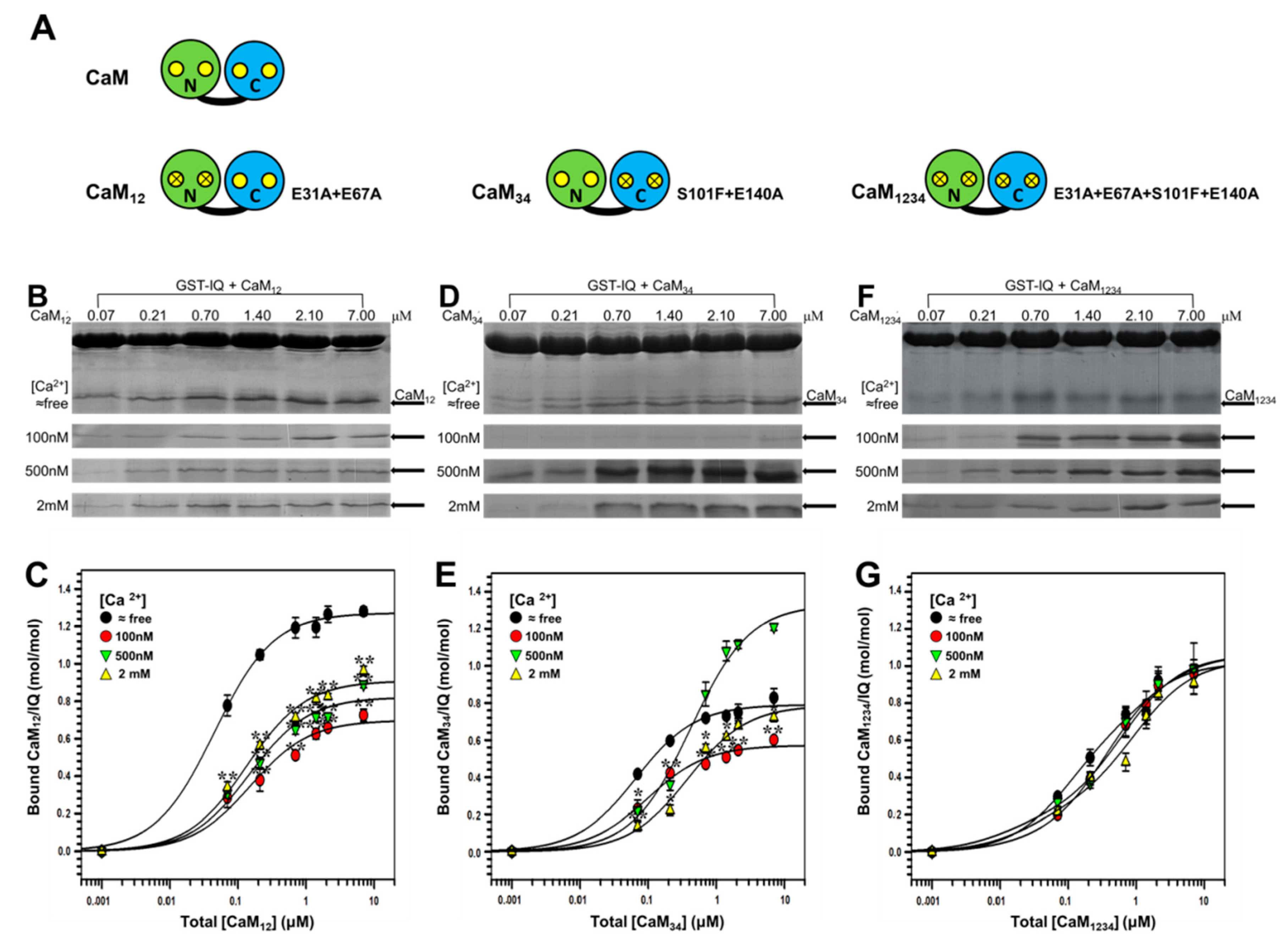
2.2. Binding of CaM Mutants to NaV1.1 IQ Domain

To further clarify the regulatory mechanism of CaM on NaV1.1 channel, we then examined the binding properties of CaM mutants to NaV1.1 IQ domain. This included CaM12 and CaM34 (Figure 3A) in which Ca2+-binding to its N- and C-lobe was eliminated, respectively, and a Ca2+-insensitive CaM mutant (CaM1234) (Figure 3A) [26]. As shown in Figure 3B,C, the binding of CaM12 to IQ was qualitatively similar to that of the wild-type (wt) CaM. The maximal binding estimated as Bmax was 1.27, 0.70, 0.82, and 0.91 mol/mol CaM12/IQ (n = 4) at ≈free, 100 nM, 500 nM and 2 mM Ca2+, respectively, showing an obvious Ca2+ dependence in the presence of Ca2+ (Table 1). It is interesting to note that the Bmax of CaM12 at ≈free Ca2+ is ~30% was greater than that at 2 mM Ca2+, suggesting that like wt CaM, CaM12 has the highest affinity for IQ in the absence of Ca2+.
Next, we examined the binding of CaM34 to IQ domain. As shown in Figure 3D,E, the binding of CaM34 to IQ was also concentration-dependent. The parameters obtained (Table 1) revealed that the maximal binding estimated as Bmax was 0.79, 0.57, 1.33, and 0.79 mol/mol CaM34/ IQ (n = 4) at ≈free, 100 nM, 500 nM, and 2 mM Ca2+, respectively. It was noted that this profile of [Ca2+] dependence was different from those of wt CaM and CaM12.
Furthermore, we examined the binding of CaM1234 to NaV1.1 IQ domain. As shown in Figure 3F, the Ca2+ dependence was totally diminished because of the mutation in four Ca2+ binding sites. As shown in Figure 3G and Table 1, the Bmax were 1.06, 1.02, 1.06, and 1.03 mol/mol (CaM1234/IQ) at ≈free, 100 nM, 500 nM, and 2 mM [Ca2+], respectively. The values of Bmax and Kd were not significantly different among different Ca2+ concentrations, confirming the Ca2+-insensitive nature of CaM1234. Our data showed that functional Ca2+ binding sites of either N- or C-lobe were required for Ca2+ dependency in CaM binding to NaV1.1 IQ domain.

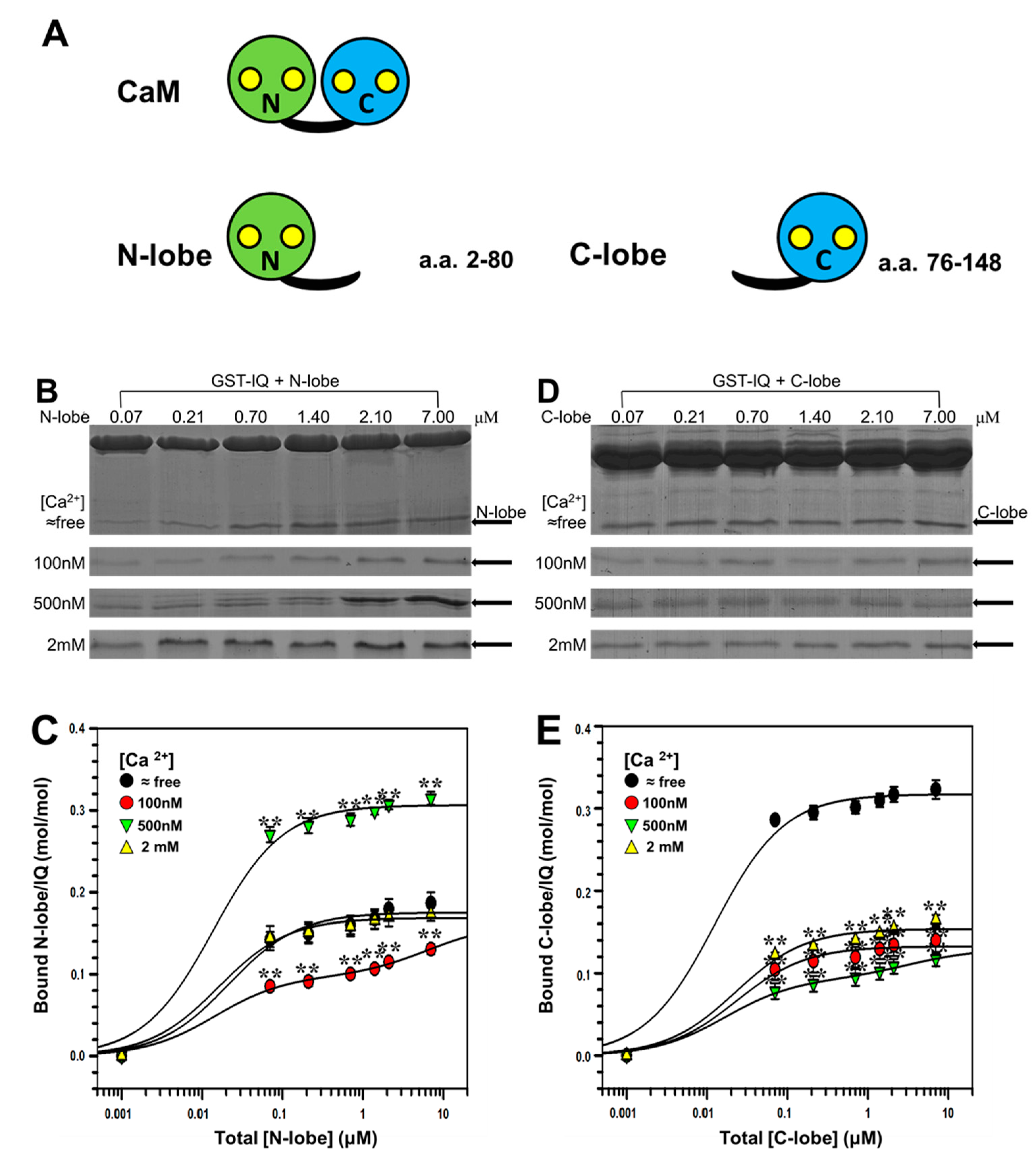
2.3. Binding of Individual N-Lobe or C-Lobe of CaM to NaV1.1 IQ Domain

In order to study the effect of specific lobe of CaM on the NaV1.1 IQ, we first computationally investigated the interactions between N-lobe or C-lobe of CaM and NaV1.1 IQ domain using Discovery Studio 2017. As shown in Figure 4A,B, the ZDock score and E_RDock for the optimal N-lobe orientation docking into NaV1.1 IQ domain were 11.28 and −23.31 kcal/mol, respectively, whereas these parameters for the best interaction of C-lobe with IQ domain were 10.66 and −26.21 kcal/mol, respectively. In addition, as shown in Figure 4C, when the N- and C- lobe were docked together into the NaV1.1 IQ domain, the best ZDock score and lowest E_RDock were 12.78 and −30.72 kcal/mol, respectively.
Next, we further examined the bindings of individual (truncated) N- and C-lobe of CaM (Figure 5A) to IQ domain under different Ca2+ concentrations by pull-down assay. As shown in Figure 5B,C and Table 2, the maximal bindings estimated as Bmax for N-lobe were 0.18, 0.16, 0.31, and 0.17 mol/mol (N-lobe/IQ) at ≈free, 100 nM, 500 nM, and 2 mM [Ca2+], respectively (n = 4). Thus the profile of [Ca2+] dependence of N-lobe was similar to that of CaM34.
We then examined the binding of C-lobe to IQ domain. As shown in Figure 5D, like wt CaM, C-lobe also had the highest affinity with IQ at ≈free [Ca2+]. The maximal binding presented by Bmax were 0.32, 0.13, 0.13, and 0.15 mol/mol (C-lobe/IQ) at ≈free, 100 nM, 500 nM, and 2 mM [Ca2+], respectively (n = 4) (Figure 5E and Table 2). Thus, the profile of [Ca2+] dependence of C-lobe was similar to those of wt CaM and CaM12.
The parameters (Table 2) revealed that binding of both N- and C-lobe to IQ was also in a Ca2+-dependent manner. Kd of N-lobe was lower than that of C-lobe in the presence of Ca2+ ([Ca2+] ≥ 100 nM), whereas Kd of C-lobe was lower than that of N-lobe in the absence of Ca2+. Additionally, Bmax of N-lobe was higher than that of C-lobe in the presence of Ca2+, whereas Bmax of C-lobe was higher than that of N-lobe in the absence of Ca2+, indicating that C-lobe was the predominant domain in apoCaM interacting with NaV1.1 IQ domain, and N-lobe was the predominant domain in Ca2+/CaM interacting with NaV1.1 IQ domain.

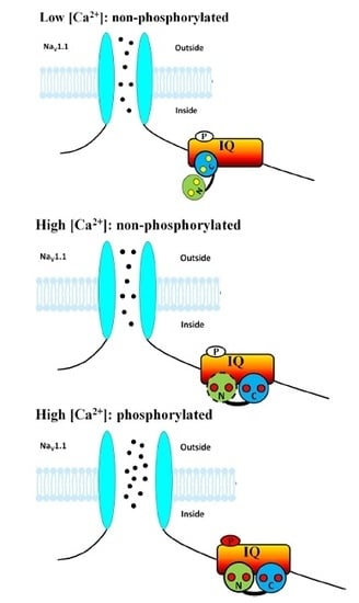
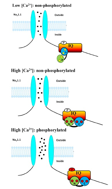
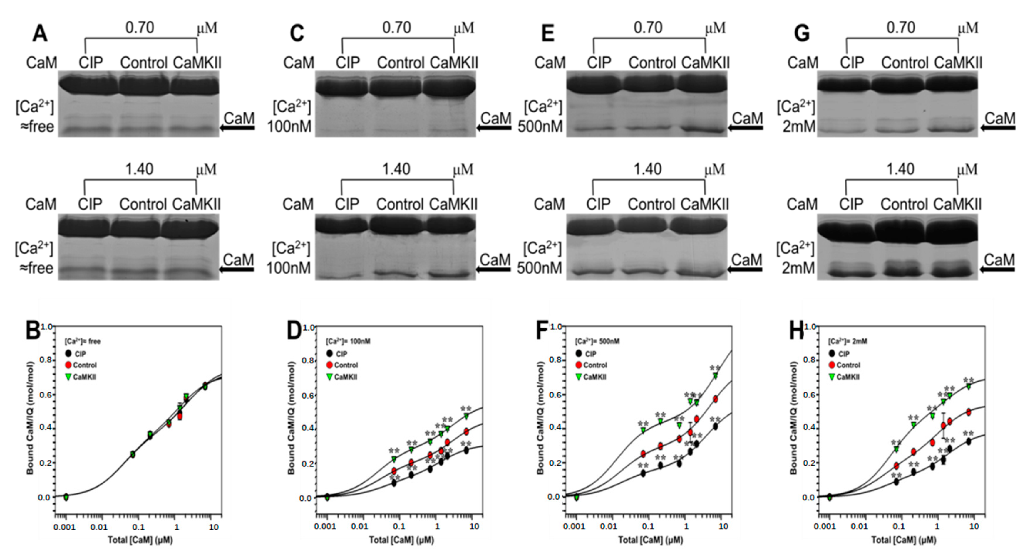
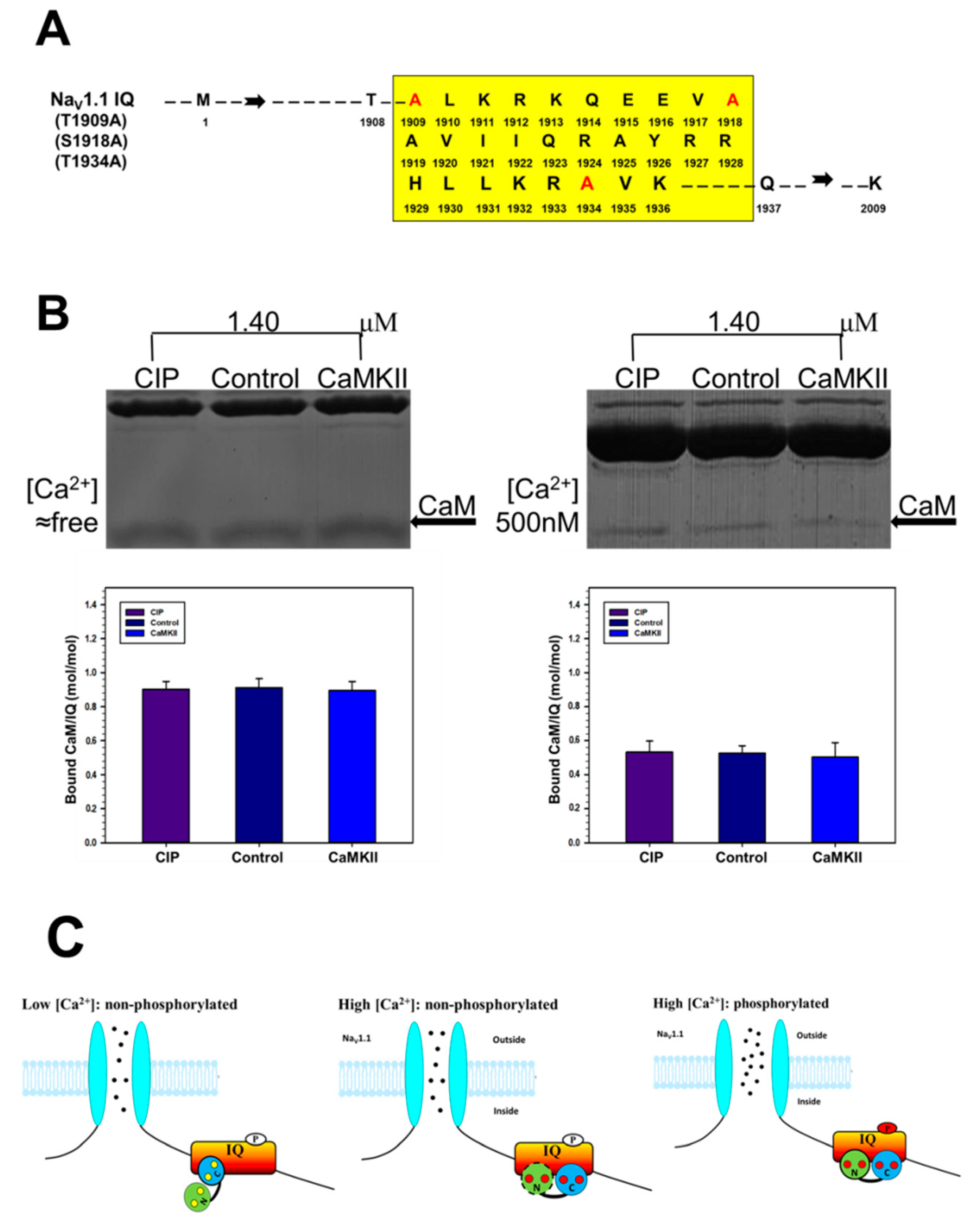
2.4. The Effect of CaMKII on CaM Binding to NaV1.1 IQ Domain

In the previous study, several CaMKII-mediated phosphorylation sites on NaV1.1 have been identified [29]. To clarify the modulation of CaMKII on NaV1.1, we further checked the effect of CaMKII on CaM binding to NaV1.1 IQ domain. As shown in Figure 6A,B, the binding of apoCaM to IQ barely changed after phosphorylation at ≈free [Ca2+] compared to that in the presence of Ca2+, suggesting that phosphorylation of IQ domain by CaMKII had little effect on its binding with apoCaM. By contrast, the binding of Ca2+/CaM to IQ was increased in the phosphorylated IQ, indicating that the effect of CaMKII on the CaM binding to NaV1.1 IQ domain could exert its regulation only in the presence of Ca2+ (Figure 6C,E,G). The parameters obtained (Figure 6D,F,H and Table 3) revealed that CaMKII-mediated phosphorylation increased the binding of CaM to IQ, while dephosphorylation by CIP decreased the affinity of CaM with IQ. In addition, Kd and Bmax showed that an increased binding of Ca2+/CaM to the channel at higher [Ca2+] was observed compared to that at 100 nM [Ca2+].
In a previous study, one CaMKII-mediated phosphorylation site S1920 on NaV1.5 IQ domain had been identified. In addition, CaMKII is a basophilic protein kinase belonging to the Ca2+/CaM dependent superfamily of serine/threonine kinases (Herren et al., 2015). Thus, we mutated three potential CaMKII-mediated phosphorylation sites—T1909, S1918, and T1934—into A (Figure 7A), then NaV1.1 IQ domain was treated with CaMKII and CIP. As shown in Figure 7B, under free [Ca2+] condition, there was no significant difference in the CaM binding to the IQ domain between the phosphorylated and dephosphorylated peptides as well as 500 nM [Ca2+] condition, indicating that CaMKII facilitated the binding of Ca2+/CaM to NaV1.1 IQ domain due to one or several phosphorylation sites in T1909, S1918, and T1934 of NaV1.1 IQ domain.

3. Materials and Methods

3.1. cDNA Construction and Site-Directed Mutagenesis

The cDNA corresponding to the IQ domain of NaV1.1 (IQ, amino acids 1909–1936) was generated by PCR using the cDNA of human NaV1.1 as the template. The primers were designed using VectorNTI software. The human CaM and its truncated proteins—N-lobe (a.a. 2–80) and C-lobe (a.a. 76–148)—were subcloned from HEK293 cells [21,34,35]. The CaMKII mutant T286D (CaMKIIT286D), which is a constitutively active type of CaMKII in the absence of CaM and Ca2+, was created with rat CaMKIIα cDNA as a template [36]. Mutants including IQ mutant (I1922E, Q1923E), potential CaMKII phosphorylation sites mutant (T1909A + S1918A + T1934A), CaM12 (E31A + E67A), CaM34 (S101F + E140A), and CaM1234 (E31A + E67A + S101F + E140A) were constructed by site-directed mutagenesis using a Quickchange™ kit (QIAGEN) [37,38]. The above DNAs were individually ligated into pGEX-6P-1 H320 expression vectors (GE Biosciences, New York, NY, USA).

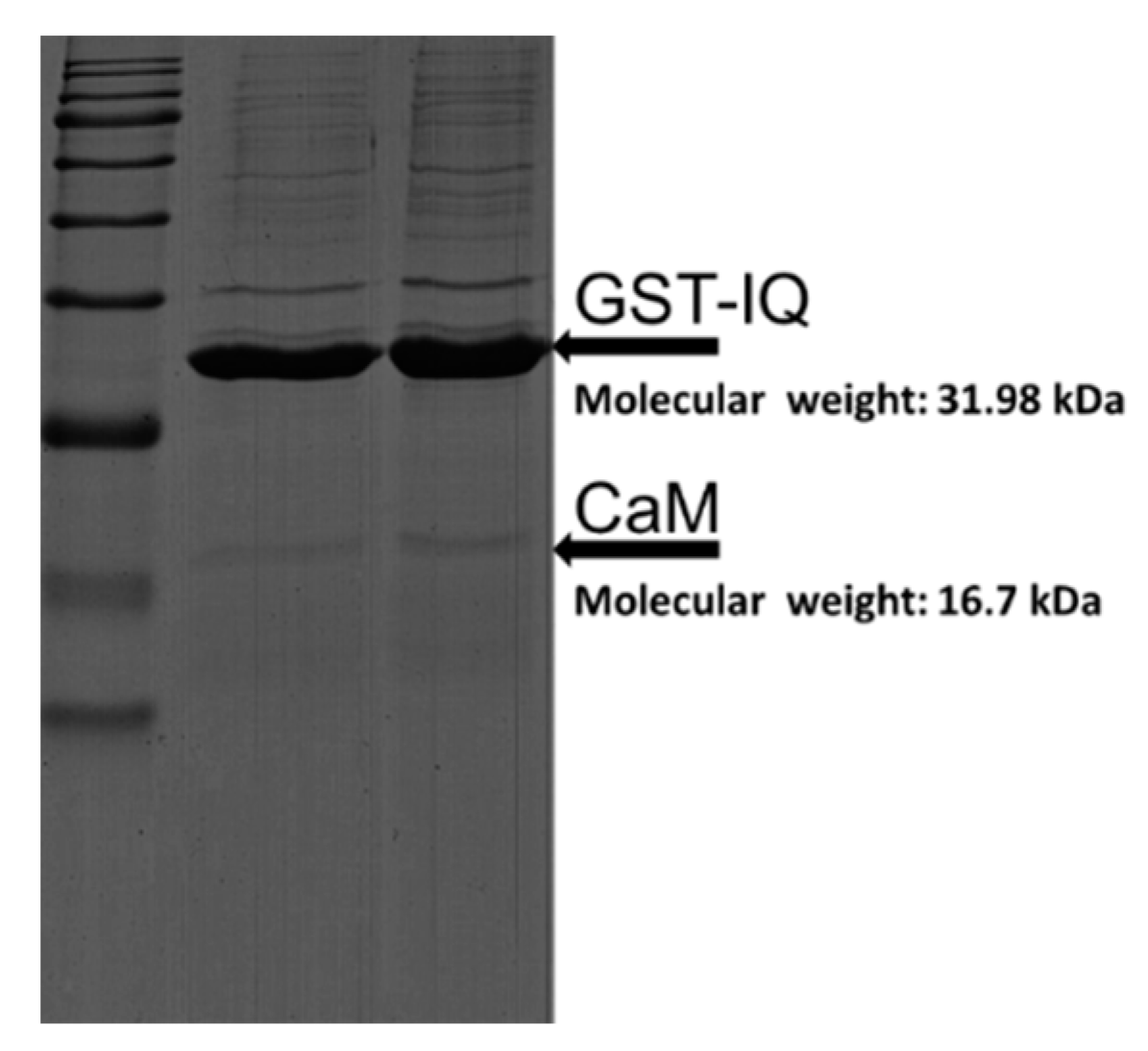
3.2. Expression and Purification of Recombinant GST Fusion Peptides

The above described vectors were transformed into Escherichia coli BL21 (DE3) to express the target peptides as glutathione-S-transferase (GST) fusion proteins. The bacteria were cultured in LB liquid medium at 37 °C overnight until an OD600 of 0.8–1.0. Then, the bacteria were induced with 1 mM isopropyl-1-thio-β-d-galactopyranoside (IPTG) and continued incubating for 4 h at 37 °C before harvesting. The ultrasonic technique was used to harvest GST-fusion peptides. Then, the fusion peptides were purified using Glutathione Sepharose 4B beads (GS-4B, GE Healthcare, New York, NY, USA). The GST regions of CaM and its mutants were cleaved with PreScission Protease (GE Healthcare). GST-IQ, CaM, and its mutants were quantified by Enhanced BCA Protein Assay Kit with BSA as standard with correction factors 1.25 (GST-IQ), 1.69 (CaM and its full-length mutants), 0.82 (N-lobe of CaM), and 0.82 (C-lobe of CaM).

3.3. GST Pull-Down Assay

GST-fusion IQ or its mutant (2–4 μg) was immobilized on GS-4B and incubated in 300 μL of Tris buffer (consisting of 150 mM NaCl, 50 mM Tris, and pH 8.0 adjusted by HCl) with increasing concentrations of CaM or its mutants (0.07, 0.21, 0.7, 1.4, 2.1, 7.0 μm) for 4 h at 4 °C under agitation in the presence of different Ca2+ concentrations ([Ca2+] ≈ free, 100, 500, and 2 mM). The [Ca2+] was calculated with MaxChelator (http://maxchelator.stanford.edu/index.html). Then, the reaction systems were gently washed twice with the same buffer. Bound CaM (or its mutant) and IQ (or EQ) were resuspended in 5× SDS-PAGE loading buffer and resolved in 15% SDS-PAGE gels. Proteins were stained by Coomassie brilliant blue R (CBB). Protein bands in the SDS-PAGE gels were digitized by the Photoshop software (Adobe, San Jose, CA, USA), and the grey level was quantified by Image J software (NIH, Bethesda, MD, USA) [34,35,36,37]. The optical density values were converted to protein contents using respective correction factors (see below).
The GST-fusion IQ (for control) immobilized to GS-4B beads (40 μL) was phosphorylated in an assay reaction (0.4 μm CaMKIIT286D) in Tris buffer containing 1 mM MgCl2 and 1 mM Na2ATP for 30 min at 30 °C. The reaction was then terminated with the addition of 1× SDS sample buffer and gently washed twice. The dephosphorylation was achieved by adding 5 U/mL calf intestinal alkaline phosphatase (CIP; New England Biolabs, Ipswich, MA, USA) into the reacting mixtures incubating at 37 °C for 30 min. CIP is a nonspecific phosphorylase that commonly exists in calf intestinal mucosa. The reaction was terminated by the addition of same Tris buffer and gently washed twice.

3.4. Computational Docking

A homology model of NaV1.1 IQ was constructed based upon the solved crystal structure of IQ motif of NaV1.2 (PDB # 2KXW) [15] using the Create Homology Model tool in Discovery Studio 2017 (BIOVIA, Boston, MA, USA). The N- and C-lobe of CaM were derived from the crystal structure of Ca2+/CaM-CaV1.2 IQ domain complex (PDB # 3DVE) [39]. Docking studies between NaV1.1 IQ and CaM N- or C-lobe were performed in Discovery Studio using the Dock Proteins protocols. The ZDOCK protocol was used for docking the IQ motif of NaV1.1 to N- or/and C-lobe of CaM and, subsequently, the RDOCK protocol was applied for further refinement of the 10 best-docked poses. For individual interactions, docking results are displayed as solid ribbons of 1 solution with the lowest RDOCK interaction energy.

3.5. Statistical Analysis

Quantified grey level was converted into molar quantities according to the mass of GST-IQ, CaM, N-lobe, and C-lobe of CaM. We found that relative optical densities of the same amount of these proteins on the gel in reference to BSA were 0.80, 0.59, 1.21, and 1.22 respectively, from which the correction factors were determined as 1.25, 1.69, 0.82, and 0.82, respectively. Curve-fitting of the total bound ligand (CaM and its mutants) was performed with the software SigmaPlot 12.0 (version 12, Sigma-Aldrich, Beijing, China). Bound ligand (y) was fitted with the following Hill’s equation for the one-site fitting model: y = Bmax·x/(Kd + x). For the two-sites model, a sum of two Hill’s equations was integrated to assume independent binding: y = Bmax1·x/(Kd1 + x) + Bmax2·x/(Kd2 + x), where Bmax1, Bmax2, Kd1, and Kd2 represents total Bmax and Kd, respectively; x is the concentration of free ligand; Kd is the apparent dissociation constant; and Bmax is the maximum binding for each binding site. We chose either one-site model or two-sites model based on the higher value of R2. Hill’s coefficient of 1.0 was assumed. Total concentration of ligand was assumed as an approximate of free ligand. The data are presented as mean ± S.E. (n = 4). The SPSS 22.0 software (version 22, Sigma-Aldrich, Beijing, China) was used to evaluate the statistical significance, and p < 0.05 by hypothesis test was considered statistically significant.

4. Discussion

Our study was aimed at clarifying the molecular mechanism underlying the modulation of Ca2+/CaM/CaMKII on NaV1.1 channel, which is a key issue in understanding the regulatory mechanism of VGSCs. Although the modulation of VGSCs has been a hot spot in ion channel research, the present study examined for the first time the effects of Ca2+ and CaM on NaV1.1 channel in a wide range of [Ca2+] using CaM mutants.
CaM is the most important Ca2+ binding protein and is involved in the regulation of numerous Ca2+-dependent pathways. Its function and structure depend strongly on Ca2+ concentration [40,41]. In our research, we checked the effect of different Ca2+ concentrations on the binding of CaM to IQ domain of NaV1.1. Our data showed that the binding of CaM to NaV1.1 IQ domain was Ca2+- and concentration-dependent. Full-length CaM switches from a simple folding structure at lower [Ca2+] to a rich and complex folding behavior at high [Ca2+] [41]. Accordingly, similar Ca2+-dependent conformational changes in CaM between NaV1.2 and NaV1.5 have previously been reported [8,40]. Our results showed that the binding of CaM to NaV 1.1 IQ was Ca2+-dependent, which reflects the Ca2+-dependent conformational change of CaM.
CaM often modulates target molecules only upon conversion to its Ca2+-bound form. However, apoCaM binding in itself markedly promotes opening of voltage-gated calcium channels (VGCCs) [9,11,34,41]. VGSCs have also been suggested to adopt a similar modulatory principle [9,16]. Our present data showed that the binding of Ca2+/CaM to NaV1.1 IQ was dramatically decreased compared to that of apoCaM, implying that apoCaM promotes activity of NaV1.1 channels. On the contrary, studies on CaV1.2 indicated that the binding of apoCaM to the channel was significantly smaller compared to that of Ca2+/CaM [21,37,42]. This may suggest that CaM binds to different channels in a channel-specific manner, meaning the detailed mechanism of CaM regulation may need to be considered on a channel-specific basis.
It has been reported that regulatory effect of CaM on VGCCs is lobe-specific, and N- and C-lobe of CaM have distinct roles in the regulation of VGCCs [7,40,43]. In addition, a single Ca2+/CaM bridges the C-terminal IQ motif of NaV1.5 to the DIII-IV linker via individual N- and C-lobes, respectively. C-lobe binds to IQ (N-lobe is free) at low [Ca2+], whereas at high [Ca2+], N-lobe binds to IQ (lobe switching) and C-lobe binds to III-IV linker, resulting in depolarization of the inactivation curve [44]. Furthermore, the most prominent Ca2+-dependent conformational change is the interaction between the calcified N-lobe of CaM and the NaV IQ domain; the CaM C-lobe remains Ca2+-free even in millimolar Ca2+, remains bound to the IQ motif, and retains its semi-open conformation. Despite similar Ca2+-dependent conformational changes between the NaV1.2 and NaV1.5 complexes, the functional effects are isoform-specific, while their mechanistic bases are not clear [40]. However, the lobe specificity of CaM modulation of NaV1.1 have not been demonstrated. In this study, we applied individual N- and C-lobe of CaM to further explore the role of individual lobes in binding to NaV1.1 IQ domain. The affinity of IQ for C-lobe binding was higher than that of N-lobe in the absence of Ca2+, whereas the affinity of IQ for N-lobe binding was higher than that of C-lobe in the presence of Ca2+, indicating distinct responses at different [Ca2+]. Thus, the property difference between the two lobes of CaM might endow the Ca2+-dependent and lobe-specific modulation of NaV1.1 channel. However, we still do not know the reason why CaM binding to the IQ domain is the largest at free Ca2+ and smaller at 100 nM Ca2+. We speculate that C-lobe of CaM might be at least partially occupied with Ca2+ at 100 nM Ca2+. In this scenario, the weaker binding of CaM to the IQ domain would be explained by the fact that the IQ domain has lower affinity for Ca2+/C-lobe than for Ca2+-free C-lobe. This point may be supported by the CaM12 and CaM34 experiments (Figure 3B–E). In the CaM12 experiment, the binding property to the IQ at free and 100 nM Ca2+ was similar to that of wild-type CaM, while this property was less pronounced in the CaM34 experiment.
One of the most intriguing findings of our research is that CaMKII-mediated phosphorylation of NaV1.1 IQ domain increased binding of CaM to the channel. It has become recognized that both the expression and function of VGSCs is under tight control of protein phosphorylation by protein kinases [45]. CaMKII activated by Ca2+/CaM maintains activity of VGCCs [36], CaMKII has emerged as a critical regulator of NaV1.5, and multiple CaMKII-mediated phosphorylation sites have been identified on NaV1.5, including S1920 and S1925, which are located on IQ domain and noncanonical CaMKII sites. [29,46]. In addition, CaMKII-enhanced INaL positively shifts inactivation curve of NaV1.2 epileptic mutant (Q54) [23,47]. Based on the structural homology of NaV1.1 and NaV1.5, we treated NaV1.1 IQ domain with CaMKII and CIP to examine a possible regulation of NaV1.1 by CaMKII. Under low [Ca2+] condition, we found there was no significant difference in the CaM binding to the IQ domain between the phosphorylated and dephosphorylated peptides. One possible reason for this may be that the phosphorylation of NaV1.1 IQ might not affect the conformation of the apoCaM binding region, which interacts mainly with C-lobe of CaM. However, in the presence of Ca2+ CaMKII-mediated phosphorylation of IQ increased the binding of CaM to IQ domain, while CIP-mediated dephosphorylation of IQ decreased the binding of CaM. Thus, it is possible that Ca2+/CaM binding region interacts mainly with N-lobe of CaM, which might be different from the apoCaM binding region. Our data has shown that CaMKII regulates the binding of Ca2+/CaM to NaV1.1 IQ domain due to one or several phosphorylation sites in T1909, S1918, and T1934 of NaV1.1 IQ domain, indicating that CaMKII-mediated phosphorylation of NaV1.1 affects CaM binding to NaV1.1. It is therefore speculated that CaMKII-mediated phosphorylation of NaV1.1 IQ domain might change the conformation of IQ domain, leading to increased binding of Ca2+/CaM to NaV1.1.
A previous study had shown that CaM overexpression in HEK1.1 cells increases the peak current of NaV1.1 in a calcium-dependent manner [12]. Our previous study has also demonstrated that neuronal VGSC activity is modulated by CaM in a concentration-dependent manner in normal and low Mg2+ condition [13]. Thus, we propose the following hypothetical model (Figure 7C) for the modulation of Ca2+/CaM/CaMKII on NaV1.1 based on our present study and other studies [12,13]: At low [Ca2+]—a nonphosphorylated state of Ca2+ concentration—C-lobe is the predominant domain for apoCaM binding to IQ domain, and the binding of apoCaM to NaV1.1 IQ is not affected by CaMKII. At high [Ca2+] but at a nonphosphorylated state of IQ domain, N-lobe becomes the predominant domain since some of the CaM binds with Ca2+. Phosphorylation of IQ by CaMKII modulates the binding of Ca2+/CaM to the channel. Meanwhile, channel activity is maintained to the basal level at 80–100 nM [Ca2+]. At high [Ca2+]—a phosphorylated state of IQ domain—N-lobe is the predominant domain for Ca2+/CaM binding to IQ domain. Meanwhile, the effect of CaMKII-mediated phosphorylation is further promoted, leading to an increased binding of Ca2+/CaM to the channel compared to that at 80–100 nM [Ca2+]. The channel activity will also be increased at high [Ca2+] compared to that at 80–100 nM [Ca2+].
In summary, we found that the binding of Ca2+/CaM to IQ was Ca2+- and concentration-dependent, and apoCaM more preferentially binds to NaV1.1 IQ domain than Ca2+/CaM. In addition, C-lobe of CaM is the predominant domain in apoCaM binding to NaV1.1 IQ domain, whereas N-lobe of CaM is the predominant domain in Ca2+/CaM binding to NaV1.1 IQ domain. In addition, CaMKII-mediated phosphorylation increases the binding of Ca2+/CaM to NaV1.1 IQ domain due to one or several phosphorylation sites in T1909, S1918, and T1934 of NaV1.1 IQ domain. Our data provides novel mechanisms for the modulation of NaV1.1 by the Ca2+/CaM/CaMKII axis. For the first time, we uncover the effect of Ca2+, lobe-specificity, and CaMKII on CaM binding to NaV1.1.

Author Contributions

F.G., M.K. and H.-L.J.: Contributed to study design and manuscript editing. J.L. (Jianing Li) and Z.Y.: Contributed to data acquisition and manuscript preparation. J.X., R.F., Q.G., T.B., J.L. (Junyan Liu), Z.L., Q.W., M.L., J.G., H.H. and E.M. Contributed to data acquisition.

Acknowledgments

This work was supported by grants from the Natural Science Foundation of China (81471323 and 31400981), Japan Society for Promotion of Science (16K08499 and 15K08181), American Heart Association (16GRNT30780002) and National Institute of Health (HL 134828).

Conflicts of Interest

The authors declare no conflict of interest.

References

  1. Adams, P.J.; Ben-Johny, M.; Dick, I.E.; Inoue, T.; Yue, D.T. Apocalmodulin itself promotes ion channel opening and Ca(2+) regulation. Cell 2014, 159, 608–622. [Google Scholar] [CrossRef] [PubMed]
  2. Asmara, H.; Minobe, E.; Saud, Z.A.; Kameyama, M. Interactions of calmodulin with the multiple binding sites of CaV1.2 Ca2+ channels. J. Pharmacol. Sci. 2010, 112, 397–404. [Google Scholar] [CrossRef] [PubMed]
  3. Baek, J.H.; Cerda, O.; Trimmer, J.S. Mass spectrometry-based phosphoproteomics reveals multisite phosphorylation on mammalian brain voltage-gated sodium and potassium channels. Semin. Cell Dev. Biol. 2011, 22, 153–159. [Google Scholar] [CrossRef] [PubMed] [Green Version]
  4. Baek, J.H.; Rubinstein, M.; Scheuer, T.; Trimmer, J.S. Reciprocal changes in phosphorylation and methylation of mammalian brain sodium channels in response to seizures. J. Biol. Chem. 2014, 289, 15363–15373. [Google Scholar] [CrossRef] [PubMed]
  5. Bahler, M.; Rhoads, A. Calmodulin signaling via the IQ motif. FEBS Lett. 2002, 513, 107–113. [Google Scholar] [CrossRef]
  6. Berendt, F.J.; Park, K.S.; Trimmer, J.S. Multisite phosphorylation of voltage-gated sodium channel alpha subunits from rat brain. J. Proteome Res. 2010, 9, 1976–1984. [Google Scholar] [CrossRef] [PubMed]
  7. Chagot, B.; Chazin, W.J. Solution NMR structure of Apo-calmodulin in complex with the IQ motif of human cardiac sodium channel NaV1.5. J. Mol. Biol. 2011, 406, 106–119. [Google Scholar] [CrossRef] [PubMed]
  8. Chen, Z.; Zhao, R.; Zhao, M.; Liang, X.; Bhattarai, D.; Dhiman, R.; Shetty, S.; Idell, S.; Ji, H.L. Regulation of epithelial sodium channels in urokinase plasminogen activator deficiency. Am. J. Physiol. Lung Cell. Mol. Physiol. 2014, 307, L609–L617. [Google Scholar] [CrossRef] [PubMed] [Green Version]
  9. Dick, I.E.; Tadross, M.R.; Liang, H.; Tay, L.H.; Yang, W.; Yue, D.T. A modular switch for spatial Ca2+ selectivity in the calmodulin regulation of CaV channels. Nature 2008, 451, 830–834. [Google Scholar] [CrossRef] [PubMed]
  10. Feldkamp, M.D.; Yu, L.; Shea, M.A. Structural and energetic determinants of apo calmodulin binding to the IQ motif of the Na(V)1.2 voltage-dependent sodium channel. Structure 2011, 19, 733–747. [Google Scholar] [CrossRef] [PubMed]
  11. Gaudioso, C.; Carlier, E.; Youssouf, F.; Clare, J.J.; Debanne, D.; Alcaraz, G. Calmodulin and calcium differentially regulate the neuronal Nav1.1 voltage-dependent sodium channel. Biochem. Biophys. Res. Commun. 2011, 411, 329–334. [Google Scholar] [CrossRef] [PubMed]
  12. Guo, F.; Minobe, E.; Yazawa, K.; Asmara, H.; Bai, X.Y.; Han, D.Y.; Hao, L.Y.; Kameyama, M. Both N- and C-lobes of calmodulin are required for Ca2+-dependent regulations of CaV1.2 Ca2+ channels. Biochem. Biophys. Res. Commun. 2010, 391, 1170–1176. [Google Scholar] [CrossRef] [PubMed]
  13. Guo, F.; Zhou, P.D.; Gao, Q.H.; Gong, J.; Feng, R.; Xu, X.X.; Liu, S.Y.; Hu, H.Y.; Zhao, M.M.; Adam, H.C.; et al. Low-Mg(2+) treatment increases sensitivity of voltage-gated Na(+) channels to Ca(2+)/calmodulin-mediated modulation in cultured hippocampal neurons. Am. J. Physiol. Cell Physiol. 2015, 308, C594–C605. [Google Scholar] [CrossRef] [PubMed]
  14. Han, D.Y.; Minobe, E.; Wang, W.Y.; Guo, F.; Xu, J.J.; Hao, L.Y.; Kameyama, M. Calmodulin- and Ca2+-dependent facilitation and inactivation of the CaV1.2 Ca2+ channels in guinea-pig ventricular myocytes. J. Pharmacol. Sci. 2010, 112, 310–319. [Google Scholar] [CrossRef] [PubMed]
  15. Han, S.; Tai, C.; Westenbroek, R.E.; Yu, F.H.; Cheah, C.S.; Potter, G.B.; Rubenstein, J.L.; Scheuer, T.; de la Iglesia, H.O.; Catterall, W.A. Autistic-like behaviour in Scn1a+/− mice and rescue by enhanced GABA-mediated neurotransmission. Nature 2012, 489, 385–390. [Google Scholar] [CrossRef] [PubMed] [Green Version]
  16. Hao, L.Y.; Wang, W.Y.; Minobe, E.; Han, D.Y.; Xu, J.J.; Kameyama, A.; Kameyama, M. The distinct roles of calmodulin and calmodulin kinase II in the reversal of run-down of L-type Ca(2+) channels in guinea-pig ventricular myocytes. J. Pharmacol. Sci. 2009, 111, 416–425. [Google Scholar] [CrossRef] [PubMed]
  17. He, G.; Guo, F.; Zhu, T.; Shao, D.; Feng, R.; Yin, D.; Sun, X.; Hu, H.; Hwang, A.; Minobe, E.; et al. Lobe-related concentration- and Ca(2+)-dependent interactions of calmodulin with C- and N-terminal tails of the CaV1.2 channel. J. Physiol. Sci. 2013, 63, 345–353. [Google Scholar] [CrossRef] [PubMed]
  18. Herren, A.W.; Weber, D.M.; Rigo, R.R.R.; Margulies, K.B.; Phinney, B.S.; Bers, D.M. CaMKII Phosphorylation of Na(V)1.5: Novel in Vitro Sites Identified by Mass Spectrometry and Reduced S516 Phosphorylation in Human Heart Failure. J. Proteome Res. 2015, 14, 2298–2311. [Google Scholar] [CrossRef] [PubMed]
  19. Hund, T.J.; Koval, O.M.; Li, J.; Wright, P.J.; Qian, L.; Snyder, J.S.; Gudmundsson, H.; Kline, C.F.; Davidson, N.P.; Cardona, N.; et al. A beta(IV)-spectrin/CaMKII signaling complex is essential for membrane excitability in mice. J. Clin. Investig. 2010, 120, 3508–3519. [Google Scholar] [CrossRef] [PubMed]
  20. Kim, E.Y.; Rumpf, C.H.; Fujiwara, Y.; Cooley, E.S.; van Petegem, F.; Minor, D.L., Jr. Structures of CaV2 Ca2+/CaM-IQ domain complexes reveal binding modes that underlie calcium-dependent inactivation and facilitation. Structure 2008, 16, 1455–1467. [Google Scholar] [CrossRef] [PubMed]
  21. Lazrak, A.; Nita, I.; Subramaniyam, D.; Wei, S.; Song, W.; Ji, H.L.; Janciauskiene, S.; Matalon, S. Alpha(1)-antitrypsin inhibits epithelial Na+ transport in vitro and in vivo. Am. J. Respir. Cell Mol. Biol. 2009, 41, 261–270. [Google Scholar] [CrossRef] [PubMed]
  22. Miao, Y.; Zhang, W.; Lin, Y.; Lu, X.; Qiu, Y. Neuroprotective effects of ischemic preconditioning on global brain ischemia through up-regulation of acid-sensing ion channel 2a. Int. J. Mol. Sci. 2010, 11, 140–153. [Google Scholar] [CrossRef] [PubMed]
  23. Minobe, E.; Asmara, H.; Saud, Z.A.; Kameyama, M. Calpastatin domain L is a partial agonist of the calmodulin-binding site for channel activation in CaV1.2 Ca2+ channels. J. Biol. Chem. 2011, 286, 39013–39022. [Google Scholar] [CrossRef] [PubMed]
  24. Pitt, G.S.; Lee, S.Y. Current view on regulation of voltage-gated sodium channels by calcium and auxiliary proteins. Protein Sci. 2016, 25, 1573–1584. [Google Scholar] [CrossRef] [PubMed] [Green Version]
  25. Potet, F.; Chagot, B.; Anghelescu, M.; Viswanathan, P.C.; Stepanovic, S.Z.; Kupershmidt, S.; Chazin, W.J.; Balser, J.R. Functional Interactions between Distinct Sodium Channel Cytoplasmic Domains through the Action of Calmodulin. J. Biol. Chem. 2009, 284, 8846–8854. [Google Scholar] [CrossRef] [PubMed] [Green Version]
  26. Sarhan, M.F.; Tung, C.C.; van Petegem, F.; Ahern, C.A. Crystallographic basis for calcium regulation of sodium channels. Proc. Natl. Acad. Sci. USA 2012, 109, 3558–3563. [Google Scholar] [CrossRef] [PubMed]
  27. Saud, Z.A.; Minobe, E.; Wang, W.Y.; Han, D.Y.; Horiuchi, M.; Hao, L.Y.; Kameyama, M. Calpastatin binds to a calmodulin-binding site of cardiac CaV1.2 Ca2+ channels. Biochem. Biophys. Res. Commun. 2007, 364, 372–377. [Google Scholar] [CrossRef] [PubMed]
  28. Scheuer, T. Regulation of sodium channel activity by phosphorylation. Semin. Cell Dev. Biol. 2011, 22, 160–165. [Google Scholar] [CrossRef] [PubMed] [Green Version]
  29. Shah, V.N.; Wingo, T.L.; Weiss, K.L.; Williams, C.K.; Balser, J.R.; Chazin, W.J. Calcium-dependent regulation of the voltage-gated sodium channel hH1: Intrinsic and extrinsic sensors use a common molecular switch. Proc. Natl. Acad. Sci. USA 2006, 103, 3592–3597. [Google Scholar] [CrossRef] [PubMed] [Green Version]
  30. Shao, D.; Zhao, M.; Xu, J.; Feng, R.; Guo, F.; Hu, H.; Sun, X.; Gao, Q.; He, G.; Sun, W.; et al. The individual N- and C-lobes of calmodulin tether to the CaV1.2 channel and rescue the channel activity from run-down in ventricular myocytes of guinea-pig heart. FEBS Lett. 2014, 588, 3855–3861. [Google Scholar] [CrossRef] [PubMed]
  31. Shifman, J.M.; Choi, M.H.; Mihalas, S.; Mayo, S.L.; Kennedy, M.B. Ca2+/calmodulin-dependent protein kinase II (CaMKII) is activated by calmodulin with two bound calciums. Proc. Natl. Acad. Sci. USA 2006, 103, 13968–13973. [Google Scholar] [CrossRef] [PubMed]
  32. Stefan, M.I.; Marshall, D.P.; le Novere, N. Structural analysis and stochastic modelling suggest a mechanism for calmodulin trapping by CaMKII. PLoS ONE 2012, 7, e29406. [Google Scholar] [CrossRef] [PubMed]
  33. Turabekova, M.A.; Rasulev, B.F.; Levkovich, M.G.; Abdullaev, N.D.; Leszczynski, J. Aconitum and Delphinium sp. alkaloids as antagonist modulators of voltage-gated Na+ channels. AM1/DFT electronic structure investigations and QSAR studies. Comput. Biol. Chem. 2008, 32, 88–101. [Google Scholar] [CrossRef] [PubMed]
  34. Stigler, J.; Rief, M. Calcium-dependent folding of single calmodulin molecules. Proc. Natl. Acad. Sci. USA 2012, 109, 17814–17819. [Google Scholar] [CrossRef] [PubMed] [Green Version]
  35. Sun, W.; Feng, R.; Hu, H.; Guo, F.; Gao, Q.; Shao, D.; Yin, D.; Wang, H.; Sun, X.; Zhao, M.; et al. The Ca(2+)-dependent interaction of calpastatin domain L with the C-terminal tail of the CaV1.2 channel. FEBS Lett. 2014, 588, 665–671. [Google Scholar] [CrossRef] [PubMed]
  36. Tadross, M.R.; Dick, I.E.; Yue, D.T. Mechanism of local and global Ca2+ sensing by calmodulin in complex with a Ca2+ channel. Cell 2008, 133, 1228–1240. [Google Scholar] [CrossRef] [PubMed]
  37. Tang, W.; Halling, D.B.; Black, D.J.; Pate, P.; Zhang, J.Z.; Pedersen, S.; Altschuld, R.A.; Hamilton, S.L. Apocalmodulin and Ca2+ calmodulin-binding sites on the CaV1.2 channel. Biophys. J. 2003, 85, 1538–1547. [Google Scholar] [CrossRef]
  38. Theoharis, N.T.; Sorensen, B.R.; Theisen-Toupal, J.; Shea, M.A. The neuronal voltage-dependent sodium channel type II IQ motif lowers the calcium affinity of the C-domain of calmodulin. Biochemistry 2008, 47, 112–123. [Google Scholar] [CrossRef] [PubMed]
  39. Thompson, C.H.; Hawkins, N.A.; Kearney, J.A.; George, A.L., Jr. CaMKII modulates sodium current in neurons from epileptic Scn2a mutant mice. Proc. Natl. Acad. Sci. USA 2017, 114, 1696–1701. [Google Scholar] [CrossRef] [PubMed] [Green Version]
  40. Van Petegem, F.; Lobo, P.A.; Ahern, C.A. Seeing the forest through the trees: Towards a unified view on physiological calcium regulation of voltage-gated sodium channels. Biophys. J. 2012, 103, 2243–2251. [Google Scholar] [CrossRef] [PubMed]
  41. Vetter, S.W.; Leclerc, E. Novel aspects of calmodulin target recognition and activation. Eur. J. Biochem. 2003, 270, 404–414. [Google Scholar] [CrossRef] [PubMed] [Green Version]
  42. Xu, J.; Pelton, R. A new route to poly(N-isopropylacrylamide) microgels supporting a polyvinylamine corona. J. Colloid Interface Sci. 2004, 276, 113–117. [Google Scholar] [CrossRef] [PubMed]
  43. Yang, L.; Li, Q.; Liu, X.; Liu, S. Roles of Voltage-Gated Tetrodotoxin-Sensitive Sodium Channels NaV1.3 and NaV1.7 in Diabetes and Painful Diabetic Neuropathy. Int. J. Mol. Sci. 2016, 17, 1479. [Google Scholar] [CrossRef] [PubMed]
  44. Yang, Y.; Liu, N.; He, Y.; Liu, Y.; Ge, L.; Zou, L.; Song, S.; Xiong, W.; Liu, X. Improved calcium sensor GCaMP-X overcomes the calcium channel perturbations induced by the calmodulin in GCaMP. Nat. Commun. 2018, 9, 1504. [Google Scholar] [CrossRef] [PubMed]
  45. Yao, L.; Fan, P.; Jiang, Z.; Viatchenko-Karpinski, S.; Wu, Y.; Kornyeyev, D.; Hirakawa, R.; Budas, G.R.; Rajamani, S.; Shryock, J.C.; et al. Nav1.5-dependent persistent Na+ influx activates CaMKII in rat ventricular myocytes and N1325S mice. Am. J. Physiol. Cell Physiol. 2011, 301, C577–C586. [Google Scholar] [CrossRef] [PubMed]
  46. Zhao, H.; Yu, Y.; Wu, X.; Liu, S.; Liu, B.; Du, J.; Li, B.; Jiang, L.; Feng, X. A Role of BK Channel in Regulation of Ca(2+) Channel in Ventricular Myocytes by Substrate Stiffness. Biophys. J. 2017, 112, 1406–1416. [Google Scholar] [CrossRef] [PubMed]
  47. Zou, J.; Salarian, M.; Chen, Y.; Veenstra, R.; Louis, C.F.; Yang, J.J. Gap junction regulation by calmodulin. FEBS Lett. 2014, 588, 1430–1438. [Google Scholar] [CrossRef] [PubMed] [Green Version]
Figure 1. The binding of calmodulin (CaM) on GST-IQ of NaV1.1 with marker on the left side. The molecular weight of CaM and GST-IQ is 16.7 kDa and 31.98 kDa, respectively.
Ijms 19 02495 g001
Figure 2. Interaction of the IQ of NaV1.1 and its mutant with CaM by pull-down assay. (A) Schematic illustrations of NaV1.1 IQ domain and its mutants EQ and IE. Amino acid sequences from human NaV1.1 IQ are presented with black letter code. The mutated amino acid of NaV1.1 EQ and IE is indicated with red letter. IQ, EQ and IE peptide contains amino acids from 1909 to 1936. (B,D,F) GST pull-down assay for the binding of CaM to IQ, EQ, or IE. (B,D,F) GST-fusion IQ, EQ, and IE was incubated with increasing concentrations of CaM (0.07 to 7 μm) at fixed [Ca2+] of ≈free, 100 nM, 500 nM, and 2 mM. Protein bands are stained by Coomassie Brilliant Blue. CaM protein bands are pointed out by arrows (B,D,F). Bound CaM (C,E,G) are plotted against total [CaM] on a molar ratio basis (CaM/IQ, EQ or IE) with mean ± S.E. (n = 4 for CaM). ** p < 0.01, compared with corresponding bindings at Ca2+-free conditions.
Ijms 19 02495 g002
Figure 3. Interaction of CaM and its mutants (CaM12/CaM34/CaM1234) with NaV1.1 IQ domain by pull-down assay. (A) Schematic illustrations of CaM and its mutant CaM12, CaM34, CaM1234. Yellow circles represent normal Ca2+-binding sites on N/C-lobe. Yellow circles with X on it represent neutralized Ca2+-binding sites. Amino acid mutations are shown with red letter code. (B,D,F) GST pull-down assay for the binding of CaM12 (B)/CaM34 (D)/CaM1234 (F) to IQ. GST-fusion IQ was incubated with increasing concentrations of CaM12/CaM34/CaM1234 (0.07–7 μm) at fixed [Ca2+] of ≈free, 100 nM, 500 nM, and 2 mM. Protein bands were stained by Coomassie Brilliant Blue. CaM12/CaM34/CaM1234 protein bands are pointed out by arrows. (C,E,G) Bound CaM12 (C)/CaM34 (E)/CaM1234 (G) are plotted against total [CaM12]/[CaM34]/[CaM1234] on a molar ratio (CaM12 or CaM34 or CaM1234/GST-IQ) basis with mean ± S.E. (n = 4 for CaM12/CaM34/CaM1234).* p < 0.05, ** p < 0.01, compared with corresponding bindings at Ca2+-free conditions.
Ijms 19 02495 g003
Figure 4. Interaction of the N-lobe/C-lobe of CaM with NaV1.1 IQ domain by docking experiment. (A) The lowest interaction energy pose for the interaction of N-lobe of CaM with NaV1.1 IQ domain; (B) The lowest interaction energy pose for the interaction of C-lobe of CaM with NaV1.1 IQ domain; and (C) The lowest interaction energy pose for the interaction of the N-lobe and C-lobe of CaM with NaV1.1 IQ domain. Red ribbon—NaV1.1 IQ; blue ribbon—N-lobe; green ribbon—C-lobe. Interaction residues and nonbond interactions are indicated as well.
Ijms 19 02495 g004
Figure 5. Interaction of the N-lobe/C-lobe of CaM with NaV1.1 IQ domain by pull-down assay. (A) Schematic illustrations of CaM and its truncated protein N-lobe (green), C-lobe (blue). N-lobe peptide contains amino acids from 2 to 80 and C-lobe peptide contains amino acids from 76 to 148. Yellow circles represent normal Ca2+-binding sites on N/C-lobe; (B,D) GST pull-down assay for the binding of N-lobe (B) or C-lobe (D) to IQ domain. GST-fusion IQ was incubated with increasing concentrations of N-lobe or C-lobe (0.07 to 7 μm) at fixed [Ca2+] of ≈free, 100 nM, 500 nM, and 2 mM. Protein bands were stained by Coomassie Brilliant Blue. N-lobe or C-lobe bands are pointed out by arrows; and (C,E) Bound N-lobe (C) or C-lobe (E) are plotted against total [N-lobe] or [C-lobe] on a molar ratio basis (N-lobe or C-lobe/GST-IQ) with mean ± S.E. (n = 4). ** p < 0.01, compared with corresponding bindings at Ca2+-free conditions.
Ijms 19 02495 g005
Figure 6. Regulation of CaMKII on CaM binding to NaV1.1 IQ domain by pull-down assay. (A,C,E,G) GST pull down assay for the binding of CaM with CaMKII-meditated phosphorylation IQ. GST-fusion IQ was phosphorylated by CaMKII, then incubated with increasing concentration of CaM (0.07 to 7 μm) at fixed [Ca2+] of ≈free, 100 nM, 500 nM, and 2 mM. Protein bands were stained by Coomassie Brilliant Blue. CaM bands are pointed out by arrows. (B,D,F,H) Bound CaM are plotted against total [CaM] on a molar ratio basis (CaM/GST-IQ) with mean ± S.E. (n = 4). ** p < 0.01, compared with corresponding bindings at control conditions.
Ijms 19 02495 g006
Figure 7. Neutralized CaMKII-mediated phosphorylation sites on NaV1.1 IQ domain. (A) Schematic illustrations of mutated NaV1.1 IQ domain with three potential CaMKII-mediated phosphorylation sites, T1909A, S1918A, and T1934A. The mutated amino acids are presented by red letter code; (B) GST pull-down assay for the binding of CaM to mutated NaV1.1 IQ. GST-fusion IQ was incubated with fixed concentration of CaM (1.4 μm) at fixed [Ca2+] of ≈free and 500 nM. Protein bands were stained by Coomassie Brilliant Blue. CaM protein bands are pointed out by arrows. (B) Bound CaM are plotted against total [CaM] on a molar ratio basis (CaM/IQ) with mean ± S.E. (n = 4 for CaM); and (C) Schematic illustrations of a hypothetical model for the modulation of Ca2+/CaM/CaMKII on NaV1.1. At low [Ca2+], a nonphosphorylated state of IQ domain, C-lobe is the predominant domain for apoCaM binding to IQ domain, and the binding of apoCaM to NaV1.1 IQ is not affected by CaMKII. At high [Ca2+], but a nonphosphorylated state of IQ domain, N-lobe becomes the predominant domain since some of CaM binds with Ca2+. Phosphorylation of IQ by CaMKII modulates the binding of Ca2+/CaM to the channel. Meanwhile, channel activity is maintained to the basal level. At high [Ca2+], a phosphorylated state of IQ domain, N-lobe is the predominant domain for Ca2+/CaM binding to IQ domain. Meanwhile, the effect of CaMKII phosphorylation is further promoted, leading to an increased binding of Ca2+/CaM to the channel compared to that at 80–100 nM [Ca2+]. The channel activity would be also increased at high [Ca2+] compared to that at low [Ca2+]. Red and black circles represent Ca2+ and Na+, respectively. Red oval with a P on it represents activated phosphorylation site.
Ijms 19 02495 g007
Table 1. Parameters for the bindings of CaM and its mutants to NaV1.1 IQ domain.
ParametersCaMCaM12CaM34CaM1234
[Ca2+] ≈ Free100 (nM)500 (nM)2 (mM)[Ca2+] ≈ Free100 (nM)500 (nM)2 (mM)[Ca2+] ≈ Free100 (nM)500 (nM)2 (mM)[Ca2+] ≈ Free100 (nM)500 (nM)2 (mM)
Bmax1 (mol/mol)1.22960.27960.25050.12581.2720.69850.8220.90930.79110.57191.33050.78750.69320.0610.19270.2423
Kd1 (μM)0.10360.0630.04330.06820.04530.15330.14810.13470.06650.09780.42310.38650.10230.16840.01320.0297
Bmax2 (mol/mol)0.82820.38380.83121.2559 0.37120.95620.86820.792
Kd2 (μM)4.58172.90541.50990.5589 1.6320.38220.61790.952
R20.98240.98610.97450.99550.99780.97250.97620.98580.99350.98410.99080.98790.99020.99840.98570.9783
p 0.0040.0020.002 0.0010.0010.001 0.0010.2960.017 0.1040.1050.031
Table 2. Parameters for the bindings of truncated CaM to NaV1.1 IQ domain.
ParametersN-LobeC-Lobe
[Ca2+] ≈ Free100 (nM)500 (nM)2 (mM)[Ca2+] ≈ Free100 (nM)500 (nM)2 (mM)
Bmax1 (mol/mol)0.08930.09730.15320.0850.14830.09280.06750.0784
Kd1 (μM)0.02140.01440.01370.0160.01240.01670.02310.0225
Bmax2 (mol/mol)0.08590.06450.15310.08360.16910.03690.06520.0755
Kd2 (μM)0.02146.37620.01370.0160.01243.30880.02310.0225
R20.97880.99370.98790.98910.98730.99540.98320.9817
p 0.0010.0010.198 0.0010.0010.001
Table 3. Parameters for the bindings of CaM to phosphorylated NaV1.1 IQ domain.
Parameters‘Phosphorylation ([Ca2+] ≈ Free)Phosphorylation ([Ca2+] = 100nM)Phosphorylation ([Ca2+] = 500nM)Phosphorylation ([Ca2+] = 2mM)
CIPControlCaMKIICIPControlCaMKIICIPControlCaMKIICIPControlCaMKII
Bmax1 (mol/mol)0.36620.37530.36230.11410.20630.29250.16240.29520.44250.13080.23040.4709
Kd1 (μM)0.03710.03760.03610.03650.0290.02510.01630.01630.0130.03830.02830.0473
Bmax2 (mol/mol)0.37130.38620.35720.19670.25720.26670.39690.48480.58940.25640.31630.24
Kd2 (μM)2.03472.49261.43811.42193.09043.0263.94515.03888.17432.0061.10431.9515
R20.99550.98910.99510.99440.99620.99870.9910.99390.97890.98610.99370.9954
p0.91 0.1750.001 0.0010.001 0.0010.002 0.001
Data from the GST pull-down assay shown in Figure 1, Figure 2, Figure 3 and Figure 4 analyzed with single or double Hill’s equations at [Ca2+] from ≈free to 2 mM. Kd—apparent dissociation constants; Bmax—the maximum bindings; R2—coefficient of determination; p—significance probability.

