Review

Acinar-to-Ductal Metaplasia (ADM): On the Road to Pancreatic Intraepithelial Neoplasia (PanIN) and Pancreatic Cancer

1
INSERM UMR1149, Inflammation Research Center (CRI), Université Paris Cité, 75018 Paris, France
2
Department of Pathology, Bichat Hospital, Université Paris Cité, 75018 Paris, France
*
Author to whom correspondence should be addressed.
Int. J. Mol. Sci. 2023, 24(12), 9946; https://doi.org/10.3390/ijms24129946
Submission received: 11 May 2023 / Revised: 2 June 2023 / Accepted: 6 June 2023 / Published: 9 June 2023
(This article belongs to the Special Issue Digestive Inflammation and New Therapeutical Targets)

Abstract

:
Adult pancreatic acinar cells show high plasticity allowing them to change in their differentiation commitment. Pancreatic acinar-to-ductal metaplasia (ADM) is a cellular process in which the differentiated pancreatic acinar cells transform into duct-like cells. This process can occur as a result of cellular injury or inflammation in the pancreas. While ADM is a reversible process allowing pancreatic acinar regeneration, persistent inflammation or injury can lead to the development of pancreatic intraepithelial neoplasia (PanIN), which is a common precancerous lesion that precedes pancreatic ductal adenocarcinoma (PDAC). Several factors can contribute to the development of ADM and PanIN, including environmental factors such as obesity, chronic inflammation and genetic mutations. ADM is driven by extrinsic and intrinsic signaling. Here, we review the current knowledge on the cellular and molecular biology of ADM. Understanding the cellular and molecular mechanisms underlying ADM is critical for the development of new therapeutic strategies for pancreatitis and PDAC. Identifying the intermediate states and key molecules that regulate ADM initiation, maintenance and progression may help the development of novel preventive strategies for PDAC.

1. Introduction

The pancreas composed of endocrine and exocrine components is an important organ for the regulation of food digestion and blood glucose balance. In human or mouse pancreas, exocrine cells account for more than 90% of the organ. Acinar cells, the main component of exocrine tissue, are polarized epithelial cells that are responsible for the production of digestive enzymes, including amylase, protease, lipase and trypsin. Terminally differentiated, secretory acinar cells are normally post-mitotic and store zymogen granules filled with these enzymes that are secreted by exocytosis. Digestive enzymes are transported in a network of ducts that discharge pancreatic juices into the duodenum. Ductal cells are responsible for producing and secreting bicarbonate ions that neutralize the acidic contents of the stomach as they enter the small intestine.
Pancreatic tissue homeostasis is a regular process of cellular renewal. The homeostatic balance in pancreas is critical for its normal functions and is disturbed during tissue injury, inflammation and tumorigenesis. Acinar-to-ductal metaplasia (ADM) is a process that corresponds to pancreatic acinar cells dedifferentiating into ductal-like cells. During ADM, the acinar cells lose their characteristic shape and function and adopt a ductal-like cell morphology. The process involves changes in the expression of genes that control cell differentiation, proliferation, and survival. It shows the ability of acinar cells to adapt to the genetic and environmental pressure. However, the exocrine cellular plasticity within the pancreas is exploited in tumorigenesis, with metaplastic, dedifferentiation and trans-differentiation processes leading to the development of pancreatic intraepithelial neoplasia (PanIN). The whole process is illustrated in Figure 1.
Pancreatic Ductal Adenocarcinoma (PDAC) is the fourth leading cause of cancer-related deaths worldwide. The projection of incidence in 2030 indicates that PDAC will become the second most prevalent cause of cancer-related death [1]. As PDAC is often diagnosed at an advanced stage, the 5-year survival rate is very low, less than 10%. Pancreatic intraepithelial neoplasia (PanIN) refers to the most frequent PDAC precursor lesions. These microscopic noninvasive epithelial preneoplastic lesions that are not detectable in humans by radiological examination exhibit varying mucin levels and degrees of cytologic atypia [2]. With oncogenic genetic insults and/or sustained environmental stress, ADM can lead to PanIN, preceding PDAC.
Here, we provide a review on the definition of ADM and the transcription factors involved, the signaling pathways triggering ADM, and the progression of ADM to PanIN.
Figure 1. Model of acinar-to-ductal metaplasia (ADM) initiation and progression to pancreatic intraepithelial neoplasia (PanIN). ADM formation is illustrated and triggering signals of ADM as well as changes in expression markers are shown. ADM is a reversible process that enables the regeneration of pancreatic acinar tissue. However, if there is sustained oncogenic Kras activation, ADM can become persistent, and acinar cell reprogramming can lead to the formation of PanIN and progression to pancreatic ductal adenocarcinoma (PDAC). Acinar cells exhibit substantial plasticity and heterogeneity while undergoing metaplasia. The corresponding histological stages in murine tissue, including normal acinar cells, ADM and PanIN, are shown with Haematoxylin/Eosin staining. Transition during ADM is observed in the second histological panel, where the lumen of acini enlarges (1), an intermediate structure is formed (2), and a ductal-like structure of ADM is framed (3). Scale bar: 100 μm. (Images adapted with permission from Ref. [3], Copyright 2023, Elsevier).
Ijms 24 09946 g001

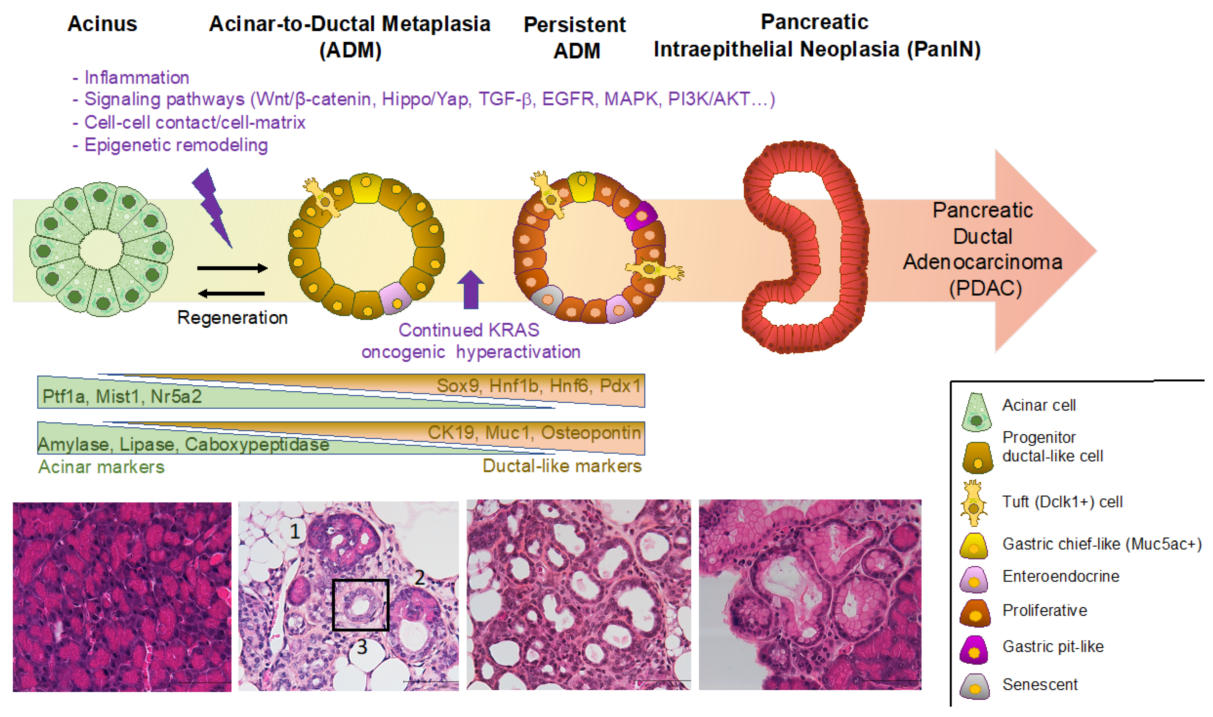
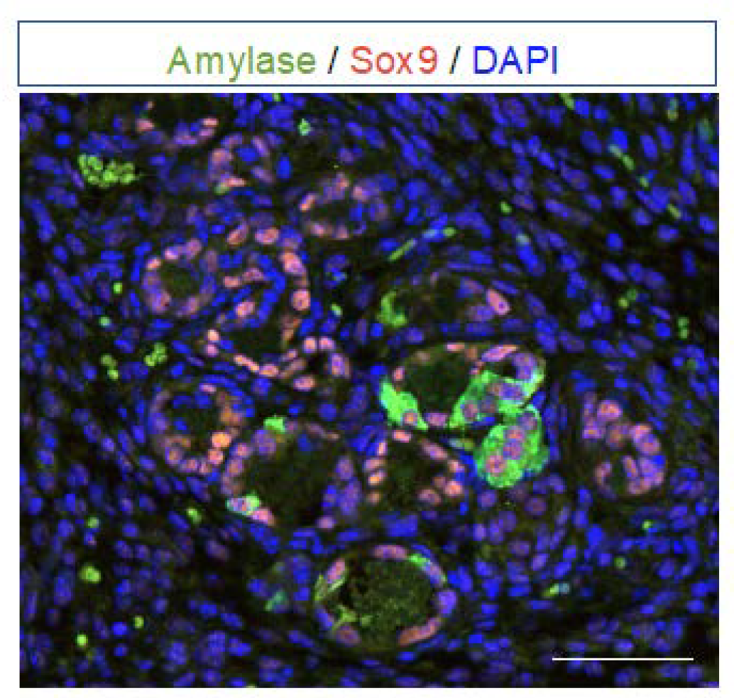
2. Definition of Acinar-to-Ductal Metaplasia (ADM) and Transcription Factors Involved

Metaplasia is a reversible change in which one differentiated cell type is replaced by another cell type. This process can occur in response to various stimuli, such as chronic inflammation or cellular injury. The metaplasia of pancreatic acinar cells manifests their ability to adapt to the genetic and environmental pressure they encounter. Many studies have shown the crucial role of acinar cells in post-injury pancreatic regeneration [4,5,6]. During this process, acinar cells undergo ADM—which is the traditionally used terminology—by losing their mature and functional characteristics as well as undergoing a morphological and transcriptional transformation into ductal-like cells with embryonic progenitor properties [7,8,9]. Like pancreatic progenitor cells, ADM cells are proliferative, whereas mature acinar and ductal cells are largely mitotically quiescent. ADM cannot be considered as a trans-differentiation event corresponding to direct conversion from acinar to ductal cells, as acinar cells dedifferentiate into an embryonic progenitor-like phenotype and differentiate into duct-like cells. The terminology of metaplasia, trans-differentiation and dedifferentiation is a matter of debate [10]. Recently, the term paligenosis, described as the biological process of converting a mature cell into a regenerative cell, was also proposed [11]. In the context of ADM, acinar cell reprogramming can englobe the different terminologies. ADM appears to be a protective mechanism that temporarily reduces extensive tissue damage caused by excessive pancreatic secretion of digestive enzymes. The damaged tissue may return to normal if the stimulus causing metaplasia is removed. Metaplasia, however, can progress to dysplasia and tumors if the stimuli that promote it persist.
The process is evolutionarily conserved as it happens in rodents (references below) and humans [12,13,14,15,16]. ADM was demonstrated with the use of genetically engineered mouse models (GEMMs) (references below) and in vitro in 3D cell culture with mouse and human primary acinar cells [17].
Morphologically, zymogene granules are gradually lost by acinar cells, which show a reduced apical cytoplasm while the lumen of the acini increases. Acinar cells lose polarity and acquire a cuboidal–columnar morphology resembling ductal precursors of the embryonic pancreas. During this process, ADM structures composed of both acinar and duct-like cells can be observed. In the later stages, ADM structures are composed only of duct-like cells, making them difficult to distinguish from pancreatic branched ducts.
The shift from acinar cells to ductal-like cells relies on a down-regulation of acinar gene expression (see Figure 1). The expression of acinar-specific transcription factors including Ptf1a, Mist1 and Nr5a2 is reduced, as well as the expression of digestive enzymes such as carboxypeptidase and amylase, leading to a gradual loss of digestive enzyme synthesis and secretory functions. During pancreas development, Ptf1a is required for the maintenance of multipotent progenitor cells (MPC) [18,19]. After E12.5, Ptf1a expression is restricted to the tip of the pancreatic epithelium adopting an acinar fate, while the cells in the trunk become restricted to a ductal or endocrine fate [20]. Ptf1a is required for maintenance of acinar cell identity by forming a complex network regulating acinar cell-specific digestive enzyme genes [7,21,22,23]. Loss of Ptf1a in acinar cells is sufficient to induce ADM and potentiate inflammation [24,25]. Mist1, restricted to acinar cells throughout development and in adult tissue [26], is a key regulator of acinar cell function, proliferation and identity maintenance [26,27]. Mist1 plays a protective role in ADM. Inhibition of Mist1 aggravated ADM [28,29,30], whereas forced expression of Mist1 significantly attenuated ADM [31]. Nr5a2, a member of the nuclear receptor family of ligand-activated transcription factors, maintains the secretory functions of acinar cells and is a key regulator of acinar cell plasticity. Loss of Nr5a2 accelerates the ADM process, and it was shown that Nr5a2 is required for maintenance of acinar identity and re-establishment of acinar fate during regeneration [32].
Co-expression of acinar markers and duct markers is used to detect ADM (Figure 2). The co-expression of various digestive enzymes is not completely lost in acinar cells during ADM and there is a concomitant upregulated expression of the duct markers including Sox9 [33], Hnf1b [3,34], Hnf6 [35], Pdx1 [36], CA19–9 [37], CAII [38], CD133 [39], and osteopontin [40] (see Figure 1).
The transcription factors Hnf1b, Hnf6, Pdx1 and Sox9 are known to play critical roles in the development and differentiation of pancreatic cells [41,42]. Hnf1b is a key member of the transcription factor network implicated in pancreatic MPCs’ control. Hnf1b deficiency in embryos leads to pancreas agenesis, showing that Hnf1b is required for pancreas morphogenesis and regional specification of the gut [43]. The sequential activation of Hnf1b, Hnf6 and Pdx1 controls the differentiation of endodermal cells into MPCs [44]. Hnf1b was shown to regulate MPC proliferation, survival and differentiation [45] and was found upregulated in ADM [12,16,23,46,47]. The transcription factor Pdx1 is essential for the specification and differentiation of MPC into endocrine and exocrine cell types. After pancreatic morphogenesis, Pdx1 is required for maintaining the identity and function of mature beta cells. Pdx1 was shown to be up-regulated in human and murine ADM, and persistent expression of Pdx1 in the pancreas causes ADM through Stat3 activation [36]. Sox9 is expressed in pancreatic MPC at E9.5 and is required for proliferation and survival of MPCs [48]. Hnf6 is also expressed in MPCs [49]. In normal adult pancreas, Sox9 and Hnf6 expression is restricted to the duct lineage. Sox9 and Hnf6 are up-regulated in human and mouse models of ADM and their overexpression in acinar cells leads to ADM. They are required for repression of acinar genes, for ADM-associated changes in cell polarity and for activation of ductal genes in acinar cells [35]. In order to study to what extent dedifferentiated acini differ from native duct cells and which genes are uniquely regulating acinar cell dedifferentiation, lineage tracing experiments and RNA sequencing were performed with human pancreatic exocrine acinar and duct cells. MECOM, regulated by Sox9, was identified as a transcription factor unique to dedifferentiated acinar cells, critical to maintain cell adhesion and to suppress acinar cell death by permitting cellular dedifferentiation [15].
If the stimuli causing ADM is removed, the acinar tissue is regenerated, and these duct-like progenitor cells formed by ADM proliferate and redifferentiate into acinar cells to replenish the damaged organ.

3. Factors Triggering ADM: Environmental and Cellular Insults

3.1. Pancreatitis and Inflammation

Pancreatitis, characterized by inflammation, ADM and fibrosis of the pancreas, can be caused by obstructions such as gallstones migration, by chronic alcohol consumption or by genetic risk factors with mutations occurring in genes encoding acinar digestive enzymes or their inhibitors such as cationic trypsinogen (PRSS1), serine protease inhibitor Kazal type 1 (SPINK1), and carboxypeptidase A1 (CPA1) [50]. These enzymes become inappropriately active in acinar cells or in their immediate microenvironment, leading to autodigestion, cell death and inflammation. Although some acinar cells are lost during acute pancreatitis through necrosis and apoptosis, other acinar cells undergo ADM. Development of pancreatitis is associated with strong up-regulation of inflammatory mediators such as tumor necrosis factor (TNF)α, Interleukin IL-1β, IL-6 and chemokines of the CXC and CC families. IL-1β plays an important role in pancreatitis, as mice overexpressing IL-1β develop ADM [51]. Activation of NF-κB also increases the severity of pancreatitis [52]. NLRP3 inflammasome is activated in pancreatitis notably by reactive oxygen species (ROS) resulting from mitochondrial damage or damage/danger-associated molecular pattern molecules (DAMPs)–cellular components [53].
Pancreatitis can be induced experimentally by injection of caerulein into rodents, which induces acinar cell death and inflammation. Caerulein-induced pancreatitis as the injury model of choice in mice has facilitated numerous insights into the cellular and molecular mechanisms of exocrine pancreas regeneration. Caerulein, as an analog of cholecystokinin, directly stimulates the production and secretion of acinar enzymes. Administered at high levels, it causes excessive production of digestive enzymes, leading to acinar cell death and transient ADM, within 1–2 days after caerulein injections. Loss of acinar cell identity in response to caerulein-induced pancreatitis is reversible. ADM are proliferative structures capable of regenerating acinar cells lost in pancreatitis. Remarkably, similar to what occurs in human acute pancreatitis, the pancreas returns to normal morphology, differentiation, and function within 1–2 weeks. Other models such as pancreatic duct ligation, which is a model of obstructive pancreatitis, were also used [54].
Obesity and a high-fat diet have been linked to an increased risk of ADM. As an example, obese rats developed ADM, which was reversible after bariatric surgery [55]. The mechanisms generally involved include metabolic dysfunction (insulin resistance and hyperglycemia), oxidative stress (characterized by the generation of reactive oxygen species (ROS) that can damage cellular components) and, most importantly, inflammation, with infiltration of immune cells, particularly macrophages, which secrete cytokines and chemokines [56]. Pro-inflammatory macrophages are activated by a variety of metabolic signals, such as free fatty acids (FFA), lipopolysaccharide (LPS), and glucose. They have been shown to drive ADM by secreting TNFα and the CCL5/RANTES chemokine, factors that activate NF-κB and its matrix metalloproteinases (MMPs) target genes which regulate the extracellular matrix [57]. A recent report also described a novel and intriguing role of cholecystokinin in obesity-associated PDAC. Obesity accelerated PDAC development through changes in the local pancreatic microenvironment driven by increased pancreatic islet (β-cells)-secreted cholecystokinin acting locally on pancreatic acinar cells to accelerate ADM formation [58].
Environmental factors such as alcohol or obesity can interfere with the initiation or completion of normal autophagy [59]. Autophagy is a cellular process that plays a critical role in maintaining cellular homeostasis by recycling and degrading intracellular components. Due to their high protein synthetic rates, acinar cells are prone to the accumulation of misfolded proteins. Insufficient autophagy and ER stress contribute to pancreatitis development [59]. Genetically engineered mice models with altered autophagy pathway components have led to chronic pancreatitis with ADM and inflammation, as is the case with ATG5 and ATG7 loss. Impaired autophagy results in ER stress, accumulation of dysfunctional mitochondria and oxidative stress [60,61,62,63].
ADM can also be induced by alteration of acinar cell polarity, cell–cell contacts and cell–matrix connections. As an example, loss of Numb, a protein that regulates integrins and cell junctions, accelerates ADM [64]. Additionally, E-cadherin is a key adherence molecule required for maintenance of structural homeostasis. Its stability in epithelial cells is regulated by p120 catenin. Deletion of p120 catenin in pancreatic MPC leads to ADM [65]. Deletion of E-cadherin also leads to ADM, associated with increased inflammation (increased inflammatory cytokines and chemokines, such as Cxcl2, Ccl2, IL-6, TNFα) and fibrosis [66].

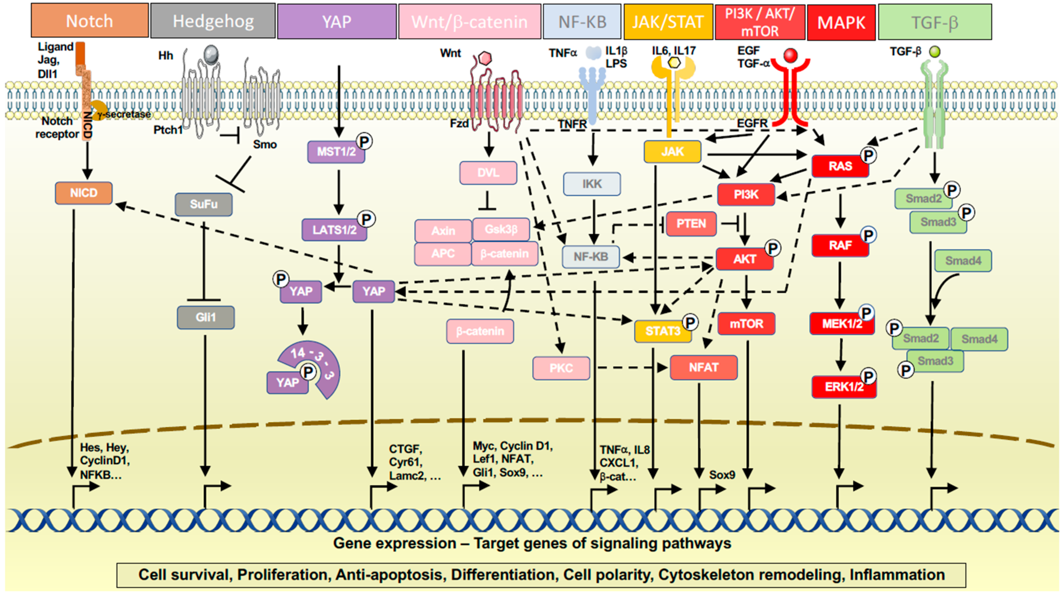
3.2. Signaling Pathways

Several pancreas developmental pathways play important roles in pancreas regeneration and ADM, including Notch, Wnt/β-catenin, Hedgehog, Hippo/YAP, EGFR, MAPK and PI3K/AKT. The ADM process is coordinated by the cross-talk between these signaling cascades. Reactivation of these developmental signaling pathways must be tightly regulated to enable the regeneration of acinar cells while preventing them from persistent ductal reprogramming (Figure 3).
Notch signaling is a key regulator of MPCs maintenance and is required during pancreas embryonic development [67]. Notch intracellular domain (NICD) translocates to the nucleus to activate the expression of target genes, especially Hes1. Active Notch signaling has been shown to be required for exocrine regeneration [68]. The loss of Hes1 led to persistent ADM after acute caerulein-induced pancreatitis and impaired regeneration of the exocrine compartment [69].
The Hedgehog (Hh) signaling pathway plays a critical role in embryonic development. As a morphogen, Hh signaling prevents pancreas formation, requiring its exclusion for pancreatic bud development [70]. Hh signaling is essential for the efficient regeneration of the exocrine pancreas, as its inhibition leads to impaired redifferentiation resulting in persistent ADM [71,72]. In vertebrates, the primary cilium is the central organelle for the transduction of Hh signaling [73]. While pancreatic ductal cells possess a single primary cilium, acinar cells lack cilia. However, when undergoing ADM, acini assemble a cilium, indicating the need for Hh signaling in the differentiation of progenitor duct-like cells [74].
Primary cilia are cellular sensors that mediate several signaling pathways important for pancreatic development and homeostasis [75]. Genetic deletion of primary cilia in embryonic progenitors leads to cystic ducts, acinar cell apoptosis and ADM. This was shown with Tg737 mutation (a gene required for cilia formation) [76], Kif3a loss (a gene required for cilia construction and maintenance) [77], and Hnf1b deficiency (associated with the absence of primary cilia by regulation of target genes such as Hnf6, Pkhd1 and Kif12) [45]. In these models, mislocalisation of β-catenin [45,49] and increased cytosolic β-catenin [76] suggested a role for Wnt signaling. Additionally, Kif3a mutants expressed high levels of Transforming Growth Factor (TGF)-β ligands and MAPK/ERK pathway activation [77].
In post-natal ducts, cilia defects caused by deletion of Hnf1b, Hnf6 or Lkb1 genes were shown to be associated with enlarged ducts, chronic pancreatitis, acinar apoptosis, ADM, inflammatory infiltrates, fibrosis and adipocyte differentiation [3,78]. Ductal deletion of Hnf1b showed enhanced signaling pathways favoring ADM including YAP, Notch, EGFR and TGF-β. This study provided molecular mechanisms by which ciliary defects in ductal cells lead to non-cell autonomous effects on acinar cells notably through the activation of the YAP mechanotransducer [3].
Hippo signaling, mediating YAP nuclear localization and downstream activation of target genes, plays an important role in regulating cell proliferation and apoptosis and is crucial to pancreas development and function, including crosstalk with Notch, WNT/β-catenin, and PI3K/AKT/mTOR signaling pathways [79]. Inactivation of Mst1/2 kinases of the Hippo pathway leading to translocation of YAP into the nucleus and activation of target genes induced ADM, immune infiltration and pancreatitis [80]. Dysregulation of the Hippo and PI3K signaling pathways has been observed in human chronic pancreatitis [81,82]. Hippo signaling inactivation associated with YAP activation and dysregulation of PI3K signaling by genetic disruption of Pten synergistically promotes ADM through the upregulation of the downstream effector pancreatic connective tissue growth factor (CTGF) [83].
YAP1/TAZ activation in cells undergoing ADM also increases the expression of the JAK–STAT3 pathway components via the upregulation of Stat3. JAK–STAT3 signaling is strongly up-regulated in mice with pancreatitis [84]. The JAK/STAT pathway regulates the downstream signaling of numerous membrane proteins and induces the transcription of genes involved in cell proliferation, apoptosis, and inflammatory factor production [85]. YAP1/TAZ signaling has been determined to be necessary and sufficient for ADM induction by regulating JAK-STAT3 signaling [84]. STAT3 is required for ADM formation. P-STAT3 is found in ADM; in addition, loss of STAT3 decreases ADM formation [86]. STAT3 is a critical component of pancreatitis as its deletion protects against pancreatitis. STAT3 supports cell proliferation, metaplasia-associated inflammation, and regulates MMP7 expression [87]. MMP7 was required for ADM in vitro [88], and MMP7-deficient mice were resistant to acinar apoptosis and ADM [89]. IL22 is another activator of STAT3 that promotes ADM [90].
Moreover, the YAP/TAZ-STAT3-PYK2-Wnt/β-catenin was shown to constitute a signaling pathway regulating ADM [91]. The Wnt/β-catenin signaling pathway is required in pancreas development [92]. The canonical pathway is activated by Wnt family ligands binding to Frizzled/LRP receptor complexes. The cascade of events that ensues prevents β-catenin degradation within the cytoplasm and allows its stabilization and nuclear translocation. β-catenin signaling was found upregulated during ADM. β-catenin null cells were incapable of undergoing ADM, showing a cell-autonomous requirement for intact β-catenin function for acinar cells to undergo ADM [93]. β-catenin was also essential for acinar cell proliferation during regeneration [94,95]. Wnt signaling was thus found to be required for ADM, as well as capable of upregulating MAPK/ERK signaling pathway also promoting ADM [93].
Members of the TGF-β superfamily are involved in embryonic development, regulation of homeostasis and diseases [96]. TGF-β signaling occurs through a receptor complex composed of TGF-β receptors which activate the canonical SMAD pathway and non-canonical pathways including MAPK/ERK and PI3K/AKT. In different mouse models, notably by using a dominant-negative type II TGF-β receptor, inhibition of TGF-β reduced caerulein-induced pancreatitis [97,98]. In vitro studies indicate that the activation of TGF-β signaling facilitated ADM of human pancreatic acinar cells, while inhibiting TGF-β signaling reduced the spontaneous ADM of human acinar cells [14,99]. Cell-autonomous expression of a constitutively activated TβRI receptor in acinar cells induced ADM. Activation of TGF-β signaling indeed disrupted acinar cell homeostasis, leading to a drastic increase in the number of ductal structures at the expense of acinar structures, with apoptosis, dedifferentiation of acinar cells and increased expression of transcription factors such as Hnfb1, Sox9 and Hes1. Hnf1b was the best TGF-β responder, and its activation was dependent on the SMAD and MAPK/ERK pathways [47].
Epidermal growth factor receptor (EGFR) is a transmembrane glycoprotein, member of the tyrosine kinase family of growth factor receptors. Its extracellular domain provides a ligand-binding site for EGF and TGF-α. The two major intracellular pathways activated by EGFR are the MAPK/ERK pathway and the P13K-AKT pathway. High-level expression of EGFR and its ligands EGF and TGFα were observed in human chronic pancreatitis specimens, as well as the activation of the EGFR signaling pathway in the areas of ADM in mice [100,101,102,103]. Several studies demonstrated that ADM requires EGFR signaling [104,105,106]. Tgf-α transgenic mice displayed ADM. Using primary explant cultures and lineage tracing studies, acinar cells were shown to undergo conversion to metaplastic ductal epithelial cells in response to TGF-α and EGFR signaling. Transition of isolated acini to ductal cells in vitro in collagen culture in the presence of TGF-α was comparable to the changes described in TGF-α transgenic mice in vivo. Whereas ADM formation was provoked by transgenic expression of EGFR ligands in vivo, genetic or pharmacological inactivation of EGFR or ligands prevented ADM [101,102]. Among the pathways that integrate EGFR signals, NFATc1/c-Jun was identified as a critical transcription factor complex induced by cell-autonomous EGFR signaling and promoting ADM through Sox9 induction [103]. Moreover, NFATc1 was recently identified as a regulatory hub mediating EGFR signaling, displacement of ARID1, a component of the SWI/SNF chromatin remodeling complex, and induction of ductal gene signature promoting acinar cell reprogramming [107].
Several signaling pathways downstream of EGFR are activated to form a complex regulatory network that coordinately regulate ADM. Kras plays a crucial role in ADM formation and progression [30]. MAPK signaling is tightly regulated and is activated by extracellular growth factor stimulation. After receptor phosphorylation, Kras binds guanosine triphosphate (GTP) and activates the serine/threonine kinase Raf. In turn, Raf phosphorylates and activates MEK1/2, which phosphorylates ERK1/2 that translocates to the nucleus where it promotes transcription and cell cycle progression [108]. Up-regulation of MAPK signaling upon induction of pancreatitis was found in isolated acinar cells [109,110] and in vivo [111]. Activation of MAPK signaling pathway has been demonstrated in human and mouse ADM [112]. ADM from human acinar cells was reduced by MAPK inhibition [12]. Inhibition of MAPK signaling in caerulein-induced pancreatitis in mice by treatment with a MEK inhibitor or by Mek1/2 knockdown blocked chronic pancreatitis development and reversed caerulein-induced damage, showing that MEK signaling is a driver of ADM required for initiation and maintenance of ADM and limiting organ regeneration [113]. Activation of KrasG12D in adult acinar cells induced ADM in vitro in a 3D collagen matrix and in vivo. Raf/MEK/ERK represents a critical downstream effector pathway through which Kras operates to induce ADM [114,115]. Lineage-tracing studies have confirmed that ADM results from adult acinar cells upon KrasG12D expression [94,114,116]. KrasG12D-induced acinar cells had elevated pERK levels [31]. Treatment of cells with a MEK inhibitor efficiently blocked accumulation of pERK, and reduced ADM and MEK1/2 inhibition blocked ADM in the 3D culture [31,112].
Reg3A and its mouse homolog Reg3B were up-regulated in human and mouse ADM, respectively, and induced ADM in a 3D culture of primary human and murine acinar cells [117]. Both Reg3B transgenic mice and Reg3B-treated mice with caerulein-induced pancreatitis promoted and sustained ADM by activating the MAPK signaling pathway [118].
The PI3K/AKT pathway was shown to be involved in ADM [119]. Cellular functions regulated by PI3K signaling include cell transformation, proliferation, growth, motility and survival [120]. Of the four class I PI3K isoforms, a family of heterodimeric lipid kinases that activates downstream kinases such as AKT, only p110α and p110β were expressed in the murine and human pancreas, in particular in the acinar compartment [121]. Constitutive activation of PI3K signaling via the expression of oncogenic p110αH1047R phenocopies KrasG12D-induced cellular plasticity by causing ADM through the canonical PI3K/AKT signaling [122]. Pik3ca activating mutation responsible for constitutive activation of PI3K was found to cause increased ADM [123]. Pharmacological selective inhibition of p110α inhibited AKT substrate phosphorylation and completely blocked ADM induced by mutated Kras. Genetic ablation of p110α blocked ADM formation induced by caerulein, whereas the ablation of p110β did not. Moreover, p110α prevented ADM induction by caerulein in a mutated Kras background. The role for p110α in actin rearrangement during ADM was found by activation of the Rho small GTPases that control F-actin network remodeling [121]. Involvement of PI3K activity in ADM was also shown by the pro-tumorigenic effect of restricted loss of the PTEN enzyme, which reverses PI3K activity. Deletion of Pten in the pancreas induced an increase in ADM structures [124]. Concomitant conditional Pten deletion and KrasG12D activation led to an accelerated and accentuated phenotype of ADM [125].
Glycogen synthase kinase-3 (GSK3) β is a serine–threonine kinase involved in several cellular functions by acting as a downstream regulatory switch for numerous signaling pathways, including Wnt/β-catenin and PI3K-AKT signaling pathways. GSK-3β protein expression was elevated following caerulein-induced pancreatitis in mice. GSK-3β promoted ADM in 3D-cultured primary acinar cells and was found necessary for ADM in vivo, contributing to proliferation of ADM cells through activation of the S6 kinase [126,127].
The transcription factor Krüppel-like factor 5 (KLF5) has been identified as being up-regulated in ADM by PI3K and MEK signaling. ADM was reduced after genetic inactivation of Klf5 in vivo through suppression of STAT3 activation. KLF5 is a possible convergence point in MEK and PI3K pathways and a potential link between ductal transformation and increased cellular proliferation during ADM process [127]. Another Krüppel-like factor, KLF4, has an essential role in ADM induction. KLF4 was upregulated and required for ADM; overexpression of KLF4 in acinar cells resulted in ADM [128].

3.3. Epigenetic Reprogramming

Epigenetic modifications refer to the chemical or physical changes that affect gene accessibility and expression without altering the DNA sequence. They include (i) DNA methylation, with involvement of DNA methyltransferases (DNMT); (ii) histone modifications (methylation, acetylation, sumoylation, ubiquitination and phosphorylation), notably with histone deacetylases (HDACs), acetylases (HATs) and methyltransferases; (iii) noncoding RNAs [129]. Given the critical role of epigenetic patterns in determining gene accessibility and transcription factor recruitment, it is not surprising that specific epigenetic events can drive acinar cell reprogramming, silencing acinar cell-specific genes and replacing them with the expression of ductal-specific genes.
As an example, HDAC Sirt1 regulates ADM by deacetylating Ptf1a and β-catenin [130]. Metaplastic acinar cells undergo chromatin reprogramming during ADM. Indeed, it has been observed that HDAC activity was up-regulated in the pancreas during pancreatitis [131]. In vivo treatment with the pan-HDAC inhibitors sodium butyrate and trichostatin A (TSA), targeting both class I and class II HDAC subfamilies, reduced ADM, inflammation and fibrosis following induced pancreatitis in rodents [132]. The class I HDAC inhibitor MS-275 modifies the expression of inflammatory molecules and suppresses the migratory properties of macrophages. Moreover, experiments using TGFα-induced acinar transformation suggested that MS-275 prevents acinar dedifferentiation into ADM in a cell-autonomous manner by a down-regulation of the expression of EGFR in acinar cells [131]. TSA was also shown to reverse ADM in mouse and primary human acinar cultures by inhibiting Spink1 and PI3K/AKT signaling [133]. However, in another study, treatment with valproic acid, another pan-HDAC inhibitor, had a detrimental effect on caerulein-induced pancreatitis, manifested by increased inflammation, decreased proliferation and persistent acinar de-differentiation with non-resolving ADM [134]. This highlights the need to further investigate the precise role of HDAC isoforms in the context of ADM.
Polycomb group (PcG) proteins are histone modifying transcriptional repressors arranged in the Polycomb Repressor Complexes 1 and 2 (PRC1, PRC2). PRC1 contains the BMI1 and the E3 ubiquitin ligase RING1B components which catalyze the mono-ubiquitination of lysine 119 of histone 2A (H2AK119ub). The catalytic subunit of PRC2, EZH2, tri-methylates lysine 27 of histone 3 (H3K27me3). High expression of PRC1 members and enrichment of histone mark H2AK119ub were found in ADM [135,136]. Elevated levels of Bmi1 and Ring1b and their catalyzed histone modification H2AK119ub in ADM were responsible for the epigenetic silencing of acinar transcription factors, such as the complex Ptf1-L containing the DNA-binding subunit Ptf1a and Rbpjl [136]. Ring1b knockout mice showed greatly impaired acinar cell dedifferentiation due to a retained expression of acinar differentiation genes [137]. This shows the role of epigenetic repression of acinar-specific differentiation genes as an essential step in ADM. Changes in H3K27Me3 catalyzed by PRC2 also occur and lead to the suppression of genes normally activated by pancreatic injury [138]. Ezh2 upregulation occurs after pancreatic injury and conditional inactivation of Ezh2 in mouse acinar cells, leading to defective regenerative response and resulting in compromised proliferation and persistent metaplastic lesions. Ezh2 is crucial for maintaining gene silencing of the p16INK4A cell cycle inhibitor in ADM during acinar cell regeneration [139].
BRG1 is a component of the SWI/SNF chromatin remodeling complexes. Deletion of Brg1 in acinar cell mice drastically attenuated the formation of ADM, and Sox9 expression was downregulated in the BRG1-depleted ADM. BRG1 bound to the Sox9 promoter to regulate its expression and was critical for recruitment of upstream regulators, including PDX1, through a local change in chromatin conformation in acinar cells [140]. Genome-wide analysis (ChIP-Seq and ATAC-Seq) in acinar cells revealed that another SWI/SNF subunit, ARID1A, remodels chromatin architecture and stabilizes acinar cell identity. Loss of Arid1a decreases chromatin accessibility of genes that produce digestive enzymes during ADM [141].
miRNA can also regulate ADM. miR-802 is a highly abundant and acinar-enriched pancreatic miRNA which suppresses ADM. Genetic ablation of mir-802 facilitated ADM in caerulein or KrasG12D-induced ADM. miR-802 deficiency resulted in de-repression of a miR-802 target network, leading to the activation of F-actin rearrangement and Sox9 expression [142].

4. Progression of ADM to Pancreatic Intraepithelial Neoplasia (PanIN)

The relevance of ADM to pancreatic ductal adenocarcinoma (PDAC) is supported by the observation that ADM is frequently associated with human PanIN lesions in PDAC patients [115,143]. Chronic pancreatitis is a well-known risk factor for PDAC development in humans. Patients with hereditary pancreatitis showed an increase in pancreatic cancer incidence [144]. In patients with pancreatitis, ADM is transient and reversible. However, persistent ADM can progress to PanIN, and finally to PDAC. The progression from ADM to PanIN has been well established both in mouse models by lineage tracing [114] and in humans [30,125,145]. It has been observed that ADM lesions are correlated with the invasive front of pancreatic cancer, contributing to desmoplasia and cancer cell invasion of the local parenchyma [146]. Mouse models, supported by human histopathological studies, demonstrated that PanIN lesions are the precursor for PDAC and often already exhibit mutations characteristic of PDAC [147,148].
The progression of ADM to PanIN is promoted by the factors that induce ductal cell identity such as Sox9, but suppressed by the factors that preserve acinar cell properties such as Ptf1a and Mist1 [25,30,128,149]. Both morphology and gene expression changes are consistent with the connection between ADM and PanIN. Ptf1a is epigenetically silenced during inflammation and oncogenic Kras-driven ADM. Ptf1a-deficient acinar cells are dramatically sensitized to Kras transformation, and loss of Ptf1a accelerates the development of invasive PDAC [25]. Forced expression of the acinar-restricted transcription factor Mist1 significantly attenuates KrasG12D-induced ADM/PanIN formation, showing that maintaining Mist1 activity in KrasG12D expressing acinar cells can partially mitigate the transformation activity of oncogenic Kras [31]. Loss of Nr5a2 cooperates with oncogenic Kras to drive ADM and PanIN development [32]. Reactivation of progenitor functions supports tumorigenicity. Transcription factors that are up-regulated in ADM, such as Sox9, Hnf6, Pdx1 and Hes1, are also up-regulated in PanIN. Loss of Hnf6 expression has been shown to correlate with human pancreatic cancer progression [150]. Sox9 stimulates gene expression that leads to ADM, PanIN and initiation of PDAC in mice [149]. Sox9 expression in patient tumor samples is elevated at all stages of PanIN lesions and PDAC, correlating with the increased expression of EGFR pathway-related genes. Similarly, in mice, the absence of Sox9 reduces EGFR signaling and pancreatic tumorigenesis [151]. Elimination of Brg1 in acinar cells impairs the formation of KrasG12D-induced ADM and PanIN, associated with the downregulation of Sox9 expression in Brg1-depleted ADMs/PanINs, showing that Brg1 is critical for PanIN initiation and progression through positive regulation of Sox9 [140].
Impaired acinar cell differentiation favors the development of PanIN in the context of oncogenic signals. Oncogenic Kras mutation represents the most frequent and earliest genetic alteration in PDAC patients, highlighting its role as a driver of PDAC [152]. The cellular plasticity in ADM is the key to pancreas regeneration as well as tumorigenesis. In the context of a physiological response to acute pancreatic insult, ADM is a reversible process with the capacity of the metaplastic cells to proliferate and replenish the damage organ. However, in the case of persistent aberrant growth factor signaling or concomitant oncogenic activation such as the KrasG12D activating mutation, the metaplastic cells cannot revert to a differentiated state, leading to irreversible ADM, facilitating the onset of PanIN and PDAC development. ADM development has been shown to precede PanIN formation in mouse KrasG12D models [115]. KrasG12D-expressing acinar cells in the 3D culture rapidly convert to ductal cells that mimic the properties associated with in vivo ADM and PanIN lesions [31]. Oncogenic Kras mutation compromises the ability of acinar cells to regenerate following acute pancreatitis and locks metaplastic cells in a persistently ADM state that can rapidly give rise to PanIN and promote PDAC development [94,153]. KrasG12D expression in adult acinar cells generates ADM lesions that progress to PanIN and PDAC [114,147,154,155]. Lineage-tracing experiments in mice have demonstrated that PanIN lesions are mainly derived from acinar cells undergoing ADM [149]. ADM is thus the earliest pre-neoplastic lesion that predisposes to PDAC, making ADM reprogramming a crucial step in pancreatic cancer initiation.
The MAPK pathway hijacks the plasticity of acinar cells to promote tumorigenesis. MAPK signaling is a key Kras effector for PanIN maintenance. MAPK activity is sufficient and required for ADM and PanIN formation [112]. MEK1/2 inhibition results in PanIN regression in Kras-activated GEMM. The MAPK pathway is required for maintaining PanIN lesions by promoting acinar dedifferentiation. Concomitant conditional Pten deletion and KrasG12D activation accelerate and accentuate the phenotype of ADM, PanIN, and malignant progression. Concurrent dysregulation of the PTEN/PI3K/AKT and MAPK pathways acts synergistically to promote PanIN initiation and progression [125]. A strong activation of PI3K signaling was observed in ADM and in PanIN [122]. The requirement for PI3K p110α in acinar cell plasticity and the initiation of cancer induced by oncogenic Kras has been established [121]. In mutant Kras-driven mouse models, the activation of the EGFR signaling pathway in the areas of ADM and PanIN was convincingly pronounced, with downstream ERK activation. EGFR or ligand inactivation using either genetic or pharmacological approaches preserves acinar cells in a well- differentiated state and almost completely abolishes ADM and PanIN induced by KrasG12D and inflammation [101,102].
Mutant Kras, injury, and stress signaling converge to activate KLF4 expression. KLF4 synergizes with mutant Kras in PanIN initiation. Overexpression of KLF4 results in sustained ADM, which promotes the initiation of PanIN in the presence of mutant Kras [128].
Important signaling pathways for pancreatic development, including Notch signaling, are involved in ADM progression to PanIN [156]. Notch signaling is also involved in tumor initiating processes, development of ADM and PanIN lesions, as well as in tumor progression [116,156]. Hes1 was shown to be required for KrasG12D-driven PanIN formation [157]. Metaplastic acinar structures notably express Notch target genes, EGFR, ErbB2 and pErk. This expression pattern parallels the expression pattern detected in PanIN, suggesting that ADM and PanIN follow similar molecular pathways [115].
Other signals contribute to the initiation or progression of ADM lesions to PanIN. They include IL6 secretion combined with downstream STAT3 activation [86,87]. REG3β expression is induced during ADM and in early PanIN by IL17, and REG3β-promoted PanIN development induced by the KrasG12D oncogene through the GP130/JAK2/STAT3 pathway [158]. Pro-inflammatory macrophages also play a critical role in ADM to PanIN transition. Depletion of macrophages in mice expressing oncogenic Kras under an acinar cell-specific promoter results in a decreased progression of ADM to PanIN [159]. The transcriptional regulators YAP/TAZ are required for KrasG12D to promote PanIN. In the absence of YAP, KrasG12D is unable to induce PanIN lesions, suggesting that acinar-to-ductal reprogramming via YAP/TAZ is a mandatory step in pancreatic cancer initiation from acinar cells [84]. Activation of TGF-β signaling in combination with oncogenic Kras activation leads to the early onset of PanIN that can naturally evolve toward high-grade/locally invasive lesions [47].
Several molecular alterations occur during the progression from ADM to PanIN, including mutations in tumor suppressor genes such as CDKN2A (also known as p16/INK4a), p53 [160] and the cyclin-dependent kinase inhibitor p21 [161], thus regulating cell cycle, genome stability, apoptosis and DNA repair. Recently, a “super-tumor suppressor” P53 mutant was found to inhibit ADM and PanIN proliferation driven by KrasG12D [162].
Altered chromatin remodeling can foster ADM to PanIN progression. Chromatin reshaping occurs in metaplastic cells and synergizes with master transcriptional regulators to render ADM irreversible. Ezh2 deficiency accelerated PanIN progression [139]. Loss of Arid1a, member of the SWI/SNF complex, counteracts acinar cell identity and cooperates with oncogenic Kras in driving pancreatic carcinogenesis [141]. As another epigenetic regulation, acinar cell-specific genetic ablation of miR-802 significantly increased the number of ADM and PanIN lesions and reduced the median survival compared with control mice [142].
Thus, ADM is a pre-neoplastic event critical for eventual malignant transformation. While development of ADM consists in an adaptative stage to environmental stress, the progression of metaplasia can be considered as an oncogenic stage. A recent study identified populations in human samples associated with ADM using single-cell RNA sequencing (scRNA-seq), and lineage trajectory analyses predicted that some ADM populations were related to PanIN, showing that the sequence ADM-PanIN-PDAC identified in mouse models can accurately reflect tumor progression in human patients [163]. Deciphering the mechanisms of metaplasia formation is important for understanding tissue homeostasis and adaptation to stress, and studying the progression of metaplasia to PanIN neoplasia may allow identification of important factors that contribute to early-stage malignancy.

5. Heterogeneity of Metaplastic Cells Revealed by scRNA-seq

Recently, scRNA-seq analyses revealed a higher epithelial heterogeneity in ADM than previously appreciated (see Figure 1). Moreover, changes between injury-induced ADM and KrasG12D oncogene-induced ADM were identified. Metaplastic cells appear to be not a homogenous population but rather can be divided into distinct metaplastic lineages that infiltrate pancreatic lesions. A recent study identified Onecut2 and Foxq1, specifically and highly expressed in metaplastic cells. Onecut2 was reported to have a major role in prostate cancer, and Foxq1 is a transcription factor that regulates Muc5ac. Their expression may be needed to regulate the transition from early metaplastic stage to late metaplastic stage. Moreover, metaplastic cells include several subpopulations with distinct transcription programs. Metaplastic cell clusters include transcription signatures of tuft-like cells (Dclk1 positive), stomach pit-like cells (Muc5ac positive), stomach chief cells, neuroendocrine-like cells and senescent cells [164]. The formation of a significant population of chemosensory tuft cells with a pancreatobiliary identity (expressing the markers Dclk1, Pou2f3, and Trpm5) was previously identified during ADM [165,166,167]. While tuft cells were rarely found in caerulein-induced transient ADM, they were found to represent 15% of ADM in TGFα- or KrasG12D-induced ADM and were also present in PanINs [165,167]. Dclk1-positive cells were characterized as a subpopulation with cancer stem cell properties [165,167,168,169,170]. Another scRNA-seq study of murine and human ADM revealed that a mucin/ductal population resembling gastric pyloric metaplasia can generate tuft and enteroendocrine cells. Then, the activation of KrasG12D in an injury-induced Hnf1b+ ADM led to neoplastic transformation and the formation of Muc5ac-positive gastric-pit-like cells [171]. KrasG12D expression was found to drive PanIN-specific changes in ADM, characterized by significantly higher expression of proto-oncogenes such as Fosl1 and Junb, known downstream targets of KrasG12D. Answering the question of which metaplastic cells within a lesion undergo malignant transformation is crucial to understanding the development of PDAC, as the specific cellular state at the time of acquiring an oncogenic mutation could be involved. Identifying this sequence of events can provide valuable insight into the origin of malignant cells and the onset of PDAC.

6. Conclusions

PDAC is a devastating disease with poor survival rates. Studies have pointed to an acinar cell origin in which acinar cells undergo ductal metaplasia at the onset of tumor formation. ADM or, as a more appropriate terminology, acinar cell reprogramming, is a mechanism needed for regeneration after inflammation or injury. It results from acinar cell identity marker silencing and activation of acinar cell dedifferentiation drivers. However, in the presence of oncogenic signaling, ADM is irreversible and leads to PanIN. Future research and clinical translation should continue to focus on using appropriate disease models, identifying suitable ADM detection markers, as well as discovering molecular targets that can impede the initiation and progression of ADM and enhance regeneration following pancreatic injury. Understanding the intermediate states of ADM and important molecules that regulate ADM formation may help the development of novel preventive strategies that could be translated to the clinical setting. The validation of whether manipulating molecular targets can hinder the formation of PanIN from ADM and using these markers for early detection of PDAC remains to be conducted preclinically and clinically. Since PanIN lesions are the most common precursor lesions and are currently clinically undetectable, understanding the mechanisms underlying formation and progression of premalignant lesions is critical for early detection and therapeutic intervention of PDAC.

Author Contributions

Acquisition of data, L.M.-D., D.L., A.C. (Anaïs Chassac) and P.N.; data analysis, L.M.-D., D.L., A.C. (Anne Couvelard), P.N. and C.H.; writing—review and editing, L.M.-D., D.L, P.N. and C.H.; supervision, C.H.; project administration, C.H.; funding acquisition, C.H. All authors have read and agreed to the published version of the manuscript.

Funding

This research was funded by Inserm, Université Paris Cité, Gefluc-Les Entreprises Contre le Cancer, la Ligue Contre le Cancer-Comité de Paris, Fondation ARC.

Institutional Review Board Statement

Not applicable.

Informed Consent Statement

Not applicable.

Data Availability Statement

Not applicable.

Conflicts of Interest

The authors declare no conflict of interest.

References

  1. Rahib, L.; Smith, B.D.; Aizenberg, R.; Rosenzweig, A.B.; Fleshman, J.M.; Matrisian, L.M. Projecting Cancer Incidence and Deaths to 2030: The Unexpected Burden of Thyroid, Liver, and Pancreas Cancers in the United States. Cancer Res. 2014, 74, 2913–2921. [Google Scholar] [CrossRef] [PubMed] [Green Version]
  2. Matsuda, Y.; Furukawa, T.; Yachida, S.; Nishimura, M.; Seki, A.; Nonaka, K.; Aida, J.; Takubo, K.; Ishiwata, T.; Kimura, W.; et al. The Prevalence and Clinicopathological Characteristics of High-Grade Pancreatic Intraepithelial Neoplasia: Autopsy Study Evaluating the Entire Pancreatic Parenchyma. Pancreas 2017, 46, 658–664. [Google Scholar] [CrossRef]
  3. Quilichini, E.; Fabre, M.; Dirami, T.; Stedman, A.; De Vas, M.; Ozguc, O.; Pasek, R.C.; Cereghini, S.; Morillon, L.; Guerra, C.; et al. Pancreatic Ductal Deletion of Hnf1b Disrupts Exocrine Homeostasis, Leads to Pancreatitis, and Facilitates Tumorigenesis. Cell Mol. Gastroenterol. Hepatol. 2019, 8, 487–511. [Google Scholar] [CrossRef] [Green Version]
  4. Puri, S.; Folias, A.E.; Hebrok, M. Plasticity and Dedifferentiation within the Pancreas: Development, Homeostasis, and Disease. Cell Stem. Cell 2015, 16, 18–31. [Google Scholar] [CrossRef] [PubMed] [Green Version]
  5. Stanger, B.Z.; Hebrok, M. Control of Cell Identity in Pancreas Development and Regeneration. Gastroenterology 2013, 144, 1170–1179. [Google Scholar] [CrossRef] [PubMed] [Green Version]
  6. Mills, J.C.; Sansom, O.J. Reserve Stem Cells: Differentiated Cells Reprogram to Fuel Repair, Metaplasia, and Neoplasia in the Adult Gastrointestinal Tract. Sci. Signal. 2015, 8, re8. [Google Scholar] [CrossRef] [PubMed] [Green Version]
  7. Pan, F.C.; Bankaitis, E.D.; Boyer, D.; Xu, X.; Van de Casteele, M.; Magnuson, M.A.; Heimberg, H.; Wright, C.V.E. Spatiotemporal Patterns of Multipotentiality in Ptf1a-Expressing Cells during Pancreas Organogenesis and Injury-Induced Facultative Restoration. Development 2013, 140, 751–764. [Google Scholar] [CrossRef] [Green Version]
  8. Murtaugh, L.C.; Keefe, M.D. Regeneration and Repair of the Exocrine Pancreas. Annu. Rev. Physiol. 2015, 77, 229–249. [Google Scholar] [CrossRef] [Green Version]
  9. Jensen, J.N.; Cameron, E.; Garay, M.V.R.; Starkey, T.W.; Gianani, R.; Jensen, J. Recapitulation of Elements of Embryonic Development in Adult Mouse Pancreatic Regeneration. Gastroenterology 2005, 128, 728–741. [Google Scholar] [CrossRef]
  10. Mills, J.C.; Stanger, B.Z.; Sander, M. Nomenclature for Cellular Plasticity: Are the Terms as Plastic as the Cells Themselves? EMBO J. 2019, 38, e103148. [Google Scholar] [CrossRef]
  11. Willet, S.G.; Lewis, M.A.; Miao, Z.-F.; Liu, D.; Radyk, M.D.; Cunningham, R.L.; Burclaff, J.; Sibbel, G.; Lo, H.-Y.G.; Blanc, V.; et al. Regenerative Proliferation of Differentiated Cells by MTORC1-Dependent Paligenosis. EMBO J. 2018, 37, e98311. [Google Scholar] [CrossRef]
  12. Houbracken, I.; de Waele, E.; Lardon, J.; Ling, Z.; Heimberg, H.; Rooman, I.; Bouwens, L. Lineage Tracing Evidence for Transdifferentiation of Acinar to Duct Cells and Plasticity of Human Pancreas. Gastroenterology 2011, 141, 731–741.e4. [Google Scholar] [CrossRef]
  13. Baldan, J.; Houbracken, I.; Rooman, I.; Bouwens, L. Adult Human Pancreatic Acinar Cells Dedifferentiate into an Embryonic Progenitor-like State in 3D Suspension Culture. Sci. Rep. 2019, 9, 4040. [Google Scholar] [CrossRef] [Green Version]
  14. Liu, J.; Akanuma, N.; Liu, C.; Naji, A.; Halff, G.A.; Washburn, W.K.; Sun, L.; Wang, P. TGF-Β1 Promotes Acinar to Ductal Metaplasia of Human Pancreatic Acinar Cells. Sci. Rep. 2016, 6, 30904. [Google Scholar] [CrossRef] [PubMed] [Green Version]
  15. Backx, E.; Wauters, E.; Baldan, J.; Van Bulck, M.; Michiels, E.; Heremans, Y.; De Paep, D.L.; Kurokawa, M.; Goyama, S.; Bouwens, L.; et al. MECOM Permits Pancreatic Acinar Cell Dedifferentiation Avoiding Cell Death under Stress Conditions. Cell Death Differ. 2021, 28, 2601–2615. [Google Scholar] [CrossRef] [PubMed]
  16. Jiang, J.; Hakimjavadi, H.; Bray, J.K.; Perkins, C.; Gosling, A.; da Silva, L.; Bulut, G.; Ali, J.; Setiawan, V.W.; Campbell-Thompson, M.; et al. Transcriptional Profile of Human Pancreatic Acinar Ductal Metaplasia. Gastro. Hep. Adv. 2023, 2, 532–543. [Google Scholar] [CrossRef]
  17. Paoli, C.; Carrer, A. Organotypic Culture of Acinar Cells for the Study of Pancreatic Cancer Initiation. Cancers 2020, 12, 2606. [Google Scholar] [CrossRef] [PubMed]
  18. Kawaguchi, Y.; Cooper, B.; Gannon, M.; Ray, M.; MacDonald, R.J.; Wright, C.V.E. The Role of the Transcriptional Regulator Ptf1a in Converting Intestinal to Pancreatic Progenitors. Nat. Genet. 2002, 32, 128–134. [Google Scholar] [CrossRef]
  19. Masui, T.; Long, Q.; Beres, T.M.; Magnuson, M.A.; MacDonald, R.J. Early Pancreatic Development Requires the Vertebrate Suppressor of Hairless (RBPJ) in the PTF1 BHLH Complex. Genes Dev. 2007, 21, 2629–2643. [Google Scholar] [CrossRef] [Green Version]
  20. Schaffer, A.E.; Freude, K.K.; Nelson, S.B.; Sander, M. Nkx6 Transcription Factors and Ptf1a Function as Antagonistic Lineage Determinants in Multipotent Pancreatic Progenitors. Dev. Cell 2010, 18, 1022–1029. [Google Scholar] [CrossRef] [Green Version]
  21. Rose, S.D.; Swift, G.H.; Peyton, M.J.; Hammer, R.E.; MacDonald, R.J. The Role of PTF1-P48 in Pancreatic Acinar Gene Expression. J. Biol. Chem. 2001, 276, 44018–44026. [Google Scholar] [CrossRef] [PubMed] [Green Version]
  22. Rodolosse, A.; Chalaux, E.; Adell, T.; Hagège, H.; Skoudy, A.; Real, F.X. PTF1alpha/P48 Transcription Factor Couples Proliferation and Differentiation in the Exocrine Pancreas [corrected]. Gastroenterology 2004, 127, 937–949. [Google Scholar] [CrossRef] [PubMed]
  23. Jiang, M.; Azevedo-Pouly, A.C.; Deering, T.G.; Hoang, C.Q.; DiRenzo, D.; Hess, D.A.; Konieczny, S.F.; Swift, G.H.; MacDonald, R.J. MIST1 and PTF1 Collaborate in Feed-Forward Regulatory Loops That Maintain the Pancreatic Acinar Phenotype in Adult Mice. Mol. Cell Biol. 2016, 36, 2945–2955. [Google Scholar] [CrossRef] [Green Version]
  24. Hoang, C.Q.; Hale, M.A.; Azevedo-Pouly, A.C.; Elsässer, H.P.; Deering, T.G.; Willet, S.G.; Pan, F.C.; Magnuson, M.A.; Wright, C.V.E.; Swift, G.H.; et al. Transcriptional Maintenance of Pancreatic Acinar Identity, Differentiation, and Homeostasis by PTF1A. Mol. Cell Biol. 2016, 36, 3033–3047. [Google Scholar] [CrossRef] [PubMed] [Green Version]
  25. Krah, N.M.; De La O, J.-P.; Swift, G.H.; Hoang, C.Q.; Willet, S.G.; Chen Pan, F.; Cash, G.M.; Bronner, M.P.; Wright, C.V.; MacDonald, R.J.; et al. The Acinar Differentiation Determinant PTF1A Inhibits Initiation of Pancreatic Ductal Adenocarcinoma. Elife 2015, 4, e07125. [Google Scholar] [CrossRef] [PubMed]
  26. Pin, C.L.; Rukstalis, J.M.; Johnson, C.; Konieczny, S.F. The BHLH Transcription Factor Mist1 Is Required to Maintain Exocrine Pancreas Cell Organization and Acinar Cell Identity. J. Cell Biol. 2001, 155, 519–530. [Google Scholar] [CrossRef] [Green Version]
  27. Direnzo, D.; Hess, D.A.; Damsz, B.; Hallett, J.E.; Marshall, B.; Goswami, C.; Liu, Y.; Deering, T.; Macdonald, R.J.; Konieczny, S.F. Induced Mist1 Expression Promotes Remodeling of Mouse Pancreatic Acinar Cells. Gastroenterology 2012, 143, 469–480. [Google Scholar] [CrossRef] [Green Version]
  28. Zhu, L.; Tran, T.; Rukstalis, J.M.; Sun, P.; Damsz, B.; Konieczny, S.F. Inhibition of Mist1 Homodimer Formation Induces Pancreatic Acinar-to-Ductal Metaplasia. Mol. Cell Biol. 2004, 24, 2673–2681. [Google Scholar] [CrossRef] [Green Version]
  29. Kowalik, A.S.; Johnson, C.L.; Chadi, S.A.; Weston, J.Y.; Fazio, E.N.; Pin, C.L. Mice Lacking the Transcription Factor Mist1 Exhibit an Altered Stress Response and Increased Sensitivity to Caerulein-Induced Pancreatitis. Am. J. Physiol. Gastrointest. Liver Physiol. 2007, 292, G1123–G1132. [Google Scholar] [CrossRef] [Green Version]
  30. Shi, G.; Zhu, L.; Sun, Y.; Bettencourt, R.; Damsz, B.; Hruban, R.H.; Konieczny, S.F. Loss of the Acinar-Restricted Transcription Factor Mist1 Accelerates Kras-Induced Pancreatic Intraepithelial Neoplasia. Gastroenterology 2009, 136, 1368–1378. [Google Scholar] [CrossRef] [Green Version]
  31. Shi, G.; DiRenzo, D.; Qu, C.; Barney, D.; Miley, D.; Konieczny, S.F. Maintenance of Acinar Cell Organization Is Critical to Preventing Kras-Induced Acinar-Ductal Metaplasia. Oncogene 2013, 32, 1950–1958. [Google Scholar] [CrossRef] [PubMed] [Green Version]
  32. von Figura, G.; Morris, J.P.; Wright, C.V.E.; Hebrok, M. Nr5a2 Maintains Acinar Cell Differentiation and Constrains Oncogenic Kras-Mediated Pancreatic Neoplastic Initiation. Gut 2014, 63, 656–664. [Google Scholar] [CrossRef] [PubMed] [Green Version]
  33. Kopp, J.L.; Dubois, C.L.; Schaffer, A.E.; Hao, E.; Shih, H.P.; Seymour, P.A.; Ma, J.; Sander, M. Sox9+ Ductal Cells Are Multipotent Progenitors throughout Development but Do Not Produce New Endocrine Cells in the Normal or Injured Adult Pancreas. Development 2011, 138, 653–665. [Google Scholar] [CrossRef] [Green Version]
  34. Roy, N.; Hebrok, M. Regulation of Cellular Identity in Cancer. Dev. Cell 2015, 35, 674–684. [Google Scholar] [CrossRef] [Green Version]
  35. Prévot, P.-P.; Simion, A.; Grimont, A.; Colletti, M.; Khalaileh, A.; Van den Steen, G.; Sempoux, C.; Xu, X.; Roelants, V.; Hald, J.; et al. Role of the Ductal Transcription Factors HNF6 and Sox9 in Pancreatic Acinar-to-Ductal Metaplasia. Gut 2012, 61, 1723–1732. [Google Scholar] [CrossRef] [PubMed]
  36. Miyatsuka, T.; Kaneto, H.; Shiraiwa, T.; Matsuoka, T.; Yamamoto, K.; Kato, K.; Nakamura, Y.; Akira, S.; Takeda, K.; Kajimoto, Y.; et al. Persistent Expression of PDX-1 in the Pancreas Causes Acinar-to-Ductal Metaplasia through Stat3 Activation. Genes Dev. 2006, 20, 1435–1440. [Google Scholar] [CrossRef] [Green Version]
  37. Gmyr, V.; Belaich, S.; Muharram, G.; Lukowiak, B.; Vandewalle, B.; Pattou, F.; Kerr-Conte, J. Rapid Purification of Human Ductal Cells from Human Pancreatic Fractions with Surface Antibody CA19-9. Biochem. Biophys. Res. Commun. 2004, 320, 27–33. [Google Scholar] [CrossRef]
  38. Inada, A.; Nienaber, C.; Fonseca, S.; Bonner-Weir, S. Timing and Expression Pattern of Carbonic Anhydrase II in Pancreas. Dev. Dyn. 2006, 235, 1571–1577. [Google Scholar] [CrossRef]
  39. Zhang, F.; Ma, D.; Liu, T.; Liu, Y.H.; Guo, J.; Song, J.; Wu, Q.; Pan, Y.; Zhang, Y.; Guo, C.; et al. Expansion and Maintenance of CD133-Expressing Pancreatic Ductal Epithelial Cells by Inhibition of TGF-β Signaling. Stem. Cells Dev. 2019, 28, 1236–1252. [Google Scholar] [CrossRef]
  40. Kilic, G.; Wang, J.; Sosa-Pineda, B. Osteopontin Is a Novel Marker of Pancreatic Ductal Tissues and of Undifferentiated Pancreatic Precursors in Mice. Dev. Dyn. 2006, 235, 1659–1667. [Google Scholar] [CrossRef] [Green Version]
  41. Pan, F.C.; Wright, C. Pancreas Organogenesis: From Bud to Plexus to Gland. Dev. Dyn. 2011, 240, 530–565. [Google Scholar] [CrossRef] [PubMed]
  42. Shih, H.P.; Wang, A.; Sander, M. Pancreas Organogenesis: From Lineage Determination to Morphogenesis. Annu. Rev. Cell Dev. Biol. 2013, 29, 81–105. [Google Scholar] [CrossRef] [PubMed] [Green Version]
  43. Haumaitre, C.; Barbacci, E.; Jenny, M.; Ott, M.O.; Gradwohl, G.; Cereghini, S. Lack of TCF2/VHNF1 in Mice Leads to Pancreas Agenesis. Proc. Natl. Acad. Sci. USA 2005, 102, 1490–1495. [Google Scholar] [CrossRef] [PubMed] [Green Version]
  44. Poll, A.V.; Pierreux, C.E.; Lokmane, L.; Haumaitre, C.; Achouri, Y.; Jacquemin, P.; Rousseau, G.G.; Cereghini, S.; Lemaigre, F.P. A VHNF1/TCF2-HNF6 Cascade Regulates the Transcription Factor Network That Controls Generation of Pancreatic Precursor Cells. Diabetes 2006, 55, 61–69. [Google Scholar] [CrossRef]
  45. De Vas, M.G.; Kopp, J.L.; Heliot, C.; Sander, M.; Cereghini, S.; Haumaitre, C. Hnf1b Controls Pancreas Morphogenesis and the Generation of Ngn3+ Endocrine Progenitors. Development 2015, 142, 871–882. [Google Scholar] [CrossRef] [Green Version]
  46. Pinho, A.V.; Rooman, I.; Reichert, M.; De Medts, N.; Bouwens, L.; Rustgi, A.K.; Real, F.X. Adult Pancreatic Acinar Cells Dedifferentiate to an Embryonic Progenitor Phenotype with Concomitant Activation of a Senescence Programme That Is Present in Chronic Pancreatitis. Gut 2011, 60, 958–966. [Google Scholar] [CrossRef]
  47. Chuvin, N.; Vincent, D.F.; Pommier, R.M.; Alcaraz, L.B.; Gout, J.; Caligaris, C.; Yacoub, K.; Cardot, V.; Roger, E.; Kaniewski, B.; et al. Acinar-to-Ductal Metaplasia Induced by Transforming Growth Factor Beta Facilitates KRASG12D-Driven Pancreatic Tumorigenesis. Cell Mol. Gastroenterol. Hepatol. 2017, 4, 263–282. [Google Scholar] [CrossRef] [Green Version]
  48. Seymour, P.A.; Freude, K.K.; Tran, M.N.; Mayes, E.E.; Jensen, J.; Kist, R.; Scherer, G.; Sander, M. SOX9 Is Required for Maintenance of the Pancreatic Progenitor Cell Pool. Proc. Natl. Acad. Sci. USA 2007, 104, 1865–1870. [Google Scholar] [CrossRef] [Green Version]
  49. Pierreux, C.E.; Poll, A.V.; Kemp, C.R.; Clotman, F.; Maestro, M.A.; Cordi, S.; Ferrer, J.; Leyns, L.; Rousseau, G.G.; Lemaigre, F.P. The Transcription Factor Hepatocyte Nuclear Factor-6 Controls the Development of Pancreatic Ducts in the Mouse. Gastroenterology 2006, 130, 532–541. [Google Scholar] [CrossRef]
  50. Mayerle, J.; Sendler, M.; Hegyi, E.; Beyer, G.; Lerch, M.M.; Sahin-Tóth, M. Genetics, Cell Biology, and Pathophysiology of Pancreatitis. Gastroenterology 2019, 156, 1951–1968.e1. [Google Scholar] [CrossRef] [Green Version]
  51. Marrache, F.; Tu, S.P.; Bhagat, G.; Pendyala, S.; Osterreicher, C.H.; Gordon, S.; Ramanathan, V.; Penz-Osterreicher, M.; Betz, K.S.; Song, Z.; et al. Overexpression of Interleukin-1beta in the Murine Pancreas Results in Chronic Pancreatitis. Gastroenterology 2008, 135, 1277–1287. [Google Scholar] [CrossRef] [PubMed] [Green Version]
  52. Huang, H.; Liu, Y.; Daniluk, J.; Gaiser, S.; Chu, J.; Wang, H.; Li, Z.-S.; Logsdon, C.D.; Ji, B. Activation of Nuclear Factor-ΚB in Acinar Cells Increases the Severity of Pancreatitis in Mice. Gastroenterology 2013, 144, 202–210. [Google Scholar] [CrossRef] [PubMed] [Green Version]
  53. Ferrero-Andrés, A.; Panisello-Roselló, A.; Roselló-Catafau, J.; Folch-Puy, E. NLRP3 Inflammasome-Mediated Inflammation in Acute Pancreatitis. Int. J. Mol. Sci. 2020, 21, 5386. [Google Scholar] [CrossRef] [PubMed]
  54. Lerch, M.M.; Gorelick, F.S. Models of Acute and Chronic Pancreatitis. Gastroenterology 2013, 144, 1180–1193. [Google Scholar] [CrossRef] [PubMed]
  55. Rebours, V.; Garteiser, P.; Ribeiro-Parenti, L.; Cavin, J.-B.; Doblas, S.; Pagé, G.; Bado, A.; Couvineau, A.; Ruszniewski, P.; Paradis, V.; et al. Obesity-Induced Pancreatopathy in Rats Is Reversible after Bariatric Surgery. Sci. Rep. 2018, 8, 16295. [Google Scholar] [CrossRef] [PubMed] [Green Version]
  56. Teper, Y.; Eibl, G. Pancreatic Macrophages: Critical Players in Obesity-Promoted Pancreatic Cancer. Cancers 2020, 12, 1946. [Google Scholar] [CrossRef] [PubMed]
  57. Liou, G.-Y.; Döppler, H.; Necela, B.; Krishna, M.; Crawford, H.C.; Raimondo, M.; Storz, P. Macrophage-Secreted Cytokines Drive Pancreatic Acinar-to-Ductal Metaplasia through NF-ΚB and MMPs. J. Cell Biol. 2013, 202, 563–577. [Google Scholar] [CrossRef]
  58. Chung, K.M.; Singh, J.; Lawres, L.; Dorans, K.J.; Garcia, C.; Burkhardt, D.B.; Robbins, R.; Bhutkar, A.; Cardone, R.; Zhao, X.; et al. Endocrine-Exocrine Signaling Drives Obesity-Associated Pancreatic Ductal Adenocarcinoma. Cell 2020, 181, 832–847.e18. [Google Scholar] [CrossRef] [PubMed]
  59. Gukovsky, I.; Li, N.; Todoric, J.; Gukovskaya, A.; Karin, M. Inflammation, Autophagy, and Obesity: Common Features in the Pathogenesis of Pancreatitis and Pancreatic Cancer. Gastroenterology 2013, 144, 1199–1209.e4. [Google Scholar] [CrossRef] [PubMed] [Green Version]
  60. Li, N.; Wu, X.; Holzer, R.G.; Lee, J.-H.; Todoric, J.; Park, E.-J.; Ogata, H.; Gukovskaya, A.S.; Gukovsky, I.; Pizzo, D.P.; et al. Loss of Acinar Cell IKKα Triggers Spontaneous Pancreatitis in Mice. J. Clin. Investig. 2013, 123, 2231–2243. [Google Scholar] [CrossRef] [Green Version]
  61. Gukovsky, I.; Gukovskaya, A.S. Impaired Autophagy Triggers Chronic Pancreatitis: Lessons from Pancreas-Specific Atg5 Knockout Mice. Gastroenterology 2015, 148, 501–505. [Google Scholar] [CrossRef] [Green Version]
  62. Diakopoulos, K.N.; Lesina, M.; Wörmann, S.; Song, L.; Aichler, M.; Schild, L.; Artati, A.; Römisch-Margl, W.; Wartmann, T.; Fischer, R.; et al. Impaired Autophagy Induces Chronic Atrophic Pancreatitis in Mice via Sex- and Nutrition-Dependent Processes. Gastroenterology 2015, 148, 626–638.e17. [Google Scholar] [CrossRef] [PubMed]
  63. Antonucci, L.; Fagman, J.B.; Kim, J.Y.; Todoric, J.; Gukovsky, I.; Mackey, M.; Ellisman, M.H.; Karin, M. Basal Autophagy Maintains Pancreatic Acinar Cell Homeostasis and Protein Synthesis and Prevents ER Stress. Proc. Natl. Acad. Sci. USA 2015, 112, E6166–E6174. [Google Scholar] [CrossRef] [PubMed] [Green Version]
  64. Greer, R.L.; Staley, B.K.; Liou, A.; Hebrok, M. Numb Regulates Acinar Cell Dedifferentiation and Survival during Pancreatic Damage and Acinar-to-Ductal Metaplasia. Gastroenterology 2013, 145, 1088–1097.e8. [Google Scholar] [CrossRef] [PubMed] [Green Version]
  65. Hendley, A.M.; Provost, E.; Bailey, J.M.; Wang, Y.J.; Cleveland, M.H.; Blake, D.; Bittman, R.W.; Roeser, J.C.; Maitra, A.; Reynolds, A.B.; et al. P120 Catenin Is Required for Normal Tubulogenesis but Not Epithelial Integrity in Developing Mouse Pancreas. Dev. Biol. 2015, 399, 41–53. [Google Scholar] [CrossRef] [Green Version]
  66. Kaneta, Y.; Sato, T.; Hikiba, Y.; Sugimori, M.; Sue, S.; Kaneko, H.; Irie, K.; Sasaki, T.; Kondo, M.; Chuma, M.; et al. Loss of Pancreatic E-Cadherin Causes Pancreatitis-Like Changes and Contributes to Carcinogenesis. Cell Mol. Gastroenterol. Hepatol. 2020, 9, 105–119. [Google Scholar] [CrossRef] [Green Version]
  67. Murtaugh, L.C.; Stanger, B.Z.; Kwan, K.M.; Melton, D.A. Notch Signaling Controls Multiple Steps of Pancreatic Differentiation. Proc. Natl. Acad. Sci. USA 2003, 100, 14920–14925. [Google Scholar] [CrossRef] [Green Version]
  68. Siveke, J.T.; Lubeseder-Martellato, C.; Lee, M.; Mazur, P.K.; Nakhai, H.; Radtke, F.; Schmid, R.M. Notch Signaling Is Required for Exocrine Regeneration after Acute Pancreatitis. Gastroenterology 2008, 134, 544–555. [Google Scholar] [CrossRef]
  69. Hidalgo-Sastre, A.; Brodylo, R.L.; Lubeseder-Martellato, C.; Sipos, B.; Steiger, K.; Lee, M.; von Figura, G.; Grünwald, B.; Zhong, S.; Trajkovic-Arsic, M.; et al. Hes1 Controls Exocrine Cell Plasticity and Restricts Development of Pancreatic Ductal Adenocarcinoma in a Mouse Model. Am. J. Pathol. 2016, 186, 2934–2944. [Google Scholar] [CrossRef] [Green Version]
  70. Hebrok, M.; Kim, S.K.; Melton, D.A. Notochord Repression of Endodermal Sonic Hedgehog Permits Pancreas Development. Genes Dev. 1998, 12, 1705–1713. [Google Scholar] [CrossRef] [Green Version]
  71. Cano, D.A.; Hebrok, M. Hedgehog Spikes Pancreas Regeneration. Gastroenterology 2008, 135, 347–351. [Google Scholar] [CrossRef] [PubMed]
  72. Fendrich, V.; Esni, F.; Garay, M.V.R.; Feldmann, G.; Habbe, N.; Jensen, J.N.; Dor, Y.; Stoffers, D.; Jensen, J.; Leach, S.D.; et al. Hedgehog Signaling Is Required for Effective Regeneration of Exocrine Pancreas. Gastroenterology 2008, 135, 621–631. [Google Scholar] [CrossRef] [PubMed] [Green Version]
  73. Gigante, E.D.; Caspary, T. Signaling in the Primary Cilium through the Lens of the Hedgehog Pathway. Wiley Interdiscip. Rev. Dev. Biol. 2020, 9, e377. [Google Scholar] [CrossRef]
  74. Bangs, F.K.; Miller, P.; O’Neill, E. Ciliogenesis and Hedgehog Signalling Are Suppressed Downstream of KRAS during Acinar-Ductal Metaplasia in Mouse. Dis. Model Mech. 2020, 13, dmm044289. [Google Scholar] [CrossRef] [PubMed]
  75. Lodh, S.; O’Hare, E.A.; Zaghloul, N.A. Primary Cilia in Pancreatic Development and Disease. Birth Defects Res. C Embryo. Today 2014, 102, 139–158. [Google Scholar] [CrossRef] [Green Version]
  76. Cano, D.A.; Murcia, N.S.; Pazour, G.J.; Hebrok, M. Orpk Mouse Model of Polycystic Kidney Disease Reveals Essential Role of Primary Cilia in Pancreatic Tissue Organization. Development 2004, 131, 3457–3467. [Google Scholar] [CrossRef] [Green Version]
  77. Cano, D.A.; Sekine, S.; Hebrok, M. Primary Cilia Deletion in Pancreatic Epithelial Cells Results in Cyst Formation and Pancreatitis. Gastroenterology 2006, 131, 1856–1869. [Google Scholar] [CrossRef]
  78. Augereau, C.; Collet, L.; Vargiu, P.; Guerra, C.; Ortega, S.; Lemaigre, F.P.; Jacquemin, P. Chronic Pancreatitis and Lipomatosis Are Associated with Defective Function of Ciliary Genes in Pancreatic Ductal Cells. Hum. Mol. Genet. 2016, 25, 5017–5026. [Google Scholar] [CrossRef] [Green Version]
  79. Wu, Y.; Aegerter, P.; Nipper, M.; Ramjit, L.; Liu, J.; Wang, P. Hippo Signaling Pathway in Pancreas Development. Front. Cell Dev. Biol. 2021, 9, 663906. [Google Scholar] [CrossRef]
  80. Gao, T.; Zhou, D.; Yang, C.; Singh, T.; Penzo-Méndez, A.; Maddipati, R.; Tzatsos, A.; Bardeesy, N.; Avruch, J.; Stanger, B.Z. Hippo Signaling Regulates Differentiation and Maintenance in the Exocrine Pancreas. Gastroenterology 2013, 144, 1543–1553.e1. [Google Scholar] [CrossRef] [Green Version]
  81. Morvaridi, S.; Dhall, D.; Greene, M.I.; Pandol, S.J.; Wang, Q. Role of YAP and TAZ in Pancreatic Ductal Adenocarcinoma and in Stellate Cells Associated with Cancer and Chronic Pancreatitis. Sci. Rep. 2015, 5, 16759. [Google Scholar] [CrossRef] [PubMed] [Green Version]
  82. Bellizzi, A.M.; Bloomston, M.; Zhou, X.-P.; Iwenofu, O.H.; Frankel, W.L. The MTOR Pathway Is Frequently Activated in Pancreatic Ductal Adenocarcinoma and Chronic Pancreatitis. Appl. Immunohistochem. Mol. Morphol. 2010, 18, 442–447. [Google Scholar] [CrossRef] [PubMed]
  83. Tamura, T.; Kodama, T.; Sato, K.; Murai, K.; Yoshioka, T.; Shigekawa, M.; Yamada, R.; Hikita, H.; Sakamori, R.; Akita, H.; et al. Dysregulation of PI3K and Hippo Signaling Pathways Synergistically Induces Chronic Pancreatitis via CTGF Upregulation. J. Clin. Investig. 2021, 131, e143414. [Google Scholar] [CrossRef]
  84. Gruber, R.; Panayiotou, R.; Nye, E.; Spencer-Dene, B.; Stamp, G.; Behrens, A. YAP1 and TAZ Control Pancreatic Cancer Initiation in Mice by Direct Up-Regulation of JAK-STAT3 Signaling. Gastroenterology 2016, 151, 526–539. [Google Scholar] [CrossRef] [PubMed] [Green Version]
  85. Hu, Q.; Bian, Q.; Rong, D.; Wang, L.; Song, J.; Huang, H.-S.; Zeng, J.; Mei, J.; Wang, P.-Y. JAK/STAT Pathway: Extracellular Signals, Diseases, Immunity, and Therapeutic Regimens. Front. Bioeng. Biotechnol. 2023, 11, 1110765. [Google Scholar] [CrossRef]
  86. Corcoran, R.B.; Contino, G.; Deshpande, V.; Tzatsos, A.; Conrad, C.; Benes, C.H.; Levy, D.E.; Settleman, J.; Engelman, J.A.; Bardeesy, N. STAT3 Plays a Critical Role in KRAS-Induced Pancreatic Tumorigenesis. Cancer Res. 2011, 71, 5020–5029. [Google Scholar] [CrossRef] [Green Version]
  87. Fukuda, A.; Wang, S.C.; Morris, J.P.; Folias, A.E.; Liou, A.; Kim, G.E.; Akira, S.; Boucher, K.M.; Firpo, M.A.; Mulvihill, S.J.; et al. Stat3 and MMP7 Contribute to Pancreatic Ductal Adenocarcinoma Initiation and Progression. Cancer Cell 2011, 19, 441–455. [Google Scholar] [CrossRef] [Green Version]
  88. Sawey, E.T.; Johnson, J.A.; Crawford, H.C. Matrix Metalloproteinase 7 Controls Pancreatic Acinar Cell Transdifferentiation by Activating the Notch Signaling Pathway. Proc. Natl. Acad. Sci. USA 2007, 104, 19327–19332. [Google Scholar] [CrossRef] [Green Version]
  89. Crawford, H.C.; Scoggins, C.R.; Washington, M.K.; Matrisian, L.M.; Leach, S.D. Matrix Metalloproteinase-7 Is Expressed by Pancreatic Cancer Precursors and Regulates Acinar-to-Ductal Metaplasia in Exocrine Pancreas. J. Clin. Investig. 2002, 109, 1437–1444. [Google Scholar] [CrossRef]
  90. Perusina Lanfranca, M.; Zhang, Y.; Girgis, A.; Kasselman, S.; Lazarus, J.; Kryczek, I.; Delrosario, L.; Rhim, A.; Koneva, L.; Sartor, M.; et al. Interleukin 22 Signaling Regulates Acinar Cell Plasticity to Promote Pancreatic Tumor Development in Mice. Gastroenterology 2020, 158, 1417–1432.e11. [Google Scholar] [CrossRef]
  91. Gao, C.; Chen, G.; Zhang, D.H.; Zhang, J.; Kuan, S.-F.; Hu, W.; Esni, F.; Gao, X.; Guan, J.-L.; Chu, E.; et al. PYK2 Is Involved in Premalignant Acinar Cell Reprogramming and Pancreatic Ductal Adenocarcinoma Maintenance by Phosphorylating β-CateninY654. Cell Mol. Gastroenterol. Hepatol. 2019, 8, 561–578. [Google Scholar] [CrossRef] [PubMed] [Green Version]
  92. Murtaugh, L.C. The What, Where, When and How of Wnt/β-Catenin Signaling in Pancreas Development. Organogenesis 2008, 4, 81–86. [Google Scholar] [CrossRef] [Green Version]
  93. Zhang, Y.; Morris, J.P.; Yan, W.; Schofield, H.K.; Gurney, A.; Simeone, D.M.; Millar, S.E.; Hoey, T.; Hebrok, M.; Pasca di Magliano, M. Canonical Wnt Signaling Is Required for Pancreatic Carcinogenesis. Cancer Res 2013, 73, 4909–4922. [Google Scholar] [CrossRef] [Green Version]
  94. Morris, J.P.; Cano, D.A.; Sekine, S.; Wang, S.C.; Hebrok, M. Beta-Catenin Blocks Kras-Dependent Reprogramming of Acini into Pancreatic Cancer Precursor Lesions in Mice. J. Clin. Investig. 2010, 120, 508–520. [Google Scholar] [CrossRef] [Green Version]
  95. Keefe, M.D.; Wang, H.; De La O, J.-P.; Khan, A.; Firpo, M.A.; Murtaugh, L.C. β-Catenin Is Selectively Required for the Expansion and Regeneration of Mature Pancreatic Acinar Cells in Mice. Dis. Model Mech. 2012, 5, 503–514. [Google Scholar] [CrossRef] [Green Version]
  96. Morikawa, M.; Derynck, R.; Miyazono, K. TGF-β and the TGF-β Family: Context-Dependent Roles in Cell and Tissue Physiology. Cold Spring Harb. Perspect Biol. 2016, 8, a021873. [Google Scholar] [CrossRef] [Green Version]
  97. Nagashio, Y.; Ueno, H.; Imamura, M.; Asaumi, H.; Watanabe, S.; Yamaguchi, T.; Taguchi, M.; Tashiro, M.; Otsuki, M. Inhibition of Transforming Growth Factor Beta Decreases Pancreatic Fibrosis and Protects the Pancreas against Chronic Injury in Mice. Lab. Investig. 2004, 84, 1610–1618. [Google Scholar] [CrossRef] [PubMed] [Green Version]
  98. Wildi, S.; Kleeff, J.; Mayerle, J.; Zimmermann, A.; Böttinger, E.P.; Wakefield, L.; Büchler, M.W.; Friess, H.; Korc, M. Suppression of Transforming Growth Factor Beta Signalling Aborts Caerulein Induced Pancreatitis and Eliminates Restricted Stimulation at High Caerulein Concentrations. Gut 2007, 56, 685–692. [Google Scholar] [CrossRef] [PubMed] [Green Version]
  99. De Waele, E.; Wauters, E.; Ling, Z.; Bouwens, L. Conversion of Human Pancreatic Acinar Cells toward a Ductal-Mesenchymal Phenotype and the Role of Transforming Growth Factor β and Activin Signaling. Pancreas 2014, 43, 1083–1092. [Google Scholar] [CrossRef]
  100. Korc, M.; Friess, H.; Yamanaka, Y.; Kobrin, M.S.; Buchler, M.; Beger, H.G. Chronic Pancreatitis Is Associated with Increased Concentrations of Epidermal Growth Factor Receptor, Transforming Growth Factor Alpha, and Phospholipase C Gamma. Gut 1994, 35, 1468–1473. [Google Scholar] [CrossRef] [Green Version]
  101. Navas, C.; Hernández-Porras, I.; Schuhmacher, A.J.; Sibilia, M.; Guerra, C.; Barbacid, M. EGF Receptor Signaling Is Essential for K-Ras Oncogene-Driven Pancreatic Ductal Adenocarcinoma. Cancer Cell 2012, 22, 318–330. [Google Scholar] [CrossRef] [PubMed] [Green Version]
  102. Ardito, C.M.; Grüner, B.M.; Takeuchi, K.K.; Lubeseder-Martellato, C.; Teichmann, N.; Mazur, P.K.; Delgiorno, K.E.; Carpenter, E.S.; Halbrook, C.J.; Hall, J.C.; et al. EGF Receptor Is Required for KRAS-Induced Pancreatic Tumorigenesis. Cancer Cell 2012, 22, 304–317. [Google Scholar] [CrossRef] [PubMed] [Green Version]
  103. Chen, N.-M.; Singh, G.; Koenig, A.; Liou, G.-Y.; Storz, P.; Zhang, J.-S.; Regul, L.; Nagarajan, S.; Kühnemuth, B.; Johnsen, S.A.; et al. NFATc1 Links EGFR Signaling to Induction of Sox9 Transcription and Acinar-Ductal Transdifferentiation in the Pancreas. Gastroenterology 2015, 148, 1024–1034.e9. [Google Scholar] [CrossRef] [Green Version]
  104. Means, A.L.; Ray, K.C.; Singh, A.B.; Washington, M.K.; Whitehead, R.H.; Harris, R.C.; Wright, C.V.E.; Coffey, R.J.; Leach, S.D. Overexpression of Heparin-Binding EGF-like Growth Factor in Mouse Pancreas Results in Fibrosis and Epithelial Metaplasia. Gastroenterology 2003, 124, 1020–1036. [Google Scholar] [CrossRef] [PubMed]
  105. Means, A.L.; Meszoely, I.M.; Suzuki, K.; Miyamoto, Y.; Rustgi, A.K.; Coffey, R.J.; Wright, C.V.E.; Stoffers, D.A.; Leach, S.D. Pancreatic Epithelial Plasticity Mediated by Acinar Cell Transdifferentiation and Generation of Nestin-Positive Intermediates. Development 2005, 132, 3767–3776. [Google Scholar] [CrossRef] [PubMed] [Green Version]
  106. Wagner, M.; Weber, C.K.; Bressau, F.; Greten, F.R.; Stagge, V.; Ebert, M.; Leach, S.D.; Adler, G.; Schmid, R.M. Transgenic Overexpression of Amphiregulin Induces a Mitogenic Response Selectively in Pancreatic Duct Cells. Gastroenterology 2002, 122, 1898–1912. [Google Scholar] [CrossRef]
  107. Zhang, Z.; Wang, X.; Hamdan, F.H.; Likhobabina, A.; Patil, S.; Aperdannier, L.; Sen, M.; Traub, J.; Neesse, A.; Fischer, A.; et al. NFATc1 Is a Central Mediator of EGFR-Induced ARID1A Chromatin Dissociation During Acinar Cell Reprogramming. Cell Mol. Gastroenterol. Hepatol. 2023, 15, 1219–1246. [Google Scholar] [CrossRef]
  108. Dhillon, A.S.; Hagan, S.; Rath, O.; Kolch, W. MAP Kinase Signalling Pathways in Cancer. Oncogene 2007, 26, 3279–3290. [Google Scholar] [CrossRef] [Green Version]
  109. Duan, R.D.; Williams, J.A. Cholecystokinin Rapidly Activates Mitogen-Activated Protein Kinase in Rat Pancreatic Acini. Am. J. Physiol. 1994, 267, G401–G408. [Google Scholar] [CrossRef]
  110. Duan, R.D.; Zheng, C.F.; Guan, K.L.; Williams, J. Activation of MAP Kinase Kinase (MEK) and Ras by Cholecystokinin in Rat Pancreatic Acini. Am. J. Physiol. 1995, 268, G1060–G1065. [Google Scholar] [CrossRef]
  111. Mazzon, E.; Impellizzeri, D.; Di Paola, R.; Paterniti, I.; Esposito, E.; Cappellani, A.; Bramanti, P.; Cuzzocrea, S. Effects of Mitogen-Activated Protein Kinase Signaling Pathway Inhibition on the Development of Caerulein-Induced Acute Pancreatitis in Mice. Pancreas 2012, 41, 560–570. [Google Scholar] [CrossRef]
  112. Collins, M.A.; Yan, W.; Sebolt-Leopold, J.S.; Pasca di Magliano, M. MAPK Signaling Is Required for Dedifferentiation of Acinar Cells and Development of Pancreatic Intraepithelial Neoplasia in Mice. Gastroenterology 2014, 146, 822–834.e7. [Google Scholar] [CrossRef] [PubMed] [Green Version]
  113. Halbrook, C.J.; Wen, H.-J.; Ruggeri, J.M.; Takeuchi, K.K.; Zhang, Y.; di Magliano, M.P.; Crawford, H.C. Mitogen-Activated Protein Kinase Kinase Activity Maintains Acinar-to-Ductal Metaplasia and Is Required for Organ Regeneration in Pancreatitis. Cell Mol. Gastroenterol. Hepatol. 2017, 3, 99–118. [Google Scholar] [CrossRef] [PubMed] [Green Version]
  114. Habbe, N.; Shi, G.; Meguid, R.A.; Fendrich, V.; Esni, F.; Chen, H.; Feldmann, G.; Stoffers, D.A.; Konieczny, S.F.; Leach, S.D.; et al. Spontaneous Induction of Murine Pancreatic Intraepithelial Neoplasia (MPanIN) by Acinar Cell Targeting of Oncogenic Kras in Adult Mice. Proc. Natl. Acad. Sci. USA 2008, 105, 18913–18918. [Google Scholar] [CrossRef] [PubMed] [Green Version]
  115. Zhu, L.; Shi, G.; Schmidt, C.M.; Hruban, R.H.; Konieczny, S.F. Acinar Cells Contribute to the Molecular Heterogeneity of Pancreatic Intraepithelial Neoplasia. Am. J. Pathol. 2007, 171, 263–273. [Google Scholar] [CrossRef] [Green Version]
  116. De La O, J.-P.; Emerson, L.L.; Goodman, J.L.; Froebe, S.C.; Illum, B.E.; Curtis, A.B.; Murtaugh, L.C. Notch and Kras Reprogram Pancreatic Acinar Cells to Ductal Intraepithelial Neoplasia. Proc. Natl. Acad. Sci. USA 2008, 105, 18907–18912. [Google Scholar] [CrossRef] [Green Version]
  117. Li, Q.; Wang, H.; Zogopoulos, G.; Shao, Q.; Dong, K.; Lv, F.; Nwilati, K.; Gui, X.-Y.; Cuggia, A.; Liu, J.-L.; et al. Reg Proteins Promote Acinar-to-Ductal Metaplasia and Act as Novel Diagnostic and Prognostic Markers in Pancreatic Ductal Adenocarcinoma. Oncotarget 2016, 7, 77838–77853. [Google Scholar] [CrossRef]
  118. Zhang, H.; Corredor, A.L.G.; Messina-Pacheco, J.; Li, Q.; Zogopoulos, G.; Kaddour, N.; Wang, Y.; Shi, B.-Y.; Gregorieff, A.; Liu, J.-L.; et al. REG3A/REG3B Promotes Acinar to Ductal Metaplasia through Binding to EXTL3 and Activating the RAS-RAF-MEK-ERK Signaling Pathway. Commun. Biol. 2021, 4, 688. [Google Scholar] [CrossRef]
  119. Baer, R.; Cintas, C.; Therville, N.; Guillermet-Guibert, J. Implication of PI3K/Akt Pathway in Pancreatic Cancer: When PI3K Isoforms Matter? Adv. Biol. Regul. 2015, 59, 19–35. [Google Scholar] [CrossRef]
  120. Vanhaesebroeck, B.; Guillermet-Guibert, J.; Graupera, M.; Bilanges, B. The Emerging Mechanisms of Isoform-Specific PI3K Signalling. Nat. Rev. Mol. Cell Biol. 2010, 11, 329–341. [Google Scholar] [CrossRef]
  121. Baer, R.; Cintas, C.; Dufresne, M.; Cassant-Sourdy, S.; Schönhuber, N.; Planque, L.; Lulka, H.; Couderc, B.; Bousquet, C.; Garmy-Susini, B.; et al. Pancreatic Cell Plasticity and Cancer Initiation Induced by Oncogenic Kras Is Completely Dependent on Wild-Type PI 3-Kinase P110α. Genes Dev. 2014, 28, 2621–2635. [Google Scholar] [CrossRef] [PubMed] [Green Version]
  122. Eser, S.; Reiff, N.; Messer, M.; Seidler, B.; Gottschalk, K.; Dobler, M.; Hieber, M.; Arbeiter, A.; Klein, S.; Kong, B.; et al. Selective Requirement of PI3K/PDK1 Signaling for Kras Oncogene-Driven Pancreatic Cell Plasticity and Cancer. Cancer Cell 2013, 23, 406–420. [Google Scholar] [CrossRef] [Green Version]
  123. Payne, S.N.; Maher, M.E.; Tran, N.H.; Van De Hey, D.R.; Foley, T.M.; Yueh, A.E.; Leystra, A.A.; Pasch, C.A.; Jeffrey, J.J.; Clipson, L.; et al. PIK3CA Mutations Can Initiate Pancreatic Tumorigenesis and Are Targetable with PI3K Inhibitors. Oncogenesis 2015, 4, e169. [Google Scholar] [CrossRef] [Green Version]
  124. Stanger, B.Z.; Stiles, B.; Lauwers, G.Y.; Bardeesy, N.; Mendoza, M.; Wang, Y.; Greenwood, A.; Cheng, K.; McLaughlin, M.; Brown, D.; et al. Pten Constrains Centroacinar Cell Expansion and Malignant Transformation in the Pancreas. Cancer Cell 2005, 8, 185–195. [Google Scholar] [CrossRef] [PubMed] [Green Version]
  125. Hill, R.; Calvopina, J.H.; Kim, C.; Wang, Y.; Dawson, D.W.; Donahue, T.R.; Dry, S.; Wu, H. PTEN Loss Accelerates KrasG12D-Induced Pancreatic Cancer Development. Cancer Res. 2010, 70, 7114–7124. [Google Scholar] [CrossRef] [Green Version]
  126. Ding, L.; Liou, G.-Y.; Schmitt, D.M.; Storz, P.; Zhang, J.-S.; Billadeau, D.D. Glycogen Synthase Kinase-3β Ablation Limits Pancreatitis-Induced Acinar-to-Ductal Metaplasia. J. Pathol. 2017, 243, 65–77. [Google Scholar] [CrossRef] [PubMed] [Green Version]
  127. He, P.; Yang, J.W.; Yang, V.W.; Bialkowska, A.B. Krüppel-like Factor 5, Increased in Pancreatic Ductal Adenocarcinoma, Promotes Proliferation, Acinar-to-Ductal Metaplasia, Pancreatic Intraepithelial Neoplasia, and Tumor Growth in Mice. Gastroenterology 2018, 154, 1494–1508.e13. [Google Scholar] [CrossRef] [Green Version]
  128. Wei, D.; Wang, L.; Yan, Y.; Jia, Z.; Gagea, M.; Li, Z.; Zuo, X.; Kong, X.; Huang, S.; Xie, K. KLF4 Is Essential for Induction of Cellular Identity Change and Acinar-to-Ductal Reprogramming during Early Pancreatic Carcinogenesis. Cancer Cell 2016, 29, 324–338. [Google Scholar] [CrossRef] [Green Version]
  129. Quilichini, E.; Haumaitre, C. Implication of Epigenetics in Pancreas Development and Disease. Best Pract. Res. Clin. Endocrinol. Metab. 2015, 29, 883–898. [Google Scholar] [CrossRef] [Green Version]
  130. Wauters, E.; Sanchez-Arévalo Lobo, V.J.; Pinho, A.V.; Mawson, A.; Herranz, D.; Wu, J.; Cowley, M.J.; Colvin, E.K.; Njicop, E.N.; Sutherland, R.L.; et al. Sirtuin-1 Regulates Acinar-to-Ductal Metaplasia and Supports Cancer Cell Viability in Pancreatic Cancer. Cancer Res. 2013, 73, 2357–2367. [Google Scholar] [CrossRef] [Green Version]
  131. Bombardo, M.; Saponara, E.; Malagola, E.; Chen, R.; Seleznik, G.M.; Haumaitre, C.; Quilichini, E.; Zabel, A.; Reding, T.; Graf, R.; et al. Class I Histone Deacetylase Inhibition Improves Pancreatitis Outcome by Limiting Leukocyte Recruitment and Acinar-to-Ductal Metaplasia. Br. J. Pharmacol. 2017, 174, 3865–3880. [Google Scholar] [CrossRef] [PubMed] [Green Version]
  132. Kanika, G.; Khan, S.; Jena, G. Sodium Butyrate Ameliorates L-Arginine-Induced Pancreatitis and Associated Fibrosis in Wistar Rat: Role of Inflammation and Nitrosative Stress. J. Biochem. Mol. Toxicol. 2015, 29, 349–359. [Google Scholar] [CrossRef]
  133. da Silva, L.; Jiang, J.; Perkins, C.; Atanasova, K.R.; Bray, J.K.; Bulut, G.; Azevedo-Pouly, A.; Campbell-Thompson, M.; Yang, X.; Hakimjavadi, H.; et al. Pharmacological Inhibition and Reversal of Pancreatic Acinar Ductal Metaplasia. Cell Death Discov. 2022, 8, 378. [Google Scholar] [CrossRef] [PubMed]
  134. Eisses, J.F.; Criscimanna, A.; Dionise, Z.R.; Orabi, A.I.; Javed, T.A.; Sarwar, S.; Jin, S.; Zhou, L.; Singh, S.; Poddar, M.; et al. Valproic Acid Limits Pancreatic Recovery after Pancreatitis by Inhibiting Histone Deacetylases and Preventing Acinar Redifferentiation Programs. Am. J. Pathol. 2015, 185, 3304–3315. [Google Scholar] [CrossRef] [PubMed]
  135. Martínez-Romero, C.; Rooman, I.; Skoudy, A.; Guerra, C.; Molero, X.; González, A.; Iglesias, M.; Lobato, T.; Bosch, A.; Barbacid, M.; et al. The Epigenetic Regulators Bmi1 and Ring1B Are Differentially Regulated in Pancreatitis and Pancreatic Ductal Adenocarcinoma. J. Pathol. 2009, 219, 205–213. [Google Scholar] [CrossRef]
  136. Benitz, S.; Regel, I.; Reinhard, T.; Popp, A.; Schäffer, I.; Raulefs, S.; Kong, B.; Esposito, I.; Michalski, C.W.; Kleeff, J. Polycomb Repressor Complex 1 Promotes Gene Silencing through H2AK119 Mono-Ubiquitination in Acinar-to-Ductal Metaplasia and Pancreatic Cancer Cells. Oncotarget 2016, 7, 11424–11433. [Google Scholar] [CrossRef] [Green Version]
  137. Benitz, S.; Straub, T.; Mahajan, U.M.; Mutter, J.; Czemmel, S.; Unruh, T.; Wingerath, B.; Deubler, S.; Fahr, L.; Cheng, T.; et al. Ring1b-Dependent Epigenetic Remodelling Is an Essential Prerequisite for Pancreatic Carcinogenesis. Gut 2019, 68, 2007–2018. [Google Scholar] [CrossRef]
  138. Johnson, C.L.; Mehmood, R.; Laing, S.W.; Stepniak, C.V.; Kharitonenkov, A.; Pin, C.L. Silencing of the Fibroblast Growth Factor 21 Gene Is an Underlying Cause of Acinar Cell Injury in Mice Lacking MIST1. Am. J. Physiol. Endocrinol. Metab. 2014, 306, E916–E928. [Google Scholar] [CrossRef]
  139. Mallen-St Clair, J.; Soydaner-Azeloglu, R.; Lee, K.E.; Taylor, L.; Livanos, A.; Pylayeva-Gupta, Y.; Miller, G.; Margueron, R.; Reinberg, D.; Bar-Sagi, D. EZH2 Couples Pancreatic Regeneration to Neoplastic Progression. Genes Dev. 2012, 26, 439–444. [Google Scholar] [CrossRef] [Green Version]
  140. Tsuda, M.; Fukuda, A.; Roy, N.; Hiramatsu, Y.; Leonhardt, L.; Kakiuchi, N.; Hoyer, K.; Ogawa, S.; Goto, N.; Ikuta, K.; et al. The BRG1/SOX9 Axis Is Critical for Acinar Cell-Derived Pancreatic Tumorigenesis. J. Clin. Investig. 2018, 128, 3475–3489. [Google Scholar] [CrossRef]
  141. Livshits, G.; Alonso-Curbelo, D.; Morris, J.P.; Koche, R.; Saborowski, M.; Wilkinson, J.E.; Lowe, S.W. Arid1a Restrains Kras-Dependent Changes in Acinar Cell Identity. Elife 2018, 7, e35216. [Google Scholar] [CrossRef] [PubMed]
  142. Ge, W.; Goga, A.; He, Y.; Silva, P.N.; Hirt, C.K.; Herrmanns, K.; Guccini, I.; Godbersen, S.; Schwank, G.; Stoffel, M. MiR-802 Suppresses Acinar-to-Ductal Reprogramming During Early Pancreatitis and Pancreatic Carcinogenesis. Gastroenterology 2022, 162, 269–284. [Google Scholar] [CrossRef]
  143. Brune, K.; Abe, T.; Canto, M.; O’Malley, L.; Klein, A.P.; Maitra, A.; Volkan Adsay, N.; Fishman, E.K.; Cameron, J.L.; Yeo, C.J.; et al. Multifocal Neoplastic Precursor Lesions Associated with Lobular Atrophy of the Pancreas in Patients Having a Strong Family History of Pancreatic Cancer. Am. J. Surg. Pathol. 2006, 30, 1067–1076. [Google Scholar] [PubMed]
  144. Klatte, D.C.F.; Wallace, M.B.; Löhr, M.; Bruno, M.J.; van Leerdam, M.E. Hereditary Pancreatic Cancer. Best Pract. Res. Clin. Gastroenterol. 2022, 58–59, 101783. [Google Scholar] [CrossRef] [PubMed]
  145. Baumgart, M.; Werther, M.; Bockholt, A.; Scheurer, M.; Rüschoff, J.; Dietmaier, W.; Ghadimi, B.M.; Heinmöller, E. Genomic Instability at Both the Base Pair Level and the Chromosomal Level Is Detectable in Earliest PanIN Lesions in Tissues of Chronic Pancreatitis. Pancreas 2010, 39, 1093–1103. [Google Scholar] [CrossRef] [PubMed]
  146. Kibe, S.; Ohuchida, K.; Ando, Y.; Takesue, S.; Nakayama, H.; Abe, T.; Endo, S.; Koikawa, K.; Okumura, T.; Iwamoto, C.; et al. Cancer-Associated Acinar-to-Ductal Metaplasia within the Invasive Front of Pancreatic Cancer Contributes to Local Invasion. Cancer Lett. 2019, 444, 70–81. [Google Scholar] [CrossRef]
  147. Guerra, C.; Schuhmacher, A.J.; Cañamero, M.; Grippo, P.J.; Verdaguer, L.; Pérez-Gallego, L.; Dubus, P.; Sandgren, E.P.; Barbacid, M. Chronic Pancreatitis Is Essential for Induction of Pancreatic Ductal Adenocarcinoma by K-Ras Oncogenes in Adult Mice. Cancer Cell 2007, 11, 291–302. [Google Scholar] [CrossRef] [Green Version]
  148. Hruban, R.H.; Adsay, N.V.; Albores-Saavedra, J.; Anver, M.R.; Biankin, A.V.; Boivin, G.P.; Furth, E.E.; Furukawa, T.; Klein, A.; Klimstra, D.S.; et al. Pathology of Genetically Engineered Mouse Models of Pancreatic Exocrine Cancer: Consensus Report and Recommendations. Cancer Res. 2006, 66, 95–106. [Google Scholar] [CrossRef] [Green Version]
  149. Kopp, J.L.; von Figura, G.; Mayes, E.; Liu, F.-F.; Dubois, C.L.; Morris, J.P.; Pan, F.C.; Akiyama, H.; Wright, C.V.E.; Jensen, K.; et al. Identification of Sox9-Dependent Acinar-to-Ductal Reprogramming as the Principal Mechanism for Initiation of Pancreatic Ductal Adenocarcinoma. Cancer Cell 2012, 22, 737–750. [Google Scholar] [CrossRef] [Green Version]
  150. Pekala, K.R.; Ma, X.; Kropp, P.A.; Petersen, C.P.; Hudgens, C.W.; Chung, C.H.; Shi, C.; Merchant, N.B.; Maitra, A.; Means, A.L.; et al. Loss of HNF6 Expression Correlates with Human Pancreatic Cancer Progression. Lab. Investig. 2014, 94, 517–527. [Google Scholar] [CrossRef] [Green Version]
  151. Grimont, A.; Pinho, A.V.; Cowley, M.J.; Augereau, C.; Mawson, A.; Giry-Laterrière, M.; Van den Steen, G.; Waddell, N.; Pajic, M.; Sempoux, C.; et al. SOX9 Regulates ERBB Signalling in Pancreatic Cancer Development. Gut 2015, 64, 1790–1799. [Google Scholar] [CrossRef] [PubMed] [Green Version]
  152. Morris, J.P.; Wang, S.C.; Hebrok, M. KRAS, Hedgehog, Wnt and the Twisted Developmental Biology of Pancreatic Ductal Adenocarcinoma. Nat. Rev. Cancer 2010, 10, 683–695. [Google Scholar] [CrossRef]
  153. De La O, J.-P.; Murtaugh, L.C. Notch and Kras in Pancreatic Cancer: At the Crossroads of Mutation, Differentiation and Signaling. Cell Cycle 2009, 8, 1860–1864. [Google Scholar] [CrossRef] [Green Version]
  154. Carrière, C.; Young, A.L.; Gunn, J.R.; Longnecker, D.S.; Korc, M. Acute Pancreatitis Markedly Accelerates Pancreatic Cancer Progression in Mice Expressing Oncogenic Kras. Biochem. Biophys. Res. Commun. 2009, 382, 561–565. [Google Scholar] [CrossRef] [PubMed] [Green Version]
  155. Collins, M.A.; Bednar, F.; Zhang, Y.; Brisset, J.-C.; Galbán, S.; Galbán, C.J.; Rakshit, S.; Flannagan, K.S.; Adsay, N.V.; Pasca di Magliano, M. Oncogenic Kras Is Required for Both the Initiation and Maintenance of Pancreatic Cancer in Mice. J. Clin. Investig. 2012, 122, 639–653. [Google Scholar] [CrossRef] [Green Version]
  156. Miyamoto, Y.; Maitra, A.; Ghosh, B.; Zechner, U.; Argani, P.; Iacobuzio-Donahue, C.A.; Sriuranpong, V.; Iso, T.; Meszoely, I.M.; Wolfe, M.S.; et al. Notch Mediates TGF Alpha-Induced Changes in Epithelial Differentiation during Pancreatic Tumorigenesis. Cancer Cell 2003, 3, 565–576. [Google Scholar] [CrossRef] [PubMed] [Green Version]
  157. Nishikawa, Y.; Kodama, Y.; Shiokawa, M.; Matsumori, T.; Marui, S.; Kuriyama, K.; Kuwada, T.; Sogabe, Y.; Kakiuchi, N.; Tomono, T.; et al. Hes1 Plays an Essential Role in Kras-Driven Pancreatic Tumorigenesis. Oncogene 2019, 38, 4283–4296. [Google Scholar] [CrossRef] [Green Version]
  158. Loncle, C.; Bonjoch, L.; Folch-Puy, E.; Lopez-Millan, M.B.; Lac, S.; Molejon, M.I.; Chuluyan, E.; Cordelier, P.; Dubus, P.; Lomberk, G.; et al. IL17 Functions through the Novel REG3β-JAK2-STAT3 Inflammatory Pathway to Promote the Transition from Chronic Pancreatitis to Pancreatic Cancer. Cancer Res. 2015, 75, 4852–4862. [Google Scholar] [CrossRef] [PubMed] [Green Version]
  159. Liou, G.-Y.; Döppler, H.; Necela, B.; Edenfield, B.; Zhang, L.; Dawson, D.W.; Storz, P. Mutant KRAS-Induced Expression of ICAM-1 in Pancreatic Acinar Cells Causes Attraction of Macrophages to Expedite the Formation of Precancerous Lesions. Cancer Discov. 2015, 5, 52–63. [Google Scholar] [CrossRef] [Green Version]
  160. Bieging, K.T.; Mello, S.S.; Attardi, L.D. Unravelling Mechanisms of P53-Mediated Tumour Suppression. Nat. Rev. Cancer 2014, 14, 359–370. [Google Scholar] [CrossRef] [Green Version]
  161. Biankin, A.V.; Kench, J.G.; Morey, A.L.; Lee, C.S.; Biankin, S.A.; Head, D.R.; Hugh, T.B.; Henshall, S.M.; Sutherland, R.L. Overexpression of P21(WAF1/CIP1) Is an Early Event in the Development of Pancreatic Intraepithelial Neoplasia. Cancer Res. 2001, 61, 8830–8837. [Google Scholar] [PubMed]
  162. Mello, S.S.; Flowers, B.M.; Mazur, P.K.; Lee, J.J.; Müller, F.; Denny, S.K.; Ferreira, S.; Hanson, K.; Kim, S.K.; Greenleaf, W.J.; et al. Multifaceted Role for P53 in Pancreatic Cancer Suppression. Proc. Natl. Acad. Sci. USA 2023, 120, e2211937120. [Google Scholar] [CrossRef] [PubMed]
  163. Cui Zhou, D.; Jayasinghe, R.G.; Chen, S.; Herndon, J.M.; Iglesia, M.D.; Navale, P.; Wendl, M.C.; Caravan, W.; Sato, K.; Storrs, E.; et al. Spatially Restricted Drivers and Transitional Cell Populations Cooperate with the Microenvironment in Untreated and Chemo-Resistant Pancreatic Cancer. Nat. Genet. 2022, 54, 1390–1405. [Google Scholar] [CrossRef]
  164. Schlesinger, Y.; Yosefov-Levi, O.; Kolodkin-Gal, D.; Granit, R.Z.; Peters, L.; Kalifa, R.; Xia, L.; Nasereddin, A.; Shiff, I.; Amran, O.; et al. Single-Cell Transcriptomes of Pancreatic Preinvasive Lesions and Cancer Reveal Acinar Metaplastic Cells’ Heterogeneity. Nat. Commun. 2020, 11, 4516. [Google Scholar] [CrossRef]
  165. Delgiorno, K.E.; Hall, J.C.; Takeuchi, K.K.; Pan, F.C.; Halbrook, C.J.; Washington, M.K.; Olive, K.P.; Spence, J.R.; Sipos, B.; Wright, C.V.E.; et al. Identification and Manipulation of Biliary Metaplasia in Pancreatic Tumors. Gastroenterology 2014, 146, 233–244.e5. [Google Scholar] [CrossRef] [Green Version]
  166. DelGiorno, K.E.; Naeem, R.F.; Fang, L.; Chung, C.-Y.; Ramos, C.; Luhtala, N.; O’Connor, C.; Hunter, T.; Manor, U.; Wahl, G.M. Tuft Cell Formation Reflects Epithelial Plasticity in Pancreatic Injury: Implications for Modeling Human Pancreatitis. Front. Physiol. 2020, 11, 88. [Google Scholar] [CrossRef]
  167. Bailey, J.M.; Alsina, J.; Rasheed, Z.A.; McAllister, F.M.; Fu, Y.-Y.; Plentz, R.; Zhang, H.; Pasricha, P.J.; Bardeesy, N.; Matsui, W.; et al. DCLK1 Marks a Morphologically Distinct Subpopulation of Cells with Stem Cell Properties in Preinvasive Pancreatic Cancer. Gastroenterology 2014, 146, 245–256. [Google Scholar] [CrossRef] [PubMed] [Green Version]
  168. Westphalen, C.B.; Takemoto, Y.; Tanaka, T.; Macchini, M.; Jiang, Z.; Renz, B.W.; Chen, X.; Ormanns, S.; Nagar, K.; Tailor, Y.; et al. Dclk1 Defines Quiescent Pancreatic Progenitors That Promote Injury-Induced Regeneration and Tumorigenesis. Cell Stem. Cell 2016, 18, 441–455. [Google Scholar] [CrossRef] [Green Version]
  169. Maruno, T.; Fukuda, A.; Goto, N.; Tsuda, M.; Ikuta, K.; Hiramatsu, Y.; Ogawa, S.; Nakanishi, Y.; Yamaga, Y.; Yoshioka, T.; et al. Visualization of Stem Cell Activity in Pancreatic Cancer Expansion by Direct Lineage Tracing with Live Imaging. Elife 2021, 10, e55117. [Google Scholar] [CrossRef]
  170. Lu, Q.; Feng, H.; Chen, H.; Weygant, N.; Du, J.; Yan, Z.; Cao, Z. Role of DCLK1 in Oncogenic Signaling (Review). Int. J. Oncol. 2022, 61, 137. [Google Scholar] [CrossRef]
  171. Ma, Z.; Lytle, N.K.; Chen, B.; Jyotsana, N.; Novak, S.W.; Cho, C.J.; Caplan, L.; Ben-Levy, O.; Neininger, A.C.; Burnette, D.T.; et al. Single-Cell Transcriptomics Reveals a Conserved Metaplasia Program in Pancreatic Injury. Gastroenterology 2022, 162, 604–620.e20. [Google Scholar] [CrossRef] [PubMed]
Figure 2. ADM stained with acinar and ductal markers. Amylase (green) and Sox9 (red) immunostainings. Nuclei are stained with DAPI (blue). Forming ADM structures show cells with co-expression of acinar (amylase, cytoplasmic staining) and ductal (Sox9, nuclear staining) markers. Completed ADM structures are stained only by the ductal Sox9 marker. Scale bar: 50 μm. Adapted with permission from Ref. [3], Copyright 2023, Elsevier).
Ijms 24 09946 g002
Figure 3. Signaling pathways regulating ADM. Schematic representations of signaling pathways implicated in ADM, namely Notch, Hedgehog, YAP, Wnt/β-catenin, NF-κB, JAK/STAT, EGFR, PI3K/AKT/mTOR, MAPK, and TGF-β, are depicted. Certain interactions between these signaling pathways are indicated. Specific target genes are showcased for some of these signaling pathways.
Ijms 24 09946 g003
Disclaimer/Publisher’s Note: The statements, opinions and data contained in all publications are solely those of the individual author(s) and contributor(s) and not of MDPI and/or the editor(s). MDPI and/or the editor(s) disclaim responsibility for any injury to people or property resulting from any ideas, methods, instructions or products referred to in the content.

