Article

Fusarium and Sarocladium Species Associated with Rice Sheath Rot Disease in Sub-Saharan Africa

by
Oluwatoyin Oluwakemi Afolabi
1,
Vincent de Paul Bigirimana
1,2,
Gia Khuong Hoang Hua
1,
Feyisara Eyiwumi Oni
1,3,
Lien Bertier
1,
John Onwughalu
4,
Olumoye Ezekiel Oyetunji
5,
Ayoni Ogunbayo
6,
Mario Van De Velde
7,
Obedi I. Nyamangyoku
2,8,
Sarah De Saeger
7 and
Monica Höfte
1,*
1
Phytopathology Laboratory, Department of Plants and Crops, Faculty of Bioscience Engineering, Ghent University, 9000 Ghent, Belgium
2
Department of Crop Sciences, School of Agriculture and Food Sciences, College of Agriculture, Animal Science and Veterinary Medicine, University of Rwanda, Musanze P.O. Box 210, Rwanda
3
Department of Phytopathology, Rijk Zwaan Breeding B.V., 2678 ZG De Lier, The Netherlands
4
Rice Research Programme, National Cereals Research Institute, P.M.B. 8, Bida 912101, Nigeria
5
Africa Rice Centre, P.M.B. 5320, Ibadan 200001, Nigeria
6
International Crops Research Institute for Semi-Arid Tropics, Bamako BP 320, Mali
7
Centre of Excellence in Mycotoxicology & Public Health, Department of Bioanalysis, Faculty of Pharmaceutical Sciences, Ghent University, 9000 Ghent, Belgium
8
Faculté des Sciences Agronomiques, Université Catholique de Bukavu, Bukavu P.O. Box 285, Congo
*
Author to whom correspondence should be addressed.
Diversity 2023, 15(10), 1090; https://doi.org/10.3390/d15101090
Submission received: 29 August 2023 / Revised: 9 October 2023 / Accepted: 12 October 2023 / Published: 17 October 2023
(This article belongs to the Section Microbial Diversity and Culture Collections)

Abstract

:
Sarocladium and Fusarium species are commonly identified as causal agents of rice sheath rot disease worldwide. However, limited knowledge exists about their genetic, pathogenic, and toxigenic diversity in sub-Saharan African (SSA) countries, where an increasing incidence of this disease has been observed. In this study, seventy fungal isolates were obtained from rice plants displaying disease symptoms in rice research programs and farmer fields in Mali, Nigeria, and Rwanda. Thus, an extensive comparative analysis was conducted to assess their genetic, pathogenic, and toxigenic diversity. The Fusarium spp. were characterized using the translation elongation factor (EF-1α) region, while a concatenation of Internal Transcribed Spacer (ITS) and Actin-encoding regions were used to resolve Sarocladium species. Phylogenetic analysis revealed four Fusarium species complexes. The dominant complex in Nigeria was the Fusarium incarnatum-equiseti species complex (FIESC), comprising F. hainanense, F. sulawesiense, F. pernambucatum, and F. tanahbumbuense, while F. incarnatum was found in Rwanda. The Fusarium fujikuroi species complex (FFSC) was predominant in Rwanda and Mali, with species such as F. andiyazi, F. madaense, and F. casha in Rwanda and F. annulatum and F. nygamai in Mali. F. marum was found in Nigeria. Furthermore, Fusarium oxysporum species complex (FOSC) members, F. callistephi and F. triseptatum, were found in Rwanda and Mali, respectively. Two isolates of F. acasiae-mearnsii, belonging to the Fusarium sambucinum species complex (FSAMSC), were obtained in Rwanda. Isolates of Sarocladium, which were previously classified into three phylogenetic groups, were resolved into three species, which are attenuatum, oryzae, and sparsum. S. attenuatum was dominant in Rwanda, while S. oryzae and S. sparsum were found in Nigeria. Also, the susceptibility of FARO44, a rice cultivar released by Africa Rice Centre (AfricaRice), was tested against isolates from the four Fusarium species complexes and the three Sarocladium species. All isolates evaluated could induce typical sheath rot symptoms, albeit with varying disease development levels. In addition, liquid chromatography-tandem mass spectrometry (LC-MS/MS) was used to determine variation in the in vitro mycotoxins of the Fusarium species. Regional differences were observed in the in vitro mycotoxins profiling. Out of the forty-six isolates tested, nineteen were able to produce one to four mycotoxins. Notably, very high zearalenone (ZEN) production was specific to the two F. hainanense isolates from Ibadan, Nigeria, while Fusarium nygamai isolates from Mali produced high amounts of fumonisins. To the best of our knowledge, it seems that this study is the first to elucidate the genetic, pathogenic, and toxigenic diversity of Fusarium species associated with the rice sheath rot disease complex in selected countries in SSA.

1. Introduction

Rice (Oryza sativa) holds significant economic importance in Africa, with Nigeria being one of the leading contributors to the continent’s global rice production share (4.2%), accounting for 24% [1]. Despite this, Nigeria remains the second-largest importer of rice worldwide, trailing only behind China. In 2018 alone, Nigeria imported approximately 3 million metric tons of milled rice, struggling to meet its demand deficit for the past decade [
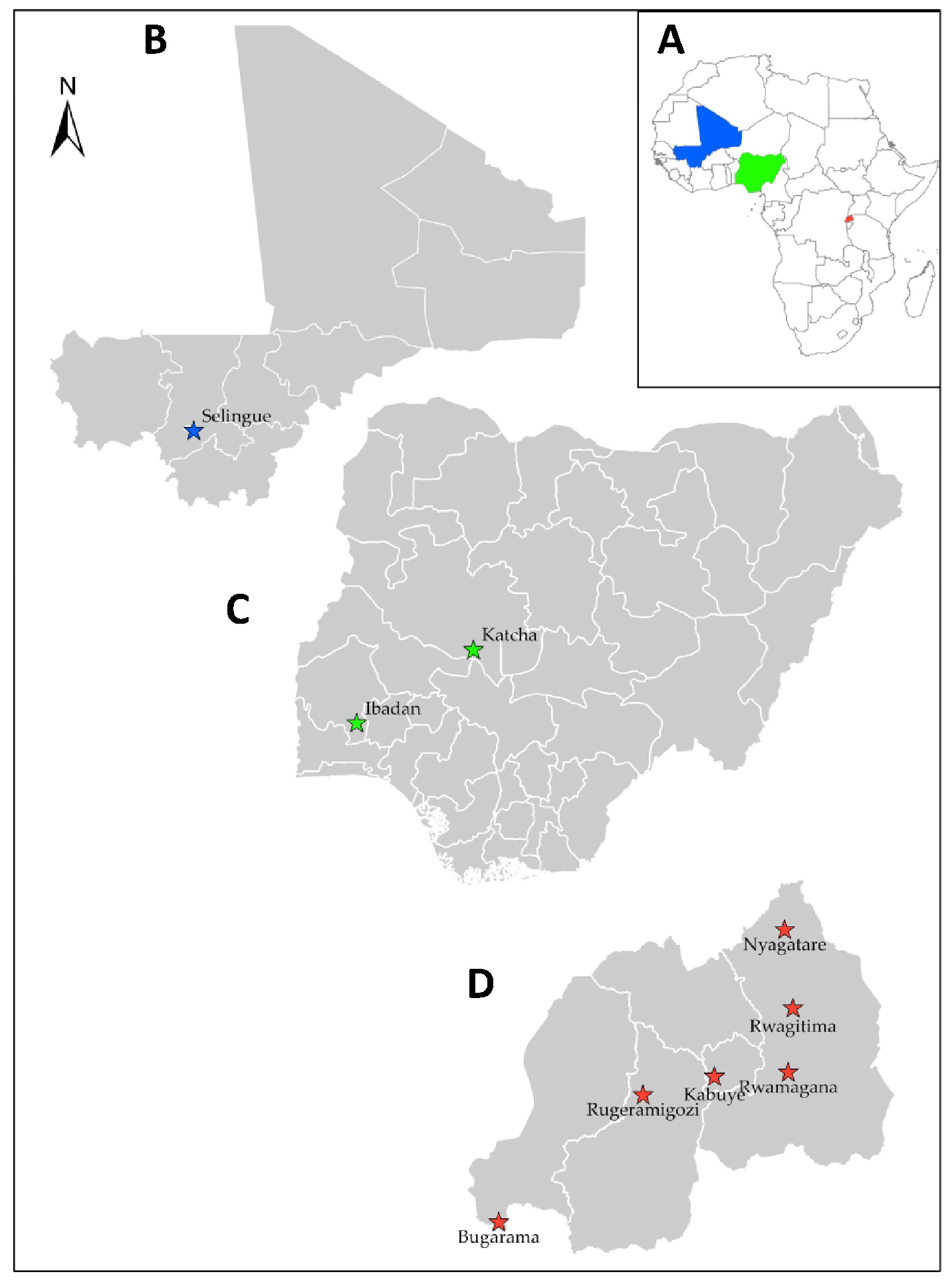
2]. Mali, a landlocked country in West Africa, ranks fifth among African nations in terms of rice production. Its rice production is being managed through irrigated systems connected to the Niger River. In Rwanda, the demand for rice is estimated at 145,000 tons per year, while national supply accounts for about 40%, creating a 60% deficit that is met through imports [3]. While the rice cultivation area is expanding in Africa, the average yields (2.35 tons/ha in 2021) are low when compared to Asia (4.95 tons/ha in 2021) [1]. Rice production is constrained by various factors, including biotic stresses such as pests and diseases. Africa has recorded a steady and substantial increase in the incidence of rice diseases such as rice yellow mottle virus (RYMV), rice blast (Pyricularia oryzae), bacterial leaf blight (Xanthomonas oryzae pv oryzae), bacterial leaf streak (Xanthomonas oryzae pv oryzicola), and rice stripe necrosis virus (RSNV) [4,5,6,7,8].
Rice sheath rot is an emerging disease worldwide [9]. Sarocladium oryzae [10], formerly Acrocylindrium oryzae, was the first organism to be associated with rice sheath rot symptoms in Taiwan in 1922 [11]. Sarocladium attenuatum was originally described as a distinct species causing rice sheath rot and was then considered a synonym of Sarocladium oryzae [12], but has recently been reestablished as a separate species causing rice sheath rot in Taiwan [13]. These authors also described a third species that causes sheath rot symptoms on rice called S. sparsum, which is closely related to S. oryzae and S. attenuatum. Sarocladium has been associated with rice sheath rot in thirty-eight countries [14].
Besides Sarocladium species, Fusarium spp. has been associated with the rice sheath rot complex. These mainly comprise isolates in the F. fujikuroi species complex (FFSC), including F. proliferatum, F. verticillioides, F. incarnatum, and F. fujikuroi [15,16,17,18,19]. In addition, various bacterial species cause rice sheath rot symptoms. The most important one is Pseudomonas fuscovaginae, which is known to cause sheath brown rot of rice at high altitudes. In Africa, this bacterium has been reported in Burundi [9,20] and Madagascar.
In West Africa, rice sheath rot has been reported in Cote D’ivoire, Gambia, Niger, Nigeria, and Senegal [14]. However, no causative fungal strains were isolated, and no detailed scientific information was provided except for Nigeria [21], where S. attenuatum was first reported as one of the causes of grain discoloration on rice [21]. In addition, an inhibitory effect of S. oryzae on seed germination was later observed [22]. Most information pertaining to the occurrence of S. oryzae in Africa relating to stored, marketed, and field seeds, especially with respect to mycotoxigenic potentials, was enumerated by [23].
Rice sheath rot can cause high yield losses of 20–80% [24,25,26]. Furthermore, an extensive survey of rice fields across West Africa enabled the identification of sheath rot symptoms in Mali and Nigeria (AfricaRice disease database). Although yield losses due to the sheath rot disease have not been estimated in Mali and Nigeria, a field survey conducted in 2011 and 2013 revealed the high incidence and severity of the disease in Rwanda [27].
Mycotoxin contamination of cereal products poses a serious concern for animal and human health. Several studies have reported Fusarium species as the major producers of mycotoxins contaminating cereals, including rice [28,29,30,31,32,33,34]. In the African region, previous studies have reported several mycotoxins being synthesized by Fusarium species isolated from rice as a serious health threat to producers and consumers [35,36,37,38,39,40].
Comprehensive information regarding the incidence and distribution of sheath rot disease is notably lacking. Additionally, there has been a notable absence of research examining the genetic, pathogenic, and toxigenic variability of pathogens associated with this disease in East and West Africa. Acquiring this crucial information will offer valuable insights into disease control and enhance management strategies for breeding programs. Therefore, this study aimed to identify, characterize, as well as assess the genetic, pathogenic, and toxigenic diversity of the pathogens associated with rice sheath rot disease in Mali, Nigeria, and Rwanda.

2. Materials and Methods

2.1. Collection of Samples

Samples were collected from rice research programs and farmer fields in Mali, Nigeria, and Rwanda (Figure 1). Naturally infected whole rice plants with sheath rot symptoms having sheath browning, necrosis, grain emptiness, and rotting, as indicated in Figure 2, were collected. Samples from farmer fields were collected during the 2017 rice growing season at the office of the rural development, Selingue village near Bamako, Mali, and from two fields located at Ibadan, Oyo State, and Katcha near Badeggi, Niger State in Nigeria. Samples were randomly collected 25 m apart at each location. The samples collected were conserved in dry paper bags, while hands were disinfected with 70% alcohol after each sampling. Samples were later stored in dry bags in the refrigerator at 4 °C in the laboratory. Isolates collected in 2011 and 2013 from Bugarama, Kabuye, Nyagatare, Rwagitima, Rugeramigozi, and Rwamagana in Rwanda were also included in this study. Agro-climatological details of the selected three countries with their various agroecologies are presented in Table 1.

2.1.1. Isolation and Purification of Sheath Rot-Associated Isolates

Infected sheath and seed samples showing symptoms of sheath rot were surface-sterilized in 2% sodium hypochlorite for two minutes and then rinsed thrice in sterile distilled water. They were drained using sterile paper towels and, thereafter, cut into small pieces of about 0.5 cm2 and plated on 90 mm diameter Petri dishes containing Potato Dextrose Agar (PDA). The cultured Petri dishes were incubated at 28 °C in darkness for 7–14 days. Cultures were further purified by plating on fresh PDA. Isolation and identification of pathogens was carried out at the Phytopathology Laboratory of Ghent University, Belgium.

2.1.2. Identification of Pathogens

Identification of fungal pathogens was obtained based on their typical structure and basic characters, as described by [41]. The incidence and diversity of fungi were observed and recorded. After 5 days, all plates were examined under a compound microscope for the presence of several fungal pathogens. Conidia of these fungi were mounted on glass slides in water and examined under a compound microscope for identification at the genus level. For Fusarium species, pure cultures were plated and stored on PDA slants at room temperature and maintained at −80 °C with 40% glycerol. A similar method of storage was used for Sarocladium species except for the use of 20% glycerol.

2.2. Molecular Characterization of Isolates

2.2.1. DNA extraction, Amplification, and Sequencing

Fungal isolates were grown on potato dextrose broth (PDB) at 28 °C for seven days. Mycelia mats were harvested by filtration, dried by blotting using sterile paper towels, frozen in liquid nitrogen, and pulverized using a Retsch MM 400, tissue lyser (Retsch GmbH, Haan, Germany).
Genomic DNA was extracted using the Wizard Genomic DNA Purification Kit (Promega Madison, WI, USA). Quantification and purity were determined using Nanodrop 3000 (Thermo Scientific, Asheville, NC, USA) and diluted to a concentration of 20 ng µL−1.
Fusarium isolates were further identified by amplifying the Translation Elongation Factor (EF-1α), using a primer pair of TEF-1-F (5′-ATG GGT AAG GAA GAC AAG AC-3′) and TEF-2-R (5′-GGA AGT ACC AGT GAT CAT GTT-3′) [42]. PCR reactions were performed in 25 µL of a solution consisting of 2 µL genomic DNA (100 ng µL−1), 5 µL PCR buffer (5×; Promega), 5 µL Q solution (Qiagen, Hilden, Germany), 0.5 µL dNTPs (10 mM; Promega), 1.75 µL of each primer (10 µM), 0.15 µL Taq DNA polymerase (5 units µL−1; Promega), and 8.85 µL ultrapure sterile water. Amplification was performed with an initial denaturation step at 94 °C for 1 min, followed by 35 cycles of denaturation at 94 °C for 30 s, primer annealing at 53 °C for 45 s, and extension at 72 °C for 1 min. Cycling ended with a final extension step at 72 °C for 5 min [42]. The amplicons were separated by horizontal electrophoresis using 1.5% agarose gels in a TAE buffer at 100 V for 25 min and visualized by ethidium bromide staining on a UV trans illuminator. Amplified products were purified with ExoSAP (Thermo Fisher Scientific, Waltam, MA, USA) and sequenced by LGC Genomics GmbH (Berlin, Germany) using Sanger sequencing.
For Sarocladium isolates, two genomic regions, the Internal Transcribed Spacer (ITS) and Actin, were amplified and sequenced. For the ITS region, primers ITS1 (5′-TCC GTA GGT GAA CCT GCG G-3′) and ITS4 (5′-TCC TCC GCT TAT TGA TAT GC -3′) were used [43]. The actin region was amplified using ACT1 (5′- TGG GAC GAT ATG GAG AAG ATC TGG CA -3′) and ACT4 (5′-TCG TCG TAT TCT TGC TTG GAG ATC CAC AT-3′) [44].
For both primer pairs, PCR reactions were performed in 25 µL of a solution consisting of 2 µL genomic DNA (100 ng µL−1), 5 µL PCR buffer (5×; Promega), 0.5 µL dNTPs (10 mM; Promega), 1.75 µL of each primer (10 µM), 0.15 µL Taq DNA polymerase (5 units µL−1, Promega), and 13.85 µL ultrapure sterile water. Amplification was performed using a Flexcycler PCR Thermal Cycler (Analytik Jena, GmbH, Jena, Germany). For ITS amplification, the thermal profile consisted of an initial denaturation step at 94 °C for 10 min, followed by 35 cycles of denaturation at 94 °C for 1 min, primer annealing at 55 °C for 1 min, and extension at 72 °C for 1 min. Cycling ended with a final extension step at 72 °C for 10 min [43]. ACT fragments were amplified using an initial denaturation step at 94 °C for 1 min, followed by 39 cycles of denaturation at 94 °C for 45 s, primer annealing at 59 °C for 30 s, and extension at 72 °C for 30 s. Cycling ended with a final extension step at 72 °C for 8 min [44].

2.2.2. Phylogenetic Analysis

The nucleotide sequences generated by the forward and reverse primers were used to obtain consensus sequences after editing via BioEdit version 7.2.5. [45]. From each duplicate identical sequence, a representative sequence from each identical set of sequences was compared to other sequences available at GenBank. Sequences were first aligned via muscle alignment in Mega V.11. [46], after which a maximum-likelihood tree was constructed based on the matrix of pairwise distances obtained using the General Time Reversible (GTR) Model. Reference sequences of Fusarium (Table A1) and Sarocladium (Table A2) species representing the three countries were used for phylogenetic analysis. For Fusarium, a Phylogenetic tree was constructed based on maximum likelihood inferred from partial EF-1α sequences of four Fusarium species complexes using IQ-Tree with GTR + G + I model and annotated using iTol software (V5) [47]. Cylindrocarpon sp. AC2011 strain CPC 13,531 was used as an outgroup. However, for Sarocladium characterization, concatenated alignments of ITS and ACT region were performed, after which a single phylogenetic tree was generated. To root the tree, Sarocladium zeae strain CBS 800.69 (Table A2) was used as an outgroup.

2.3. Pathogenicity Assay

The location of the isolates and genetic groupings were used to select a subset of twenty-nine isolates for pathogenicity studies on rice plants. For pathogenicity tests with Fusarium species, representative isolates from the four Fusarium species complexes recorded in all the locations (FIESC, FFSC, FOSC, FSAMSC) were used for rice inoculation. To evaluate the pathogenicity of Sarocladium species, thirteen isolates comprising S. attenuatum (3), S. oryzae (6), and S. sparsum (4) were used. Indica rice cultivar (FARO 44) released by Africa Rice Center was used for the assay.
Inoculum was prepared according to the standard grain inoculum technique [48]. Briefly, rice grains were soaked in water for 60 min, excess water was removed, and the grains were autoclaved twice on two different days. For 4 g of rice grains, 1 plug (diameter = 5 mm) from the edge of a 14-day-old fungal colony was added together with 1 mL of sterile distilled water. Every two days, the grain inoculum was shaken to prevent the formation of clumps. After 10 days of incubation at 28 °C, the inoculum was fully colonized.
The rice seeds were dehulled and surface sterilized in 2% sodium hypochlorite solution for 25 min, rinsed five times in sterile distilled water, and placed in Petri dishes containing sterile moistened filter papers (Whatman, grade 3). Following seedling emergence, six seedlings were transplanted into perforated plastic trays (22 × 15 × 6 cm) containing potting soil (Structural; Snebbout, Kaprijke, Belgium). Plants were watered daily, fertilized weekly with 0.2% iron sulfate and 0.1% ammonium sulfate, and maintained in a growth chamber (28 °C, 60% relative humidity). Six-week-old plants were used for the inoculation.
One fully colonized fungal grain was introduced in the junction point between the sheath of the second youngest plant leaf and the stem. Inoculation points were covered with moist cotton wool and wrapped with parafilm to maintain humidity. High humidity was maintained for 24 h post-inoculation by incubating in a controlled room at 28 °C day² and night, 16/12 light regime, and 85% relative humidity. Subsequently, incubation was maintained at 65% relative humidity, temperature, and light regime as above for 2–10 days. The disease development was evaluated eight days after inoculation by measuring the lesion length on the flag leaf sheath. Three trays containing six rice plants were used in each treatment (n = 18). The experiment was conducted once.

2.4. Statistical Analysis

Lesion length was used as a measure of isolate virulence on rice plants. Since the conditions of normality were not met, a non-parametric analysis was carried out. The lesion length of the infected sheath was quantified using Kruskal–Wally’s Rank Sum test followed by a post hoc Mann–Whitney test. Statistical significance was defined as p = 0.05.

2.5. Mycotoxin Analysis

2.5.1. Culture Preparation

Pure cultures of seventy-seven identified isolates (Fusarium—46, Sarocladium—31) were sub-cultured on Petri dishes with PDA. The medium was poured into 90 mm Petri dishes. Two mm of clean and pure cultured isolates were sub-cultured on fresh PDA plates and incubated at 25 °C for 3 weeks. Each isolate was grown in triplicates.

2.5.2. Reagents and Standards

Ethyl acetate and dichloromethane (DCM) were purchased from (Thermofisher Scientific, Merelbeke, Belgium). Analytical grade formic acid (100%) and ammonium acetate were from (Merck, Darmstadt, Germany). Purified water was from the Arium pro-VF system (Millipore, Brussels, Belgium). LC-MS grade acetic acid and methanol were from Biosolve (Valkenswaard, The Netherlands).
Certified mycotoxin standard solutions, more specifically aflatoxin mix (AFB1, AFB2, AFG1 and AFG2), deoxynivalenol (DON), fumonisin mix (FB1 and FB2), nivalenol (NIV), neosolaniol (NEO), OTA, T2, HT2, 3-acetyldeoxynivalenol (3-ADON), diacetoxyscirpenol (DAS), 15- acetyldeoxynivalenol (15-ADON), fusarenon-X (F-X), sterigmatocystin (STC), zearalenone (ZEN), and deepoxy-deoxynivalenol (DOM) were purchased from Biopure (RomerLabs, Getzersdorf, Austria). Fumonisin B3 (FB3) and enniatin B (ENN B) were obtained from Fermentek (Jerusalem, Israel). Alternariol (AOH) and alternariol monomethylether (AME) were purchased from Sigma-Aldrich (Bornem, Belgium), and roquefortine (ROQ-C) was purchased from Alexis Biochemicals (Enzo Life Sciences BVBA, Zandhoven, Belgium).
Working solutions were prepared by diluting the stock solutions in methanol and stored at −20 °C. A standard mixture consisting of the above mycotoxins (without DOM) in a concentration range between 0.5 ng/µL and 40 ng/µL was prepared as well and stored at −20 °C.
Mobile phase A (94% water, 5% methanol, 1% acetic acid, and 5 mM ammonium acetate) and mobile phase B (97% methanol, 2% water, 1% acetic acid, and 5 mM ammonium acetate) were prepared.

2.5.3. Sample Preparation and Extraction

The extraction process started with the preparation of the quality control samples. Briefly, three plugs each of blank agar (uninoculated) were removed and placed into each of the three 50 mL Falcon tubes (spike 1, spike 2, and blank) and were macerated into pieces using a sterile scalpel blade. Then, 50 µL DOM internal standard (50 ng/µL) was added into each tube, after which 25 µL and 100 µL of the standard mixture were added to spike 1 and spike 2, respectively. The mixtures (spikes and blank) were left in the dark for 15 min.
Following the control sample preparation, Fusarium mycotoxins were extracted from pure cultures of different isolates by using a sterile 9 mm cork borer and scalpel to take three plugs (2 sides + center). The plugs were transferred into 50 mL Falcon tubes and macerated into pieces using a sterile scalpel blade. Then, 50 µL DOM internal standard (50 ng/µL) was added into each tube and left in the dark for 15 min. The samples, together with the quality control samples, were extracted by adding ethyl acetate + 1% formic acid. The content was agitated gently on a vertical shaker for 20 min and centrifuged at 3000 g for 15 min. Then, a folded filter paper (VWR International, Zaventem, Belgium) moistened with ethyl acetate + 1% formic was placed on a new extraction tube to collect the upper layer of the filtrate. Thereafter, 5 mL of dichloromethane (DCM) was added to each of the samples. The mixtures were agitated on a vertical shaker for 20 min and centrifuged at 3000 g for 15 min. Following centrifugation, the bottom layer (DCM phase) was collected in the same Falcon tube with the same filter paper. The filtrates were evaporated to dryness at 40 °C under a gentle nitrogen stream. The dissolved residue was reconstituted in 200 µL injection solvent (60% mobile phase A and 40% mobile phase B), well-vortexed, and ultra-centrifuged for 5 min at 10,000 rpm. Finally, 100 µL of the filtrates was transferred into HPLC vials for LC-MS/MS analysis.

2.5.4. Multi-Metabolite Analysis (LC-MS/MS)

The samples were analyzed using a Quattro Premier XE triple quadrupole mass spectrometer coupled with a Waters Acquity UPLC system (Waters, Milford, MA, USA).
Liquid chromatography conditions and MS parameters were followed, as described by [49]. The analytical column used was a symmetry C18, 5 µm, 2.1 × 150 mm with a guard column of the same material (3.5 µm, 10 mm × 2.1 mm) (Waters, Zellik, Belgium) kept at room temperature. The injection volume was 10 µL. The capillary voltage was set at 3.2 kV with a source block temperature and desolvation temperature of 120 and 400 °C, respectively. Data processing was performed using the Masslynx and Quanlynx software (version 4.2).

3. Results

3.1. Sampling and Isolation

Information on Fusarium isolates obtained from diseased rice plants in Nigeria, Mali, and Rwanda is presented in Table 2. Of the 46 Fusarium isolates evaluated in this study, nine isolates were obtained from seeds, while 37 isolates originated from the rice sheath. The highest number was obtained from Rwanda (24 isolates), followed by Nigeria (15 isolates), while Mali (seven isolates) had the least.
Information on Sarocladium isolates obtained from diseased rice plants in Nigeria, Mali, and Rwanda is given in Table 3. Out of the 24 Sarocladium isolates obtained, four were from seeds and 20 from the rice sheath. The highest number of isolates were obtained from Nigeria (nine from Katcha, seven from Ibadan), six isolates were from Rwanda, and only two from Mali (Table 3).
Altogether, seventy isolates comprising Sarocladium species (24) and Fusarium species (46) were obtained.

3.2. Phylogenetic Analysis of Fusarium and Sarocladium-like spp.

3.2.1. Fusarium Species

Partial sequences of the TEF-1a gene revealed the identity of all 46 Fusarium isolates used. Similarities to DNA sequences in the Fusaroid-ID and GenBank database ranged from 99 to 100%. Members of four species complexes were identified: F. fujikuroi species complex (FFSC—48%); F. incarnatum-equiseti species complex (FIESC—35%); F. oxysporum species complex (FOSC—13%); and F. sambucinum species complex (FSAMSC—4%) (Table 2). The phylogenetic analysis of the 46 Fusarium isolated is presented in Figure 3. The origin and Genbank accession numbers of the reference isolates used are given in Table A1.
FFSC isolates from Rwanda are clustered with F. andiyazi (7 isolates), F. madaense (six isolates), and F. casha (one isolate). FFSC isolates from Mali were identified as F. nygamai (five isolates) and F. annulatum (one isolate), while in Nigeria, two F. marum isolates were found.
FIESC isolates were found in Nigeria and Rwanda and belong to five different species (sulawesiense, pernambucatum, tanahbumbuense, hainanense, and incarnatum) previously classified from rice, cereals, insects, and human samples [31,35,36,50,51,52,53] (Figure 3). Eight of our FIESC isolates were nested within the F. sulawesiense clade, including seven isolates from Ibadan and an isolate from Katcha. One isolate from Ibadan clustered with members of F. pernambucatum; two isolates from Katcha nested within the F. tanahbumbuense clade, and two isolates from Ibadan nested within the F. hainanense group. Three isolates from Rwanda were found in F. incarnatum (Figure 3). None of our isolates clustered with the F. equiseti species clade (Figure 3).
The FOSC was found in Rwanda and Mali with members belonging to F. callistephi (five isolates from Rwanda) and F. triseptum (one isolate from Mali). Two isolates belonging to F. acasia-mearnsii in the FSAMSC were obtained from Rwanda.

3.2.2. Sarocladium Species

Thirty-one S. oryzae-like isolates were used for phylogenetic analysis (Nigeria = 16, Mali = 2, Rwanda = 6, reference isolates =10 (Table 3 and Table A2). We sequenced the ITS and ACT regions of all isolates from Nigeria, Rwanda, and Mali and four of the Sarocladium reference isolates obtained from the Fungal Biodiversity Institute (Centraalbureau voor Schimmelcultures, CBS), Utrecht, The Netherlands (sequence references listed in Table 3). Sequences of the other reference isolates were obtained from Genbank (Table A2).
Partial sequences and concatenation of both ITS and ACT regions showed the identities of all the isolates.
They were further subjected to BLASTn comparison with isolates in GenBank. Results revealed that all 31 isolates had 98–100% identity with Sarocladium species. A concatenated tree, in which reference sequences from GenBank were included (see Table A2), clearly delineated the Sarocladium isolates into three distinct phylogenetic groups with high bootstrap values (Figure 4). Most isolates from Nigeria (11 of 16), one isolate from Mali (SEMA0013A), and one isolate from Rwanda (RFRG2) clustered together with reference isolates CBS 180.74 from India, CBS 361.75 from Kenya, and CBS120.817 from Panama and were identified as S. oryzae. The second Mali isolate (SEMA0029) and five of the six Rwandan isolates clustered with reference isolates CBS 101.61 from Mexico and CBS 399.73 from India and belong to the S. attenuatum lineage. Five isolates from Nigeria clustered with reference isolate CBS 414.81 from Nigeria and with the S. sparsum isolate 18,042 from Taiwan (Figure 4). They mostly occurred in Ibadan, Nigeria (DS), except for an isolate (BDNG0025) found on infected seeds in Katcha (SGS). Finding a substantial number of this group in Ibadan was not unusual because it clustered with a reference isolate CBS 414.81 of Ibadan origin, collected, and reported [21], and later deposited into the GenBank [44], and recently reclassified as S. sparsum.

3.3. Pathogenicity Testing

Sixteen isolates from the dominant Fusarium groups recorded in Nigeria, Rwanda, and Mali were used for pathogenicity testing on the FARO 44 rice variety. All four Fusarium species complexes could induce rice sheath rot symptoms on the rice cultivar, but the degree of aggressiveness of all the Fusarium species tested showed significant variations on the rice cultivar (Figure 5). Specifically, one of the two isolates of F. marum IBNGF0016 from Ibadan in Nigeria caused the highest disease severity on FARO 44, followed by the second F. marum isolate IBNGF0012, and an FIESC isolate F. sulawesiense BDNGF0002 from Katcha in Nigeria. On the contrary, F. nygamai originated from Mali and one of the FIESC isolates, F. tanahbumbuense BDNGF0001, from Ibadan, Nigeria, were the least aggressive isolates.
For the pathogenicity tests with Sarocladium species, disease evaluation at 8 days post-inoculation (DPI) showed that all thirteen isolates tested could induce typical sheath rot symptoms on FARO 44, albeit with varying disease development levels (Figure 6). Isolates affiliated with S. sparsum, all of Nigeria origin, were the most aggressive (p < 0.05). In contrast, isolates affiliated with S. oryzae and S. attenuatum were less aggressive (Figure 6). S. oryzae isolate IBNG0011 from Nigeria is the most aggressive among the group, while SEMA0013A from Mali appears to be the least aggressive.

3.4. Mycotoxin Profiling (In Vitro)

Forty-six Fusarium and thirty-one Sarocladium species obtained from rice with sheath rot symptoms were investigated for multi-mycotoxin production using LC-MS/MS. None of the Sarocladium isolates produced mycotoxins. Our results revealed that the Fusarium species were able to produce eight mycotoxins. The most detected mycotoxins include type A (DAS and NEO) and B (NIV and FUS-X) trichothecenes, which were produced by FIESC isolates obtained across the two regions of Nigeria, albeit at different concentrations (Table 4). Furthermore, zearalenone (ZEN) at a very high concentration of 26,173 and 32,529 µg/kg was detected in the two Ibadan Nigerian F. hainanense isolates IBNGF0005 and IBNGF0003, respectively. Two Rwandan isolates, F. madaense RFRM18 and F. acasiae-mearnsii RFRM19, produced ZEN at the concentration of 1349 and 329 µg/kg, respectively. The predominant mycotoxins detected from Malian F. nygamai isolates were fumonisins (FB1, FB2, and FB3), which occurred at a very high concentration. FB1 concentrations ranged from 53,118 to 141,102 µg/kg, followed by FB2, ranging from 3391 to 5122 µg/kg, while FB3 occurred at a lower concentration, which ranged between 355 and 692 µg/kg. Furthermore, F. nygamai and F. annulatum SEMAF17-225A from Mali produced DAS, FUS-X, NIV, and NEO at different concentrations (Table 4).

4. Discussion

This study shows that both Fusarium and Sarocladium species are associated with rice sheath rot disease in Nigeria, Rwanda, and Mali and provides insight into their genetic, pathogenic, and toxigenic diversity. Molecular characterization using the EF-1α gene enabled the delineation of Fusarium isolates into four distinct Fusarium species complexes, whereas concatenation of ITS and ACT sequences delineated Sarocladium into three species. Phylogenetic analysis showed that isolates grouped differently according to their geographical location (Figure 3 and Figure 4).
Four Fusarium species complexes (FIESC, FFSC, FOSC, and FSAMSC) were found to be associated with rice sheath rot in SSA. Previous research has resolved FIESC species as a mere complex indicated by numbers, but current studies further updated them according to the new nomenclature, well elucidated from numbers to names [50,51,52]. Additionally, they were characterized using the recently updated Fusarium ID database (Fusarioid-ID). Dominant in our findings were members of the FIESC previously classified from rice, cereals, insects, and human samples [29,33,34,52,53,54,55], including F. sulawesiense (FIESC 16), F. pernambucatum (FIESC 17), F. tanahbumbuense (FIESC 24), F. hainanense (FIESC 26), and F. incarnatum (FIESC 38). This is similar to those found to be associated with rice sheath rot in Indonesia [19], India [17], and the USA [18]. A larger part of the isolates clustered with F. sulawesiense, which supports the findings in Brazil and China [29,56], while the abundance of F. hainanense and a few others, such as F. pernambucatum and F. tanahbumbuense, were among the FIESC reported on Brazilian rice. There is a wide variation among the species obtained within the FIESC complex, and the observed variation could be correlated with variation in agro-ecological zones. Notable is the fact that some of the F. sulawesiense isolates from Ibadan (Nigeria) are closely related to strains NTB 1 (rice sheath rot, Indonesia), LC6936 (rice, China), F1 (Sweet potato, US), BT48, and PRT6 (oil palm, Indonesia), and ITEM7547 (Musa, Bahamas), all of which originated from climates characterized by high temperature and humidity. Isolate F1 originated from Louisiana, USA, which is known for its humid subtropical climate with long, hot, and humid summer, similar to the climate of ITEM7547 from the Bahamas [34,57]. Based on our findings, we can hypothesize that the environmental factors could be the driving forces to be considered in the distribution of the FIESC isolates. Notable differences in climate and farming practices could not be underestimated as the three F. incarnatum isolates from a higher altitude in Rwanda formed part of recently classified FIESC 38 isolates from a similar altitude and climate in Brazil [29]. Within the FIESC clades, none of the studied isolates was found among the F. equiseti clade, which disagrees with the previous studies of [54,55,58], whereby variable percentages of both F. equiseti and F. incarnatum were reported from rice samples. Members of the F. equiseti clade are frequent in cereals grown in Western Europe, Turkey, and North America [34].
FFSC species have been implicated as another causal agent of various rice diseases, including sheath rot [51,59]. F. andiyazi and F. madaense represent the principal species recovered in Rwanda; F. nygamai was only isolated from Mali, while F. marum was recovered from Nigeria. Isolates of F. casha (Rwanda) and F. annulatum (Mali) were also found among our FFSC. The peculiarity in the clustering of F. andiyazi, F. madaense, and F. marum, clades observed in our studies is comparable to the typical phylogenetic pattern observed in [60]. F. andiyazi and F. madaense are typically associated with tropical grasses, including sorghum, maize, millet, and rice, in various parts of the world [60]. The two virulent F. marum isolates from Nigeria clearly resolved into a separate clade and clustered with two F. marum isolates obtained from sorghum in Cameroon [60]. Isolates clustering with F. nygamai, the most dominant species in Mali, are closely related to isolates obtained from cereals from regions with similar warm and dry climates (Figure 4), such as Australia [61], Italy [28], Mexico [62], and Tunisia [36]. These FIESC and FFSC findings are consistent with the recent report of rice sheath rot disease in Indonesia [19] and rice disease in China [56,63].
Furthermore, isolates clustering with F. callistephi were found in Rwanda, while F. triseptatum was found in Mali. Both are members of the FOSC. F. callistephi is mainly known as a wilt pathogen on ornamentals from the Asteraceae family [64]. Lastly, members of FSAMSC, which includes two isolates clustering with F. acasiae-mearnsii of Rwanda origin, were also identified (Figure 4). F. acasiae-mearnsii isolates have previously been found in Australia and South Africa and can cause head blight on wheat [65].
Reports on mycotoxins produced by Fusarium species isolated from rice sheath rot disease in sub-Saharan Africa are very limited. In consequence, this is the first study to investigate the toxigenic potentials of Fusarium isolates from rice sheath rot disease in Mali, Nigeria, and Rwanda. The predominant mycotoxins found in FIESC isolates from Nigeria were trichothecenes, while the three F. incarnatum isolates from Rwanda did not produce mycotoxins. Among the 12 FIESC isolates collected from Ibadan (derived savannah region) in Nigeria, type A (DAS, NEO) and B (NIV, FUS-X) trichothecenes were detected in seven samples. This confirms the previous mycotoxins findings on cereals [29,58,66]. In addition, a huge ZEN production of 32,529 and 26,173 µg/kg was detected from the two F. hainanense isolates collected from Ibadan. Similar results for F. hainanense were obtained in Brazilian rice [29]. This also corroborates the study of [67], which demonstrated that ZEN production by Fusarium species is greater in moldy samples, which is favored by wet climates with high rainfall and high humidity. Within the FFSC, F. andiyazi isolates did not produce mycotoxins, while the F. annulatum isolate and three out of five F. nygamai isolates from the dry and hot Sudan Guinea Savannah of Mali produced trichothecenes and fumonisins (FB1, FB2, and FB3), respectively. This is consistent with the findings of [68] reporting high levels of toxins for F. nygamai, while F. andiyazi isolates produced little or no mycotoxins. Moreover, fumonisin producers were not detected among the isolates collected from Nigeria and Rwanda. Thus, fumonisin contamination may be expected to be higher in samples collected in the Sahel with a warm and dry climate. The development of fumonisins in cereal crops prior to harvest might increase due to heat and water stress that characterized the environmental drought [69]. Only two of the 23 Fusarium isolates from Rwanda produced mycotoxins: trichothecenes (NIV and Fus-X) and ZEN were detected in a F. madaense isolate and a F. acaciae-mearnsii isolate, both obtained from the Rwamagana district. It has been shown before that F. acacia-mearnsii isolates can produce NIV [70] and ZEN [71].
According to the research of [72], which elucidated the presence and absence of biosynthetic gene clusters responsible for the synthesis of mycotoxins and secondary metabolites in FIESC, further studies are necessary to investigate if the mycotoxin production potentials of our isolates agree with their genetic profile or assess if there are differences in expression level. In conclusion, mycotoxin production is common in rice-derived Fusarium isolates from Nigeria (12 out of 15) and Mali (5 out of 7) but rare in Rwanda (2 out of 25).
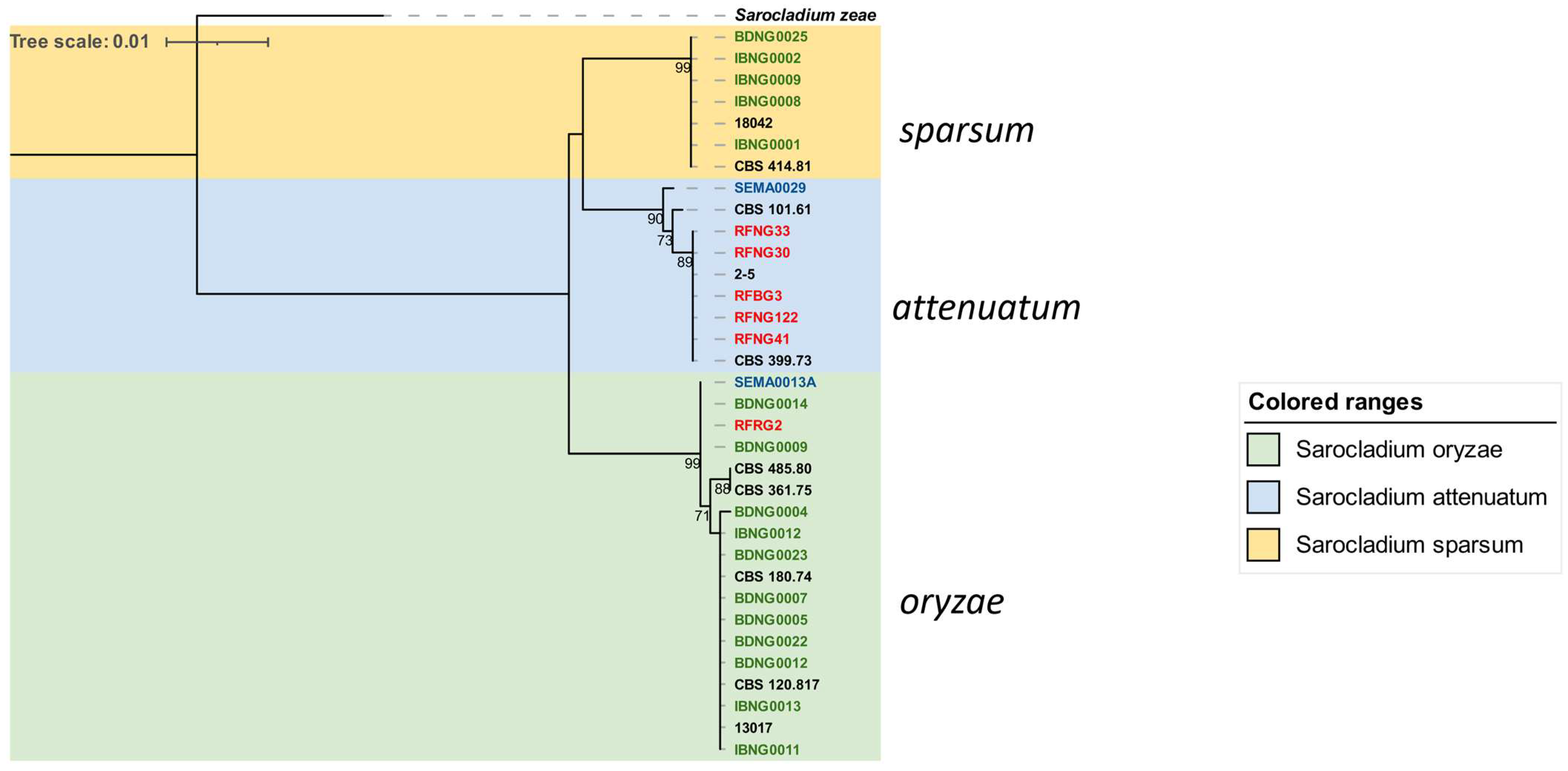
In contrast to the heterogeneity observed among Fusarium species, three clearly delineated Sarocladium species were recovered from the three countries of study, but with a lower frequency of occurrence in Mali and Rwanda. Following the characterization of Sarocladium species causing rice sheath rot in Taiwan by [13] and using a concatenation of two genes, we were able to resolve our isolates into three species, namely, attenuatum, oryzae, and sparsum (Figure 4). Isolates belonging to S. sparsum were only found in Nigeria and mostly originated from Ibadan (DS), except for an isolate (BDNG0025) from an infected seed in Katcha (SGS). Finding a larger part of this group in Ibadan was not unexpected because it clusters with an Ibadan-origin reference isolate CBS 414.81, collected and reported as S. attenuatum [21], and later deposited into the Genbank [44]. Our results clearly show, however, that isolate CBS 414.81 belongs to sparsum species. Surprisingly, this group was not found in Mali and Rwanda. There occurs a notable correlation between this group and the collection region, which proves that geographical area and climate are the most crucial factors that influence the occurrence of these pathotypes and their virulence. This agrees with the hypothesis that isolates from different locations may also vary in their level of aggressiveness [73,74]. S. oryzae isolates (Figure 4) showed a strong intra-species similarity that is not phylogeographically based. Isolates in this species were the most predominant and widely distributed. It consists of 18 similar isolates from nearly all the rice-growing regions in the world. Most isolates from Nigeria (11 of 16) belong to this group. An isolate from Mali (SEMA0013A) generated from this study, isolates from previous studies on rice, such as 13017 from Taiwan [13], CBS 180.74 from India, African isolates CBS 361.75 from Kenya [75], RFRG2 from Rwanda (this study), Central American CBS 120.817 from Panama, and CBS 485.80 Australia [76], are part of this group. They were found in two agro-ecological zones of Nigeria, although more frequently in Katcha than Ibadan.
The presence of S. oryzae in all the rice-growing regions of the world is a signal of its flexibility to adjust to various agro-ecological zones. It also suggests a link between its dispersal, rice movement, and international trade. A potential quarantine threat is of great concern with the rapid distribution of this group. This might also imply that the origin of the isolate may be connected to Asia. It should be noted that African countries, including Mali, Nigeria, and Rwanda, are major importers of rice from Asia despite Nigeria being the highest producer on the continent.
A third distinct group, S. attenuatum, was dominated by Rwandan isolates; the second Mali isolate, SEMA0029, also formed part of the group.
It is important to note that the two Sarocladium isolates obtained from Mali formed two of the three species found in this study despite the small sample size. Several reasons might be responsible, from accession variability, as local rice is mostly cultivated in Mali [77], to toxigenic variability among the competing Fusarium species, and multiple cropping system variations, among others. Larger-scale surveys across the country are necessary to have a wider knowledge of genetic diversity and distribution.
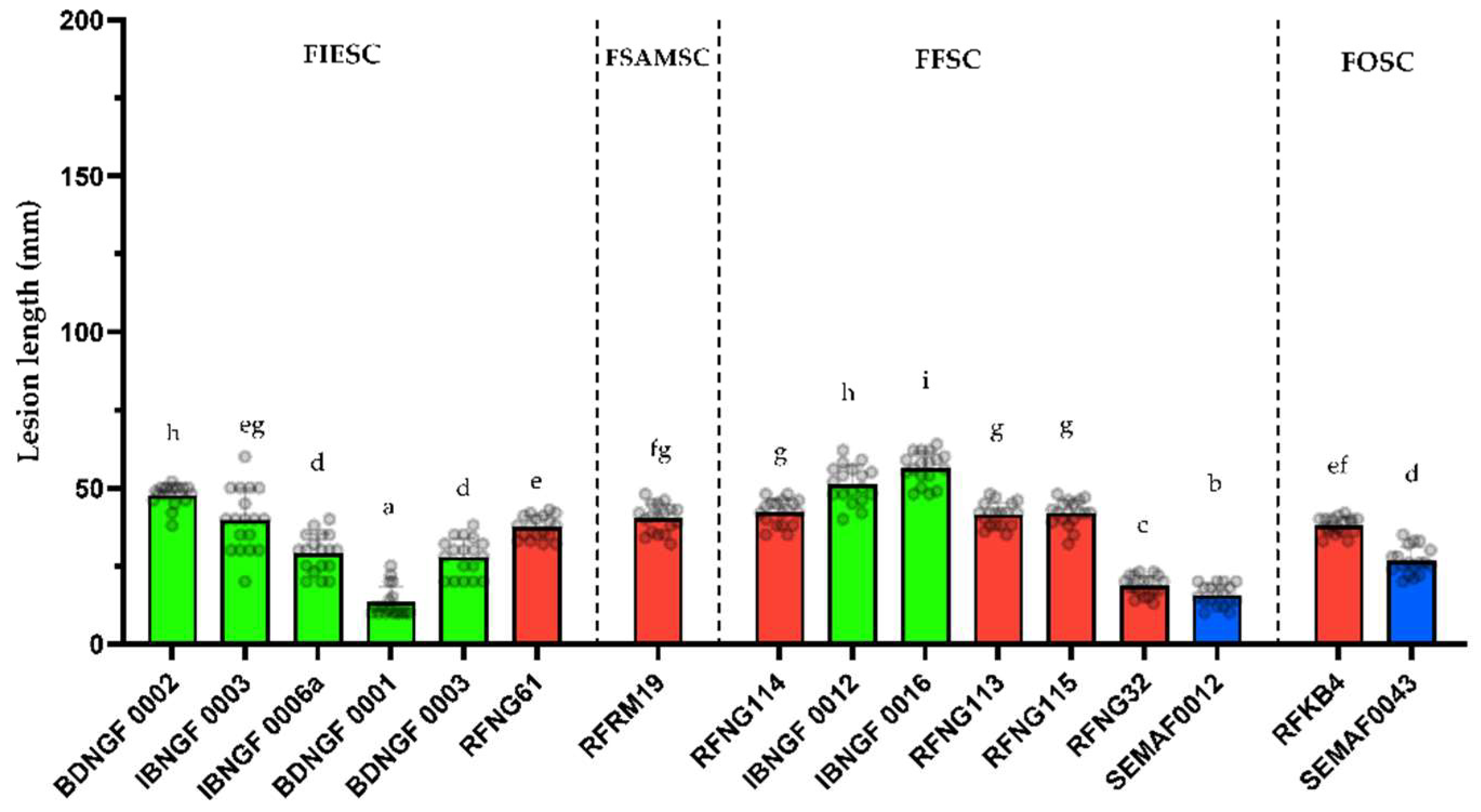
It is a well-known phenomenon that several factors such as variations in climate, topography, and farming practices, among others, are the drivers of variation in pathogen populations. To verify this claim, this study has revealed a strong ecology-driven diversification among the Sarocladium species used. It also exposed how environmental variation was able to influence genetic and virulence relatedness. The S. sparsum isolates that are mainly found in Ibadan (DS) are clearly more aggressive on the rice FARO44 cultivar used in this study than the S. oryzae and S. attenuatum strains, which are more common in the savannah region in Katcha-Badeggi and in Rwanda. The same trend of aggressiveness was reported in the study of [21], who used four isolates (presumably S. sparsum) collected from the southern region (DS and Humid Forest) of Nigeria. This confirms previous work showing that group 3 strains (=S. sparsum) are more aggressive on the rice japonica cultivar Kitaake than group 1 (=S. oryzae) or group 2 (=S. attenuatum) strains. S. sparsum isolates also produce high amounts of the toxin helvolic acid in planta which is clearly correlated with disease severity [78].

5. Conclusions

In conclusion, our study shows that despite the limited sampling size, diversity occurs within the East (Rwanda) and West African (Mali and Nigeria) isolates of Fusarium and Sarocladium with clear regional differences. The toxigenic profile of both pathogens was elucidated, and we found that most Fusarium isolates from Nigeria and Mali were able to produce one or more mycotoxins. In contrast, only two out of 24 isolates from Rwanda were able to produce mycotoxins. Further investigations with a broader geographic scope and a larger collection of samples are necessary to examine pathogenic variability and the population’s genetic structure. Meanwhile, based on the distinct groups of isolates from different regions, breeders in various agro-ecological regions should take note of the variations in virulence. This information can serve as a basis for selecting strains useful for identification and selecting effective sources of resistance for local rice breeding programs.
Moreover, additional studies are required to determine whether Sarocladium and Fusarium individually contribute to the observed symptoms in the field or if there is an interplay between both pathogens in the rice sheath rot complex. To the best of our knowledge, this research provides the first comprehensive dataset on the distribution, genetics, pathogenicity, and toxigenic profile of Fusarium species associated with rice sheath rot disease in sub-Saharan Africa.

Author Contributions

Conceptualization, V.d.P.B., O.O.A., O.I.N. and M.H.; methodology, O.O.A., V.d.P.B., M.V.D.V., F.E.O. and G.K.H.H.; validation, O.O.A. and V.d.P.B.; formal analysis, O.O.A.; investigation, O.O.A., V.d.P.B., L.B. and G.K.H.H.; resources, O.O.A., V.d.P.B., J.O., O.E.O. and A.O.; curation, O.O.A.; writing—original draft preparation, O.O.A.; writing, review, and editing, M.H.; visualization, O.O.A. and V.d.P.B.; supervision, M.H. and S.D.S.; project administration, O.O.A., V.d.P.B. and M.H.; funding acquisition, O.O.A., V.d.P.B., S.D.S. and M.H. All authors have read and agreed to the published version of the manuscript.

Funding

V.P. Bigirimana received a doctoral grant from the Belgian Technical Cooperation (BTC) (project reference: 10RWA/0018). This work was funded by a grant from the Special Research Fund of Ghent University (GOA 01GB3013) and by the Fund for Scientific Research Flanders (FWO G031317N).

Institutional Review Board Statement

Not applicable.

Data Availability Statement

All sequences generated in this study have been deposited in GenBank.

Acknowledgments

The authors wish to appreciate Christ’l Detavernier, Frédéric Dumoulin (UGent, Faculty of Pharmaceutical Sciences) for their help with the LC-MS/MS analysis. We also acknowledged the contributions of Aderonke Oludare and Opeyemi Ogedengbe (AfricaRice) for their coordination of rice sampling and transportation to Belgium.

Conflicts of Interest

The authors declare no conflict of interest.

Appendix A

Table A1. Sequences of reference strains of Fusarium obtained from GenBank used for building phylogenetic tree.
Species ComplexSpeciesIsolate NameHostOriginAccession NumberReferences
FIESCF. equisetiNL19-045005SoilNetherlandsMZ921835[50]
CPC 35262Human toenailCzech republicQED42271[79]
F. hainanense (26)LC11638Oryza spChinaMK289581[80]
15Ar057RiceBrazilMK298120[29]
LB2Oryza sativaPhilippinesJF715935[31]
PCO2Oil palmIndonesiaHM770725[81]
NRRL 28714Clinical samplesUSAGQ505604[54]
JBR 10Oryza sativa sheathIndonesiaMT138474[19]
F. nanum (25)F685WheatSpainKF962950[53]
NRRL 22244Clinical samplesUSAGQ505596[54]
F. tanahbumbuense (24)LC13726Digitaria spChinaMW594396[80]
NTT 6Oryza sativa sheathIndonesiaMT138460[19]
F. incarnatum (38)ITEM 7155Trichosanthe dioicaMalawiLN901581[34]
F. pernambucanum (17)NRRL 32864Clinical samplesUSAGQ505613[54]
CBS 791.70Musa sampientumNetherlandsMN170491[79]
F. sulawesiense (16)CBS 622.87Bixa orellanaBrazilMN170503[79]
ITEM7547Musa sampientumBahamasLN901580[34]
LC12173Luffa aegypticaChinaMK289605[80]
MS3369Wild riceBrazilMT682685[82]
LC6936Oryza sativaChinaMK289621[80]
F1Sweet potatoUSAKC820972[57]
BT48Oil palmIndonesiaHM770722[81]
PRT6Oil palmIndonesiaHM770723[81]
NTB 1IndonesiaOryza sativa sheathMT138458[19]
FFSCF. andiyaziLLC 1152Striga hermonthica seedEthiopiaOP486864[83]
MO5-1946S-3_PCNB Sorghum grainUSAKM462919[84]
F.marumKSU 15077SorghumCamerounMT374735[60]
KSU15074SorghumCamerounMT374736[60]
E432Rice seedsItalyGU827420[85]
E439Rice seedsItalyGU827419[85]
30ALHOryza sativa seedChinaFN252387[15]
F. madaenseCBS 146669Arachis hypogaeaNigeriaMW402098[40]
44ALHOryza sativa seedTanzaniaFN252390[15]
IALHOryza sativa seedBurkina FasoFN252388[15]
CML3853SorghumbicolorNigeriaMK895723[60]
CML3895SorghumbicolorTanzaniaMK895727[60]
PB-2SugarcaneChinaKP314282[86]
38ALHOryza sativa seedIndiaFN252389[15]
F. cashaPPRI20462Amaranthus cruentusSouth AfricaMF787262[87]
F. nygamaiB5Hp1g3B1BarleyTunisiaMG452941[36]
KC 13TomatoKenyaKT357537[88]
ENTO90Wild riceAustraliaMG873156[61]
M7491RiceItalyHM243236[28]
F. annulatumLC11670Oryza sativaChinaMW580517[63]
34ALHOryza sativa seedChinaFN252396[15]
LC13675Syzygium samarangenseChinaMW580542[63]
F. proliferatumCBS 480.96SoilPapua New GuineaMN534059[89]
FOSCF. triseptatumLC13771Deep sea sedimentChinaMW594358[63]
F. oxysporumFoc230BananaNigeriaAY217161Unpublished
F. callistephiCBS 187.53Callistephus chinensisNetherlandsMH484966[83]
SRRC1630Cooked riceNigeriaKT950251[90]
CBS 115423Agathosma betulinaSouth Africa MH484996[83]
FSAMSCF. acaciae-mearnsiiLC13786Musa nanaChinaMW620091[63]
Table A2. Sequences of reference strains of Sarocladium spp. on rice obtained from GenBank used for building phylogenetic trees.
GenusSpeciesIsolateOriginAccession Number ITSReference ITSAccession Number ACTReference ACT
SarocladiumattenuatumCBS 399.73IndiaHG965027[44]HG964979[44]
2-5TaiwanLC461444[13]LC464336[13]
oryzaeCBS 180.74IndiaHG965026[44]HG964978[44]
13017TaiwanLC461506[13]LC464380[13]
sparsumCBS 414.81NigeriaHG965028[44]HG964980[44]
18042TaiwanLC461520[13]LC464308[13]
SarocladiumzeaeCBS 800.69USAFN691451[91]HG965000[44]

References

  1. FAOSTAT. Available online: https://www.fao.org/faostat/en/#home (accessed on 10 August 2022).
  2. KPMG Nigeria. Rice Industry Review; KPMG: Abuja, Nigeria, 2019; Available online: www.kpmg/ng (accessed on 28 August 2022).
  3. MINICOM. Trade & Industry E-Newsletter; Ministry of Trade & Industry: Kigali, Rwanda, 2021; Volume 3, Issue 38. Available online: https://www.minicom.gov.rw/ (accessed on 28 August 2022).
  4. Afolabi, O.; Milan, B.; Amoussa, R.; Koebnik, R.; Poulin, L.; Szurek, B.; Habarugira, G.; Bigirimana, J.; Silue, D.; Ongom, J.; et al. First Report of Xanthomonas oryzae pv oryzicola Causing Bacterial Leaf Streak of Rice in Uganda. Plant Dis. 2014, 98, 1579. [Google Scholar] [CrossRef] [PubMed]
  5. Afolabi, O.; Amoussa, R.; Bilé, M.; Oludare, A.; Gbogbo, V.; Poulin, L.; Koebnik, R.; Szurek, B.; Silué, D. First Report of Bacterial Leaf Blight of Rice Caused by Xanthomonas oryzae pv. oryzae in Benin. Plant Dis. 2016, 100, 515. [Google Scholar] [CrossRef]
  6. Oludare, A.; Sow, M.; Afolabi, O.; Pinel-Galzi, A.; Hébrard, E.; Silué, D. First Report of Rice stripe necrosis virus Infecting Rice in Benin. Plant Dis. 2015, 99, 735. [Google Scholar] [CrossRef]
  7. Séré, Y.; Onasanya, A.; Afolabi, A.; Abo, E. Evaluation and Potential of Double Immunodifusion Gel Essay for Serological Characterization of Rice yellow mottle virus Isolates in West Africa. Afr. J. Biotechnol. 2005, 4, 197–205. [Google Scholar]
  8. Traoré, O.; Pinel-Galzi, A.; Sorho, F.; Sarra, S.; Rakotomalala, M.; Sangu, E.; Kanyeka, Z.; Séré, Y.; Konaté, G.; Fargette, D. A Reassessment of the Epidemiology of Rice yellow mottle virus Following Recent Advances in Field and Molecular Studies. Virus Res. 2009, 141, 258–267. [Google Scholar] [CrossRef]
  9. Bigirimana, V.d.P.; Hua, G.K.H.; Nyamangyoku, O.I.; Höfte, M. Rice Sheath Rot: An Emerging Ubiquitous Destructive Disease Complex. Front. Plant Sci. 2015, 6, 1066. [Google Scholar] [CrossRef]
  10. Gams, W.; Hawksworth, D.L. The Identity of Acrocylindrium oryzae Sawada and a Similar Fungus Causing Sheath-Rot of Rice. Kavaka 1976, 3, 57–61. [Google Scholar]
  11. Mew, T.W.; Gonzales, P. A Handbook of Rice Seedborne Fungi; Los Banos (Phillippines) International Rice Research Institute, and Enfield, N.H. (USA): Los Baños, CA, USA; Science Publishers: Hauppauge, NY, USA, 2002; 83p, ISBN 1578082552. [Google Scholar]
  12. Bridge, P.D.; Hawksworth, D.L.; Kavishe, D.F.; Farnell, P.A. A Revision of the Species Concept in Sarocladium, the Causal Agent of Sheath-rot in Rice and Bamboo Blight, Based on Biochemical and Morphometric Analyses. Plant Pathol. 1989, 38, 239–245. [Google Scholar] [CrossRef]
  13. Ou, J.H.; Lin, G.C.; Chen, C.Y. Sarocladium Species Associated with Rice in Taiwan. Mycol. Prog. 2020, 19, 67–80. [Google Scholar] [CrossRef]
  14. CABI Sarocladium oryzae (Rice Sheath Rot). Available online: https://www.cabi.org/isc/datasheet/48393 (accessed on 10 August 2022).
  15. Wulff, E.G.; Sørensen, J.L.; Lübeck, M.; Nielsen, K.F.; Thrane, U.; Torp, J. Fusarium spp. Associated with Rice Bakanae: Ecology, Genetic Diversity, Pathogenicity and Toxigenicity. Env. Environ. Microbiol. 2010, 12, 649–657. [Google Scholar] [CrossRef]
  16. Abbas, H.K.; Cartwright, R.D.; Shier, W.T.; Abouzied, M.M.; Bird, C.B.; Rice, L.G.; Ross, P.F.; Sciumbato, G.L.; Meredith, F.I. Natural Occurrence of Fumonisins in Rice with Fusarium Sheath Rot Disease. Plant Dis. 1998, 82, 22–25. [Google Scholar] [CrossRef]
  17. Prabhukarthikeyan, S.R.; Keerthana, U.; Nagendran, K.; Yadav, M.K.; Parameswaran, C.; Panneerselvam, P.; Rath, P.C. First Report of Fusarium proliferatum Causing Sheath Rot Disease of Rice in Eastern India. Plant Dis. 2021, 105, 704. [Google Scholar] [CrossRef]
  18. Imran, M.; Khanal, S.; Zhou, X.; Anthony-Babu, S.; Atiq, M. First report of Fusarium sheath rot of rice caused by Fusarium incarnatum-equiseti species complex in the United States. Plant Dis. 2022, 106, 3206. [Google Scholar] [CrossRef]
  19. Pramunadipta, S.; Widiastuti, A.; Wibowo, A.; Suga, H.; Priyatmojo, A. Identification and Pathogenicity of Fusarium Spp. Associated with the Sheath Rot Disease of Rice (Oryza sativa) in Indonesia. J. Plant Pathol. 2022, 104, 251–267. [Google Scholar] [CrossRef]
  20. Musonerimana, S.; Bez, C.; Licastro, D.; Habarugira, G.; Bigirimana, J.; Venturi, V. Pathobiomes Revealed That Pseudomonas Fuscovaginae and Sarocladium oryzae Are Independently Associated with Rice Sheath Rot. Microb. Ecol. 2020, 80, 627–642. [Google Scholar] [CrossRef]
  21. Ngala, G. Sarocladium attenuatum as One of the Causes of Rice Grain Spotting in Nigeria. Plant Pathol. 1983, 32, 289–293. [Google Scholar] [CrossRef]
  22. Ekefan, E.J.; Ibrnenge Vange, T. Effect of Seed Treatment with Tolchlofos-Methyl on Percentage Incidence of Seed-Borne Fungi and Germination of Rice (Oryza sativa) in Makurdi, Nigeria. Indian J. Agric. Sci. 2006, 76, 450–453. [Google Scholar]
  23. Makun, H.A.; Dutton, M.F.; Njobeh, P.B.; Phoku, J.Z.; Yah, C.S. Incidence, Phylogeny and Mycotoxigenic Potentials of Fungi Isolated from Rice in Niger State, Nigeria. J. Food Saf. 2011, 31, 334–349. [Google Scholar] [CrossRef]
  24. Ghosh, M.K.; Amudha, R.; Jayachandran, S.; Sakthivel, N. Detection and Quantification of Phytotoxic Metabolites of Sarocladium oryzae in Sheath Rot-Infected Grains of Rice. Lett. Appl. Microbiol. 2002, 34, 398–401. [Google Scholar] [CrossRef]
  25. Naeimi, S.; Okhovvat, S.M.; Hedjaroude, G.A.; Khosravi, V. Sheath Rot of Rice in Iran. Abstract. Commun. Agric. Appl. Biol. Sci. 2003, 68, 681–684. [Google Scholar] [PubMed]
  26. Mvuyekure, S.M.; Sibiya, J.; Derera, J.; Nzungize, J.; Nkima, G. Genetic Analysis of Mechanisms Associated with Inheritance of Resistance to Sheath Rot of Rice. Plant Breed. 2017, 136, 509–515. [Google Scholar] [CrossRef]
  27. Bigirimana, V.P. Characterization of Sheath rot Pathogens from Major Rice-Growing Areas in Rwanda. Ph.D. Thesis, Ghent University, Ghent, Belgium, 2016. [Google Scholar]
  28. Amatulli, M.T.; Spadaro, D.; Gullino, M.L.; Garibaldi, A. Conventional and Real-Time PCR for the Identification of Fusarium fujikuroi and Fusarium proliferatum from Diseased Rice Tissues and Seeds. Eur. J. Plant Pathol. 2012, 134, 401–408. [Google Scholar] [CrossRef]
  29. Avila, C.F.; Moreira, G.M.; Nicolli, C.P.; Gomes, L.B.; Abreu, L.M.; Pfenning, L.H.; Haidukowski, M.; Moretti, A.; Logrieco, A.; Del Ponte, E.M. Fusarium incarnatum-equiseti species complex Associated with Brazilian Rice: Phylogeny, Morphology and Toxigenic Potential. Int. J. Food Microbiol. 2019, 306, 108267. [Google Scholar] [CrossRef] [PubMed]
  30. Choi, J.H.; Lee, S.; Nah, J.Y.; Kim, H.K.; Paek, J.S.; Lee, S.; Ham, H.; Hong, S.K.; Yun, S.H.; Lee, T. Species Composition of and Fumonisin Production by the Fusarium fujikuroi species complex Isolated from Korean Cereals. Int. J. Food Microbiol. 2018, 267, 62–69. [Google Scholar] [CrossRef] [PubMed]
  31. Cruz, A.; Marín, P.; González-Jaén, M.T.; Aguilar, K.G.I.; Cumagun, C.J.R. Phylogenetic Analysis, Fumonisin Production and Pathogenicity of Fusarium fujikuroi Strains Isolated from Rice in the Philippines. J. Sci. Food Agric. 2013, 93, 3032–3039. [Google Scholar] [CrossRef]
  32. Desjardins, A.E.; Manandhar, H.K.; Plattner, R.D.; Manandhar, G.G.; Poling, S.M.; Maragos, C.M. Fusarium Species from Nepalese Rice and Production of Mycotoxins and Gibberellic Acid by Selected Species. Appl. Environ. Environ. Microbiol. 2000, 66, 1020–1025. [Google Scholar] [CrossRef]
  33. Leyva-Madrigal, K.Y.; Larralde-Corona, C.P.; Apodaca-Sánchez, M.A.; Quiroz-Figueroa, F.R.; Mexia-Bolaños, P.A.; Portillo-Valenzuela, S.; Ordaz-Ochoa, J.; Maldonado-Mendoza, I.E. Fusarium species from the Fusarium fujikuroi Species Complex Involved in Mixed Infections of Maize in Northern Sinaloa, Mexico. J. Phytopathol. 2015, 163, 486–497. [Google Scholar] [CrossRef]
  34. Villani, A.; Moretti, A.; De Saeger, S.; Han, Z.; Di Mavungu, J.D.; Soares, C.M.G.; Proctor, R.H.; Venâncio, A.; Lima, N.; Stea, G.; et al. A Polyphasic Approach for Characterization of a Collection of Cereal Isolates of the Fusarium incarnatum-equiseti species complex. Int. J. Food Microbiol. 2016, 234, 24–35. [Google Scholar] [CrossRef] [PubMed]
  35. Amadi, J.; Adeniyi, D. Mycotoxin Production by Fungi Isolated from Stored Grains. Afr. J. Biotechnol. 2009, 8, 1219–1221. [Google Scholar]
  36. Jedidi, I.; Soldevilla, C.; Lahouar, A.; Marín, P.; González-Jaén, M.T.; Said, S. Mycoflora Isolation and Molecular Characterization of Aspergillus and Fusarium species in Tunisian cereals. Saudi J. Biol. Sci. 2018, 25, 868–874. [Google Scholar] [CrossRef]
  37. Madbouly, A.K.; Ibrahim, M.I.M.; Sehab, A.F.; Abdel-Wahhab, M.A. Co-Occurrence of Mycoflora, Aflatoxins and Fumonisins in Maize and Rice Seeds from Markets of Different Districts in Cairo, Egypt. Food Addit. Contam Part B 2012, 5, 112–120. [Google Scholar] [CrossRef]
  38. Makun, H.; Gbodi, T.; Akanya, O. Fungi and Some Mycotoxins Contaminating Rice (Oryza sativa) in Niger State, Nigeria. Afr. J. Biotechnol. 2007, 6, 99–108. [Google Scholar]
  39. Rofiat, A.-S.; Fanelli, F.; Atanda, O.; Sulyok, M.; Cozzi, G.; Bavaro, S.; Krska, R.; Logrieco, A.F.; Ezekiel, C.N. Fungal and Bacterial Metabolites Associated with Natural Contamination of Locally Processed Rice (Oryza sativa L.) in Nigeria. Food Addit. Contam. Part. A Chem. Anal. Control Expo. Risk Assess. 2015, 32, 950–959. [Google Scholar] [CrossRef]
  40. Ezekiel, C.N.; Kraak, B.; Sandoval-Denis, M.; Sulyok, M.; Oyedele, O.A.; Ayeni, K.I.; Makinde, O.M.; Akinyemi, O.M.; Krska, R.; Crous, P.W.; et al. Diversity and Toxigenicity of Fungi and Description of Fusarium madaense Sp. Nov. From Cereals, Legumes and Soils in North-Central Nigeria. MycoKeys 2020, 67, 95–124. [Google Scholar] [CrossRef] [PubMed]
  41. Barnett, H.L.; Hunter, B.B. Illustrated Genera of Imperfect Fungi. Fourth Edition; APS Press: St. Paul, MN, USA, 1998. [Google Scholar]
  42. O’Donnell, K.; Kistler, H.C.; Cigelnik, E.; Ploetz, R.C. Multiple Evolutionary Origins of the Fungus Causing Panama Disease of Banana: Concordant Evidence from Nuclear and Mitochondrial Gene Genealogies. Proc. Nat. Acad. Sci. USA 1998, 95, 2044–2049. [Google Scholar] [CrossRef] [PubMed]
  43. White, T.J.; Bruns, T.; Lee, S.; Taylor, J. Amplification and Direct Sequencing of Fungal Ribosomal RNA Genes for Phylogenetics. In PCR Protocols: A Guide to Methods and Applications; Innes, M.A., Gefland, D.H., Sninsky, J.J., White, T.J., Eds.; Academic Press Inc.: San Diego, CA, USA, 1990; pp. 315–322. [Google Scholar]
  44. Giraldo, A.; Gené, J.; Sutton, D.A.; Madrid, H.; de Hoog, G.S.; Cano, J.; Decock, C.; Crous, P.W.; Guarro, J. Phylogeny of Sarocladium (Hypocreales). Persoonia 2015, 34, 10–24. [Google Scholar] [CrossRef]
  45. Hall, T. Bioedit Version 7 2 5|Bioedit Company|Bioz. Available online: https://www.bioz.com/result/bioedit%20version%207%202%205/product/Bioedit%20Company (accessed on 10 August 2022).
  46. Tamura, K.; Stecher, G.; Kumar, S. MEGA11: Molecular Evolutionary Genetics Analysis Version 11. Mol. Biol. Evol. 2021, 38, 3022–3027. [Google Scholar] [CrossRef]
  47. Letunic, I.; Bork, P. Interactive Tree of Life (ITOL) v5: An Online Tool for Phylogenetic Tree Display and Annotation. Nucleic Acids Res. 2021, 49, W293–W296. [Google Scholar] [CrossRef]
  48. Sakthivel, N.; Gnanamanickam, S.S. Evaluation of Pseudomonas fluorescens for Suppression of Sheath Rot Disease and for Enhancement of Grain Yields in Rice (Oryza sativa L.). Appl. Environ. Microbiol. 1987, 53, 2056–2059. [Google Scholar] [CrossRef]
  49. Monbaliu, S.; Van Poucke, C.; Detavernier, C.; Dumoulin, F.; Van De Velde, M.; Schoeters, E.; Van Dyck, S.; Averkieva, O.; Van Peteghem, C.; De Saeger, S. Occurrence of Mycotoxins in Feed as Analyzed by a Multi-Mycotoxin LC-MS/MS Method. J. Agric. Food Chem. 2010, 58, 66–71. [Google Scholar] [CrossRef]
  50. Crous, P.W.; Lombard, L.; Sandoval-Denis, M.; Seifert, K.A.; Schroers, H.J.; Chaverri, P.; Gené, J.; Guarro, J.; Hirooka, Y.; Bensch, K.; et al. Fusarium: More than a Node or a Foot-Shaped Basal Cell. Stud. Mycol. 2021, 98, 100116. [Google Scholar] [CrossRef]
  51. Han, S.L.; Wang, M.M.; Ma, Z.Y.; Raza, M.; Zhao, P.; Liang, J.M.; Gao, M.; Li, Y.J.; Wang, J.W.; Hu, D.M.; et al. Fusarium Diversity Associated with Diseased Cereals in China, with an Updated Phylogenomic Assessment of the Genus. Stud. Mycol. 2023, 104, 87–148. [Google Scholar] [CrossRef]
  52. Wang, J.-H.; Zhang, J.-B.; Li, H.-P.; Gong, A.-D.; Xue, S.; Agboola, R.S.; Liao, Y.-C. Molecular Identification, Mycotoxin Production and Comparative Pathogenicity of Fusarium temperatum Isolated from Maize in China. J. Phytopathol. 2014, 162, 147–157. [Google Scholar] [CrossRef]
  53. Castellá, G.; Cabañes, F.J. Phylogenetic Diversity of Fusarium incarnatum-equiseti species complex Isolated from Spanish Wheat. Anton Leeuw Int. J. Gen. Mol. Microbiol. 2014, 106, 309–317. [Google Scholar] [CrossRef] [PubMed]
  54. O’Donnell, K.; Gueidan, C.; Sink, S.; Johnston, P.R.; Crous, P.W.; Glenn, A.; Riley, R.; Zitomer, N.C.; Colyer, P.; Waalwijk, C.; et al. A Two-Locus DNA Sequence Database for Typing Plant and Human Pathogens within the Fusarium oxysporum Species Complex. Fungal Genet. Biol. 2009, 46, 936–948. [Google Scholar] [CrossRef]
  55. O’Donnell, K.; Humber, R.A.; Geiser, D.M.; Kang, S.; Park, B.; Robert, V.A.R.G.; Crous, P.W.; Johnston, P.R.; Aoki, T.; Rooney, A.P.; et al. Phylogenetic Diversity of Insecticolous Fusaria Inferred from Multilocus DNA Sequence Data and Their Molecular Identification via FUSARIUM-ID and Fusarium MLST. Mycologia 2012, 104, 427–445. [Google Scholar] [CrossRef] [PubMed]
  56. Lu, Y.; Qiu, J.; Wang, S.; Xu, J.; Ma, G.; Shi, J.; Bao, Z. Species Diversity and Toxigenic Potential of Fusarium incarnatum-equiseti species complex isolates from Rice and Soybean in China. Plant Dis. 2021, 105, 2628–2636. [Google Scholar] [CrossRef] [PubMed]
  57. da Silva, W.L.; Clark, C.A. Infection of Sweetpotato by Fusarium solani and Macrophomina phaseolina Prior to Harvest. Plant Dis. 2013, 97, 1636–1644. [Google Scholar] [CrossRef] [PubMed]
  58. Kosiak, E.B.; Holst-Jensen, A.; Rundberget, T.; Gonzalez Jaen, M.T.; Torp, M. Morphological, Chemical and Molecular Differentiation of Fusarium equiseti Isolated from Norwegian Cereals. Int. J. Food Microbiol. 2005, 99, 195–206. [Google Scholar] [CrossRef]
  59. Nicolli, C.P.; Haidukowski, M.; Susca, A.; Gomes, L.B.; Logrieco, A.; Stea, G.; Del Ponte, E.M.; Moretti, A.; Pfenning, L.H. Fusarium fujikuroi Species Complex in Brazilian Rice: Unveiling Increased Phylogenetic Diversity and Toxigenic Potential. Int. J. Food Microbiol. 2020, 330, 108667. [Google Scholar] [CrossRef]
  60. Costa, M.M.; Saleh, A.A.; Melo, M.P.; Guimarães, E.A.; Esele, J.P.; Zeller, K.A.; Summerell, B.A.; Pfenning, L.H.; Leslie, J.F. Fusarium mirum Sp. Nov, Intertwining Fusarium madaense and Fusarium andiyazi, Pathogens of Tropical Grasses. Fungal Biol. 2022, 126, 250–266. [Google Scholar] [CrossRef]
  61. Petrovic, T.; Burgess, L.W.; Cowie, I.; Warren, R.A.; Harvey, P.R. Diversity and Fertility of Fusarium Sacchari from Wild Rice (Oryza australiensis) in Northern Australia, and Pathogenicity Tests with Wild Rice, Rice, Sorghum and Maize. Eur. J. Plant Pathol. 2013, 136, 773–788. [Google Scholar] [CrossRef]
  62. Velarde-Félix, S.; Garzón-Tiznado, J.A.; Hernández-Verdugo, S.; López-Orona, C.A.; Retes-Manjarrez, J.E. Occurrence of Fusarium oxysporum Causing Wilt on Pepper in Mexico. Can. J. Plant Pathol. 2018, 40, 238–247. [Google Scholar] [CrossRef]
  63. Wang, M.M.; Crous, P.W.; Sandoval-Denis, M.; Han, S.L.; Liu, F.; Liang, J.M.; Duan, W.J.; Cai, L. Fusarium and Allied Genera from China: Species Diversity and Distribution. Persoonia 2022, 48, 1–53. [Google Scholar] [CrossRef]
  64. Edel-Hermann, V.; Lecomte, C. Current Status of Fusarium oxysporum Formae Speciales and Races. Phytopathology 2019, 109, 512–530. [Google Scholar] [CrossRef]
  65. Starkey, D.E.; Ward, T.J.; Aoki, T.; Gale, L.R.; Kistler, H.C.; Geiser, D.M.; Suga, H.; Tóth, B.; Varga, J.; O’Donnell, K. Global Molecular Surveillance Reveals Novel Fusarium Head Blight Species and Trichothecene Toxin Diversity. Fungal Genet. Biol. 2007, 44, 1191–1204. [Google Scholar] [CrossRef] [PubMed]
  66. Hartman, G.L.; McCormick, S.P.; O’donnell, K. Trichothecene-Producing Fusarium species Isolated from Soybean Roots in Ethiopia and Ghana and Their Pathogenicity on Soybean. Plant Dis. 2019, 103, 2070–2075. [Google Scholar] [CrossRef] [PubMed]
  67. Makun, H.A.; Hussaini, A.M.; Timothy, A.G.; Olufunmilayo, H.A.; Ezekiel, A.S.; Godwin, H.O. Fungi and Some Mycotoxins Found in Mouldy Sorghum in Niger State, Nigeria. World J. Agric. Sci. 2009, 5, 5–17. [Google Scholar]
  68. Leslie, J.F.; Summerell, B.A. The Fusarium Laboratory Manual; Blackwell Publishing Ltd.: Oxford, UK, 2006. [Google Scholar]
  69. Atehnkeng, J.; Ojiambo, P.S.; Donner, M.; Ikotun, T.; Sikora, R.A.; Cotty, P.J.; Bandyopadhyay, R. Distribution and Toxigenicity of Aspergillus species Isolated from Maize Kernels from Three Agro-Ecological Zones in Nigeria. Int. J. Food Microbiol. 2008, 122, 74–84. [Google Scholar] [CrossRef]
  70. Goswami, R.S.; Kistler, H.C. Pathogenicity and in Planta Mycotoxin Accumulation among Members of the Fusarium graminearum Species Complex on Wheat and Rice. Phytopathology 2005, 95, 1397–1404. [Google Scholar] [CrossRef]
  71. Beukes, I.; Rose, L.J.; van Coller, G.J.; Viljoen, A. Disease Development and Mycotoxin Production by the Fusarium graminearum Species Complex Associated with South African Maize and Wheat. Eur. J. Plant Pathol. 2018, 150, 893–910. [Google Scholar] [CrossRef]
  72. Villani, A.; Proctor, R.H.; Kim, H.S.; Brown, D.W.; Logrieco, A.F.; Amatulli, M.T.; Moretti, A.; Susca, A. Variation in Secondary Metabolite Production Potential in the Fusarium incarnatum-equiseti species Complex Revealed by Comparative Analysis of 13 Genomes. BMC Genom. 2019, 20, 314. [Google Scholar] [CrossRef]
  73. Ayyadurai, N.; Kirubakaran, S.I.; Srisha, S.; Sakthivel, N. Biological and Molecular Variability of Sarocladium oryzae, the Sheath Rot Pathogen of Rice (Oryza sativa L.). Curr. Microbiol. 2005, 50, 319–323. [Google Scholar] [CrossRef]
  74. Tajul Islam Chowdhury, M.; Salim Mian, M.; Taher Mia, M.A.; Rafii, M.Y.; Latif, M.A. Agro-Ecological Variations of Sheath Rot Disease of Rice Caused by Sarocladium oryzae and DNA Fingerprinting of the Pathogen’s Population Structure. Genet. Mol. Res. 2015, 14, 18140–18152. [Google Scholar] [CrossRef] [PubMed]
  75. Bills, G.F.; Platas, G.; Gams, W. Conspecificity of the Cerulenin and Helvolic Acid Producing “Cephalosporium caerulens”, and the Hypocrealean Fungus Sarocladium oryzae. Mycol. Res. 2004, 108, 1291–1300. [Google Scholar] [CrossRef]
  76. Lanoiselet, V.; You, M.P.; Li, Y.P.; Wang, C.P.; Shivas, R.G.; Barbetti, M.J. First Report of Sarocladium oryzae Causing Sheath Rot on Rice (Oryza sativa) in Western Australia. Plant Dis. 2012, 96, 1382. [Google Scholar] [CrossRef] [PubMed]
  77. Demont, M.; Ndour, M. Upgrading Rice Value Chains: Experimental Evidence from 11 African Markets. Glob. Food Secur. 2015, 5, 70–76. [Google Scholar] [CrossRef]
  78. Peeters, K.J.; Haeck, A.; Harinck, L.; Afolabi, O.O.; Demeestere, K.; Audenaert, K.; Höfte, M. Morphological, Pathogenic and Toxigenic Variability in the Rice Sheath Rot Pathogen Sarocladium oryzae. Toxins 2020, 12, 109. [Google Scholar] [CrossRef]
  79. Xia, J.W.; Sandoval-Denis, M.; Crous, P.W.; Zhang, X.G.; Lombard, L. Numbers to Names—Restyling the Fusarium incarnatum-equiseti species complex. Persoonia 2019, 43, 186–221. [Google Scholar] [CrossRef]
  80. Wang, M.M.; Chen, Q.; Diao, Y.Z.; Duan, W.J.; Cai, L. Fusarium incarnatum-equiseti Complex from China. Persoonia 2019, 43, 70–89. [Google Scholar] [CrossRef]
  81. Suwandi; Akino, S.; Kondo, N. Common Spear Rot of Oil Palm in Indonesia. Plant Dis. 2012, 96, 537–543. [Google Scholar] [CrossRef] [PubMed]
  82. Tralamazza, S.M.; Piacentini, K.C.; Savi, G.D.; Carnielli-Queiroz, L.; de Carvalho Fontes, L.; Martins, C.S.; Corrêa, B.; Rocha, L.O. Wild Rice (O. Latifolia) from Natural Ecosystems in the Pantanal Region of Brazil: Host to Fusarium incarnatum-equiseti species Complex and Highly Contaminated by Zearalenone. Int. J. Food Microbiol. 2021, 345, 109127. [Google Scholar] [CrossRef]
  83. Lombard, L.; Sandoval-Denis, M.; Lamprecht, S.C.; Crous, P.W. Epitypification of Fusarium oxysporum—Clearing the Taxonomic Chaos. Persoonia 2019, 43, 1–47. [Google Scholar] [CrossRef] [PubMed]
  84. Funnell-Harris, D.L.; Sattler, S.E.; O’Neill, P.M.; Eskridge, K.M.; Pedersen, J.F. Effect of Waxy (Low Amylose) on Fungal Infection of Sorghum Grain. Phytopathology 2015, 105, 786–796. [Google Scholar] [CrossRef]
  85. Prà, M.D.; Tonti, S.; Pancaldi, D.; Nipoti, P.; Alberti, I. First Report of Fusarium andiyazi Associated with Rice Bakanae in Italy. Plant Dis. 2010, 94, 1070. [Google Scholar] [CrossRef]
  86. Yao, Z.; Zou, C.; Peng, N.; Zhu, Y.; Bao, Y.; Zhou, Q.; Wu, Q.; Chen, B.; Zhang, M. Virome Identification and Characterization of Fusarium sacchari and F. andiyazi: Causative Agents of Pokkah Boeng Disease in Sugarcane. Front. Microbiol. 2020, 11, 240. [Google Scholar] [CrossRef] [PubMed]
  87. Vermeulen, M.; Rothmann, L.A.; Swart, W.J.; Gryzenhout, M. Fusarium casha Sp. Nov. and F. curculicola Sp. Nov. in the Fusarium fujikuroi Species Complex Isolated from Amaranthus cruentus and Three Weevil Species in South Africa. Diversity 2021, 13, 472. [Google Scholar] [CrossRef]
  88. Bogner, C.W.; Kariuki, G.M.; Elashry, A.; Sichtermann, G.; Buch, A.K.; Mishra, B.; Thines, M.; Grundler, F.M.W.; Schouten, A. Fungal Root Endophytes of Tomato from Kenya and Their Nematode Biocontrol Potential. Mycol. Prog. 2016, 15, 30. [Google Scholar] [CrossRef]
  89. Yilmaz, N.; Sandoval-Denis, M.; Lombard, L.; Visagie, C.M.; Wingfield, B.D.; Crous, P.W. Redefining species limits in the Fusarium fujikuroi species complex. Persoonia 2021, 46, 129–162. [Google Scholar] [CrossRef]
  90. Moore, G.G.; Fapohunda, S.O. Molecular Investigations of Food-Borne Cladosporium and Fusarium Species from Nigeria. J. Bacteriol. Mycol. 2016, 3, 1024. [Google Scholar]
  91. Perdomo, H.; Sutton, D.A.; García, D.; Fothergill, A.W.; Cano, J.; Gené, J.; Summerbell, R.C.; Rinaldi, M.G.; Guarro, J. Spectrum of Clinically Relevant Acremonium species in the United States. J. Clin. Microbiol. 2011, 49, 243–256. [Google Scholar] [CrossRef] [PubMed]
Figure 1. Locations in Mali, Nigeria, and Rwanda where rice samples were taken. A. Location of Mali (blue), Nigeria (green), and Rwanda (red) in Africa; B. Location of the area where samples were collected in Mali; C. Locations in Nigeria; D. Locations in Rwanda.
Diversity 15 01090 g001
Figure 2. Diseased rice plants showing typical sheath rot disease symptoms. (A) Typical sheath browning characteristic of sheath rot disease on rice field at AfricaRice experimental field, Ibadan, Nigeria. (B) Greyish-brown lesions on the leaf flags enclosing the panicle observed during the screen house experiment. (C) Emerged brownish panicles, chaffy, and sterile grains, showing typical sheath rot disease symptoms.
Diversity 15 01090 g002
Figure 3. Phylogenetic tree based on maximum likelihood inferred from partial EF-1α sequences of four Fusarium species complexes using IQ-Tree with GTR + G + I model and annotated using the iTOL software (V5). Cylindrocarpon sp. AC2011 strain CPC 13,531 was used as an outgroup. Isolates in color and bold were obtained in this study (Blue—Mali, Green—Nigeria, and Red—Rwanda). FIESC: Fusarium incarnatum-equiseti species complex; FSAMSC: Fusarium sambucinum species complex; FOSC: Fusarium oxysporum species complex; FFSC: Fusarium fujikuroi species complex.
Diversity 15 01090 g003
Figure 4. Phylogenetic tree based on the concatenation of both ITS and Actin region of Sarocladium species obtained in this study. This tree was generated using the Jukes–Cantor model and the maximum likelihood method in MEGA. Sarocladium zeae strain CBS 800.69 was used as an outgroup. Sequences in color and bold were obtained in this study (Blue—Mali, Green—Nigeria, and Red—Rwanda).
Diversity 15 01090 g004
Figure 5. Mean lesion length (millimeters) at 8 dpi on FARO44 rice cultivar inoculated with isolates from four Fusarium species 6 weeks after planting. Origin isolates: Blue—Mali, Green—Nigeria, and Red—Rwanda. Different letters indicate statistically significant differences based on Kruskal–Wallis Rank Sum test followed by a post hoc Mann–Whitney test. Statistical significance was defined as p = 0.05.
Diversity 15 01090 g005
Figure 6. Mean lesion length (millimeters) at 8 dpi on FARO44 rice cultivar inoculated with Sarocladium species after 6 weeks of planting. Origin isolates: Blue—Mali, Green—Nigeria, and Red—Rwanda. Different letters indicate statistically significant differences based on Kruskal–Wally’s Rank Sum test followed by a post hoc Mann–Whitney test. Statistical significance was defined as p = 0.05.
Diversity 15 01090 g006
Table 1. Agro-ecological details of the sampling regions in Mali, Nigeria, and Rwanda.
LocationEcologyAnnual Precipitation (mm)Temperature (°C)EcosystemElevation (m)
Nigeria
IbadanDerived Savannah1300–150025–35Irrigated lowland225
KatchaSouthern Guinea Savannah900–100028–40Rainfed lowland123
Mali
SelingueSudan Guinea Savannah≤60035–50Irrigated lowland351
Rwanda
BugaramaMosaic Vegetation and Forest (West)109824Irrigated marshland900
KabuyeMosaic Vegetation and Forest (Central)95122Irrigated marshland1270
Nyagatare/RwagitimaSavannah (East)78320Irrigated marshland1470
RwamaganaSavannah (East)97919Irrigated marshland1680
RugeramigoziMosaic Vegetation and Forest (South)115419Irrigated marshland1706
Table 2. Fusarium isolates obtained from rice plants showing sheath rot disease symptoms in Nigeria, Mali, and Rwanda.
OriginStrain CodeSpecies Species ComplexHost PartYear of IsolationGenbank EF-1α
Nigeria
IbadanIBNGF0001F. sulawesienseFIESC 16Seed2017MN539083
IbadanIBNGF0002F. pernambucanumFIESC 17Sheath2017MN539084
IbadanIBNGF0003F. hainanenseFIESC 26Seed2017MN539085
IbadanIBNGF0004F. sulawesienseFIESC 16Sheath2017MN539086
IbadanIBNGF0005F. hainanenseFIESC 26Sheath2017MN539087
IbadanIBNGF0006AF. sulawesienseFIESC 16Sheath2017MN539088
IbadanIBNGF0006BF. sulawesienseFIESC 16Sheath2017MN539089
IbadanIBNGF0007AF. sulawesienseFIESC 16Sheath2017MN539090
IbadanIBNGF0012F. marumFFSCSheath2017MN539096
IbadanIBNGF0013F. sulawesienseFIESC 16Sheath2017MN539091
IbadanIBNGF0016F. marumFFSCSheath2017MN539097
IbadanIBNGF0019F. sulawesienseFIESC 16Sheath2017MN539092
KatchaBDNGF0001F. tanahbumbuenseFIESC 24Sheath2017MN539091
KatchaBDNGF0002F. sulawesienseFIESC 16Seed2017MN539094
Katcha BDNGF0003F. tanahbumbuenseFIESC 24Seed2017MN539095
Mali
SelingueSEMAF0004F. nygamaiFFSCSeed2017MN539098
SelingueSEMAF0010F. nygamaiFFSCSeed2017MN539099
SelingueSEMAF0012AF. nygamaiFFSCSeed2017MN539100
SelingueSEMAF0012BF. nygamaiFFSCSheath2017MN539101
SelingueSEMAF17-225AF. annulatumFFSCSheath2017MN539103
SelingueSEMAF17-225BF. nygamaiFFSC Seed2017MN539102
SelingueSEMAF0043F. triseptatumFOSCSheath2017MN539104
Rwanda
KabuyeRFKB4F. callistephiFOSCSeed2013KX424544
KabuyeRFKB6F. madaenseFFSCSheath2013KX424545
NyagatareRFNG10F. andiyaziFFSCSheath2011KX424546
NyagatareRFNG13F. andiyaziFFSCSheath2011KX424552
NyagatareRFNG16F. andiyaziFFSCSheath2011KX424553
NyagatareRFNG20F. andiyaziFFSC Sheath2011KX424554
NyagatareRFNG32F. andiyaziFFSCSheath2011KX424555
NyagatareRFNG54F. callistephiFOSCSheath2011OQ909428
NyagatareRFNG57F. madaenseFFSCSheath2011KX424556
NyagatareRFNG59F. callistephiFOSCSheath2011KX424557
NyagatareRFNG60F. callistephiFOSCSheath2011OQ909429
NyagatareRFNG61F. incarnatumFIESC 38Sheath2011OQ909431
NyagatareRFNG72F. andiyaziFFSC Sheath2011OQ909425
NyagatareRFNG96F. callistephiFOSCSheath2011OQ909430
NyagatareRFNG110F. madaenseFFSCSheath2011OQ909426
NyagatareRFNG113F. madaenseFFSCSheath2011KX424548
NyagatareRFNG114F. madaenseFFSCSheath2011KX424549
NyagatareRFNG115F. andiyaziFFSCSheath2011KX424550
NyagatareRFNG127F. acasiae mearnsiiFSAMSCSheath2013KX424551
RwamaganaRFRM13F. incarnatumFIESC 38Sheath2013OQ867255
RwamaganaRFRM17F. incarnatumFIESC 38Sheath2013OQ909427
RwamaganaRFRM18F. madaenseFFSCSheath2013KX424559
RwamaganaRFRM19F. acasiae-mearnsiiFSAMSCSheath2013KX424560
RwamaganaRFRM35F. cashaFFSCSheath2013KX424561
Table 3. Sarocladium isolates obtained from rice plants showing sheath rot disease symptoms in Nigeria, Mali, and Rwanda.
Strain CodeSpeciesHost/PartYear of IsolationGenbank ITSGenbank ACTIN
Nigeria
IbadanIBNG0001S. sparsumSheath2017MN389594MN783308
IbadanIBNG0002S. sparsumSheath2017MN389595MN783309
IbadanIBNG0008S. sparsumSheath2017MN389596MN783310
IbadanIBNG0009S. sparsumSeed2017MN389597MN783311
IbadanIBNG0011S. oryzaeSheath2017MN389589MN783312
IbadanIBNG0012S. oryzaeSheath2017MN389590MN783313
IbadanIBNG0013S. oryzaeSheath2017MN389591MN783314
KatchaBDNG0004S. oryzaeSeed2017MN389581MN783299
KatchaBDNG0005S. oryzaeSheath2017MN389582MN783300
Katcha BDNG0007S. oryzaeSeed2017MN389583MN783301
KatchaBDNG0009S. oryzaeSheath2017MN389584MN783302
KatchaBDNG0012S. oryzaeSheath2017MN389585MN783303
KatchaBDNG0014S. oryzaeSheath2017MN389586MN783304
Katcha BDNG0022S. oryzaeSheath2017MN389587MN783305
KatchaBDNG0023S. oryzaeSheath2017MN389588MN783306
KatchaBDNG0025S. sparsumSeed2017MN389593MN783307
Mali
SelingueSEMA0013AS. oryzaeSheath2017MN641009MN783315
SelingueSEMA0029S. attenuatumSheath2017MN641010MN783316
Rwanda
BugaramaRFBG3S. attenuatumSheath2011KX424828OP374130
NyagatareRFNG30S. attenuatumSheath2011KX424536OP374131
NyagatareRFNG33S. attenuatumSheath2011KX424537OP374132
NyagatareRFNG41S. attenuatumSheath2011KX424538OP374133
NyagatareRFNG122S. attenuatumSheath2011 KX424531OP374134
RugeramigoziRFRG2S. oryzaeSheath2013KX424542OP374135
CBS isolates *
MexicoCBS 101.61S. attenuatumNA1959MN389592MN783317
KenyaCBS 361.75S. oryzaeNANAMN389580MN783318
PanamaCBS 120.817S. oryzaeNANAMN389579MN783319
AustraliaCBS 485.80S. oryzaeSheath1980MN389598MN783320
* CBS (Centraalbureau voor Schimmelcultures) Fungal Biodiversity Centre, Utrecht, The Netherlands.
Table 4. Mycotoxins produced by the Fusarium spp. in vitro. The values are concentrations in (µg/kg) using LC-MS/MS. All strains were tested, and only mycotoxin producers are reported.
Fusarium sp. Strain CodeMycotoxin (µg/kg)
NIVNEOFXDASFB1FB2FB3ZEN
FIESC
F. hainanenseIBNGF0003 32,432
F. hainanenseIBNGF0005 29,681
F. sulawesienseIBNGF000473 106
F. sulawesienseIBNGF0006A11520196
F. sulawesienseIBNGF0006B95
F. sulawesienseIBNGF0007A56
F. sulawesienseIBNGF00134716211
F. sulawesienseIBNGF000192575118137053
F. sulawesienseBDNGF000281
F. pernambucanumIBNGF000212225355
F. tanahbumbuenseBDNGF0001 12
F. tanahbumbuenseBDNGF000353
FFSC
F. annulatumSEMAF17-225A5673415977
F. madaenseRFRM18195 1120 1349
F. nygamaiSEMAF0010 69,6794234573
F. nygamaiSEMAF0012A 118,0249325702
F. nygamaiSEMAF0012B 53,1183389355
FOSC
F. triseptatumSEMAF004340
FSAMSC
F. acasiae-mearnsiiRFRM1982 178 330
Disclaimer/Publisher’s Note: The statements, opinions and data contained in all publications are solely those of the individual author(s) and contributor(s) and not of MDPI and/or the editor(s). MDPI and/or the editor(s) disclaim responsibility for any injury to people or property resulting from any ideas, methods, instructions or products referred to in the content.

