
The Lichen Genus Letrouitia (Brigantiaeaceae, Ascomycota) in China

College of Life Sciences, Liaocheng University, Liaocheng 252059, China
*
Author to whom correspondence should be addressed.
Diversity 2024, 16(5), 254; https://doi.org/10.3390/d16050254
Submission received: 16 March 2024
/
Revised: 17 April 2024
/
Accepted: 18 April 2024
/
Published: 23 April 2024
(This article belongs to the Special Issue Phylogeny, Taxonomy and Ecosystems of Lichens)
Abstract
:Based on morphological, chemical and molecular studies, two new species of the lichen genus Letrouitia are newly described from China. Letrouitia arcuata is distinguished by its arcuate ascospores [8–10(–12)-locular, (28–)33–50(–62.5) × (8–)10–14.5 µm] and L. sinuosa by its ascomata with wavy margins and ascospores with lens-shaped locules [6–8-locular, (18–)19.5–32(–34) × (6.5–)8–14 μm]. In addition, L. magenta is reported for the first time as a new record in China, characterized by small and round ascomata. The descriptions, distribution and phylogenetic analysis of the respective species have been actualized and a key to the Letrouitia species known from China is provided.
1. Introduction
The taxonomic classification of the lichen genus Letrouitia Hafellner & Belleme. has been controversial. In 1970, Santesson placed Letrouitia (as Brigantiaea p.p.) in the family Teloschistaceae based on the presence of anthraquinones [1]. This position remained unchanged until Hafellner and Bellemere established the genus Letrouitia, with Letrouitia domingensis (Pers.) Hafellner & Bellem. as the type species, based on anthraquinones, thick spore septa and a Letrouitia-type ascus [2]. Letrouitia was placed in the new family Letrouitiaceae, which was affiliated with the Teloschistaceae in the suborder Teloschistineae [3]. In 2022, Letrouitia was placed in the family Brigantiaeaceae (Teloschistales, Lecanoromycetes, Ascomycota, Fungi) [4]. To date, a total of 20 species have been reported, mostly growing epiphytically on bark and occurring mainly in the tropics and subtropics [4,5,6,7,8,9,10].
Letrouitia is characterized by a crustose, olive-grey to greenish thallus; round to somewhat distorted apothecia with a well-developed excipulum and a prominent margin, which is often orange in colour due to anthraquinones crystals; (1–)8-spored asci with a diffuse, I+ outer ascus wall; and hyaline ascospores with three types, normal transversely septate, muriform or screw-formed [5,6,7,11,12,13].
Prior to this study, only four species of Letrouitia had been reported from China. L. subvulpina (Nyl.) Hafellner was first reported from Taiwan Province [5]. Subsequently, three species were reported, L. aureola (Tuck.) Hafellner & Bellem., L. parabola (Nyl.) R. Sant. & Hafellner known from Taiwan Province [14] and L. transgressa (Malme) Hafellner & Bellem. known from Yunnan Province [15]. During the survey of the lichen diversity in the south of China, two new species and a new record of Letrouitia were discovered. The aim of this study was to provide the three species together with photographs of their external morphology and internal anatomical features. Phylogenetic trees were constructed from ITS, nuLSU and mtSSU sequences. Overall, three species of Letrouitia were reported here. This result greatly increased the diversity of Letrouitia in China and is an important addition to the study of Letrouitia.
2. Materials and Methods
2.1. Specimens and Morphology
The specimens were collected from Yunnan and Hainan Provinces, China, and deposited in the Fungarium of College of Life Sciences, Liaocheng University (LCUF), after drying and low-temperature treatment.
A dissecting microscope (Olympus SZX16, Olympus Corporation, Tokyo, Japan) was used to observe the structure of the apothecia, and a compound microscope (Olympus BX53 with OLYMPUS DP74 digital camera, Olympus Corporation, Tokyo, Japan) was used to observe microscopic characteristics. Measurements of the apothecia, epithecium, paraphysis, asci and ascospores were taken from mature vertical sections of fruit bodies mounted in water. The specific methods of anatomical study were as follows: First, well-developed ascomata were removed on white card, and the apothecia were cut longitudinally as thinly and completely as possible by hand with a blade. Second, a complete and thin section was placed on a slide moistened with sterile water, a coverslip was applied and excess water was absorbed with absorbent paper. The characteristics of the epithecium, hymenium, hypothecium, asci and ascospores were observed. Photographs and notes were made. Finally, Lugol’s iodine solution (1% iodine solution) was added to check for the amyloidity of the ascospores.
2.2. Chemistry
Spot tests were performed on the thallus surface (10% KOH, saturated aqueous NaOCl and saturated p-phenylenediamine in ethanol). The lichen substances of the thallus were detected and identified by thin-layer chromatography (TLC), using solvent C [16,17,18]. The specific procedures were as follows: First, the solvent was prepared according to the formula (toluene/acetic acid = 200:30 mL) and the silicone glass plate was prepared; then, a small amount of thallus cortex and medulla was scraped with a blade (75% alcohol disinfectant), placed separately in centrifuge tubes and soaked with acetone for about 15 min, and the sample was spotted with the capillary according to the number, with Lethariella cladonioides as the partition standard sample. Second, the silicone plate was placed in a chromatography cylinder at 1 cm below the C system solvent, with the origin spots above the solvent, and the silicone resin plate was removed before the solvent reached 1 cm from the end of the chromatography plate. The solvent was dried with a dryer. Next, the silicone plate was sprayed with 10% sulfuric acid and baked at 85 °C for 10–15 min, and the colour and position of the spots were recorded under white, 365 nm and 254 nm ultraviolet light, respectively. Finally, the partitions were as follows: zone 1: between the upper and lower tangents of the chromatographic origin; zone 4 and 7: the tangent lines at the upper and lower borders of the norstictic acid and atranorin acid spots; zone 2 and 3: equal parts between zone 1 and zone 4 divided by a line; zone 5 and 6: equal parts between zones 4 and 7 divided by a line; zone 8: above zone 7. The colour and position of each spot were noted.
2.3. DNA Extraction and PCR Sequencing
Total genomic DNA was extracted from specimens collected for this study by using the Hi-DNA-secure Plant Kit (Tiangen, Beijing, China). The ITS, nuLSU and mtSSU regions were amplified using ITS1F/ITS4 [19,20], AL2R/LR6 and mtSSU1/mtSSU3R [21,22] primer pairs, respectively. Reactions were performed in a 50 µL reaction system containing 2 µL of each primer solution, 2 µL of genomic DNA, 19 µL of ddH2O and 25 µL of 2×Taq PCR MasterMix (Tiangen, Beijing, China). The PCR conditions for ITS included an initial denaturation at 94 °C (3 min), 35 cycles of denaturation at 94 °C (30 s), annealing at 52 °C (30 s), extension at 72 °C (90 s) and a final extension at 72 °C (10 min). For nuLSU, the conditions included an initial denaturation at 94 °C (5 min), 35 cycles of denaturation at 94 °C (30 s), annealing at 58 °C (30 s), extension at 72 °C (90 s) and a final extension at 72 °C (10 min). For mtSSU, the conditions included an initial denaturation at 94 °C (5 min), 35 cycles of denaturation at 94 °C (45 s), annealing at 50 °C (30 s), extension at 72 °C (90 s) and a final extension at 72 °C (10 min). The PCR target product was confirmed by electrophoresis on 1% agarose gels and sequenced by Biosune Inc. (Shanghai, China).
2.4. Phylogenetic Analysis
Twenty-six related sequences for phylogenetic tree construction were downloaded from GenBank (Table 1). Twenty-three sequences covering the reported species of Letrouitia were retrieved from GenBank. According to previous research, Lecanora contractula AFTOL-877 was selected as an outgroup [23], while its current name is Polyozosia contractula (Nyl.) S.Y. Kondr., Lőkös & Farkas [24], as shown in Table 1. A multi-locus (ITS, mtSSU and nuLSU) phylogenetic analysis was performed. The combined analysis included 40 sequences representing 9 in-group taxa and 1 out-group taxon (Table 1). The alignment was performed using MAGA 11 with the MUSCLE option [25]. The three single-locus alignments were concatenated in Geneious v. 9.2.0. The concatenated data matrix contained 3298 nucleotide sites (ITS 863 bp, nuLSU 1459 bp and mtSSU 976 bp). To check the consistency between the three loci, an incongruence length difference (ILD) test was performed using PAUP* v. 4.0. The P value of the ILD test was 0.7 (>0.05), so the three loci were suitable for polygenic phylogeny. Phylogenetic relationships were inferred using maximum likelihood (ML) and Bayesian inference (BI) analyses on the CIPRES Scientific Gateway portal (http://www.phylo.org/portal2/, accessed on 25 July 2023) [26]. The ML analysis was performed using RAxML-HPC BlackBox v. 8.2.12 [27], with a GTRGAMMA model and bootstrap statistics calculated from 1000 bootstrap replicates. For the BI analysis, jModelTest 2.1.6 [28] was used to determine the best fitting model for each partition. For the ITS region, we used GTR+I+G, for nuLSU, we used SYM+G, and for mtSSU, we used GTR+G. The BI analysis was performed using MrBayes on XSEDE (3.2.7a) on CIPRES with 2 independent runs, searching for 10,000,000 generations [29]. Each run included 4 independent chains and sampling every 1000 generations [30]. After discarding the burn-in, the remaining 75% was used to compute the consensus tree [27,31]. ML bootstrap values (BS) ≥ 70% and Bayesian posterior probabilities (PP) ≥ 0.95 were considered as significantly supported. The generated phylogenetic trees were plotted using FigTree v. 1.4.3.
3. Results
3.1. Phylogenetic Results
Since the topologies of the Bayesian inference tree and the maximum likelihood tree were similar, Bayesian posterior probabilities (PP) and ML bootstrap values (BS) were combined and placed at the node of the BI tree (Figure 1). Within the phylogenetic tree, the new species Letrouitia arcuata YN18225 was clearly separated from other Letrouitia species. L. sinuosa HN19607 was shown to be sister to the clade consisting of L. magenta A.H. Ekanayaka & K.D. Hyde (posterior probability = 1; bootstrap value = 100%). Based on the differences in phylogeny and morphology compared to other species, which were described in detail below, they were classified as two new species, as shown in bold in Figure 1.
Two specimens of L. magenta from China were clustered with the material from Thailand, forming a highly supported clade (posterior probability = 1; bootstrap value = 100%). In combination with the morphological similarity, we identified these specimens as L. magenta, which was characterized for the first time in China. A specimen of Letrouitia from China and L. subvulpina from Costa Rica were clustered together in the phylogenetic tree (posterior probability = 1; bootstrap value = 99%). Based on the morphological similarity, we identified this specimen as L. subvulpina, which has been reported in China [5]. Polyozosia contractula was the out-group taxon. Bayesian posterior probabilities and ML bootstrap values are shown next to the nodes.
3.2. Taxonomy
Letrouitia arcuata C. Cui & Z.F. Jia, sp. nov., Figure 2.
MycoBank: MB 852825.
Diagnosis: This species differs from Letrouitia parabola in having more locules and longer ascospores with a flexural shape.
Type: China. Yunnan Province: Mengla County, Rainforest Valley Xishuangbanna National Park of Tropical Rainforests, 21°54′51″ N, 101°11′28″ E, alt. 626 m, on bark, 26 January 2018, X.H. Wu YN18225 (Holotype, LCUF; GenBank OR395215 for ITS and OR395220 for LSU).
Etymology: The specific epithet from latin arcuatus refers to the arcuate ascospores. The definition of arcuate is curved like a bow.
Description: Thallus grey to greenish, smooth, crack; soredia and isidia absent. Ascomata distorted when mature, sessile, constricted at the base, 0.5–1.5 mm wide; disc reddish-brown, ±plane; margin prominent, yellow, elevated above disc; epithecium brown, 11–35 μm; hymenium hyaline, 76–142 μm; hypothecium pale brown, 35–73 μm; asci clavate, 4–6-spored, 75–80 × 25–35 µm; ascospores hyaline, flexural, transversely septate, 8–10(–12)-locular, locules lens-shaped, (28–)33–50(–62.5) × (8–)10–14.5 µm, I–. Pycnidia not seen.
Chemistry: Thallus K–, C–, KC– and P–. Disc margin K+, deep reddish brown. TLC: parietinic acid.
Ecology and distribution: Found on bark in a tropical rainforest, Yunnan, in the southwest of China. So far, this species is only known from the type locality in China, Asia.
Notes: The new species is characterized by its arcuate, transversely septate ascospores and lens-shaped locules. Letrouitia arcuata is easily distinguished from the three other species known in China, L. subvulpina, L. parabola and L. transgressa, by its transversely septate ascospores with no vertical septate (muriform ascospores in L. subvulpina and submuriform in both L. parabola and L. transgressa) [5]. It differs from L. aureola, known in China, in that the latter has 8-spored asci and smaller ascospores of 19–27 × 5–6 µm [5]. It is similar to L. magenta, but the latter differs in having round and smaller ascomata when mature (0.2–0.8 mm diam.), 6–8-spored asci and shorter ascospores sized (21–)22.5–30 × (7–)8–13 µm. It is also similar to L. leprolyta (Nyl.) Hafellner, but the latter differs in having short, warty or erumpent isidia, shorter ascospores (19–30 × 7–14 µm) with 6–8-locules, and the thallus and apothecia K+ purple [5].
Letrouitia sinuosa S.H. Jiang & Z.F. Jia, sp. nov., Figure 3.
MycoBank: MB 852828.
Diagnosis: This species differs from Letrouitia transgressa by distorted apothecia, smaller ascospores and lens-shaped locules at maturity.
Type: China. Hainan Province: Baoting County, Qixianling Hot Spring National Forest Park, 18°42′29″ N, 109°42′00″ E, alt. 609 m, on bark, 13 December 2019, Y.H. Ju HN19607 (Holotype, LCUF; GenBank OR395219 for ITS, OR395224 for LSU and OR395227 for SSU).
Etymology: The specific epithet from latin sinuosus refers to the wavy margin of disc.
Description: Thallus grayish yellow to grayish green, crustose, ±smooth to cracked, shiny; soredia and isidia absent. Ascomata with wavy and inward-folding margin, sessile, constricted at base, 0.5–2.5 mm diam.; disc reddish-brown, plane; margin prominent, orange, shiny, wavy and inward-folding, elevated above disc; epithecium reddish-brown, 15–35 μm; hymenium hyaline, 89–145 μm; hypothecium pale brown, 20–50 μm; asci clavate, 4–8-spored, 70–110 × 15–25 µm; ascospores hyaline, transversely septate, 6–8-locular, locules lens-shaped, no vertical septa, (18–)19.5–32(–34) × (6.5–)8–14 μm, I–. Pycnidia not seen.
Chemistry: Thallus K+ brown, C–, KC+ yellowish brown and P–. Disc margin K+, deep reddish-brown. TLC: fragilin, parietin and parietinic acid.
Ecology and distribution: Found on bark in a tropical rainforest, Hainan, in the south of China. So far, this species only known from the type locality in China, Asia.
Notes: The new species is characterized by its ascomata with a wavy and inward-folding margin, which differs from Letrouitia magenta. L. sinuosa differs from L. subvulpina, L. parabola and L. transgressa reported in China in that the latter have muriform or submuriform ascospores [5]. Although L. sinuosa is similar to L. aureola in having transversely septate ascospores, the latter differs by its narrower ascospores (19–27 × 5–6 µm) [5]. It differs from L. arcuata in that the latter has arcuate and larger ascospores with more locules. It resembles L. assamana S.Y. Kondr., G.K. Mishra & D.K. Upreti, but differs in that the latter has 4–6-spored asci, with 4(–5) spiral cells and submuriform ascospores with 1 longitudinal septum in (1–)3–4-locules [32]. It also resembles L. muralis Hafellner, but the latter differs in having 2–4-spored asci and submuriform ascospores, 8–10/1–4-septate [5].
Letrouitia magenta Ekanayaka & K.D. Hyde, in Ekanayaka, Jones, Zhao & Hyde, Asian Journal of Mycology 2(1): 79 (2019), Figure 4.
Description: Thallus grey, greenish to greenish yellow, crustose, smooth, slightly shiny, crack; soredia and isidia absent. Ascomata round, sessile, constricted at base, 0.2–0.8 mm diam.; disc reddish-brown, plane to more or less convex; margin prominent, orange, elevated above disc; epithecium reddish-brown, 12–34 μm; hymenium hyaline, 72–120 μm; hypothecium pale brown, 25–42 μm; asci clavate, 6–8-spored, 60–100 × 13–30 µm; ascospores hyaline, transversely septate, 6–8 lens-shaped locules, (21–)22.5–30 × (7–)8–13 μm, I–. Pycnidia not seen.
Chemistry: Thallus K+ brown, C–, KC+ brown and P–. Disc margin K+, deep reddish-brown. TLC: fragilin, parietin and parietinic acid.
Ecology and distribution: Found on bark in tropical rainforests. Previously known only in Thailand [8] and new to China.
Specimens examined: China. Yunnan Province: Mengla County, Rainforest Valley Xishuangbanna National Park of Tropical Rainforests, 21°54′56″ N, 101°11′13″ E, alt. 640 m, on bark, 1 July 2021, X.X. He YN210757 (LCUF; GenBank OR395216 for ITS, OR395221 for LSU and OR395225 for SSU), YN210758 (LCUF; GenBank OR395217 for ITS, OR395222 for LSU and OR395226 for SSU).
Notes: The two Chinese specimens (YN210757 and YN210758) clustered with Letrouitia magenta from Thailand, which received a high support value (posterior probability = 1; bootstrap = 100%), as shown in Figure 1. The morphology, anatomy and chemical characteristics of the Chinese specimens are similar to those of the type specimen [8], but the ascospores are smaller [(21–)22.5–30 × (7–)8–13 μm vs. 28–45 × 10–15 μm], and the disc colour is brown in the Chinese specimens. L. magentas is easily distinguished from L. arcuata by round and smaller ascomata when mature (0.2–0.8 mm diam.), 6–8-spored asci and shorter ascospores sized (21–)22.5–30 × (7–)8–13 μm. Although the ascospores of L. magenta are similar to those of L. sinuosa, the tree showed that they are in different clades, and the ascomata of them are different. L. magenta has smaller and round ascomata, whereas L. sinuosa has larger and distorted ascomata (0.5–2.5 mm diam.) with wavy and inward-folding margins. L. domingensis differs in the presence of conidia (3 × 1 µm), longer ascospores (20–40 × 10–14 µm), more locules (6–10) and K+ purple [7]. L. aureola differs in having 8-spored asci, narrower ascospores (19–27 × 5–6 µm) and K+ purple of the thallus [6]. L. parabola differs in having the thallus and apothecia K+ purple, and wider ascospores (25–35 × 12–18 µm) that are spirally septate and submuriform [5]. L. magentas resembles L. muralis with an olive-grey thallus and brown disc with an orange margin, but the latter can be distinguished by asci with 2–4 spores, ascospores submuriform at maturity and the thallus and apothecia K+ purple [5]. The ascospores of L. magenta are similar to those of L. leprolytoides S.Y. Kondr. & Elix (23–31 × 8–14 µm), but the latter has cylindrical, finger-like to coralloid-branched isidia (50–70 µm wide and 0.3–0.4 mm long), and the thallus K+ purple-violet [33].
3.3. Previously Reported Species of China
Letrouitia aureola (Tuck.) Hafellner & Bellemère, Nova Hedwigia 35 (2 & 3): 281 (1982) [1981].
≡ Lecidea aureola Tuck., Proc. Amer. Acad. Arts & Sci. 6: 281 (1866) [1864].
Specimens: China. Taiwan Province: Pingdong County: Kenting Forest Recreation Area, near guesthouse, alt. 200 m, 51QTE743300, on Vitex quinquefolia, 17 October 2001, Sparrius 5375 (Herb. Sparrius) [16].
Letrouitia parabola (Nyl.) R. Sant. & Hafellner, in Hafellner & Bellemère, Nova Hedwigia 35(2 & 3): 281 (1982) [1981].
≡ Lecidea parabola Nyl., Bull. Soc. Linn. Normandie, sér. 2 2: 90 (1868).
Specimens: China. Taiwan Province: Pingdong County: Kenting Forest Recreation Area, near guesthouse, 200 m alt., 51QTE743300, on Ficus, 17 October 2001, Aptroot 53298 (ABL) [16].
Letrouitia subvulpina (Nyl.) Hafellner, Nova Hedwigia 35(4): 705 (1983) [1981].
≡ Lecidea subvulpina Nyl., Bull. Soc. Linn. Normandie, sér. 2 2: 89 (1868).
Specimens: China. Hainan Province: Baoting County, Qixianling Hot Spring National Forest Park, 18°42′29″ N, 109°42′00″ E, alt. 609 m, on bark, 13 December 2019, Y.H. Ju HN19636 (LCUF; GenBank OR395218 for ITS and OR395223 for LSU); Taiwan Province: Gaoxiong Pref., Shanping, 750 m, 6 February 1965, Kurokawa 2705 (TNS) [5].
Letrouitia transgressa (Malme) Hafellner & Bellem., in Hafellner, Nova Hedwigia 35(4): 710 (1983) [1981].
≡ Bombyliospora domingensis f. transgressa Malme, Ark. Bot. 18 (12): 5 (1923).
Specimens: China. Yunnan Province: Mengla County, Menglun Town, Rainforest Valley Xishuangbanna National Park of Tropical Rainforests, 21°55′25.1″ N, 101°15′17.3″ E, alt. 541 m, on bark of Ficus, 27 December 2006, a.s.l. Coll.: Hur, J.S., KoLRI-006468 (CH-060696) [17].
Additional note: Letrouitia domingensis was reported from China by Asahina under the name Bombyliospora domingensis (Pers.) Zahlbr. v. glaucocarpa (Nyl.) Vain. [34,35,36]. However, Hafellner pointed out on page 673 that B. domingensis reported by Asahina was not L. domingensis [5]. After consulting the literature and examining photographs, we found that the appearance of B. domingensis reported by Asahina was different from L. domingensis reported by Hafellner [5]. B. domingensis is characterized by a K– thallus, 8-spored asci, muriform ascospores, and conidia not seen, whereas L. domingensis is characterized by a K+ purple thallus, (6–)8-spored asci, transversely septate ascospores, and conidia present [7,34]. Therefore, L. domingensis is no longer found in China now, and we correct this here. A working key is provided for the Chinese species.
| Key to the species of Letrouitia known in China |
 |
4. Discussion
The chemotypes of Letrouitia are divided into two major groups by Johansson et al. [11]. Group 1 chemotypes contain anthraquinones, which are found in L. aureola, L. bifera (Nyl.) Hafellner, L. corallina (Müll. Arg.) Hafellner, L. coralloidea (Müll. Arg.) Hafellner, L. domingensis, L. flavidula (Tuck.) Hafellner, L. leprolyta, L. muralis, L. parabola, L. pseudomuralis Hafellner, L. spiralis Hafellner and L. transgressa. Group 2 chemotypes contain anthraquinones, dibenzofuran derivatives and/or possibly chlorodepsidones, which are found in L. flavocrocea (Nyl.) Hafellner & Bellem., L. subvulpina and L. vulpina (Tuck.) Hafellner & Bellem. In Figure 1, the phylogenetic tree is divided into clade A (anthraquinones, dibenzofuran derivatives and/or possibly chlorodepsidones) and clade B (anthraquinones), which is in agreement with the previous results of Johansson et al. [11]. Clade A consists of L. subvulpina and L. vulpina, which have muriform spores. Although the ascospores of L. transgressa and L. parabola are submuriform, L. transgressa and L. parabola belong to clade B. Perhaps because of their chemotypes, they differ from the other two species (L. subvulpina and L. vulpina). Clade B consists of five reported species (L. magenta, L. transgressa, L. domingensis, L. parabola and L. flavidula) and two new species (L. arcuata and L. sinuosa) containing only anthraquinones. Meanwhile, L. arcuate formed a single clade, without fragilin and parietin. Based on these facts, we speculate that there may be a relationship between the secondary chemistry and the phylogenetic evolution of Letrouitia. This hypothesis requires further investigation to validate.
The seven species of Letrouitia from China are all distributed in tropical regions, in Yunnan (Mengla), Taiwan (Pingdong and Gaoxiong) and Hainan (Baoting). Altitudes range from 200 m to 750 m, which are generally considered low or intermediate altitudes [5,14,15]. In fact, species from subtropical regions at the same level of altitude were reported as well [4,5,6,7,8,9,10]. The species reported from Europe were rare, possibly due to the temperate climate of Europe. Thus, the distribution pattern of Letroitia is more likely shaped by the climate, rather than by altitude. In tropical or subtropical regions, the abundance of Letroitia specimens should be higher.
Author Contributions
Conceptualization, C.C., M.D. and Z.J.; methodology, C.C. and S.J.; software, C.C. and M.D.; validation, C.C., M.D. and Z.J.; formal analysis, C.C., M.D., S.J. and Z.J.; investigation, C.C. and S.J.; resources, Z.J.; data curation, C.C. and Z.J.; writing—original draft preparation, C.C.; writing—review and editing, S.J., M.D. and Z.J.; visualization, C.C.; supervision Z.J.; project administration, C.C., M.D. and Z.J.; funding acquisition, C.C., M.D. and Z.J. All authors have read and agreed to the published version of the manuscript.
Funding
This study was supported by Shandong Provincial Natural Science Foundation, China (ZR2023QC245), the National Natural Science Foundation of China (31750001 and 32300005) and the Doctoral Initiation Fund of Liaocheng University (318052130).
Institutional Review Board Statement
Not applicable.
Informed Consent Statement
Not applicable.
Data Availability Statement
Publicly available datasets were analyzed in this study. These data can be found at http://www.ncbi.nlm.nih.gov/ (accessed on 19 January 2024).
Acknowledgments
We are very grateful to all of the specimen collectors mentioned in this study.
Conflicts of Interest
The authors declare no conflicts of interest.
References
- Santesson, J. Neuere Probleme der Flechtenchemie. Dtsch. Bot. Ges. Neue Folge 1970, 4, 5–21. [Google Scholar]
- Hafellner, J.; Bellemere, A. Elektronenoptische Untersuchungen an Arten der Flechtengattung Letrouitia gen. nov. Nova Hedwig. 1981, 35, 263–312. [Google Scholar]
- Eriksson, O.; Baral, H.-O.; Currah, R.S.; Hansen, K.; Kurtzman, C.P.; Rambold, G.; Læssøe, T. Outline of Ascomycota—2001. Myconet 2001, 7, 1–88. [Google Scholar]
- Wijayawardene, N.N.; Hyde, K.D.; Dai, D.Q.; Sánchez-García, M.; Goto, B.T.; Saxena, R.K.; Erdoğdu, M.; Selçuk, F.; Rajeshkumar, K.C.; Aptroot, A.; et al. Outline of Fungi and fungus-like taxa—2021. Mycosphere 2022, 13, 53–453. [Google Scholar] [CrossRef]
- Hafellner, J. Monographie der Flechtengattung Letrouitia (Lecanorales, Teloschistineae). Nova Hedwig. 1981, 35, 645–729. [Google Scholar]
- Awasthi, D.D.; Srivastava, P. Lichen genera Brigantiaea and Letrouitia from India. Proc. Indian Acad. Sci. (Plant Sci.) 1989, 99, 165–177. [Google Scholar] [CrossRef]
- Shi, H.X.; Qian, Z.G.; Wang, X.Y.; Liu, D.; Zhang, Y.Y.; Ye, X.; Harada, H.; Wang, L.S. The genus Letrouitia (Letrouitiaceae: Lichenized Ascomycota) new to Cambodia. Mycobiology 2015, 43, 163–165. [Google Scholar] [CrossRef] [PubMed]
- Ekanayaka, A.H. New and known discolichens from Asia and eastern Europe. Asian J. Mycol. 2019, 2, 48–86. [Google Scholar] [CrossRef]
- Gogoi, R.; Joseph, S.; Nayaka, S.; Yasmin, F. Additions to the lichen biota of Assam State, India. J. Threat. Taxa 2019, 11, 13765–13781. [Google Scholar] [CrossRef]
- Lücking, R.; Hodkinson, B.P.; Leavitt, S.D. The 2016 classification of lichenized fungi in the Ascomycota and Basidiomycota—Approaching one thousand genera. Bryologist 2017, 119, 361–416. [Google Scholar] [CrossRef]
- Johansson, S.; Schting, U.; Elix, J.A.; Wardlaw, J.H. Chemical variation in the lichen genus Letrouitia (Ascomycota, Letrouitiaceae). Mycol. Prog. 2005, 4, 139–148. [Google Scholar] [CrossRef]
- Gaya, E.; Navarro-Rosinés, P.; Llimona, X.; Hladun, N.; Lutzoni, F. Phylogenetic reassessment of the Teloschistaceae (lichen-forming Ascomycota, Lecanoromycetes). Mycol. Res. 2008, 112, 528–546. [Google Scholar] [CrossRef] [PubMed]
- Joshi, S.; Nguyen, T.T.; Dzung, N.A.; Jayalal, U.; Oh, S.-O.; Hur, J.-S. New records of corticolous lichens from Vietnam. Mycotaxon 2013, 123, 479–489. [Google Scholar] [CrossRef]
- Aptroot, A.; Sparrius, L.B. New microlichens from Taiwan. Fungal Divers. 2003, 14, 1–50. [Google Scholar]
- Kondratyuk, S.; Lőkös, L.; Tschabanenko, S.; Haji Moniri, M.; Farkas, E.; Wang, X.; Oh, S.O.; Hur, J.S. New and noteworthy lichen-forming and lichenicolous fungi. Acta Bot. Hung. 2013, 55, 275–349. [Google Scholar] [CrossRef]
- Culberson, C.F.; Kristinsson, H. A standardized method for the identification of lichen products. J. Chromatogr. A 1970, 46, 85–93. [Google Scholar] [CrossRef]
- Culberson, C.F. Improved conditions and new data for identification of lichen products by standardized thin-layer chromatographic method. J. Chromatogr. A 1972, 72, 113–125. [Google Scholar] [CrossRef] [PubMed]
- Orange, A.; James, P.; White, F. Microchemical Methods for the Identification of Lichens; British Lichen Society: London, UK, 2010; p. 101. [Google Scholar] [CrossRef]
- White, T.J.; Bruns, S.; Lee, S.; Taylor, J. Amplification and direct sequencing of fungal ribosomal RNA genes for phylogenetics. PCR Protoc. Guide Methods Appl. 1990, 1, 315–322. [Google Scholar] [CrossRef]
- Gardes, M.; Bruns, T.D. ITS primers with enhanced specificity for basidiomycetes-application to identification of mycorrhizae and rusts. Mol. Ecol. 1993, 2, 113–118. [Google Scholar] [CrossRef]
- Vilgalys, R.; Hester, M. Rapid genetic identification and mapping of enzymatically amplified ribosomal DNA from several Cryptococcus species. J. Bacteriol. 1990, 172, 4238–4246. [Google Scholar] [CrossRef]
- Scheidegger, C.; Sperisen, C.; Zoller, S. PCR primers for the amplification of mitochondrial small subunit ribosomal DNA of lichen-forming Ascomycetes. Lichenologist 1999, 31, 511–516. [Google Scholar] [CrossRef]
- Kondratyuk, S.Y.; Lőkös, L.; Jang, S.H.; Hur, J.S.; Farkas, E. Phylogeny and taxonomy of Polyozosia, Sedelnikovaea and Verseghya of the Lecanoraceae (Lecanorales, lichen-forming Ascomycota). Acta Bot. Hung. 2019, 61, 137–184. [Google Scholar] [CrossRef]
- Miadlikowska, J.; Kauff, F.; Hofstetter, V.; Fraker, E.; Grube, M.; Hafellner, J.; Reeb, V.; Hodkinson, B.P.; Kukwa, M.; Lücking, R.; et al. New insights into classification and evolution of the Lecanoromycetes (Pezizomycotina, Ascomycota) from phylogenetic analyses of three ribosomal RNA- and two protein-coding genes. Mycologia 2006, 98, 1088–1103. [Google Scholar] [CrossRef] [PubMed]
- Tamura, K.; Stecher, G.; Kumar, S. MEGA11: Molecular evolutionary genetics analysis version 11. Mol. Biol. Evol. 2021, 38, 3022–3027. [Google Scholar] [CrossRef] [PubMed]
- Miller, M.A.; Pfeiffer, W.; Schwartz, T. Creating the CIPRES Science Gateway for inference of large phylogenetic trees. In Proceedings of the 2010 Gateway Computing Environments Workshop (GCE), New Orleans, LA, USA, 14 November 2010; pp. 1–8. [Google Scholar] [CrossRef]
- Stamatakis, A. RAxML version 8: A tool for phylogenetic analysis and post-analysis of large phylogenies. Bioinformatics 2014, 30, 1312–1313. [Google Scholar] [CrossRef] [PubMed]
- Darriba, D.; Taboada, G.L.; Doallo, R.; Posada, D. jModelTest 2: More models, new heuristics and parallel computing. Nature Methods 2012, 9, 772. [Google Scholar] [CrossRef] [PubMed]
- Ronquist, F.; Teslenko, M.; van der Mark, P.; Ayres, D.L.; Darling, A.; Hohna, S.; Larget, B.; Liu, L.; Suchard, M.A.; Huelsenbeck, J.P. MrBayes 3.2: Efficient Bayesian phylogenetic inference and model choice across a large model space. Syst. Biol. 2012, 61, 539–542. [Google Scholar] [CrossRef] [PubMed]
- Ronquist, F.; Huelsenbeck, J.P. MrBayes 3: Bayesian phylogenetic inference under mixed models. Bioinformatics 2003, 19, 1572–1574. [Google Scholar] [CrossRef] [PubMed]
- Hillis, D.M.; Bull, J.J. An empirical test of bootstrapping as a method for assessing confidence in phylogenetic analysis. Syst. Biol. 1993, 42, 182–192. [Google Scholar] [CrossRef]
- Kondratyuk, S.Y.; Upreti, D.K.; Mishra, G.K.; Nayaka, S.; Hur, J.S. New and noteworthy lichen-forming and lichenicolous fungi 10. Acta Bot. Hung. 2020, 62, 69–108. [Google Scholar] [CrossRef]
- Elix, J.A.; Kondratyuk, S.Y. Two new species of Letrouitia (Letrouitiaceae: Ascomycota) from Australia. Australas. Lichenol. 2008, 62, 16–19. Available online: https://www.anbg.gov.au/abrs/lichenlist/AL_62.pdf (accessed on 5 January 2023).
- Asahina, Y. Lichenologische notizen (V). J. Jpn. Bot. 1934, 10, 352–357. [Google Scholar] [CrossRef]
- Asahina, Y. Lichenologische notizen (XXV). J. Jpn. Bot. 1944, 20, 129–134. [Google Scholar] [CrossRef]
- Wang Yang, J.R.; Lai, M.J. A checklist of the lichens of Taiwan. Taiwania 1973, 18, 83–104. [Google Scholar]
Figure 1.
The Bayesian phylogenetic tree generated from the analysis of combined ITS, nuLSU and mtSSU. Support values are given to the right of the nodes as posterior probability (PP)/bootstrap value (BS). Only significant values (PP ≥0.95 and BS ≥70%) are shown. Newly generated sequences are in bold.

Figure 2.
Letrouitia arcuata (holotype, YN18225): (A) thallus with ascoma; (B) thallus with ascomata; (C) apothecium section; (D) ascus with ascospores; (E) ascospore. Scale bars: (A) = 1 mm; (B) = 1 mm; (C) = 200 µm; (D) = 50 µm; (E) = 20 µm.
Figure 2.
Letrouitia arcuata (holotype, YN18225): (A) thallus with ascoma; (B) thallus with ascomata; (C) apothecium section; (D) ascus with ascospores; (E) ascospore. Scale bars: (A) = 1 mm; (B) = 1 mm; (C) = 200 µm; (D) = 50 µm; (E) = 20 µm.

Figure 3.
Letrouitia sinuosa (holotype, HN19607): (A,B) thallus with ascomata; (C) apothecium section; (D) ascus with ascospores; (E) ascospore. Scale bars: (A) = 1 mm; (B) = 1 mm; (C) = 200 µm; (D) = 20 µm; (E) = 10 µm.
Figure 3.
Letrouitia sinuosa (holotype, HN19607): (A,B) thallus with ascomata; (C) apothecium section; (D) ascus with ascospores; (E) ascospore. Scale bars: (A) = 1 mm; (B) = 1 mm; (C) = 200 µm; (D) = 20 µm; (E) = 10 µm.

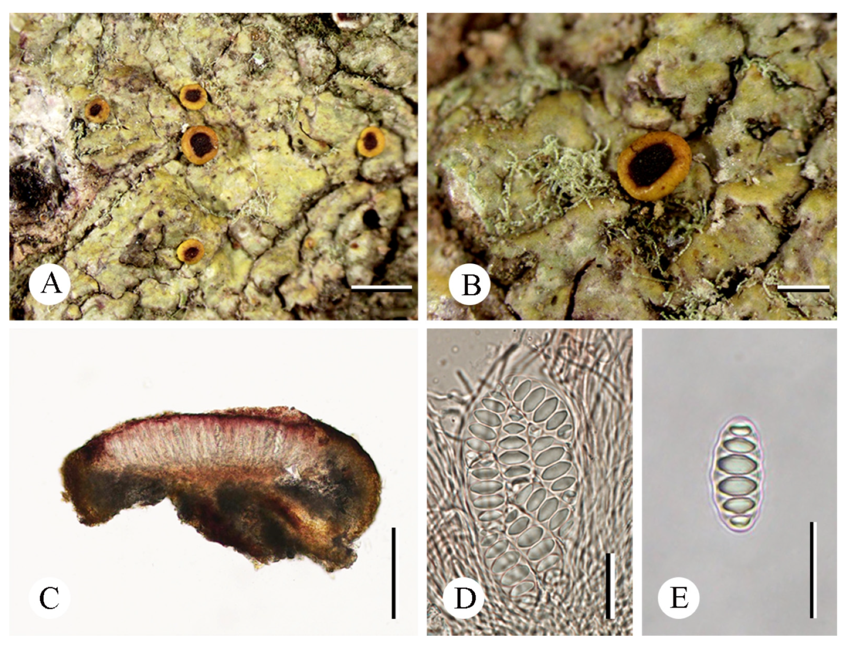
Figure 4.
Letrouitia magenta (YN210757): (A) thallus with ascomata; (B) thallus with ascoma; (C) apothecium section; (D) ascus with ascospores; (E) ascospore. Scale bars: (A) = 1 mm; (B) = 0.5 mm; (C) = 200 µm; (D) = 20 µm; (E) = 25 µm.
Figure 4.
Letrouitia magenta (YN210757): (A) thallus with ascomata; (B) thallus with ascoma; (C) apothecium section; (D) ascus with ascospores; (E) ascospore. Scale bars: (A) = 1 mm; (B) = 0.5 mm; (C) = 200 µm; (D) = 20 µm; (E) = 25 µm.


Table 1.
The specimens and sequences used in the phylogenetic analysis.
| Species | Specimen | Locality | ITS | nuLSU | mtSSU |
|---|---|---|---|---|---|
| Polyozosia contractula | AFTOL-877 | – | HQ650604 | DQ986746 | DQ986898 |
| Letrouitia arcuata | YN18225 | China Yunnan | OR395215 | OR395220 | – |
| Letrouitia domingensis | AFTOL-102 | – | HQ650700 | AY584648 | AY584619 |
| Letrouitia domingensis | Gaya 55 | Dominican Republic | JQ301673 | JQ301569 | JQ301505 |
| Letrouitia domingensis | MES-3181 FLAS-F-63803 | Belize | ON383441 | – | – |
| Letrouitia flavidula | Gaya 35 | Costa Rica | JQ301674 | – | JQ301506 |
| Letrouitia magenta | MFLU 18-0693 | Thailand | MK499353 | MK499365 | – |
| Letrouitia magenta | YN210757 | China Yunnan | OR395216 | OR395221 | OR395225 |
| Letrouitia magenta | YN210758 | China Yunnan | OR395217 | OR395222 | OR395226 |
| Letrouitia parabola | Gaya 11 | USA | JQ301675 | JQ301570 | JQ301507 |
| Letrouitia sinuosa | HN19607 | China Hainan | OR395219 | OR395224 | OR395227 |
| Letrouitia subvulpina | Gaya 44 | Costa Rica | JQ301676 | – | – |
| Letrouitia subvulpina | HN19636 | China Hainan | OR395218 | OR395223 | – |
| Letrouitia transgressa | MFLU 18-0689 | Thailand | MK499352 | MK499364 | – |
| Letrouitia vulpina | Gaya 72 | Reunion | JQ301677 | JQ301571 | JQ301509 |
| Letrouitia vulpina | USE419 | France | KC179452 | KC179209 | KC179543 |
Note: newly generated sequences are shown in bold.
Disclaimer/Publisher’s Note: The statements, opinions and data contained in all publications are solely those of the individual author(s) and contributor(s) and not of MDPI and/or the editor(s). MDPI and/or the editor(s) disclaim responsibility for any injury to people or property resulting from any ideas, methods, instructions or products referred to in the content. |
© 2024 by the authors. Licensee MDPI, Basel, Switzerland. This article is an open access article distributed under the terms and conditions of the Creative Commons Attribution (CC BY) license (https://creativecommons.org/licenses/by/4.0/).
Share and Cite
MDPI and ACS Style
Cui, C.; Dou, M.; Jiang, S.; Jia, Z. The Lichen Genus Letrouitia (Brigantiaeaceae, Ascomycota) in China. Diversity 2024, 16, 254. https://doi.org/10.3390/d16050254
AMA Style
Cui C, Dou M, Jiang S, Jia Z. The Lichen Genus Letrouitia (Brigantiaeaceae, Ascomycota) in China. Diversity. 2024; 16(5):254. https://doi.org/10.3390/d16050254
Chicago/Turabian StyleCui, Can, Mingzhu Dou, Shuhao Jiang, and Zefeng Jia. 2024. "The Lichen Genus Letrouitia (Brigantiaeaceae, Ascomycota) in China" Diversity 16, no. 5: 254. https://doi.org/10.3390/d16050254
Note that from the first issue of 2016, this journal uses article numbers instead of page numbers. See further details here.