Article

Investigation of the Association between Bilateral Selective Anterograde Cerebral Perfusion and Postoperative Ischemic Stroke in Obese Patients with Emergency Surgery for Acute Type A Aortic Dissection

1
Faculty of Medicine, Carol Davila University of Medicine and Pharmacy, 050474 Bucharest, Romania
2
Emergency Institute for Cardiovascular Diseases “Prof. Dr. Iliescu”, 022322 Bucharest, Romania
3
Department of Cardiac Surgery, Emergency Clinical Hospital for Children “Maria Skłodowska Curie”, 077120 Bucharest, Romania
4
Department of Cardiovascular Surgery, Emergency Clinical Hospital Bucharest, 014461 Bucharest, Romania
5
Academy of Romanian Scientists, 050711 Bucharest, Romania
*
Authors to whom correspondence should be addressed.
Medicina 2024, 60(4), 661; https://doi.org/10.3390/medicina60040661
Submission received: 5 March 2024 / Revised: 2 April 2024 / Accepted: 18 April 2024 / Published: 19 April 2024
(This article belongs to the Section Cardiology)

Abstract

:
Background and objectives: The relationship between cerebral perfusion and new postoperative ischemic stroke in obese patients is not well defined. The aim of this study was to investigate the association between selective bilateral anterograde cerebral perfusion and new postoperative ischemic stroke in obese patients with emergency surgery for acute type A aortic dissection. Materials and methods: A total of 292 patients with emergency surgery for acute type A aortic dissection were included in this study. Patients with hemorrhagic stroke or ischemic stroke with severe neurological dysfunction at admission that were not candidates for surgery; patients who died in the first 48 h after intensive care admission and patients with incomplete medical records were excluded. Results: The mean age was 59.42 ± 10.68 years and the mean Euroscore was 9.12 ± 1.63. Obesity was present in 76.4%, the incidence of new postoperative ischemic stroke was 27.5%, and the postoperative mortality rate was 26.7%. The mean cardiopulmonary bypass time was 206.81 ± 75.48 min, the aortic cross-clamp time was 118.2 ± 46.42 min, and 90% of cases required cerebral perfusion. The mean cerebral perfusion time was 30.8 ± 24.41 min. Obese patients had a higher frequency of in-hospital death (p = 0.009), smoking (p = 0.036), hypertension (p = 0.023), left common carotid artery dissection (p < 0.001), right common carotid artery dissection (p = 0.029), femoral artery cannulation (p = 0.026), aortic root replacement (p = 0.009), aortic valve replacement (p = 0.005) and early reintervention for bleeding (p = 0.004). Using logistic regression, selective bilateral anterograde cerebral perfusion over 40 min in obese patients was independently associated with new postoperative ischemic stroke (OR = 2.35; 95%CI = 1.36–4.86; p = 0.021). Conclusions: A patient-tailored strategy for cerebral perfusion should be considered in obese patients, considering the high atheromatous burden of the supra-aortic vessels in these patients and the potential risk of atheromatous embolization associated with this technique.

1. Introduction

Acute type A aortic dissection (ATAAD) is a complex cardiovascular disease, with a 5.8% mortality rate at 48 h with medical treatment alone [1,2]. Emergency central aortic surgery (Figure 1, Figure 2 and Figure 3) with the main goal of resecting the primary intimal tear and restoring flow in the true lumen is the preferred choice of treatment [3]. Complete arch replacement, a hemiarch approach, and open distal anastomosis necessitate a period of circulatory arrest. Antegrade cerebral perfusion (ACP) is the preferred strategy for brain protection during circulatory arrest and can increase the time of hypothermic circulatory arrest (HCA) [4,5]. From a technical point of view, selective ACP is performed with two cannulas inserted under direct vision into the innominate and left common carotid arteries. This technique provides superior brain protection when the HCA exceeds 40–50 min [6].
Despite advances in strategies for brain protection, the incidence of neurological complications in these patients is high and ranges from 17 to 48% [7,8,9]. One of the most dreaded neurological complications regarding mortality and morbidity is ischemic stroke, with an incidence of up to 24.8% [10].
The incidence of obesity is increasing in all industrialized countries and frequent comorbid conditions such as diabetes mellitus, hypertension, hyperlipidemia, and obesity-induced systemic inflammation pose a challenge to care during cardiac surgery, especially those with cardiopulmonary bypass [11]. Patients with obesity are more predisposed to developing ATAAD [12]. Moreover, obese patients with emergency surgery for ATAAD had higher operative mortality and an increased risk of low cardiac output syndrome and pulmonary complications [12,13].
The relationship between obesity and postoperative NC in patients with surgery for ATAAD is not well defined. This study investigated the association between obesity and postoperative ischemic stroke in patients with emergency surgery for ATAAD with HCA and selective ACP.

2. Materials and Methods

Between January 2015 and May 2023, 319 patients were transferred to our center for ATTAD management. The diagnosis was based on chest computer tomography (CT) with intravenous contrast before admission. After a cardiology consult in the emergency department with mandatory transthoracic echocardiography, patients who were candidates for emergency surgery were transferred into the operating room. Clinical characteristics and demographic data were collected from medical records and the electronic health system.
Inclusion criteria: patients with acute type A aortic dissection according to the Stanford classification are considered for emergency surgery.
Exclusion criteria: (1) patients with hemorrhagic stroke documented on computed tomography (CT) scans before admission who were not candidates for surgery; (2) patients with ischemic stroke documented on computed tomography (CT) scans before admission and severe neurological dysfunction who were not candidates for surgery; (3) patients who died in the operating room or within the first 48 h after intensive care admission; (4) patients in the postoperative setting for whom the neurological status could not be evaluated; (5) patients with incomplete medical records.
Obesity was defined as a BMI over 30 kg/m2.
Neurological examination and head CT were the methods used for the diagnosis of a new postoperative ischemic stroke. The Modified Rankin Scale (mRS) was the tool used for quantification of the degree of disability at discharge, based on data collected from clinical charts.
The European System for Cardiac Operative Risk (EuroSCORE) was utilized to assess operative mortality.

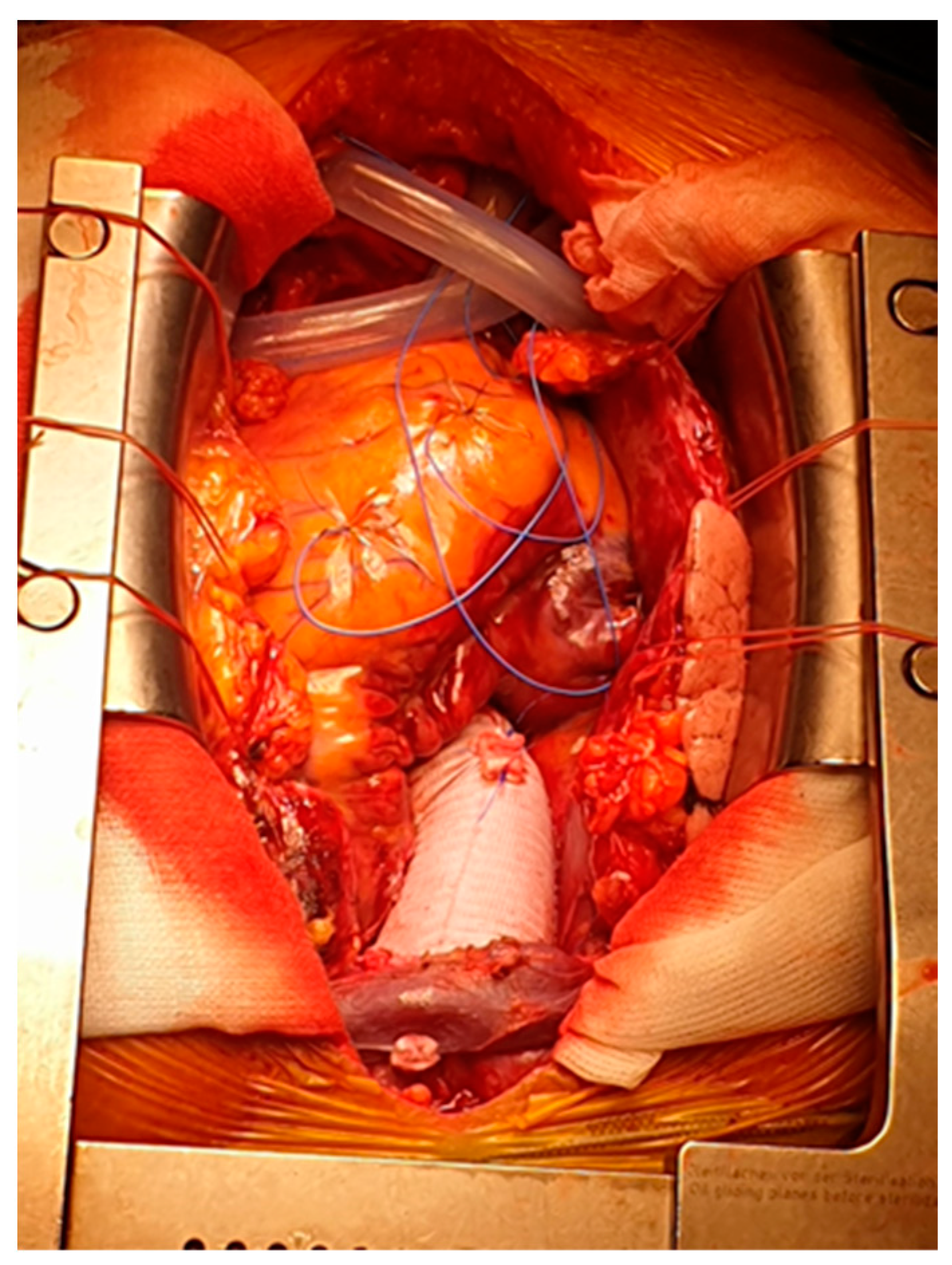
2.1. Surgical Technique

Cardiopulmonary bypass (CPB) was achieved with a two-stage venous cannula and axillary or femoral artery cannulation under direct vision after general anesthesia and median sternotomy. Another strategy for arterial cannulation was inserting a cannula directly into the dissected ascending aorta when the axillary or femoral arteries were not suitable, under transoesophageal echography guidance. We use cold (4 °C), crystalloid cardioplegia, which is usually administered in a retrograde manner. Venting of the left ventricle was achieved with a cannula inserted via the superior left pulmonary vein inserted through the left atrium and mitral valve. Also, the pericardial well was flooded with carbon dioxide early in the intervention to minimize the risk of air embolism. Systemic cooling of the patient to 25–28 °C was applied after CPB was started.
The first step was performing an open distal anastomosis with circulatory arrest and BSACP. The hemiarch approach is usually the preferred method, with total arch replacement being reserved for cases where a primary entry tear cannot be excluded with this strategy. We prefer the hemiarch technique because of the higher risk of neurological complications when total arch replacement is selected [10]. Cerebral oxygenation was monitored with near-infrared spectroscopy (NIRS) in all patients.
The BSACP protocol was the following: 13F or 15F ballon-tip cannulas were used to administer retrograde cardioplegia and were inserted into the innominate artery and left common carotid artery under direct vision. An individual pump and pressure line were used for each cannula. Perfusion parameters for a target value of 50–70% on NIRS are for the left side: pressure of 60–70 mmHg with a flow of 180–230 mL/min and for the right side: perfusion pressure of 60–70 mmHg with a flow between 150–200 mL/min. After open distal anastomosis was performed, CPB was restarted, and the supra-aortic vessels were unclamped after de-airing. During the rewarming of the patient, the proximal repair was completed.
A special consideration should be mentioned regarding cardiac de-airing after aortic repair of dissection. A “Y” cannula generally used for anterograde cardioplegia administration in elective cases is inserted directly into the aortic prosthesis and deairing is performed via this cannula. Also, this cannula is utilized to vent the aortic root until completion of the cardiopulmonary bypass.

2.2. Statistical Analysis

Wizard 2 Statistical Software for Mac OS (Wizard–Statistics & Analysis®, Raipur, Chattisgarh, India) was used for statistical analysis. Summary statistics are presented as absolute numbers and percentages for categorical values and as mean ± standard deviation for continuous values.
The incidence of new postoperative ischemic stroke in patients with emergency surgery for ATAAD was investigated. Patients with a BMI over 30 kg/m2 were divided into two groups: patients with selective ACP below and over 40 min, considering that data from the literature suggests that ACP is safer when DHCA exceeds 40 min [6]. In order to investigate the association of selective ACP time in obese patients undergoing emergency surgery for ATAAD and the development of new postoperative ischemic stroke, multivariable analysis was performed using logistic regression and taking into account a model that included variables achieving a p-value < 0.5 in univariate analysis. A predictive modeling strategy with the backward stepwise method of entering data was then used.
Logistic regression results are presented as odds ratios (OR) with confidence limits and p-values. p < 0.05 was considered statistically significant. An independent t-test or Chi-square test was used for comparing the neurologically complicated and uncomplicated groups.

3. Results

3.1. General Characteristics of the Study Population

A total of 292 patients with ATAAD and emergency surgery were included in the study. Of these, 27 patients were excluded (3 patients with hemorrhagic stroke and 2 patients with massive ischemic stroke that were not candidates for surgery, 4 patients with unresponsive cardiac arrest at admission, 5 patients with irreversible peripheric ischemia, 13 patients died on the operating table, 10 because of hemorrhagic shock and 3 patients had myocardial infarction). The preoperative data are summarized in Table 1.
The mean age was 59.42 ± 10.68 years and the mean Euroscore was 9.12 ± 1.63. There was a 3.89 ± 2.37 h mean time from diagnosis of ATAAD to surgery. Obesity was present in 76.4% of cases. The most frequent complaint at admission was thoracic pain in 92.1% of patients followed by abdominal pain in 12.3% and 11% of cases presented for syncope. Preoperative ischemic stroke was documented on head CT in 7 patients (2.4%), 30 patients presented with lower leg ischemia, and mesenteric ischemia was documented based on clinical examination and contrast abdominal CT in 5 patients (2%). A bicuspid aortic valve was present in 9.24% of cases, severe acute aortic regurgitation in 25.3% of patients, and cardiac tamponade in 12.7% of cases. The innominate artery was the most frequently dissected supra-aortic artery in 19.2% of cases, followed by the left common carotid artery in 12.9% of patients. The right common carotid artery was dissected in 7.7% of cases. In 48.6% of cases, the primary entry tear was at the level of the ascending aorta. Intramural hematoma was diagnosed in 8.6% of cases and 2.1% of patients had penetrating aortic ulcers.
Postoperative death occurred in 26.7% (78 patients). Causes of death were the following: septic shock in 36 patients (46.15%) cardiogenic shock in 12 patients (15.38%), mixed shock in 9 cases (11.53%), and hemorrhagic shock in 21 patients (26.92%).

3.2. Intraoperative and Postoperative Data

Table 2 summarises the intraoperative and postoperative data.
The incidence of a new postoperative ischemic stroke was 27.5% (81 patients). The incidence of in-hospital death in patients with ischemic stroke was 31.8% (28 patients) and 81.8% (73 patients) were obese. The mean cardiopulmonary bypass time was 206.81 ± 75.48 min and the aortic cross-clamp time was 118.2 ± 46.42 min. In 90% of cases, cerebral perfusion was applied with a mean time of 30.8 ± 24.41 min. The supracoronary ascending aorta was replaced in 96.9% of cases. At the level of the aortic arch, hemiarch replacement was performed in 45.9% of patients and total arch replacement was selected in 5.5% of cases. Root replacement was necessary for 23.3% of patients. Concomitant procedures were required in 72 patients: coronary artery bypass grafting in 5.5% of cases, mitral valve repair in 1% of cases, aortic valve replacement in 16.8% of cases, peripheral V-A ECMO in two patients, one patient required a femoral-femoral bypass and one patient had aortic coarctation repair. Axillary artery cannulation was performed in 64.5% of cases, followed by femoral artery cannulation in 31.5% of patients. Early reintervention for mediastinal bleeding was necessary in 24.1% of patients. The mean intensive care stay was 11.2 ± 13.6 days, prolonged more than 24 h mechanical ventilation was necessary in 74% of patients, and dialysis was required in 51% of cases. Postoperative mesenteric ischemia was seen in 22 patients (7.5%) while 6 patients (2.1%) developed deep sternal wound infections. Treatment for mesenteric ischemia was open abdominal surgery with resection of affected bowel in 21 patients while 1 patient received interventional treatment with stenting of the superior mesenteric artery.

3.3. Characteristics of Obese Patients

Table 3 shows the comparison between the patients with and without obesity. Regarding preoperative characteristics, obese patients had a higher frequency of in-hospital death (p = 0.009), hyperlipidemia (p = 0.03), smoking (p = 0.036), hypertension (p = 0.023), left common carotid artery dissection (p < 0.001) and right common carotid artery dissection (p = 0.029). A higher frequency of femoral artery cannulation (p = 0.026), aortic root replacement (p = 0.009), aortic valve replacement (p = 0.005), and early reintervention for bleeding (p = 0.004) were observed in obese patients. No differences between the two groups were observed regarding postoperative characteristics.

3.4. Characteristics of Patients with and without Ischemic Stroke

Table 4 shows the comparison of different intraoperative parameters in patients with and without ischemic stroke. Patients with stroke had significantly higher cardiopulmonary bypass times (p = 0.018) and cerebral perfusion times (p = 0.18). No significant difference was observed in cerebral perfusion pressure and flow. Also, no difference was observed regarding the site of arterial cannulation. Table 5 presents the Modified Rankin Scale at discharge. No patient had a full recovery, while 32 patients with ischemic stroke died.

3.5. Logistic Regression

Variables included in the univariate analysis were the following: age, smoking, hypertension, female sex, diabetes, chronic obstructive lung disease, family history of aortic dissection, anemia, renal dysfunction, hepatic dysfunction, lactic acidosis, BMI, at admission: thoracic pain, abdominal pain, syncope, lower leg ischemia, mesenteric ischemia, cardiac tamponade, severe aortic regurgitation, severe ventricular disfunction, intimal tear in the ascending aorta, intimal tear in the aortic arch, innominate artery dissection, right common carotid artery dissection, right subclavian artery dissection, left common carotid artery dissection, left subclavian artery dissection, cardiopulmonary bypass time, aortic cross clamp time, cerebral perfusion time, femoral artery cannulation, axillary artery cannulation, aortic root replacement, supracoronary ascending aorta replacement, hemiarch replacement, total arch replacement, coronary artery bypass grafting, aortic valve replacement, mitral valve repair, and early reintervention for bleeding.
Results of the univariate analysis of variables associated with new postoperative ischemic stroke achieving a p-value < 0.5 are presented in Table 6. Cardiac tamponade at admission (OR = 6.27; 95%CI = 2.57–15.25; p < 0.01) and dissection of innominate artery (OR = 0.21; 95%CI = 0.06–0.72; p = 0.013) were included in the final model after backward selection. Selective ACP over 40 min in obese patients was associated with new postoperative ischemic stroke (OR = 2.18; 95%CI = 1.12–4.22; p = 0.021) in univariate analysis and after model adjustment was independently associated with new postoperative ischemic stroke (OR = 2.35; 95%CI = 1.14–4.86; p = 0.021).

4. Discussion

The main findings of this study are that selective ACP over 40 min is associated with new postoperative ischemic stroke in obese patients (BMI > 30 kg/m2) undergoing emergency surgery for ATAAD with HCA. This result is consistent with data from the literature regarding risk factors for neurological complications (Table 7). Obese patients had a higher frequency of in-hospital death, femoral artery cannulation, left and right carotid artery dissection, aortic root replacement, aortic valve replacement, and early reintervention for bleeding.
The incidence of ischemic stroke in this study was 27.5%. This is in accordance with previous studies that reported an incidence of up to 32.8% in NC [2,16,17,18,19]. Despite advances in brain protection methods, this is still a high incidence. Out of 89 patients with new postoperative ischemic stroke, 28 (21.8%) patients died in the intensive care unit, confirming the high in-hospital mortality previously reported [19,20,21]. Out of 223 patients with a BMI over 30 kg/m2, 75.7% had selective ACP, and 66 (29.6%) patients developed ischemic stroke. Data from the literature suggests that an increased risk of air or atheromatous embolism exists when the arch vessels are manipulated during surgery [22]. This could explain the high incidence of ischemic stroke in our study, considering the association between obesity and atherosclerosis. Inflammation is the link between the two and it is explained by the fact that adipose tissue releases adipokine, which in turn induces insulin resistance, endothelial dysfunction, and hypercoagulability, all of which promote atherosclerosis [23]. There are reports of longer circulatory arrest times when using BSACP because this technique is technically more complex, with an increased risk of stroke over 30 min [24,25]. Also, there are reports that operation times are higher in obese patients [26,27] and coupled with the technical difficulties of selective ACP institution could increase the rate of ischemic stroke. Operation times were not significantly longer in obese patients in our study (CPB time: 192.02 ± 73.18 vs. 211.39 ± 75.76; p = 0.558, aortic cross-clamp time: 113.53 ± 48.23 vs. 119.65 ± 45.86; p = 0.392, cerebral perfusion time: 31.58 ± 23.54 vs. 30.53 ± 24.78; p = 0.38). The mean cerebral perfusion time in our study was 30.8 ± 24.41 min and 30.53 ± 24.78 min in obese patients. Considering this, our data do not support the statement that BSACP is technically more complex.
Other risk factors promoting atherosclerosis are smoking, hyperlipidemia, and diabetes. Smoking is a well-known factor in the development of cardiovascular disease via the initiation and progression of atherosclerosis. Mechanisms include oxidative stress induction, vascular inflammation, platelet coagulation, and vascular dysfunction [28]. Moreover, a study of 1320 autopsied specimens revealed atherosclerotic involvement of the aorta and coronary arteries in heavy smokers compared to nonsmokers [29]. The relationship between smoking and obesity is controversial. Some studies report no association between active smoking and BMI [30], while others found an association between smoking and lower BMI [30] and smoking cessation with increased BMI [31]. However, the association of smoking and obesity defined by BMI is described as a reverse causation, due to obese patients trying to lose weight by starting smoking [32]. Several studies report an association of smoking with obesity defined by waist circumference [33,34]. In our study, 29.1% of total patients and 22.2% of patients who developed ischemic stroke after surgery were active smokers. There was a significant difference between obese and non-obese patients regarding the frequency of smokers (p = 0.036), with 26% of obese patients being smokers. Smoking-induced atheromatosis at the level of the aortic arch could contribute to the high incidence of stroke in our study.
Atherosclerosis is a well-known complication of diabetes. It is estimated that 90% of patients with type 2 diabetes are attributable to obesity [35]. Common mechanisms include endothelial activation and inflammation, mitochondrial oxidative stress, changes in the extracellular matrix, and disruption of cellular defense systems [36]. One study reports a “common soil” between the two conditions named “nuclear factor KB” [37]. This “master regulator” triggers either hyperglycemia-induced endothelial dysfunction, or various micro RNAs that favor atherosclerosis [37]. Inflammation induced by chronic hyperglycemia induces endothelium dysfunction that promotes atherosclerosis [38]. Particularly relevant to our study, The Insulin Resistance Atherosclerosis Study (IRAS) reported that there is an association between increased atherosclerosis of the internal carotid artery with diabetes [39]. In our study, there were no significant differences between obese and non-obese patients regarding the incidence of diabetes, and only 7.9% of patients had diabetes. However, 30.4% of patients who developed stroke after surgery had diabetes and the increased carotid atherosclerosis described in these patients could contribute to the risk of stroke.
Hyperlipidemia is another well-known factor for atherosclerosis and its association with obesity is well described [40,41]. A higher level of cholesterol is an independent risk factor for atherosclerosis of the carotid artery. Patients without any plaque had a significantly lower level of cholesterol compared with those with any degree of internal carotid artery stenosis [42]. Moreover, patients with untreated hyperlipidemia had more severe carotid artery plaques and a higher risk of plaque rupture according to morphology studies [42]. In our study, the incidence of hyperlipidemia was 69.5% and there was a significant difference between obese and non-obese patients regarding the incidence of hyperlipidemia (p = 0.023). Also, 79.2% of patients with stroke had hyperlipidemia. These results could suggest that hyperlipidemia contributes to atherosclerosis, especially at the level of the carotid arteries, and could raise the risk of stroke during aortic arch manipulation and selective cerebral perfusion.
Obese patients with emergency surgery for ATAAD had a significantly higher in-hospital mortality (14.5% vs. 30.5%; p = 0.009). This finding is supported by data from several studies. Xiao Xu and al reported that morbid obesity is associated with an increased risk of in-hospital mortality in this group of patients [28]. Lio et al. reported a higher perioperative mortality rate and reduced 5-year survival rates in obese patients with emergency surgery for ATAAD [43]. Xiaogao et al. found that although obesity was more common in younger patients, it was more likely to cause death in elderly patients (age > 60 years) [13]. The high rate of mortality could be explained by the increased risk of low cardiac output syndrome and pulmonary complications in obese patients [43]. Postoperative acute lung injury is reported to have a higher incidence in obese patients and the risk of severe postoperative hypoxia seems to be of concern, especially in the female group [44,45]. Oxidative stress and an inflammatory response seem to be involved in the process of lung injury in obese patients [45].
In our study, aortic root replacement (11.6% vs. 26.9%, p = 0.009) and aortic valve replacement (5.8% vs. 20.2%; p = 0.005) had a higher frequency in obese patients. Several studies described an association between markers of obesity and the presence of an aortic aneurysm [14,46,47]. Inflammation caused by an imbalance of interleukin 6, 8 and TNF-alpha secreted by adipose tissue is thought to weaken and dilate vessels and may explain the role of obesity in aneurysm formation and weight loss may reduce the aortic diameter [15]. Obesity is also a risk factor for the development of calcified aortic stenosis and subsequent surgery. Two major studies including 71,817 patients from Sweden [48] and the Copenhagen General Population Study [49] support our findings. Also, obesity is a risk factor for calcified aortic stenosis progression [50,51]. Structural and metabolic changes are the link between obesity and calcified aortic stenosis. Obesity-related hypertension causes endothelial injury, while increased levels of atherogenic lipoproteins are deposited on the aortic leaflets, leading to calcification of the aortic valve [52,53].
Obesity may play a protective role in perioperative bleeding and transfusion needs [54]. Studies report this “obesity paradox” in patients undergoing myocardial revascularization [36,55]. Obese patients with ATAAD have better long-term survival compared to normal-weight patients and this could be explained by a younger age at dissection or a more typical presentation at admission [36,56,57]. Because of their increased body surface area and total blood volume, obese patients are less susceptible to hemodilution and CPB-related coagulopathy. Also, compression of mediastinal fat on minor bleeding sites could reduce the bleeding risk [54]. In our study, this does not seem to be the case, with obese patients having higher rates of early reintervention for bleeding (37.3% vs. 20.2%; p = 0.004). This could be explained by a higher incidence of coagulopathy when considering the complexity of ATAAD surgery, with HCA and longer CPB times, compared to myocardial revascularization alone or combined with valvular procedures.

5. Conclusions

Data from 292 patients with emergency surgery for ATAAD were analyzed. More than 40 min of bilateral selective ACP in obese patients (BMI > 30 kg/m2) with emergency surgery for ATAAD was independently associated with a new postoperative ischemic stroke using multivariable analysis. Also, obese patients had a higher rate of early reintervention for bleeding, aortic root replacement, aortic valve replacement, femoral artery cannulation, left and right common carotid artery dissection, and higher hospital mortality. This result could be explained by the heavy atheromatous burden more frequent in obese patients at the level of the supra-aortic vessels, coupled with the risk of atheromatous embolism when selective bilateral ACP is used. When choosing a cerebral perfusion strategy in obese patients, this risk should be balanced with the fact that bilateral selective ACP is safer when HCA is more than 30 min, and a patient-tailored strategy should be considered.

Author Contributions

Conceptualization, M.R., I.M.M., C.R., V.A.I. and H.M.; Data curation, M.R., B.R. and A.I.; Formal analysis, M.R., A.I., S.P. and C.V.; Funding acquisition, M.R., A.I., C.V., S.P. and M.C.; Investigation, M.R., B.R. and M.C.; Methodology, M.R., V.A.I. and H.M.; Project administration, M.R., I.M.M. and C.R.; Resources, M.R. and B.R.; Software, M.R. and C.R.; Supervision, C.R., S.P. and C.V.; Validation, O.S., V.A.I. and H.M.; Visualization, M.R., C.R., A.I., M.C., S.P., C.V. and V.A.I.; Writing—original draft, M.R. and I.M.M.; Writing—review and editing, M.R., O.S., V.A.I. and H.M. All authors have read and agreed to the published version of the manuscript.

Funding

Publication of this paper was supported by the University of Medicine and Pharmacy Carol Davila, through the institutional program Publish not Perish.

Institutional Review Board Statement

This study was conducted in accordance with the Declaration of Helsinki and approved by the Romanian National Ethics Committee—approval Code: 852; Approval Date: 3 January 2017.

Informed Consent Statement

Informed consent was obtained from all subjects involved in this study.

Data Availability Statement

The data presented in this study are available on reasonable request from the corresponding author.

Conflicts of Interest

The authors declare no conflicts of interest.

References

  1. Harris, K.M.; Nienaber, C.A.; Peterson, M.D.; Woznicki, E.M.; Braverman, A.C.; Trimarchi, S.; Myrmel, T.; Pyeritz, R.; Hutchison, S.; Strauss, C.; et al. Early Mortality in Type A Acute Aortic Dissection: Insights From the International Registry of Acute Aortic Dissection. JAMA Cardiol. 2022, 7, 1009–1015. [Google Scholar] [CrossRef] [PubMed] [PubMed Central]
  2. Conzelmann, L.O.; Hoffmann, I.; Blettner, M.; Kallenbach, K.; Karck, M.; Dapunt, O.; Borger, M.A.; Weigang, E. GERAADA Investigators. Analysis of risk factors for neurological dysfunction in patients with acute aortic dissection type A: Data from the German Registry for Acute Aortic Dissection type A (GERAADA). Eur. J. Cardio-Thorac. Surg. 2012, 42, 557–565. [Google Scholar] [CrossRef] [PubMed]
  3. Carrel, T.; Sundt, T.M., 3rd; von Kodolitsch, Y.; Czerny, M. Acute aortic dissection. Lancet 2023, 401, 773–788. [Google Scholar] [CrossRef] [PubMed]
  4. Svensson, L.G.; Crawford, E.S.; Hess, K.R.; Coselli, J.S.; Raskin, S.; Shenaq, S.A.; Safi, H.J. Deep hypothermia with circulatory arrest. Determinants of stroke and early mortality in 656 patients. J. Thorac. Cardiovasc. Surg. 1993, 106, 19–28; Discussion 28–31. [Google Scholar] [CrossRef] [PubMed]
  5. Dong, S.B.; Xiong, J.X.; Zhang, K.; Zheng, J.; Xu, S.D.; Liu, Y.M.; Sun, L.Z.; Pan, X.D. Different hypothermic and cerebral perfusion strategies in extended arch replacement for acute type an aortic dissection: A retrospective comparative study. J. Cardiothorac. Surg. 2020, 15, 236. [Google Scholar] [CrossRef] [PubMed] [PubMed Central]
  6. Malvindi, P.G.; Scrascia, G.; Vitale, N. Is unilateral antegrade cerebral perfusion equivalent to bilateral cerebral perfusion for patients undergoing aortic arch surgery? Interact. Cardiovasc. Thorac. Surg. 2008, 7, 891–897. [Google Scholar] [CrossRef] [PubMed]
  7. Mészáros, I.; Mórocz, J.; Szlávi, J.; Schmidt, J.; Tornóci, L.; Nagy, L.; Szép, L. Epidemiology and clinicopathology of aortic dissection. Chest 2000, 117, 1271–1278. [Google Scholar] [CrossRef] [PubMed]
  8. Blanco, M.; Díez-Tejedor, E.; Larrea, J.L.; Ramírez, U. Neurologic complications of type I aortic dissection. Acta Neurol. Scand. 1999, 99, 232–235. [Google Scholar] [CrossRef] [PubMed]
  9. Hagan, P.G.; Nienaber, C.A.; Isselbacher, E.M.; Bruckman, D.; Karavite, D.J.; Russman, P.L.; Evangelista, A.; Fattori, R.; Suzuki, T.; Oh, J.K.; et al. The International Registry of Acute Aortic Dissection (IRAD): New insights into an old disease. JAMA 2000, 283, 897–903. [Google Scholar] [CrossRef] [PubMed]
  10. Robu, M.; Marian, D.R.; Margarint, I.; Radulescu, B.; Știru, O.; Iosifescu, A.; Voica, C.; Cacoveanu, M.; Ciomag Ianula, R.; Gașpar, B.S.; et al. Association between Bilateral Selective Antegrade Cerebral Perfusion and Postoperative Ischemic Stroke in Patients with Emergency Surgery for Acute Type A Aortic Dissection-Single Centre Experience. Medicina 2023, 59, 1365. [Google Scholar] [CrossRef] [PubMed] [PubMed Central]
  11. Ushakumari, D.S.; Machovec, K.A. Cardiac surgery in obese patients. In Oxford Textbook of Anaesthesia for the Obese Patient; Oxford University Press: Oxford, UK, 2021; p. 127. [Google Scholar]
  12. Xu, X.; Yin, R.; Zhi, K.; Qin, Y.; Tu, B.; Wu, S.; Dong, Z.; Liu, D.; He, J. Morbid obesity impacts mortality among inpatients with type a aortic dissection: An analysis of the national inpatient sample. J. Cardiothorac. Surg. 2023, 18, 14. [Google Scholar] [CrossRef] [PubMed] [PubMed Central]
  13. Pan, X.; Xing, Z.; Yang, G.; Ding, N.; Zhou, Y.; Chai, X. Obesity Increases In-Hospital Mortality of Acute Type A Aortic Dissection Patients Undergoing Open Surgical Repair: A Retrospective Study in the Chinese Population. Front. Cardiovasc. Med. 2022, 9, 899050. [Google Scholar] [CrossRef] [PubMed] [PubMed Central]
  14. Apoloni, R.C.; Zerati, A.E.; Wolosker, N.; Saes, G.F.; Wolosker, M.; Curado, T.; Puech-Leão, P.; De Luccia, N. Analysis of the Correlation Between Central Obesity and Abdominal Aortic Diseases. Ann. Vasc. Surg. 2019, 54, 176–184. [Google Scholar] [CrossRef] [PubMed]
  15. Cronin, O.; Walker, P.J.; Golledge, J. The association of obesity with abdominal aortic aneurysm presence and growth. Atherosclerosis 2013, 226, 321–327. [Google Scholar] [CrossRef] [PubMed]
  16. Ehrlich, M.P.; Ergin, M.A.; McCullough, J.N.; Lansman, S.L.; Galla, J.D.; Bodian, C.A.; Apaydin, A.; Griepp, R.B. Results of immediate surgical treatment of all acute type A dissections. Circulation 2000, 102 (Suppl. S3), III248–III252. [Google Scholar] [CrossRef] [PubMed]
  17. Easo, J.; Weigang, E.; Hölzl, P.P.; Horst, M.; Hoffmann, I.; Blettner, M.; Dapunt, O.E. Influence of operative strategy for the aortic arch in DeBakey type I aortic dissection—Analysis of the German Registry for Acute Aortic Dissection type A (GERAADA). Ann. Cardiothorac. Surg. 2013, 2, 175–180. [Google Scholar] [CrossRef] [PubMed] [PubMed Central]
  18. Hanganu, A.R.; Constantin, A.; Moise, E.S.; Niculae, C.M.; Olaru, I.D.; Băicuș, C.; Hristea, A. Peripheral nervous system involvement associated with COVID-19. A systematic review of literature. PLoS ONE 2023, 18, e0283827. [Google Scholar] [CrossRef] [PubMed] [PubMed Central]
  19. Ghoreishi, M.; Sundt, T.M.; Cameron, D.E.; Holmes, S.D.; Roselli, E.E.; Pasrija, C.; Gammie, J.S.; Patel, H.J.; Bavaria, J.E.; Svensson, L.G.; et al. Factors associated with acute stroke after type A aortic dissection repair: An analysis of the Society of Thoracic Surgeons National Adult Cardiac Surgery Database. J. Thorac. Cardiovasc. Surg. 2020, 159, 2143–2154.e3. [Google Scholar] [CrossRef] [PubMed]
  20. Dumfarth, J.; Kofler, M.; Stastny, L.; Plaikner, M.; Krapf, C.; Semsroth, S.; Grimm, M. Stroke after emergent surgery for acute type A aortic dissection: Predictors, outcome and neurological recovery. Eur. J. Cardio-Thorac. Surg. 2018, 53, 1013–1020. [Google Scholar] [CrossRef] [PubMed]
  21. Filip, C.; Cirstoveanu, C.; Bizubac, M.; Berghea, E.C.; Căpitănescu, A.; Bălgrădean, M.; Pavelescu, C.; Nicolescu, A.; Ionescu, M.D. Pulse Wave Velocity as a Marker of Vascular Dysfunction and Its Correlation with Cardiac Disease in Children with End-Stage Renal Disease (ESRD). Diagnostics 2021, 12, 71. [Google Scholar] [CrossRef] [PubMed] [PubMed Central]
  22. Kazui, T.; Inoue, N.; Komatsu, S. Surgical treatment of aneurysms of the transverse aortic arch. J. Cardiovasc. Surg. 1989, 30, 402–406. [Google Scholar] [PubMed]
  23. Rocha, V.Z.; Libby, P. Obesity, inflammation, and atherosclerosis. Nat. Rev. Cardiol. 2009, 6, 399–409. [Google Scholar] [CrossRef] [PubMed]
  24. Song, S.J.; Kim, W.K.; Kim, T.H.; Song, S.W. Unilateral versus bilateral antegrade cerebral perfusion during surgical repair for patients with acute type A aortic dissection. JTCVS Open 2022, 11, 37–48. [Google Scholar] [CrossRef] [PubMed] [PubMed Central]
  25. Preventza, O.; Simpson, K.H.; Cooley, D.A.; Cornwell, L.; Bakaeen, F.G.; Omer, S.; Rodriguez, V.; de la Cruz, K.I.; Rosengart, T.; Coselli, J.S. Unilateral versus bilateral cerebral perfusion for acute type A aortic dissection. Ann. Thorac. Surg. 2015, 99, 80–87. [Google Scholar] [CrossRef] [PubMed]
  26. Vassiliades TAJr Nielsen, J.L.; Lonquist, J.L. Effects of obesity on outcomes in endoscopically assisted coronary artery bypass operations. Heart Surg. Forum 2003, 6, 99–101. [Google Scholar] [CrossRef] [PubMed]
  27. Choi, J.C.; Bakaeen, F.G.; Cornwell, L.D.; Dao, T.K.; Coselli, J.S.; LeMaire, S.A.; Chu, D. Morbid obesity is associated with increased resource utilization in coronary artery bypass grafting. Ann. Thorac. Surg. 2012, 94, 23–28. [Google Scholar] [CrossRef] [PubMed]
  28. Siasos, G.; Tsigkou, V.; Kokkou, E.; Oikonomou, E.; Vavuranakis, M.; Vlachopoulos, C.; Verveniotis, A.; Limperi, M.; Genimata, V.; Papavassiliou, A.G.; et al. Smoking and atherosclerosis: Mechanisms of disease and new therapeutic approaches. Curr. Med. Chem. 2014, 21, 3936–3948. [Google Scholar] [CrossRef] [PubMed]
  29. Strong, J.P.; Richards, M.L. Cigarette smoking and atherosclerosis in autopsied men. Atherosclerosis 1976, 23, 451–476. [Google Scholar] [CrossRef] [PubMed]
  30. Zbikowski, S.M.; Jack, L.M.; McClure, J.B.; Deprey, M.; Javitz, H.S.; McAfee, T.A.; Catz, S.L.; Richards, J.; Bush, T.; Swan, G.E. Utilization of services in a randomized trial testing phone- and web-based interventions for smoking cessation. Nicotine Tob. Res. 2011, 13, 319–327. [Google Scholar] [CrossRef] [PubMed] [PubMed Central]
  31. Munafò, M.R.; Tilling, K.; Ben-Shlomo, Y. Smoking status and body mass index: A longitudinal study. Nicotine Tob. Res. 2009, 11, 765–771. [Google Scholar] [CrossRef] [PubMed]
  32. Chiolero, A.; Faeh, D.; Paccaud, F.; Cornuz, J. Consequences of smoking for body weight, body fat distribution, and insulin resistance. Am. J. Clin. Nutr. 2008, 87, 801–809. [Google Scholar] [CrossRef] [PubMed]
  33. Slagter, S.N.; van Vliet-Ostaptchouk, J.V.; Vonk, J.M.; Boezen, H.M.; Dullaart, R.P.; Kobold, A.C.; Feskens, E.J.; van Beek, A.P.; van der Klauw, M.M.; Wolffenbuttel, B.H. Associations between smoking, components of metabolic syndrome and lipoprotein particle size. BMC Med. 2013, 11, 195. [Google Scholar] [CrossRef] [PubMed Central]
  34. Sun, M.; Jiang, Y.; Sun, C.; Li, J.; Guo, X.; Lv, Y.; Yu, Y.; Yao, Y.; Jin, L. The associations between smoking and obesity in northeast China: A quantile regression analysis. Sci. Rep. 2019, 9, 3732. [Google Scholar] [CrossRef] [PubMed] [PubMed Central]
  35. Verma, S.; Hussain, M.E. Obesity and diabetes: An update. Diabetes Metab. Syndr. 2017, 11, 73–79. [Google Scholar] [CrossRef] [PubMed]
  36. Stamou, S.C.; Nussbaum, M.; Stiegel, R.M.; Reames, M.K.; Skipper, E.R.; Robicsek, F.; Lobdell, K.W. Effect of body mass index on outcomes after cardiac surgery: Is there an obesity paradox? Ann. Thorac. Surg. 2011, 91, 42–47. [Google Scholar] [CrossRef] [PubMed]
  37. Lawrence, T. The nuclear factor NF-kappaB pathway in inflammation. Cold Spring Harb. Perspect. Biol. 2009, 1, a001651. [Google Scholar] [CrossRef] [PubMed] [PubMed Central]
  38. La Sala, L.; Prattichizzo, F.; Ceriello, A. The link between diabetes and atherosclerosis. Eur. J. Prev. Cardiol. 2019, 26, 15–24. [Google Scholar] [CrossRef] [PubMed]
  39. Wagenknecht, L.E.; Zaccaro, D.; Espeland, M.A.; Karter, A.J.; O’Leary, D.H.; Haffner, S.M. Diabetes and progression of carotid atherosclerosis: The insulin resistance atherosclerosis study. Arterioscler. Thromb. Vasc. Biol. 2003, 23, 1035–1041. [Google Scholar] [CrossRef] [PubMed]
  40. Sullivan, P.W.; Ghushchyan, V.H.; Ben-Joseph, R. The impact of obesity on diabetes, hyperlipidemia and hypertension in the United States. Qual. Life Res. 2008, 17, 1063–1071. [Google Scholar] [CrossRef] [PubMed]
  41. Dang, S.W.; Gao, L.; Li, Y.J.; Zhang, R.; Xu, J. Metabolic characteristics of non-obese and obese metabolic dysfunction-associated fatty liver disease in type 2 diabetes mellitus and its association with diabetic peripheral neuropathy and diabetic retinopathy. Front. Med. 2023, 10, 1216412. [Google Scholar] [CrossRef] [PubMed] [PubMed Central]
  42. Kerenyi, L.; Mihalka, L.; Csiba, L.; Bacso, H.; Bereczki, D. Role of hyperlipidemia in atherosclerotic plaque formation in the internal carotid artery. J. Clin. Ultrasound 2006, 34, 283–288. [Google Scholar] [CrossRef] [PubMed]
  43. Lio, A.; Bovio, E.; Nicolò, F.; Saitto, G.; Scafuri, A.; Bassano, C.; Chiariello, L.; Ruvolo, G. Influence of Body Mass Index on Outcomes of Patients Undergoing Surgery for Acute Aortic Dissection: A Propensity-Matched Analysis. Tex. Heart Inst. J. 2019, 46, 7–13. [Google Scholar] [CrossRef] [PubMed] [PubMed Central]
  44. Gong, M.; Wu, Z.; Xu, S.; Li, L.; Wang, X.; Guan, X.; Zhang, H. Increased risk for the development of postoperative severe hypoxemia in obese women with acute type a aortic dissection. J. Cardiothorac. Surg. 2019, 14, 81. [Google Scholar] [CrossRef] [PubMed] [PubMed Central]
  45. Wu, Z.; Wang, Z.; Wu, H.; Hu, R.; Ren, W.; Hu, Z.; Chang, J. Obesity is a risk factor for preoperative hypoxemia in Stanford A acute aortic dissection. Medicine 2020, 99, e19186. [Google Scholar] [CrossRef] [PubMed] [PubMed Central]
  46. Stoll, S.; Sowah, S.A.; Fink, M.A.; Nonnenmacher, T.; Graf, M.E.; Johnson, T.; Schlett, C.L.; von Stackelberg, O.; Kirsten, R.; Bamberg, F.; et al. Changes in aortic diameter induced by weight loss: The HELENA trial- whole-body MR imaging in a dietary intervention trial. Front. Physiol. 2022, 13, 976949. [Google Scholar] [CrossRef] [PubMed] [PubMed Central]
  47. Alpert, M.A.; Omran, J.; Bostick, B.P. Effects of Obesity on Cardiovascular Hemodynamics, Cardiac Morphology, and Ventricular Function. Curr. Obes. Rep. 2016, 5, 424–434. [Google Scholar] [CrossRef] [PubMed]
  48. Larsson, S.C.; Wolk, A.; Håkansson, N.; Bäck, M. Overall and abdominal obesity and incident aortic valve stenosis: Two prospective cohort studies. Eur. Heart J. 2017, 38, 2192–2197. [Google Scholar] [CrossRef] [PubMed] [PubMed Central]
  49. Kaltoft, M.; Langsted, A.; Nordestgaard, B.G. Obesity as a Causal Risk Factor for Aortic Valve Stenosis. J. Am. Coll. Cardiol. 2020, 75, 163–176. [Google Scholar] [CrossRef] [PubMed]
  50. Ngo, M.V.; Gottdiener, J.S.; Fletcher, R.D.; Fernicola, D.J.; Gersh, B.J. Smoking and Obesity Are Associated with the Progression of Aortic Stenosis. Am. J. Geriatr. Cardiol. 2001, 10, 86–90. [Google Scholar] [CrossRef]
  51. Iosifescu, A.G.; Moldovan, H.; Iliescu, V.A. Aortic prosthesis-patient mismatch strongly affects early results of double valve replacement. J. Heart Valve Dis. 2014, 23, 149–157. [Google Scholar] [PubMed]
  52. Pawade, T.A.; Newby, D.E.; Dweck, M.R. Calcification in Aortic Stenosis: The Skeleton Key. J. Am. Coll. Cardiol. 2015, 66, 561–577. [Google Scholar] [CrossRef] [PubMed]
  53. Varbo, A.; Benn, M.; Smith, G.D.; Timpson, N.J.; Tybjaerg-Hansen, A.; Nordestgaard, B.G. Remnant cholesterol, low-density lipoprotein cholesterol, and blood pressure as mediators from obesity to ischemic heart disease. Circ. Res. 2015, 116, 665–673. [Google Scholar] [CrossRef] [PubMed]
  54. De Santo, L.S.; Moscariello, C.; Zebele, C. Implications of obesity in cardiac surgery: Pattern of referral, physiopathology, complications, prognosis. J. Thorac. Dis. 2018, 10, 4532–4539. [Google Scholar] [CrossRef] [PubMed] [PubMed Central]
  55. Scott, B.H.; Seifert, F.C.; Glass, P.S.A.; Grimson, R. Blood use in patients undergoing coronary artery bypass surgery: Impact of cardiopulmonary bypass pump, hematocrit, gender, age, and body weight. Obstet. Anesth. Dig. 2003, 97, 958–963. [Google Scholar] [CrossRef] [PubMed]
  56. Feldeisen, T.; Hughes, G.C.; Brinster, D.; Braverman, A.C.; Leshnower, B.; Bekeredjian, R.; Myrmel, T.; Arnaoutakis, G.J.; Pai, C.-W.; Khoynezhad, A.B.; et al. The impact of obesity on outcomes in acute aortic dissection. J. Am. Coll. Cardiol. 2022, 79 (Suppl. S9), 1776. [Google Scholar] [CrossRef]
  57. Preda, S.; Câlmâc, L.; Nica, C.; Cacoveanu, M.; Țigănașu, R.; Badea, A.; Zăman, A.; Ciomag Ianula, R.; Nistor, C.; Gașpar, B.S.; et al. TAVI in a Heart Transplant Recipient-Rare Case Report and Review of the Literature. Biomedicines 2023, 11, 2634. [Google Scholar] [CrossRef] [PubMed] [PubMed Central]
Figure 1. Supra-coronary ascending aorta replacement.
Medicina 60 00661 g001
Figure 2. Ascending aorta and hemiarch replacement.
Medicina 60 00661 g002
Figure 3. Total aortic arch replacement with a Dacron graft and separated reconnection of supra-aortic vessels.
Medicina 60 00661 g003
Table 1. General characteristics of patients with emergency surgery for ATAAD; mean ± SD; n (%).
PreoperativeN = 292
Age (years)59.42 ± 10.68
Euroscore 9.12 ± 1.63
Time from diagnosis to surgery (hours)3.89 ± 2.37
Obesity (BMI > 30 kg/m2)223 (76.4)
Hyperlipidemia 202 (69.5)
Smoke85 (29.1)
Female gender92 (31.5)
Hypertensive 269 (92.1)
Diabetes23 (7.9)
COPD14 (4.8)
Aortic diameter (mm)6.36 ± 1.29
Family history of aortic dissection7 (2.4)
Thoracic pain269 (92.1)
Abdominal pain36 (12.3)
Syncope32 (11)
Lower leg ischemia30 (10.3)
Mesenteric ischemia5 (2)
Ischemic stroke7 (2.4)
Cardiac tamponade37 (12.7)
Cardiogenic shock17 (5.8)
ST segment depression32 (11)
ST segment elevation25 (8.6)
Bicuspid aortic valve27 (9.24)
Severe aortic regurgitation74 (25.3)
Severe left ventricle dysfunction14 (4.8)
Pericardial effusion158 (54.1)
Intramural hematoma 25 (8.6)
Penetrating aortic ulcers 6 (2.1)
Ascending aorta intimal tear 142 (48.6)
Aortic arch intimal tear13 (4.5)
Innominate artery dissection55 (19.2)
Right common carotid artery dissection22 (7.7)
Left common carotid artery dissection37 (12.9)
Right subclavian artery dissection13 (4.5)
Left subclavian artery dissection24 (8.4)
Table 2. Intraoperative and postoperative data; mean ± SD; n (%).
N = 292
Intraoperative
Cardiopulmonary bypass time (min)206.81 ± 75.48
Aortic cross clamp time (min)118.2 ± 46.42
Cerebral perfusion time (min)30.8 ± 24.41
Hypothermic circulatory arrest and cerebral perfusion262 (90)
Axillary artery cannulation144 (49.3)
Femoral artery cannulation140 (47.9)
Ascending aorta cannulation8 (2.74)
Aortic root replacement68 (23.3)
Supracoronary ascending aorta replacement283 (96.9)
Hemiarch replacement134 (45.9)
Total arch replacement16 (5.5)
Combined procedures68 (23.28)
Aortic valve replacement49 (16.8)
Coronary artery bypass grafting16 (5.5)
Mitral valve repair3 (1)
Femoro-femoral bypass1 (0.34)
Coarctation repair1 (0.34)
Peripheral V-A ECMO2 (0.68)
Postoperative
Early reintervention for bleeding70 (24.1)
Mean intensive care stay (days)11.2 ± 13.6
Mechanical ventilation over 24 h216 (74)
Dialysis 149 (51)
Ischemic stroke81 (27.5)
Hemiplegia 63 (21.57)
Coma11 (3.76)
Aphasia5 (2.6)
Visual field deficits2 (0.68)
Mesenteric ischemia22 (7.5)
Deep sternal wound infection6 (2.1)
V-A ECMO: veno-arterial extracorporeal membrane oxygenator.
Table 3. Comparison between the patients with type A aortic dissection with and without obesity; mean ± SD; n (%).
BMI < 30 kg/m2
n = 69
BMI > 30 kg/m2
n = 223
p
Preoperative
Age59.46 ± 8.1659.41 ± 11.360.828
Smoke27 (39.1)56 (26)0.036
Female sex27 (39.1)65 (29.1)0.119
Hypertension68 (98,6)201 (90.1)0.023
Hyperlipidemia26 (37.7)176 (79)0.03
Diabetes4 (5.8)19 (8.5)0.463
COPD2 (2.9)12 (5.4)0.399
Family history of aortic dissection2 (2.9)5 (2.2)0.755
Thoracic pain65 (94.2)204 (91.5)0.463
Abdominal pain8 (11.6)28 (12.6)0.832
Syncope12 (17.4)20 (9)0.05
Lower limb ischemia6 (8.7)24 (10.8)0.621
Mesenteric ischemia1 (1.7)5 (2.1)0.825
Stroke1 (1.4)6 (2.7)0.556
Cardiac tamponade9 (13)28 (12.6)0.915
Cardiogenic shock3 (4.3)14 (6.3)0.550
ST depression10 (14.5)22 (9.9)0.282
ST elevation7 (10.1)18 (8.1)0.591
Aortic regurgitation (TTE)65 (94.2)190 (85.2)0.05
Pericardial effusion (TTE)32 (46.4)126 (56.5)0.14
Severe left ventricular disfunction (TTE)1 (1.4)13 (5.8)0.137
Ascending aorta Intimal flap (TEE)8 (11.9)50 (22.4)0.06
Intramural hematoma (TEE)4 (6)21 (9.4)0.378
Aortic plaque (TEE)2 (3)4 (1.8)0.536
Innominate artery dissection (ADU)34 (25)39 (17.5)0.178
Right common carotid artery dissection (ADU)10 (14.1)13 (5.8)0.029
Left common carotid artery dissection (ADU)18 (26.6)20 (9)<0.001
Right subclavian artery dissection4 (6.2)9 (4)0.453
Left subclavian artery dissection9 (12.5)16 (7.2)0.175
Intraoperative
Cardiopulmonary bypass time (min)192.02 ± 73.18211.39 ± 75.760.558
Aortic cross clamp time (min)113.53 ± 48.23119.65 ± 45.860.392
Cerebral perfusion time (min)31.58 ± 23.5430.53 ± 24.780.38
Femoral artery cannulation25 (36.2)115 (51.6)0.026
Axillary artery cannulation41 (59.4)103 (46.2)0.055
Cerebral perfusion45 (65.2)145 (64.9)0.957
Aortic root replacement 8 (11.6)154 (26.9)0.009
Ascending aorta replacement64 (92.8)219 (98.2)0.022
Hemiarch replacement32 (46.4)102 (45.7)0.926
Aortic arch replacement2 (2.9)14 (6.3)0.281
Aortic valve replacement4 (5.8)45 (20.2)0.005
Coronary artery bypass grafting5 (7.2)11 (4.9)0.461
Mitral valve repair0(0)3 (1.3)0.333
Early reintervention for bleeding26 (37.3)45 (20.2)0.004
Postoperative
Days in intensive care13.18 ± 14.4110.62 ± 13.370.859
In hospital death10 (14.5)68 (30.5)0.009
Mechanical ventilation more than 24 h56 (81.2)160 (71.7)0.12
Dialysis30 (43.5)119 (53.4)0.151
Multiple system organ failure17 (24.6)81 (36.3)0.072
Stroke 15 (21.7)66 (29.6)0.203
Mesenteric ischemia 6 (8.7)16 (7.2)0.676
Mediastinitis 1 (1.4)5 (2.2)0.685
ADU: arterial Doppler ultrasound; TTE: transthoracic echocardiography, TEE: transesophageal echocardiography.
Table 4. Comparison between the patients with type A aortic dissection with and without ischemic stroke; mean ± SD; n (%).
Stroke +
n = 81
Stroke −
n = 211
p
Cardiopulmonary bypass time (min)222.62 ± 70.8200.75 ± 76.510.018
Aortic cross clamp time (min)124.53 ± 48.3115.782 ± 45.560.107
Cerebral perfusion time (min)38.5 ± 28.927.92 ± 21.90.018
Cerebral perfusion pressure65 ± 1.366 ± 2.10.761
Cerebral perfusion flow190 ± 12.76210 ± 5.390.271
Femoral artery cannulation40 (49.4)100 (47.4)0.761
Axillary artery cannulation39 (48.1)105 (49.8)0.805
Table 5. Modified Rankin Scale (mRS) at discharge.
mRSN = 81
00 (0)
12 (1.62)
27 (5.67)
311 (8.91)
418 (14.58)
511 (8.91)
632 (25.92)
Table 6. Factors associated with ischemic stroke (multivariable statistics).
OR95%CIpOR95%CIp
Family history of aortic dissection6.871.31–36.180.023
Lower leg ischemia0.070.01–0.580.013
Tamponade at admission4.872.37–9.96<0.0016.272.57–15.25<0.001
Ascending aorta entry tear0.170.05–0.560.004
Innominate artery dissection0.260.11–0.650.0040.210.06–0.720.013
Left common subclavian artery dissection0.20.06–0.680.01
CPB time1.0041.001–1.0070.028
Cerebral perfusion time0.010.005–0.0310.008
Aortic root replacement0.420.21–0.850.017
Hemiarch replacement3.0381.78–5.18<0.001
Reintervention for bleeding1.931.09–3.41<0.001
BMI over 30 kg/m2 and BSACP over 40 min2.181.12–4.220.0212.351.36–4.860.021
Table 7. Risk factors associated with neurological complications [14,15].
Risk FactorOR95% CIp
German registry of acute aortic dissection [14]Total surgery time1.0021.001–1.0030.0001
CPB time1.0021.001–1.0040.0005
CA time1.0091.003–1.0150.0017
Malperfusion of 3 or more organs2.2061.278–3.8100.038
Nordic consortium of acute aortic disection [15]CBP time1.19 1.11–1.26<0.001
Cerebral ischemia4.28 2.56–7.17<0.001
Tamponade 1.85 1.12–3.050.015
Cardiogenic shock2.45 1.20–4.980.013
Disclaimer/Publisher’s Note: The statements, opinions and data contained in all publications are solely those of the individual author(s) and contributor(s) and not of MDPI and/or the editor(s). MDPI and/or the editor(s) disclaim responsibility for any injury to people or property resulting from any ideas, methods, instructions or products referred to in the content.

