Article

The Influence of Maturity, Storage, and Embryo Size on Coconut Callus Induction Success

1
School of Breeding and Multiplication (Sanya Institute of Breeding and Multiplication), Hainan University, Sanya 572025, China
2
Hainan Seed Industry Laboratory, 7 Yumin Road, Yazhou, Sanya 572025, China
3
Hainan State Farms Academy of Sciences Group Co., Ltd., Haikou 570226, China
4
School of Agriculture and Food Sustainability, The University of Queensland, Hartley Teakle Building, St Lucia, QLD 4072, Australia
5
Applied Biotechnology for Crop Development Research Unit, International University, Quarter 6, Linh Trung Ward, Thu Duc City, Ho Chi Minh City 70000, Vietnam
6
Vietnam National University, Ho Chi Minh City 70000, Vietnam
*
Author to whom correspondence should be addressed.
These authors contributed equally to this work.
Forests 2024, 15(5), 764; https://doi.org/10.3390/f15050764
Submission received: 24 March 2024 / Revised: 22 April 2024 / Accepted: 23 April 2024 / Published: 26 April 2024
(This article belongs to the Special Issue Somatic Embryogenesis and Other Vegetative Propagation Technologies)

Abstract

:
Coconut palms (Cocos nucifera L.) are globally significant palms with both economic and cultural value. Despite the increasing demand for coconut products, production is decreasing globally due to palm senility, pests, and diseases. It has been estimated that over half of the world’s coconut palms need to be replaced immediately. The coconut industry has acknowledged that conventional propagation methods are unlikely to yield sufficient high-quality planting material. Therefore, coconut tissue culture is considered a potential solution to this problem. By using coconut tissue culture, a large number of plantlets can be obtained in a short period of time. In this study, the quality of explants and the development stage (visible shoot/non-visible shoot) of coconut used for micropropagation were examined. To our knowledge, little research has been undertaken on this aspect of coconut micropropagation. Our results indicated that tender coconut fruit exhibited an advantage over mature fruits. In addition, coconut plumule explants subjected to an extended storage of 15 days demonstrated enhanced development compared to those without storage. Notably, smaller embryos utilized as explants displayed superior callus formation compared to their larger counterparts. Finally, embryos possessing shoots exhibited improved callus initiation, albeit accompanied by a more pronounced browning effect. Further investigations are required to obtain more knowledge about the most suitable conditions for plumule explants that lead to optimal callus initiation.

1. Introduction

The coconut (Cocos nucifera L.) belongs to the palm tree family (Arecaceae) and is the only surviving species within the Cocos genus [1]. This tree holds great significance economically and culturally worldwide [2]. It is called the “Tree of Life” due to the many valuable uses of its various parts. For example, coconuts serve as a primary source of dietary fat in the diets of people in South Asia and Southeast Asia [3], while coconut water is a nutrient-rich beverage loved by people worldwide [4]. The husk and shell are utilized to produce activated charcoal and fiber [5], and the dried kernel is processed into copra for the extraction of crude coconut oil and copra meal [6]. The coconut palm plays a vital role in various cultural and traditional contexts, particularly within the realms of Hinduism and Buddhism [7]. However, the growing population has caused a substantial rise in the need for both producing and consuming coconuts. The global market value of coconut products amounted to approximately $13 billion in 2019, and it is projected to potentially reach $31 billion by 2024 [8]. The sustainability and expansion of the industry face noteworthy challenges due to the existence of aged and unproductive coconut trees in these plantations, environmental pressures, and the absence of high-yield coconut varieties [9]. The international coconut water market is expanding swiftly, with the United States taking the lead, closely trailed by Europe, Brazil, and India, but the scarcity of young coconuts has been an obstacle for a period of time [10]. A similar situation can be found in the seedling market as well. However, high-quality seedlings have hindered the industry’s sustainable development [9]. A number of elite varieties, such as, the aromatics and makapuno, cannot be obtained from seeds, for instance [11]. The inherent growth structure of the monocot coconut palm inhibits the initiation of shoots through branching [12]. Thus, traditional coconut breeding methods are not able to meet the growing need for high-performance varieties and disease-resistant seedlings because of the limitations mentioned above. In vitro methods (micropropagation, embryo culture, and protoplast-based approaches [13]) were created to address this issue. The induction of calluses during the micropropagation process is one of the most important stages in somatic embryogenesis, directly linked to the final multiplication rate [2,14]. The micropropagation of the coconut palm is influenced by various factors, including the composition of the culture medium, the type of explants used [15], the presence of plant growth regulators, heterogeneous responses, and the acclimatization procedure [16]. The quality of the explant is certainly important, as indicated by Nwite et al. [16]. Specifically, vigor, physiological state and pre-treatment could influence the development and growth of callus [2,17]. Using healthy, vigorous explants increases the chances of successful culture initiation and development [18,19]. However, the impact of coconut embryo explants on the initiation of callus formation has not yet been studied. The aim of this research is to closely look into the influence of size and age on plumule-based embryogenic callus initiation. The initiation of callus and its conditions are crucial for the subsequent generation of somatic embryos and in vitro plantlets. By optimizing tissue culture conditions, a large quantity of plantlets can be produced, providing continuous momentum for the development of the global coconut industry.

2. Materials and Methods

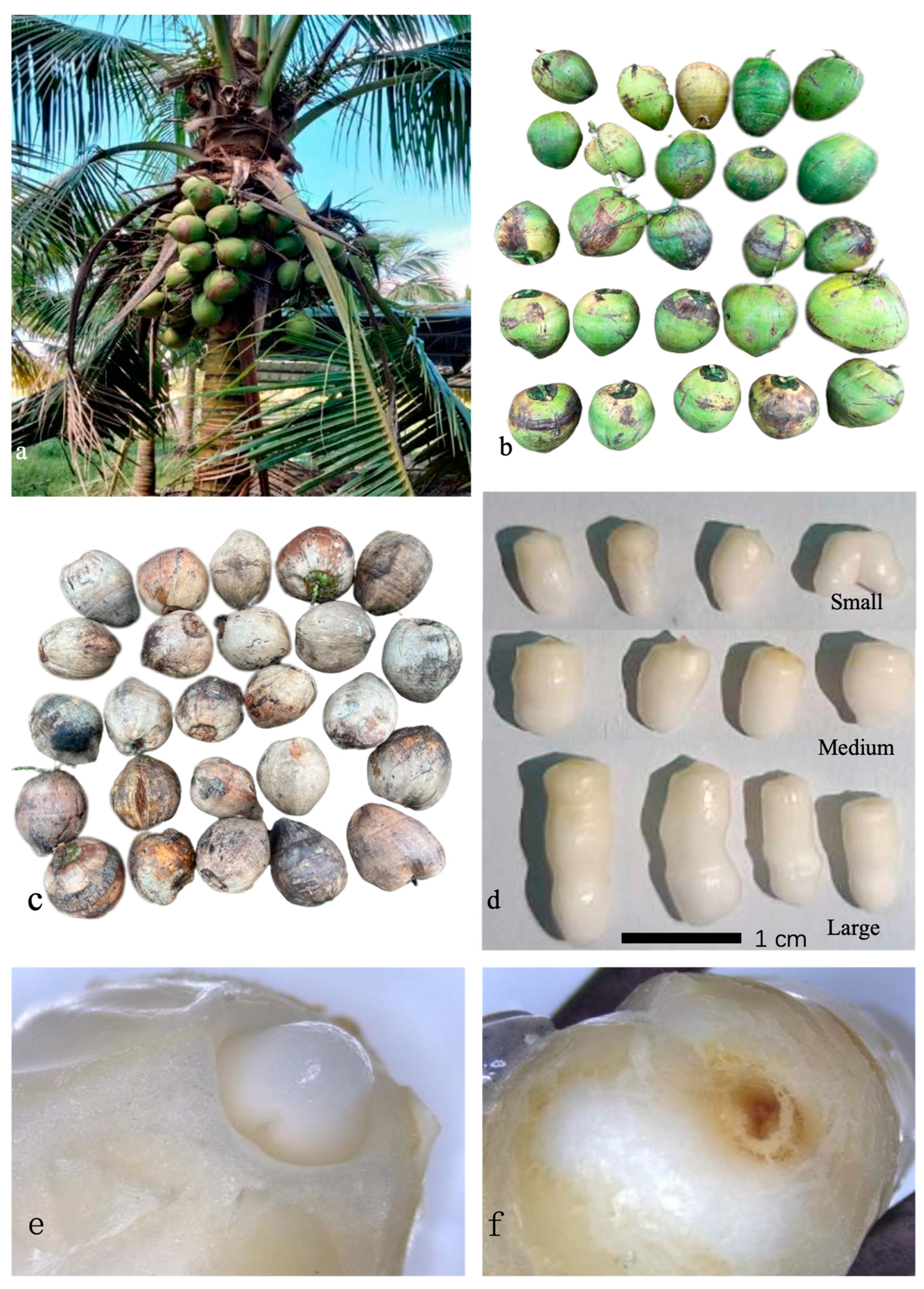
2.1. Plant Materials Preparations

Approximately 300 Hainan Tall coconut fruits, aged between 8 and 12 months (Figure 1a), were gathered from Wenzi Village, Qionghai City, located in Hainan Province, China. Twenty-five green (younger) fruits and 25 brown (older) fruits were individually selected for the next step of processing. Another 50 fruits were taken as experimental material to test the impact of different storage times on the final results of callus formation. The coconut fruits underwent the process of dehusking and were subsequently split using a machete in the field. The endosperm plugs, each containing an embryo, were extracted from each fruit using a cork borer that had been sterilized with a 70% ethanol solution. In batches of 10, these plugs were washed with tap water and transferred to clean glass jars. All fruits and endosperm plugs were promptly transported to the laboratory at the Sanya Nanfan Research Institute of Hainan University in Yazhou, Hainan Province, China, for further preparation. Upon arrival, these endosperm plugs were placed in a new sterile container and positioned within a laminar airflow hood. Subsequently, the endosperm plugs were rinsed with a 70% (v/v) ethanol solution for 3 min. Then, the embryos were isolated using forceps and a fine scalpel blade. These isolated embryos were then placed in sterile glass jars (120 × 80 mm). Following this, the embryos were subjected to a one-minute rinse in 70% (v/v) ethanol and were then immersed in a 2.1% (v/v) sodium hypochlorite (NaOCl) solution, with agitation, for a duration of 15 min. The embryos were subsequently washed three times with sterile distilled water. After the surface sterilization process, all embryos were transferred from the glass jars to sterile, autoclaved paper sheets for drainage. Following a 5 to 10 min drying period, the embryos were individually placed into micro-centrifugation tubes for further use. Embryos were then individually placed onto a fresh pre-culture medium (PCM, 5 mL) in thin glass bottles (50 mL, Falcon®, Corning, NY, USA) and moved into a darkened incubator (27 ± 1 °C). This method was described in detail by Mu (2020) [20].

2.2. Medium Preparations

All the culture media were prepared three days prior to their use, and all tissue-culture-approved chemicals were sourced from Guangzhou Chemical Reagent Factory; the media were autoclaved at 121 °C for 30 min before use. The detailed compositions of the media were in accordance with the description by Mu (2022) [2]. The basal medium (BM) employed for embryo culture was Y3 medium [21], comprising Y3 macro- and micro-nutrients, MS vitamins [22], agar (6 g L−1), and activated charcoal (AC; 2.5 g L−1). The pre-culture medium (PCM) was derived from BM, with the addition of ferrous sulphate heptahydrate (10 mL FeEDTA; 41.7010 mg/L Fe2SO4·7H2O), 6-benzylaminopurine (BAP; 5 μM), sucrose (60 g L−1), agar (6 g L−1), and AC (2.5 g L−1). The callus induction medium (CIM) was prepared using BM supplemented with FeEDTA (10 mL), BAP (5 μM), sucrose (30 g L−1), agar (6 g L−1), AC (2.5 g L−1), and 2,4-D (600 μM). Once prepared, each medium (approximately 30 mL) was poured into Petri dishes (90 × 20 mm). The pH was adjusted to 5.7 before adding the gelling agent and activated charcoal (AC).

2.3. Experiment Set-Up

Experiment on the age of coconuts: The external husk color of a coconut can serve as a visual indicator of its age and maturity. Generally, older coconuts have a drier, brown, or tan husk, while younger coconuts have a greener husk. In this study, 8-month-old coconuts (Figure 1b) and 10-month-old coconuts (Figure 1c) were employed. The treating method of embryos was described above. Twenty-five green (younger) fruits and 25 brown (older) fruits were randomly selected for each treatment level. Five embryos were inoculated in one vessel, and this process was replicated 5 times.
Experiment on storage time: Experimental coconuts were stored in a cool, shaded area for 15 days, while the control group was processed immediately upon arrival at the laboratory. Fifty fruits were selected as experimental material to test the impact of different storage times on the final results of callus formation. Twenty-five fruits were randomly picked for use in one treatment with 5 replicates. This experiment simulates the situation where coconuts, acting as implants, need to be stored and transported over long distances. This scenario commonly happens in many coconut tissue culture research institutes worldwide.
Experiment on embryo size: After the sterilization processes, experimental embryos are selected according to their size and divided into three groups (Figure 1d): 4–8 mm length, 8–12 mm length, and beyond 12 mm. Forty-five fruits were randomly selected and used in this study; each treatment consisted of 5 replicate vessels, each containing 3 explants.
Experiment on embryogenic shoot/non-shoot formation: The timing of germination varies, with the appearance of the plumule (ca. 2 mm in length) occurring between 10 and 15 days. Germinated embryos were then removed. After 15 days, all embryos were separated into two groups, those with shoot or without shoot (Figure 1e,f). Upon emergence, the plumules were isolated with a scalpel blade and moved to the CIM mentioned above. Plumules from the ungerminated embryos cannot be seen from the outside, but they were extracted under microscope, as described by Nguyen (2018) [18]. Fifty embryos were used in this study, with 25 used for each treatment. Five explants were cultured per vessel, and each treatment was designed with 5 replicates.

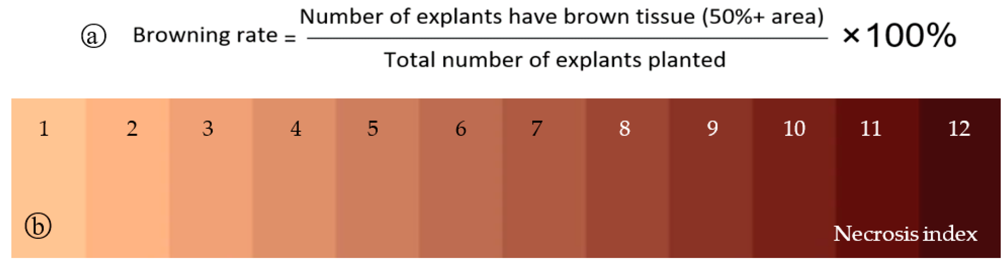
2.4. Assessment and Data Analysis
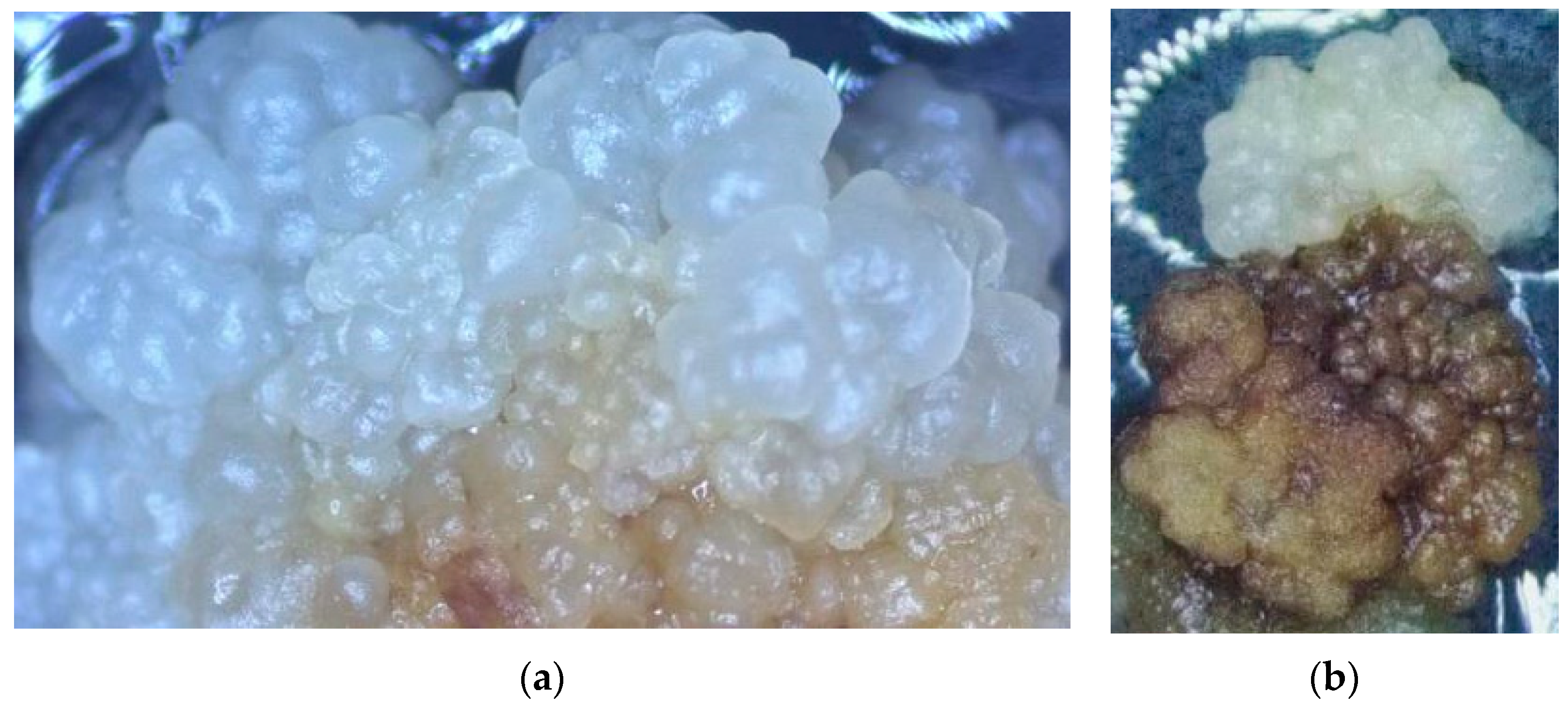
After 60 days of treatments without light and under 27 ± 2 °C, the rate of callus initiation, the proportion of cultures forming somatic embryogenic callus, and culture necrosis (by browning level) were assessed (as shown in Figure 2a). The rate of callus initiation was measured by counting the number of plumule cultures that formed callus after one month of culture. Subsequently, during the subculture, which occurred at three months, the rate of somatic embryogenic callus formation was determined by calculating the proportion of callus cultures producing somatic embryos. At the same third subculture, an evaluation of culture necrosis was conducted. Explants that demonstrated a browning index of 6 or higher (as shown in Figure 2b) were categorized as undergoing necrosis [2]. If more than 80% of the area showed signs of necrosis, we considered it nonviable. Statistical analysis for all experimental data involved the application of Student’s t-test and analysis of variance (ANOVA).

3. Results

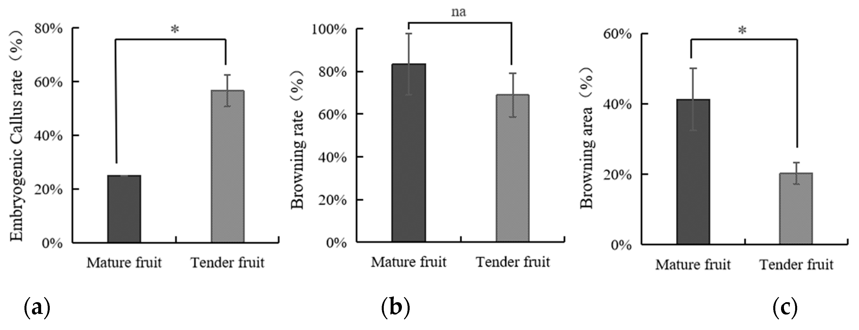
3.1. The Impact of Coconut Fruit Age on the Initiation of Callus

Firstly, the identification and definition of embryogenic callus and browned callus are quite important in this study. Embryogenic callus (Figure 3a) has the capacity of generating somatic embryos, which are distinguished by their smooth, translucent, and clearly defined somatic structure. On the other hand, the non-embryogenic callus (Figure 3c) exhibits a rough, yellowish, and sponge-like appearance [14]. Non-embryogenic callus refers to a type of callus in plant tissue culture that does not possess the ability to develop into embryos. The occurrence of browning in coconut tissue culture is a significant drawback in both embryogenic and non-embryogenic callus, leading to the deterioration of the tissues [23]. The definition and criteria for browned tissue were described above (Figure 2), and the appearances of browned tissue are revealed in Figure 3b. The browned callus tissue will quickly spread, negatively affecting the normal callus tissue, as demonstrated in Figure 3d.
This experiment was conducted to determine the optimal developmental stage of coconut nut as a type of explant. In this study, coconut nuts at different growth stages (mature or tender) were used. Similarly, explants from tender coconut formed significantly more embryogenic callus (56.7%) compared to the mature coconut fruits (25.0%). It was also found that using mature fruits as explants leads to greater necrosis (83.3%) and a larger necrotic area (41.3%), as indicated in Figure 4. Overall, our results demonstrate that the developmental stage is a critical factor for the initiation of embryogenic callus.

3.2. The Influence of Storage Time of Coconut on the Production of Callus

An experiment was undertaken to examine the influence of coconut storage time on the initiation and development of the embryogenic callus of coconut. A 15-day storage period may elevate the germination rate of coconut’s zygotic embryos from 76.2% to 91.7%. A storage period of 15 days could elevate the generation of embryogenic callus from 52.4% to 75.0%, as shown in Figure 5. Meanwhile, coconut explants from the non-stored fruits demonstrated a higher level of necrosis and larger necrotic areas. Our results demonstrate that storing coconut fruits for 15 days significantly elevated the formation of embryogenic callus.

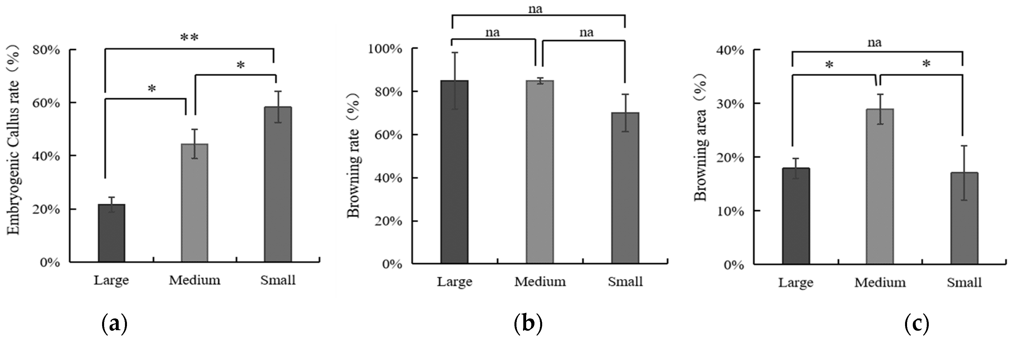
3.3. The Influence of Embryo Size of Coconut Fruits on Callus Initiation

The results of this study indicate that the callus generated from embryos of all sizes demonstrated similar germination rates, namely 100% (large), 90% (medium), and 93% (small). Specifically, the embryogenic callus rate of embryos of different sizes was measured three months after callus initiation. The small embryos showed a significantly higher induction rate of embryogenic callus (ca. 53%) compared to the medium and large embryos (ca. 40% and 22%, respectively, as shown in Figure 6). Regarding necrosis, embryos of all sizes did not show any significant difference, while the medium embryos led to a significantly larger necrotic area (ca. 29%) compared to the others. Our results suggest that smaller zygotic embryos of coconut may lead to a higher embryogenic callus rate. As the size of the explant increases, there is a decreasing trend in the embryogenic callus rate. However, the size of the embryos does not exhibit a correlation with the degree of callus necrosis.

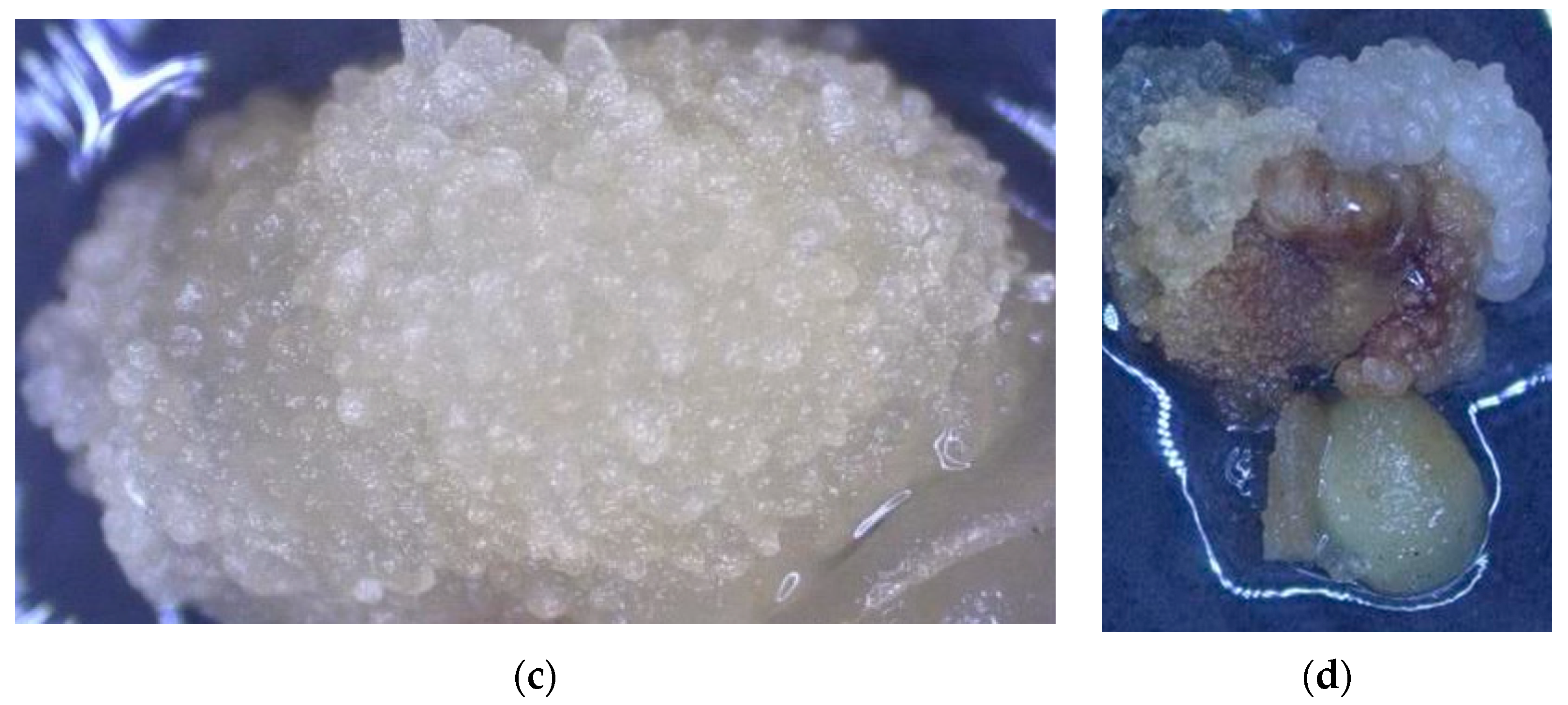
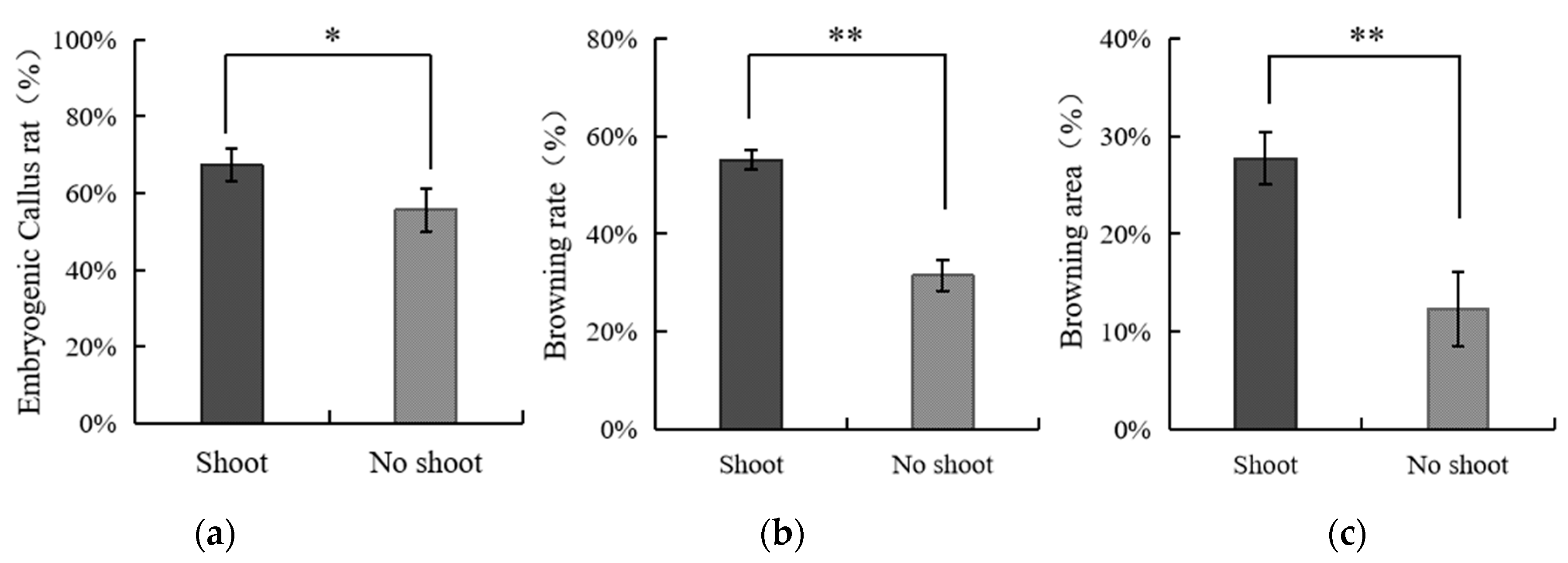
3.4. The Effect of Applying Embryos with Shoot or Non-Shoot as the Explant for Callus Initiation

Our results indicate that when using embryos with shoots as explants, an improved embryogenic rate and more browning could be achieved. Callus generated from those embryos with/without shoots demonstrated similar survival rates (ca. 94% for both). However, embryos with shoots had a significantly higher embryogenic callus rate (ca. 67%) compared to the embryos without shoots (ca. 56%), as demonstrated in Figure 7. On the contrary, the application of embryos with shoots may lead to a significantly higher browning rate (ca. 55%) than that of embryos without shoots (ca. 32%), as well as a greater browning area (ca. 28% to 12%). In Figure 8, it is clearly observed that callus derived from embryos with shoots showed more severe browning while exhibiting rapid growth and producing more embryogenic callus. On the other hand, the embryos without shoots showed the opposite trend.

4. Discussion

While protocols for coconut somatic embryogenesis (SE) from plumule tissues have been established for the last 20 years, the ideal developmental stage and physiological condition of the explant tissue from coconut are still unknown. To achieve an optimized callogenesis pathway, we revealed influences of the explant quality and physiological conditions on the initiation of primary callus.
The success of callus production is significantly influenced by the maturity of the explant [24]. A comprehensive investigation into the impact of maturity on the success of coconut tissue culture was conducted in this study. The research on maturity was divided into two parts: the maturity of coconut fruit and the maturity of in vitro tissues (explants) and their influence on tissue culture. The maturity of the zygotic embryo (age of the coconut fruit) is an important factor that impacts somatic embryogenesis (SE), as it can influence the generation and expansion of callus, ultimately affecting the process of SE. Embryos at the age of 8 months exhibited callus formation in approximately only 30 to 40%, with the resulting callus typically displaying slow growth in culture. On the other hand, embryos aged 9 months failed to generate any callus [25]. In current coconut tissue culture protocols, mature embryos aged 8–10+ months are commonly employed as explants [18,26,27,28], although some studies also utilize embryos aged 10–12 months [2]. These studies often use the estimated age of the embryo as a metric, but time measurements are subjective and rough, and visual observability is not applied. In this study, the appearance of the fruit was used as a more intuitive assessment of maturity. We also found that when using coconut fruits aged 12–14 months as explants, germinated coconut fruits (unsuitable for tissue culture) cannot be determined visually by external appearance. This results in the wastage of a significant number of fruits. The method described in this study can address this issue by utilizing green (or half green/half brown) coconut fruits. Additionally, experimental findings indicate that green coconut fruits can achieve a higher somatic embryogenic rate compared to mature coconuts. To ensure the maturation of each green coconut, we subjected experimental coconuts to a 15-day pre-treatment. The results indicated that pre-treatment could further enhance the success of tissue culture. It was speculated that the storage pre-treatment might activate the internal germination mechanism of the coconuts, imparting greater vitality to the explants [29]. Storing the coconut fruits for 15 days during shipment would not pose an issue and could even boost the callusing rate, which links back to the previous concern regarding long-distance shipment. Therefore, the optimal treatment for coconut is to harvest them when the outer skin still retains some green color, followed by pre-treatment (approximately 15 days of storage in a cool, shaded area at room temperature). However, the underlying mechanism behind this process remains unclear and requires further investigation.
To locate and maturate the plumule, a pre-culture was required, but previous studies did not provide clear information regarding the maturity of the plumule tissues at the time of excision. To reach an optimal pre-culture, scholars placed more emphasis on the pre-culture time rather than the growth stage of the plumule. However, this study leans towards using the plumule growth stage as the parameter, rather than relying on pre-culture time, to determine whether to extract the plumule for somatic embryogenesis. Due to genetic differences of varieties worldwide, the pre-culture time to reach the same developmental stage may vary. A study conducted by López-Villalobos (2002) suggested that 10 days may be the optimal pre-culture period for coconut somatic embryogenesis [30], while Fernando et al. (2004) pointed out that pre-culture mature zygotic embryos in a germination media for 15–17 days before plumule extraction could increase the successful rate of somatic embryogenesis of coconut [31]. Nguyen (2018) indicated that a two-week pre-culture of embryos containing plumules leads to the best callus initiation. The plumule must be excised as an explant for coconut somatic embryogenesis when the tissue culture reaches a size recognizable to the naked eye [18]. Mu (2022) also employed a 14-day pre-culture before plumule isolation [2]. In other coconut plumule culture protocols, the duration of pre-culturing the embryo was not clearly documented, thereby reducing the reproducibility of these methods [27,32]. In this study, we clearly found that coconut zygotic embryos with small shoots on the top (it usually takes 14–20 days to reach this developmental stage) demonstrated a significantly higher embryogenic rate than those without shoots. Our results are supported by other studies. Nguyen (2018) also suggested that the emergence of small shoots from zygotic embryos signals the need for plumule isolation [18]. This phenomenon may relate to the meristematic competence of developing tissues, leading to variability in the callus response [33,34]. Similar reactions were found in callus initiation utilizing immature inflorescences of coconut at various stages of development [17,35]. However, the molecular mechanism still remains unknown, further research being required with regard to this scientific issue. A storage process can also be considered as a pre-treatment for embryos, facilitating germination processes during the storage period [29]. This may be the reason why stored coconut demonstrated a better performance regarding somatic embryogenesis.
At the early stages of plant tissue culture, scientists observed that the size of the explant is closely linked to the final outcome of tissue culture [36]. The selection of explant size is a crucial factor that affects the success of tissue culture procedures [37,38]. The size of the explant can impact nutrient absorption, growth rate, and differentiation [39]. These assertions have been justified in coconut tissue culture. In their study on coconut, Malaurie et al. indicated that explants, such as plumules, with a small size (ca. 1 mm) lead to a higher density of meristematic cells in coconut tissue culture [40]. Bazrafshan (2022) justified that the size of the embryo may have notable impacts on the embryos’ germination ability, as well as on subsequent steps, such as tissue culture and cryopreservation [19]. In our research, we found that utilizing smaller embryos may lead to a greater amount of primary callus, while generating an acceptable portion of necrotic tissue. This result could be attributed to the development stage of the embryos, which is connected to their size. More studies are needed to explain the mechanism behind this phenomenon.

5. Conclusions

In conclusion, our findings shed light on key factors influencing the success of plumule-based coconut micropropagation. Immature (tender) coconut fruits emerged as the optimal starting material, displaying a heightened capacity for generating embryogenic callus while minimizing browning. The 15-day pre-treatment, facilitating self-maturation, proved instrumental in achieving superior rates of embryogenic callus formation, coupled with reduced browning. Notably, the use of smaller embryos during the in vitro stage yielded the most favorable outcomes, with no significant differences observed between large and medium embryos in terms of browning. Intriguingly, embryos with small shoots exhibited a higher embryogenic rate, albeit with an associated increase in browning tissue. Therefore, for an optimal coconut tissue culture protocol, it is recommended to utilize tender coconut, subject it to a 15-day pre-treatment, select smaller embryos during the in vitro cultivation, and prioritize those with small shoots to attain the most successful results in coconut tissue culture using plumule tissue.

Author Contributions

Data curation, H.P.; formal analysis, J.C.; funding acquisition, J.L.; investigation, S.Y. and X.W.; methodology, H.X. and Z.Y.; resources, M.M.H.; supervision, J.L.; validation, Z.M.; visualization, B.-M.T.; writing—original draft, Z.M. All authors have read and agreed to the published version of the manuscript.

Funding

This research was supported by Hainan Province Science, Technology and Innovation Project for Talent (KJRC2023L09 and KJRC2023D16), Hainan Yazhou Bay Seed Lab. + (JBGS + B21HJ0903), Postdoctoral Research Funding Program of Hainan Province (No. B23B10015), Hainan Provincial Natural Science Foundation (324QN360), and the “111”Project (No. D20024).

Data Availability Statement

The original contributions presented in the study are included in the article further inquiries can be directed to the corresponding author.

Conflicts of Interest

Author Shuya Yang was employed by the company Hainan State Farms Academy of Sciences Group Co., Ltd. The remaining authors declare that the research was conducted in the absence of any commercial or financial relationships that could be construed as a potential conflict of interest.

References

  1. Alshawwa, I.A.; Elsharif, A.A.; Abu-Naser, S.S. An Expert System for Coconut Diseases Diagnosis. Int. J. Acad. Eng. Res. 2019, 3, 8–13. [Google Scholar]
  2. Mu, Z. Overcoming Bottlenecks in the Pathway of Clonal Propagation of Coconut (Cocos nucifera L.). Ph.D. Thesis, School of Agriculture and Food Sciences, The University of Queensland, Brisbane City, QLD, Australia, 2022. [Google Scholar]
  3. Deen, A.; Visvanathan, R.; Wickramarachchi, D.; Marikkar, N.; Nammi, S.; Jayawardana, B.C.; Liyanage, R. Chemical composition and health benefits of coconut oil: An overview. J. Sci. Food Agric. 2021, 101, 2182–2193. [Google Scholar] [CrossRef] [PubMed]
  4. Sudalaimuthu, S.; Senthilkumar, P.; Sivakumar, B. Coconut industry in a nutshell. Retrieved Novemb. 2012, 23. [Google Scholar]
  5. Yang, Z.; Liu, Z.; Xu, H.; Li, Y.; Huang, S.; Cao, G.; Shi, M.; Zhu, J.; Zhou, J.; Li, R. ArecaceaeMDB: A comprehensive multi-omics database for Arecaceae breeding and functional genomics studies. Plant Biotechnol. J. 2023, 21, 11. [Google Scholar] [CrossRef] [PubMed]
  6. De Silva, H.W.S. The Coconut Industry in Sri Lanka: An Analysis of Government Intervention Measures. Master’s Thesis, The Australian National University, Canberra, Australia, 1979. [Google Scholar]
  7. Nayar, N. Origin and Domestication. In Coconut; Daya Publishing House A Division of Astral International Pvt. Ltd.: New Delhi, India, 2017; pp. 39–72. [Google Scholar]
  8. Zainol, F.A.; Arumugam, N.; Daud, W.N.W.; Suhaimi, N.A.M.; Ishola, B.D.; Ishak, A.Z.; Afthanorhan, A. Coconut Value Chain Analysis: A Systematic Review. Agriculture 2023, 13, 1379. [Google Scholar] [CrossRef]
  9. Yousefi, K.; Abdullah, S.N.A.; Hatta, M.A.M.; Ling, K.L. Genomics and Transcriptomics Reveal Genetic Contribution to Population Diversity and Specific Traits in Coconut. Plants 2023, 12, 1913. [Google Scholar] [CrossRef]
  10. Thadathil, S.T. A comprehensive review of chemical composition and nutritional health benefits of coconut water. Pharma Innov. J. 2023, 12, 343–550. [Google Scholar]
  11. Nguyen, Q.T.; Bandupriya, H.D.; López-Villalobos, A.; Sisunandar, S.; Foale, M.; Adkins, S.W. Tissue culture and associated biotechnological interventions for the improvement of coconut (Cocos nucifera L.): A review. Planta 2015, 242, 1059–1076. [Google Scholar] [CrossRef]
  12. Wilms, H.; Swennen, R.; Panis, B. Developing New In Vitro Micropropagation and Cryopreservation Techniques in Coconut; KU Leuven: Leuven, Belgium, 2023. [Google Scholar]
  13. Guo, Q.; Wang, Y.; Zou, J.; Jing, H.; Li, D. Efficient isolation and transformation of protoplasts in coconut endosperm and leaves for gene function studies. Trop. Plants 2023, 2, 16. [Google Scholar] [CrossRef]
  14. Kong, E.Y.; Biddle, J.; Kalaipandian, S.; Adkins, S.W. Coconut Callus Initiation for Cell Suspension Culture. Plants 2023, 12, 968. [Google Scholar] [CrossRef]
  15. Mu, Z.; Tran, B.-M.; Xu, H.; Yang, Z.; Qamar, U.Z.; Wang, X.; Xiao, Y.; Luo, J. Exploring the potential application of coconut water in healthcare and biotechnology: A review. Beverage Plant Res. 2024, 1–9. [Google Scholar] [CrossRef]
  16. Nwite, P.A.; Eke, C.; Osemwegie, Q.; Ikhajiagbe, B. Assessment of media modification and induction of calli on coconut (Cocos nucifera L.) palm explants. Res. J. Agric. Sci. 2023, 2320, 6063. [Google Scholar]
  17. Antonova, I. Somatic Embryogenesis for Micropropagation of Coconut (Cocos nucifera L.). Ph.D. Thesis, School of Land, Crop and Food Sciences, The University of Queensland, Brisbane City, QLD, Australia, 2009. [Google Scholar]
  18. Nguyen, T.Q. Clonal Propagation of Coconut (Cocos nucifera L.) for Elite Seedling Production and Germplasm Exchange. Ph.D. Thesis, School of Agriculture and Food Sciences, The University of Queensland, Brisbane City, QLD, Australia, 2018. [Google Scholar]
  19. Bazrafshan, A. Ex situ conservation of coconut (Cocos nucifera L.) germplasm using cryopreservation. Cryobiology 2022, 97, 297. [Google Scholar]
  20. Mu, Z. Optimization of Coconut Micropropagation via Somatic Embryogenesis. Multidiscip. Digit. Publ. Inst. Proc. 2020, 36, 55. [Google Scholar]
  21. Eeuwens, C. Mineral requirements for growth and callus initiation of tissue explants excised from mature coconut palms (Cocos nucifera) and cultured in vitro. Physiol. Plant. 1976, 36, 23–28. [Google Scholar] [CrossRef]
  22. Murashige, T.; Skoog, F. A revised medium for rapid growth and bio assays with tobacco tissue cultures. Physiol. Plant. 1962, 15, 473–497. [Google Scholar] [CrossRef]
  23. EL-Gioushy, S.; Liu, R.; Fan, H. A complete protocol to reduce browning during coconut (Cocos nucifera L.) tissue culture through shoot tips and inflorescence explants. Plant Arch. 2020, 20, 2196–2204. [Google Scholar]
  24. Bhojwani, S.S.; Dantu, P.K. Plant Tissue Culture: An Introductory Text; Springer: Berlin/Heidelberg, Germany, 2013; Volume 318. [Google Scholar]
  25. Adkins, S.W.; Samosir, Y.M.; Nikmatullah, A.; Ogle, H. Coconut (Cocos nucifera) in vitro ecology: Modifications of headspace and medium additives can optimize somatic embryogenesis. In Proceedings of the II International Symposium on Biotechnology of Tropical and Subtropical Species, Taipei, Taiwan, 5–9 November 2001; pp. 21–32. [Google Scholar]
  26. Fernando, S.; Santha, E.; Hewarathna, D. Activated coconut shell charcoal as a component of tissue culture media of Cocos nucifera L. J. Natl. Sci. Found. Sri Lanka 2010, 38. [Google Scholar] [CrossRef]
  27. Pérez-Núñez, M.; Chan, J.; Saenz, L.; González, T.; Verdeil, J.-L.; Oropeza, C. Improved somatic embryogenesis from Cocos nucifera (L.) plumule explants. Vitr. Cell. Dev. Biol. Plant 2006, 42, 37–43. [Google Scholar] [CrossRef]
  28. Sáenz, L.; Chan, J.L.; Narvaez, M.; Oropeza, C. Protocol for the micropropagation of coconut from plumule explants. In Plant Cell Culture Protocols; Humana Press: New York, NY, USA, 2018; Volume 1815, pp. 161–170. [Google Scholar]
  29. Beveridge, F.C.; Kalaipandian, S.; Yang, C.; Adkins, S.W. Fruit Biology of Coconut (Cocos nucifera L.). Plants 2022, 11, 3293. [Google Scholar] [CrossRef]
  30. Lopez Villalobos, A. Roles of Lipids in Coconut (Cocos nucifera L.) Embryogenesis. Ph.D. Thesis, University of London, London, UK, 2002. [Google Scholar]
  31. Fernando, S.; Weerakoon, L.; Gunathilake, T. Micropropagation of coconut through plumule culture. Print. Sri Lanka 2004, 16, 1–10. [Google Scholar] [CrossRef]
  32. Chan, J.; Saenz, L.; Talavera, C.; Hornung, R.; Robert, M.; Oropeza, C. Regeneration of coconut (Cocos nucifera L.) from plumule explants through somatic embryogenesis. Plant Cell Rep. 1998, 17, 515–521. [Google Scholar] [CrossRef]
  33. Feher, A.; Pasternak, T.P.; Dudits, D. Transition of somatic plant cells to an embryogenic state. Plant Cell Tissue Organ Cult. 2003, 74, 201–228. [Google Scholar] [CrossRef]
  34. Solís-Ramos, L.Y.; Andrade-Torres, A.; Sáenz-Carbonell, L.; Oropeza-Salín, C.; Castaño de la Serna, E. Somatic embryogenesis in recalcitrant plants. In Embryogenesis; Intech Open: London, UK, 2012; pp. 597–618. [Google Scholar]
  35. Verdeil, J.-L.; Huet, C.; Grosdemange, F.; Buffard-Morel, J. Plant regeneration from cultured immature inflorescences of coconut (Cocos nucifera L.): Evidence for somatic embryogenesis. Plant Cell Rep. 1994, 13, 218–221. [Google Scholar] [CrossRef] [PubMed]
  36. Buchsbaum, R.M. Size of explant and volume of medium in tissue cultures. J. Exp. Zool. 1932, 63, 483–508. [Google Scholar] [CrossRef]
  37. Dal Vesco, L.L.; de Almeida Pinto, A.; Zaffari, G.R.; Nodari, R.O.; Dos Reis, M.S.; Guerra, M.P. Improving pineapple micropropagation protocol through explant size and medium composition manipulation. Fruits 2001, 56, 143–154. [Google Scholar] [CrossRef]
  38. Singh, B.; Sood, N. Significance of explant preparation and sizing in Aloe vera L.—A highly efficient method for in vitro multiple shoot induction. Sci. Hortic. 2009, 122, 146–151. [Google Scholar] [CrossRef]
  39. Nhut, D.T.; An, T.T.T.; Huong, N.T.D.; Don, N.T.; Hai, N.T.; Thien, N.Q.; Vu, N.H. Effect of genotype, explant size, position, and culture medium on shoot generation of Gerbera jamesonii by receptacle transverse thin cell layer culture. Sci. Hortic. 2007, 111, 146–151. [Google Scholar] [CrossRef]
  40. Malaurie, B.; Tregear, J.; N’Nan, O.; Bandupriya, H.; Borges, M.; Verdeil, J.-L. Cryopreservation as a tool for the management of coconut germplasm. In Proceedings of the I International Symposium on Cryopreservation in Horticultural Species, Leuven, Belgium, 5–8 April 2009; pp. 461–466. [Google Scholar]
Figure 1. The plant material employed in this study. (a) Hainan Tall coconut tree used as the material resource. (b) Coconut fruits at the age of 8 months. (c) Coconut fruits at the age of 10+ months. (d) Coconut embryos in different sizes. (e) An image of a coconut embryo with a small shoot. (f) An image of a coconut embryo without a shoot.
Forests 15 00764 g001
Figure 2. Assessments applied in this study and the calculation of (a) the browning rate; (b) the browning index palette used to assess coconut explant necrosis.
Forests 15 00764 g002
Figure 3. Different types of calluses are mentioned in this study: (a) embryogenic callus with a translucent surface; (b) browned callus with embryogenic callus attached; (c) non-embryogenic callus with a yellowish, rough surface. (d) An explant that has embryogenic callus, non-embryogenic callus, and browned callus. The spreading of the browning area can be observed.
Forests 15 00764 g003aForests 15 00764 g003b
Figure 4. (a) Coconut embryogenic callus rate, (b) browning rate, and (c) browning area of mature/tender fruits. Bars represent standard errors, and the * sign indicates significant differences (p ≤ 0.05). Na represent no significant difference.
Forests 15 00764 g004
Figure 5. (a) Coconut embryogenic callus rate, (b) browning rate, and (c) browning area varied among coconut fruits with different storage periods. Bars represent standard errors, and the * sign indicates significant differences (p ≤ 0.05). Na represent no significant difference.
Forests 15 00764 g005
Figure 6. (a) Somatic embryogenic callus rate, (b) browning rate, and (c) browning area of callus generated from zygotic embryos in different sizes. Bars represent standard errors, the * sign indicates significant differences (p ≤ 0.05), and the ** sign means very significant differences (p ≤ 0.01). Na represent no significant difference.
Forests 15 00764 g006
Figure 7. (a) Somatic embryogenic callus rate, (b) browning rate, and (c) browning area of callus generated from zygotic embryos with shoots and without shoots. Bars represent standard errors, the * sign indicates significant differences (p ≤ 0.05), and the ** sign indicates a very significant difference (p ≤ 0.01).
Forests 15 00764 g007
Figure 8. The differences between callus derived from coconut zygotic embryos with and without shoots. (a) Callus from zygotic embryos with shoots, 1 month after callus initiation; (b) callus from zygotic embryos with shoots, 2 months after callus initiation: severe browning can be observed; (c) callus derived from zygotic embryos without shoots, 1 month after callus initiation; (d) callus derived from zygotic embryos without shoots, 2 months after callus initiation: less browning tissue can be found. All pictures were taken using the same scale.
Forests 15 00764 g008
Disclaimer/Publisher’s Note: The statements, opinions and data contained in all publications are solely those of the individual author(s) and contributor(s) and not of MDPI and/or the editor(s). MDPI and/or the editor(s) disclaim responsibility for any injury to people or property resulting from any ideas, methods, instructions or products referred to in the content.

