Article

Pre-Infection Stages of Austropuccinia psidii in the Epidermis of Eucalyptus Hybrid Leaves with Different Resistance Levels

1
Departamento de Produção Vegetal, Faculdade de Ciências Agronômicas–UNESP, Botucatu 18610-307, São Paulo, Brasil
2
Departamento de Fitopatologia, Universidade Federal de Viçosa, Viçosa 36570-900, Minas Gerais, Brasil
3
Departamento de Botânica, Instituto de Biociências de Botucatu–UNESP, Botucatu 18618-000, São Paulo, Brasil
4
Departamento de Biologia Geral, Universidade Federal de Viçosa, Viçosa 36570-900, Minas Gerais, Brasil
5
Departamento de Entomologia/BIOAGRO, Universidade Federal de Viçosa, Viçosa 36570-900, Minas Gerais, Brasil
*
Author to whom correspondence should be addressed.
Forests 2017, 8(10), 362; https://doi.org/10.3390/f8100362
Submission received: 25 July 2017 / Revised: 19 September 2017 / Accepted: 20 September 2017 / Published: 17 October 2017
(This article belongs to the Special Issue Forest Pathology and Plant Health)

Abstract

:
Rust is a major Eucalyptus spp. disease, which is especially damaging for early-stage plants. The aim of this study was to verify the pre-infection process of Austropuccinia psidii (A. psidii) in the leaves of three phenological stages of Eucalyptus clones with different resistance levels. Plants from the hybrids of Eucalyptus urophylla × Eucalyptus grandis (E. grandis) with variable levels of resistance to this disease were used. The pathogen was inoculated in vitro on abaxial leaf discs of first, third, and fifth leaf stages and maintained under conditions suitable for disease development. Subsequently, samples from these discs were collected 24 and 120 h after inoculation and processed using scanning electron microscopy analysis. No symptoms were seen in any leaf stage of the resistant clone. Additionally, a low incidence of A. psidii germination (1.3–2%) and appressoria (0–0.5%) in three leaf stages was observed. However, the first leaf stage of the susceptible clone presented germination of large numbers of urediniospores (65%) with appressoria (55%) and degradation of the cuticle and wax. From the third stage, the percentage of germinated urediniospores (<15%) and appressoria (<2%) formation of this clone decreased. Protrusions on the leaf surface, associated with the pathogen, were observed on the first and third leaf stages of the resistant clone and on the fifth stage of the susceptible clone, suggesting a possible defensive plant reaction.

1. Introduction

The expansion of eucalypt plantations to new areas around the world is confronting abiotic and biotic diseases that can limit certain genetic materials of this plant [1]. Rust, caused by Austropuccinia psidii (A. psidii) (G. Winter) Beenken comb. nov., is one of the most notable diseases of Eucalyptus spp., and is mainly associated with its immature tissues [1,2,3]. This fungus infects both native and exotic Myrtaceae [4,5,6].
Rust is found mainly on early stage plants [1,7]. Most damage is found in nurseries, but it can also attack plants in the field. Eucalyptus spp. are more susceptible to A. psidii infection up to two years of age, due to the higher number of shoots and tender tissues, which is ideal for the establishment of this pathogen [7,8,9].
The initial infection events of fungi that cause rust include adherence to the cuticle and direct germ tube growth on host plant surface [10]. The A. psidii urediniospores often directly penetrate through the host cuticle and epidermis, between the anticlinal walls of the epidermal cells, by forming appressoria [11]. The process of intercellular colonization by the pathogen starts after its penetration through intracellular haustoria that draw nutrients from the host cells [1,7,12].
Resistant genetic plant materials are the most widely used control method to manage rust, but many cultivated eucalypt clones are susceptible to this disease. As with other rust species [13], the pathogenesis of this fungus depends on its thigmotropic response for host recognition [14]. Young leaves and shoots of susceptible eucalypt plants are infected by A. psidii, while mature or older leaves are resistant [1,7]. The resistance to A. psidii infection of older Eucalyptus leaves occurs at the pre-penetration stage and is due, amongst other inherent factors, to their higher wax quantity [15]. However, the resistance of young leaves in resistant plants needs to be investigated. The study of A. psidii initial infection processes on leaf surfaces of the clones with different resistance levels is important to understand the behavior of these eucalypt clones.
Thus, the objective of this study was to evaluate the pre-infection process of A. psidii in leaves at different phenological stages of eucalypt clones with different resistance levels.

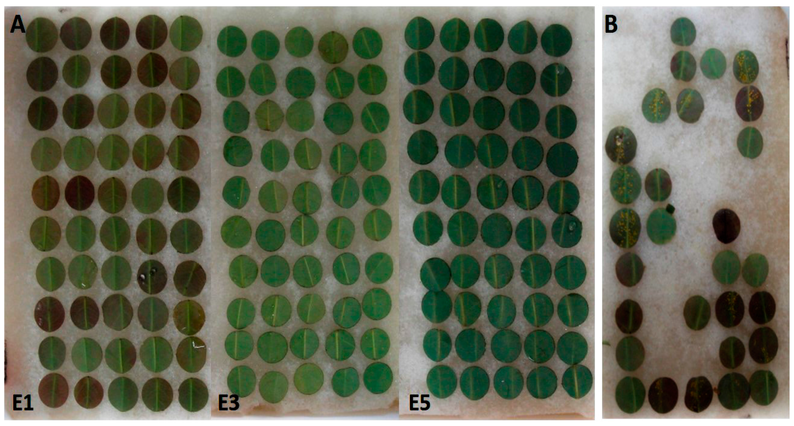
2. Materials and Methods
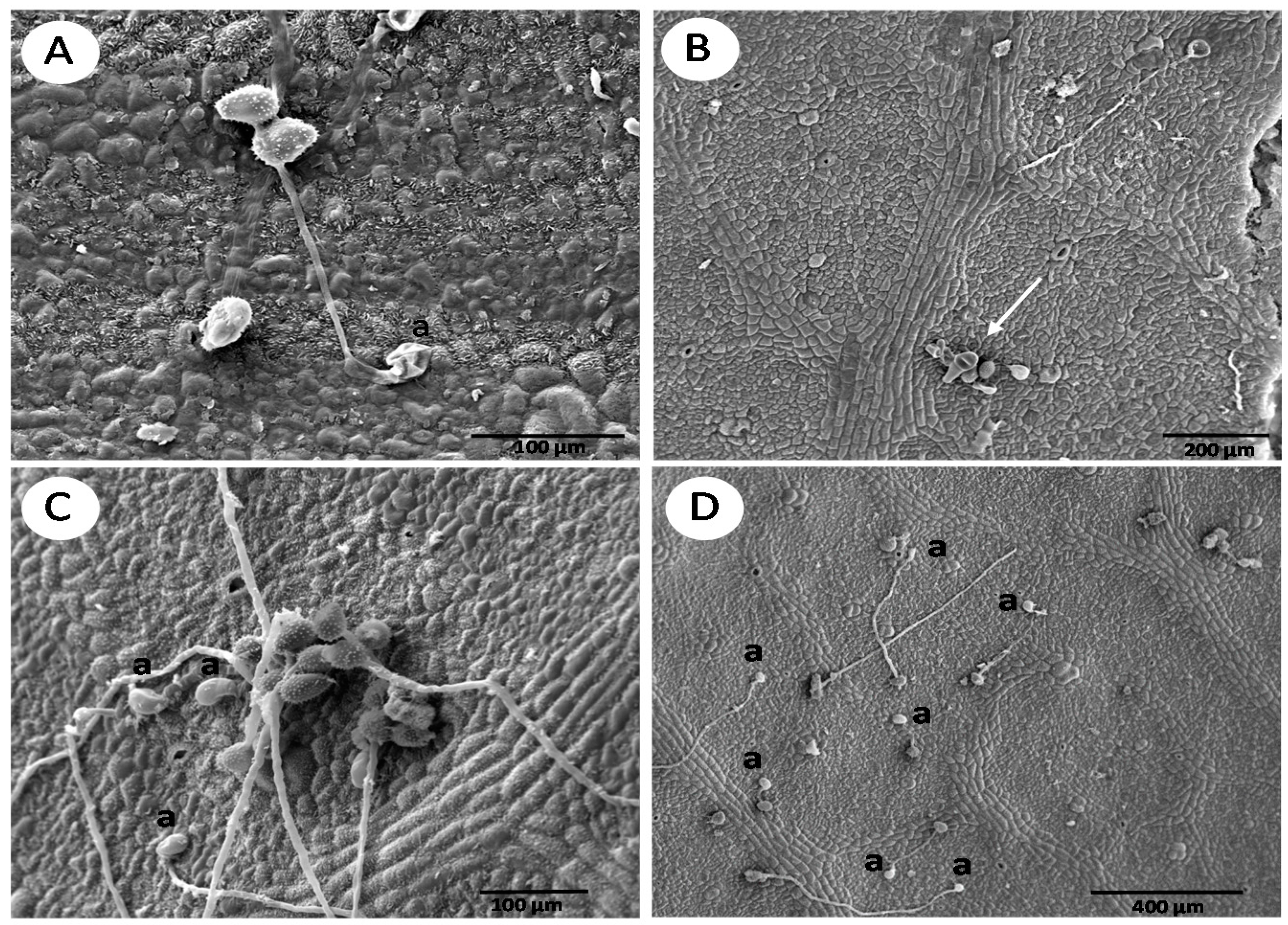
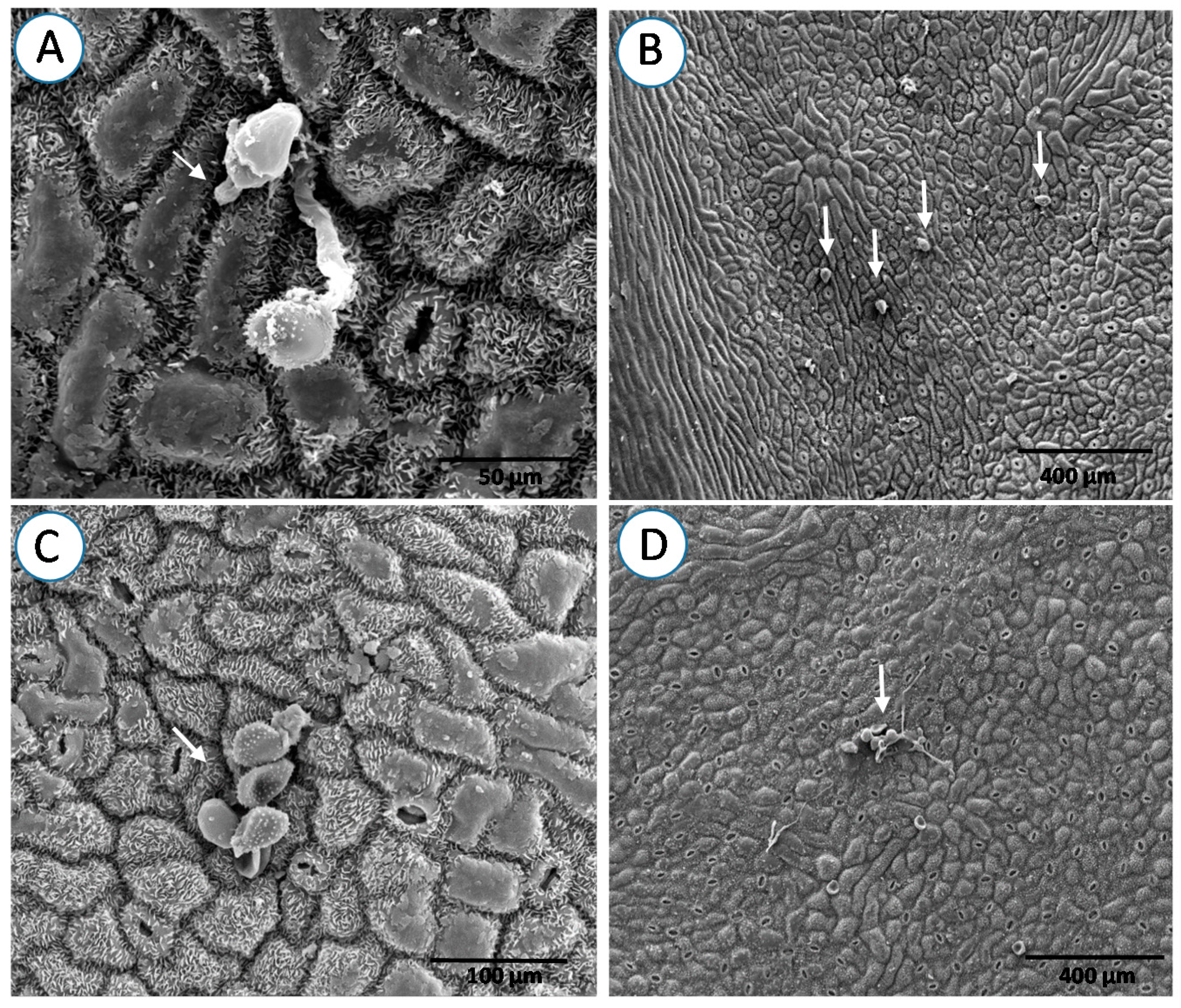
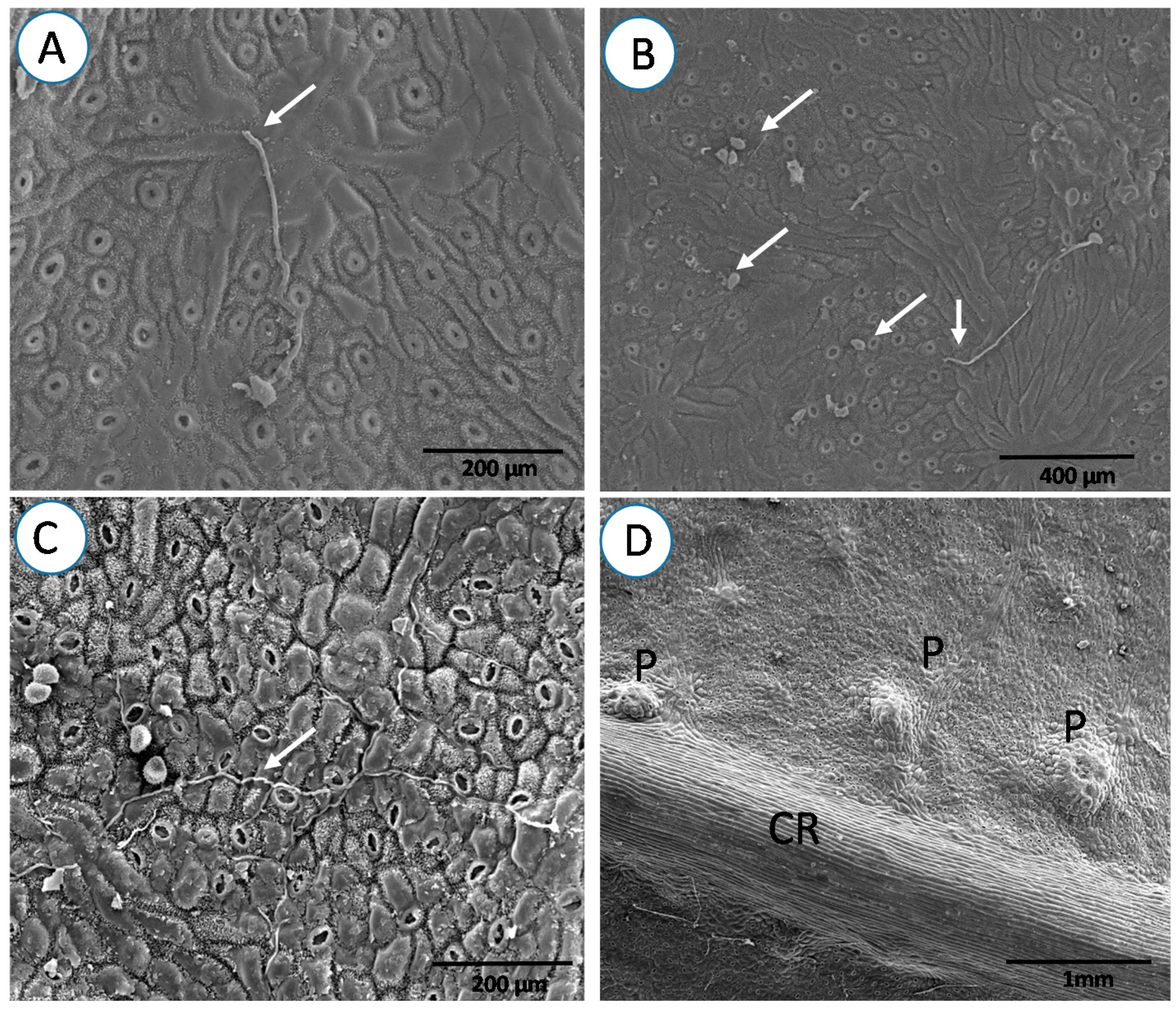
Seedlings of rust-susceptible and resistant Eucalyptus urophylla S. T. Blake × Eucalyptus grandis (E. grandis) W. Hill ex Maiden “urograndis” were grown in 2 L capacity pots, containing Carolina Soil® (Carolina Soil do Brasil, Santa Cruz do Sul, RS, Brazil) substrate enriched with simple superphosphate (6 kg/m3) and Osmocote® (Tecnutri do Brasil, Tietê, SP, Brazil) (NPK 19:06:10) and kept in a greenhouse at 20–30 °C for three months. These clones were selected based on their response to A. psidii, and their seedlings were supplied by Votorantim Celulose e Papel S.A, located in Jacareí, São Paulo State, Brazil. A total of 50 discs per leaf stage with a 1.5 cm diameter were cut with a punch from the first, third, and fifth leaf pairs from branches of six plants of each clone. These discs were placed on water-saturated foam in plastic trays (Figure 1A). An isolate of A. psidii (FCA-PP2303), obtained from the mycological collection of the Laboratory of Forest Pathology at the Universidade Estadual Paulista (UNESP-Botucatu), was inoculated on Syzygium jambos L. (Alston) and after sporulation of the fungus, the urediniospores were collected to prepare the inoculum [16]. An inoculum suspension in water containing 1% Tween 20 was prepared at a concentration of 9 × 10⁴ spores/mL [17]. The concentration of spore suspension was determined with a haemocytometer. Five microliters of the urediniospores suspension were inoculated on the central part of the abaxial surface of leaf discs for easy identification of the inoculum deposition site for scanning electron microscopy (SEM) analysis. The trays with the inoculated materials were covered with a glass lid to reduce moisture loss and placed in an incubation chamber at 20 ± 1 °C in continuous darkness for the first 24 h, followed by a 12 h photoperiod at approximately 10 μM photons/s/m2, which is ideal for rust development, according to previous studies [15,16]. Samples were taken from the first leaf stage of the susceptible clone and put in a humid chamber for 12 days in order to confirm that the inoculation method was able to cause the disease (Figure 1B). The experiment was set up using a completely randomized design, with four replications. The assays were repeated once.
Leaf samples (5 mm2) were collected at 24 and 120 h after pathogen inoculation for SEM. The preparation and analysis of the samples followed the previously described methodology [18] with some adaptations. Leaf samples were fixed in 2.5% glutaraldehyde in 0.1 M phosphate buffer (pH 7.3) for 24 h. Fragments used per treatment were transferred to 0.05 M cacodylate buffer and washed three times for 10 min. These fragments were placed into a 1% solution of osmium tetroxide in 0.1 M phosphate buffer (pH 7.3) for 1 h, washed with distilled water three times, and dehydrated in an acetone series (25%, 50%, 75%, 90%, and 100% (3×) for 10 min each stage). After dehydration, samples were taken at the critical point apparatus (Balzers CPD 030) to replace the acetone by CO2 and complete drying. Specimens were mounted on aluminum stubs and covered with gold (SCD 050, Balzers) for observation under a scanning electron microscope (Philips SEM 515). The images generated at 20 kV and 9 mm working distance were digitally recorded. Percentage of urediniospores germination and appressoria formation were quantified through SEM images in leaf samples collected 24 and 120 h after inoculation. A total of 50 urediniospores were observed. The experiment was distributed as a double factorial in split plot in time. The factorial was constituted by the combination of two clones differing in resistance level × three leaf stages. The data were subjected to analysis of variance (ANOVA). When the results were significant, the average values were separated using a Tukey test at 5% probability. An AIC (Akaike Information Criteria) analysis using generalized linear models via likelihood was performed [19] because discrete counting data was used. The generalized model of normal distribution was the best fit for germination and appressoria formation data in comparison to a model of Poisson distribution. The statistical analyses were carried out using the R program.

3. Results

ANOVA showed that only the clone × leaf stage interaction was a significant source of variation (Table 1). There was no difference in the percentage of urediniospores germination and appressoria formation at 24 and 120 h after inoculation of the pathogen. A higher urediniospore germination and appressoria formation of A. psidii was observed in the first leaf stage of the susceptible clone (Table 2 and Figure 2). Dehydration of urediniospores and formation of germ tubes and appressoria were occasionally observed in the resistant clone at 24 h after its inoculation (Figure 2A,B). On the other hand, urediniospores germinated and formed intact germ tubes of various sizes, producing appressoria, beginning the infectious process in the susceptible clone (Figure 2C,D). In the third leaf stage, the incidence of germinated urediniospores was lower on the leaf surface of the resistant clone and appressorium formation was rarely observed (Table 2, Figure 3A,B). A low incidence of urediniospore germination with appressoria on the leaf of the susceptible clone in relation to the first leaf stage was observed (Table 2, Figure 3C,D). In the fifth leaf stage, the number of germinated urediniospores was lower in the resistant clone and no appressorium formation was observed (Table 2, Figure 4A,B). However, the susceptible clone had urediniospores with long germ tubes without appressoria (Table 2 and Figure 4C). Skin cells formed protrusions on its leaf surface (Figure 4D). The third and fifth leaf stages had higher wax quantity on the leaf surface compared to that of the first stage.
SEM images showed the presence of protrusions on the resistant clone leaves involving urediniospores as a possible plant reaction with dehydration of appressoria and urediniospores (Figure 5A,B). These reactions were observed in all leaf stages of the resistant clone, 120 h after inoculation, and in the fifth stage of the susceptible clone, 24 h after inoculation. Cuticle and wax degradation, indicating probable onset of A. psidii colonization in the host, were observed in the leaves of the first leaf stage of the susceptible clone (Figure 5C,D). The resistant clone in this stage did not exhibit symptoms on the leaf surface and presented a lower incidence of appressoria (Figure 5A,B). Leaves of the third leaf stage of the resistant clone had a low quantity of germinated urediniospore and protrusions involving urediniospores (Figure 6A,B). This was characterized by protrusions on the leaf surface, similar to those observed during the first leaf stage, with low urediniospores germination. However, the susceptible clone showed extensive germ tube formation and damage by the pathogen (Figure 6C,D). The fifth leaf stage of the resistant clone had a lower quantity of urediniospore germinated, without appressoria and urediniospore wilting (Figure 7A,B). In the susceptible clone, the pathogen presented long germ tubes without appressoria and penetration in the leaves (Figure 7C,D).

4. Discussion

In the first leaf stage, the susceptible clone showed a higher degree of urediniospore germination with appressoria and degradation of the cuticle and wax. From the third leaf stage, the inhibition of the pre-infection fungus process in leaves of the susceptible Eucalyptus urophylla × E. grandis clone showed that there is some plant defense response related to leaf age. In the resistant clone, these mechanisms occurred from the first leaf stage. In the fifth leaf stage, the susceptible clone showed superficial growth, but without penetration of the pathogen, while the resistant clone had many inactive urediniospores. This resembles the pattern observed on Eucalyptus grandis susceptible to A. psidii [15]. The germination, appressoria formation, and penetration of this fungus gradually decreased from the first to the fourth leaf stage, but without penetrating in the fifth leaf stage [15].
In the first leaf stage of the resistant clone, dehydration of urediniospores and formation of germ tubes and appressoria were occasionally observed, as reported for the Phakopsora pachyrhizi (P. pachyrhizi) Syd. & P. Syd. urediniospores in soybean [20]. The susceptible clone already had many urediniospores germinated with appressorium followed by cuticle and wax degradation. Different to other Puccinia species, in that penetration occurs via stomata [21,22,23], the penetration of A. psidii occurs between the anticlinal walls of the leaf epidermis directly into the mesophyll of the leaf after appressoria formation [24]. Once formed, appressoria adhere tightly to the leaf surface and secrete extracellular enzymes, or generate physical force, or use a combination of both factors to bring about cuticle penetration [25]. The leaf surface is the first line of defense against plant invaders, where the adhesion of the pathogen occurs, followed by penetration and infection [26]. In the interaction Puccinia recondita f. sp. tritici–wheat, the resistance is not related to the pre-infectious processes at the leaf surface level [23], different to what was observed in our research.
In the fifth leaf stage of the susceptible clone, the urediniospores with long germ tubes without appressoria, and some with short germ tubes with appressoria, are similar to P. pachyrhizi urediniospores on soybean [20]. Germ tube extension and differentiation can occur in response to signals including surface hardness, hydrophobicity, plant signals, and surface topography [27]. The short germ tubes decrease the amount of endogenous energy required for growth, which thus can be used to penetrate the cells [24]. These results demonstrate that the fungus germinates on the surface of leaves of plants in the fifth leaf stage, mainly on those of the susceptible clone, but without penetrating or colonizing its tissues.
The presence of epicuticular waxes on leaves of the two clones, especially in the third and fifth leaf stages, agrees with that reported for some eucalypt species as an important factor for resistance to A. psidii infection [15]. Appressorium formation in P. pachyrhizi and Phakopsora apoda (Har. & Pat.) Mains occurred in a place with lower wax deposition on the leaf surface [20,28]. Variability in the quantity of wax on the surface may also modify fungal behavior and interfere with the infection process [29]. Recognition of the cuticle surface by the fungus, necessary for appressorium differentiation, depends on the wax distribution pattern on leaves and also on the pathogen capacity to degrade it [30]. Thus, the increase in the quantity of wax on the leaf surface may have interfered with appressorium development, as seen in the pathosystem Hordeum chilense Roem. et Schult.-Puccinia hordei G.H. Otth [31]. Surface contact is essential for appressorium induction [32] and germination and germ tube growth requires fewer stimuli than appressorium formation [33].
In the fifth leaf stage, the presence of long germ tubes and absence of appressoria and penetration by the pathogen in the susceptible clone is similar to that reported for Alternaria solani Sorauer on the surface of Solanum lycopersicum L. leaves resistant and susceptible to this pathogen [34]. The higher chitinase and peroxidase activity on older eucalypt leaves [26] can be one of the reasons for the fungal wilt structures because chitinases increase plant resistance to pathogens by catalyzing chitin polymer hydrolysis, the main components of fungal cell walls [35]. Peroxidases are also involved in numerous cellular processes including the final suberin and lignin biosynthesis steps [36] and the metabolism of phenylpropanoids [37]. In addition, peroxidases are involved in the oxidation of phenols and compounds toxic to pathogenic organisms [38] and the formation of papillae that can block fungal entry [39].
Protrusions associated with the pathogen and observed on the leaf surface of first and third leaf stages of the resistant clone and in the fifth leaf stage of the susceptible clone suggest a possible defensive plant reaction. This was also shown by the protrusions involving urediniospores and dehydration of appressoria and urediniospores. This may be due to chemical compounds produced by leaves, such as essential oils, and extracted from eucalypt leaves with high antimicrobial activity [40,41,42,43].
The inoculation method using detached eucalypt leaf discs was effective for SEM studies and for evaluating the resistance of the plant to rust, with the onset of symptoms in the first leaf stage of the susceptible eucalypt clone. The efficiency of this method had also been reported for bean [44,45,46] and soybean [47].
The results obtained in this research will be of great importance for the international forestry industry. From the results observed in this study, we recommend the use of the genetic materials with a lower leaf maturation period and with higher wax content in the younger leaves to be tested in the breeding programs for the control of this disease. Future studies could be conducted to analyze the anatomy and chemical composition of leaves of different phenological stages of the resistant and susceptible plants, before and after the infectious process of A. psidii in order to identify other possible resistance mechanisms of eucalypt plants to this disease.

5. Conclusions

Resistance and susceptibility of eucalypt clones occur in the pre-infection process. The failure of the pathogen to recognize an infection site on the leaves of the resistant clone and on old leaves of the susceptible clone and initiate appressorium formation appears to be the key factor explaining such resistance. The urediniospores germination with appressoria and degradation of the cuticle waxes had higher values in the first leaf stages of the susceptible Eucalyptus urophylla × E. grandis clone. From the third leaf stage in the susceptible clone, the germination and appressorium formation of the fungus was prevented by the defense mechanisms, while this took place in the first leaf stage of the resistant clone. Protrusions on the leaf surface were associated with the pathogen in the first and third leaf stages of the resistant clone, at 120 h after inoculation, and in the fifth leaf stage of the susceptible clone, 24 h after inoculation, suggesting a possible plant defense reaction. The results presented in this work help to explain the resistance of old and young eucalypt leaves to A. psidii.

Acknowledgments

The authors wish to acknowledge researcher Donizete Dias Costa of Votorantim Celulose e Papel, “Conselho Nacional de Desenvolvimento Científico e Tecnológico (CNPq)”, “Coordenação de Aperfeiçoamento de Pessoal de Nível Superior (CAPES)” and “Fundação de Amparo à Pesquisa do Estado de Minas Gerais (FAPEMIG)” and “Programa Cooperativo sobre Proteção Florestal-Instituto de Pesquisas e Estudos Florestais” (PROTEF-IPEF) for financial support. Global Edico Services and Phillip John Villani (The University of Melbourne, Australia) revised and corrected the English language used in this manuscript.

Author Contributions

R.R.S. conceived and designed the study, performed the sampling, processed and analyzed the data, and wrote the paper; A.C.S. discussed results and wrote the paper; R.A.R. conceived and designed the study, and reviewed drafts of the paper; J.E.S. reviewed drafts and wrote of the paper; J.C.Z. reviewed drafts and wrote of the paper; E.L.F. conceived and designed the study, reviewed drafts of the paper, and contributed reagents/materials/analysis tools.

Conflicts of Interest

The authors declare no conflict of interest. The funding sponsors had no role in the design of the study; in the collection, analyses, or interpretation of data; in the writing of the manuscript, and in the decision to publish the results.

References

  1. Alfenas, A.C.; Zauza, E.A.V.; Mafia, R.G.; Assis, T.F. Clonagem e Doenças do Eucalipto, 2nd ed.; Editora UFV: Viçosa, Brazil, 2009. [Google Scholar]
  2. Miranda, A.C.; Moraes, M.L.T.; Tambarussi, E.V.; Furtado, E.L.; Mori, E.S.; Silva, P.H.M.; Sebbenn, A.M. Heritability for resistance to Puccinia psidii Winter rust in Eucalyptus grandis Hill ex Maiden in Southwestern Brazil. Tree Genet. Genomes 2013, 9, 321–329. [Google Scholar] [CrossRef]
  3. Beenken, L. Austropuccinia: A new genus name for the myrtle rust Puccinia psidii placed within the redefined family Sphaerophragmiaceae (Pucciniales). Phytotaxa 2017, 297, 53–61. [Google Scholar] [CrossRef]
  4. Figueiredo, M.B. Doenças fúngicas emergentes em grandes culturas. Biológico 2001, 63, 29–32. [Google Scholar]
  5. Morin, L.; Aveyard, R.; Lidbetter, J.R.; Wilson, P.G. Investigating the host-range of the rust fungus Puccinia psidii sensu lato across tribes of the family Myrtaceae present in Australia. PLoS ONE 2012, 7, e35434. [Google Scholar] [CrossRef] [PubMed]
  6. Yamaoka, Y. Recent outbreaks of rust diseases and the importance of basic biological research for controlling rusts. J. Gen. Plant Pathol. 2014, 80, 375–388. [Google Scholar] [CrossRef]
  7. Ferreira, F.A. Patologia Florestal–Principais Doenças Florestais do Brasil; Sociedade de Investigações Florestais: Viçosa, Brazil, 1989. [Google Scholar]
  8. Demuner, N.L.; Alfenas, A.C. Fungicidas sistêmicos para o controle da ferrugem causada por Puccinia psidii em Eucalyptus cloeziana. Fitopatol. Bras. 1991, 16, 174–177. [Google Scholar]
  9. Coutinho, T.A.; Wingfield, M.J.; Alfenas, A.C.; Crous, P.W. Eucalyptus rust: A disease with the potential for serious international implications. Plant Dis. 1998, 82, 819–825. [Google Scholar] [CrossRef]
  10. Mendgen, K.; Hahn, M.; Deising, H. Morphogenesis and mechanisms of penetration by plant pathogenic fungi. Annu. Rev. Phytopathol. 1996, 34, 367–386. [Google Scholar] [CrossRef] [PubMed]
  11. Glen, M.; Alfenas, A.C.; Zauza, E.A.V.; Wingfield, M.J.; Mohammed, C. Puccinia psidii: A threat to the Australian environment and economy—A review. Australas. Plant Pathol. 2007, 36, 1–16. [Google Scholar] [CrossRef]
  12. Bedendo, I.P. Ferrugens. In Manual de Fitopatologia: Princípios e Conceitos; Bergamin Filho, A., Kimati, H., Amorim, L., Eds.; Agronômica Ceres: São Paulo, Brazil, 1995; pp. 872–880. [Google Scholar]
  13. Terhune, B.T.; Hoch, H. Substrate hydrophobicity and adhesion of Uromyces urediospores and germlings. Exp. Mycol. 1993, 17, 241–252. [Google Scholar] [CrossRef]
  14. Bailey, J.A.; O’Connell, R.J.; Pring, R.J.; Nash, C. Infection strategies of Colletotrichum species. In Colletotrichum: Biology, Pathology and Control; Bailey, J.A., Jeger, M.J., Eds.; CAB International: Wallingford, UK, 1992; pp. 88–120. [Google Scholar]
  15. Xavier, A.A.; da Silva, A.C.; Guimarães, L.M.S.; Matsuoka, K.; Hodges, C.S.; Alfenas, A.C. Infection process of Puccinia psidii in Eucalyptus grandis leaves of different ages. Trop. Plant Pathol. 2015, 40, 318–325. [Google Scholar] [CrossRef]
  16. Ruiz, R.A.R.; Alfenas, A.C.; Ferreira, F.A. Influência da temperatura, do tempo de molhamento foliar, fotoperíodo e intensidade de luz sobre a infecção de Puccinia psidii em eucalipto. Fitopatol. Bras. 1989, 14, 55–61. [Google Scholar]
  17. Pinto, C.S.; Costa, R.M.L.; Moraes, C.B.; Pieri, C.; Tambarussi, E.V.; Furtado, E.L.; Mori, E.S. Genetic variability in progenies of Eucalyptus dunnii Maiden for resistance to Puccinia psidii. Crop Breed. Appl. Biotechnol. 2014, 14, 187–193. [Google Scholar] [CrossRef]
  18. Medice, R.; Alves, E.; Assis, R.T.; Magno, R.G., Jr.; Lopes, E.A.G.L. Óleos essenciais no controle da ferrugem asiática da soja Phakopsora pachyrhizi Syd. & P. Syd. Cienc. Agrotec. 2007, 31, 83–90. [Google Scholar]
  19. Akaike, H. A new look at the statistical model identification. IEEE Trans. Automat. Control 1974, 19, 716–723. [Google Scholar] [CrossRef]
  20. Magnani, E.B.Z.; Alves, E.; Araújo, D.V. Eventos dos processos de pré-penetração, penetração e colonização de Phakopsora pachyrhizi em folíolos de soja. Fitopatol. Bras. 2007, 32, 156–160. [Google Scholar] [CrossRef]
  21. Hughes, F.L. Scanning electron microscopy of early infection in the uredial stage of Puccinia sorghi in Zea mays. Plant Pathol. 1985, 34, 61–68. [Google Scholar] [CrossRef]
  22. Lennox, C.L.; Rijkenberg, F.H.J. Scanning electron microscopy study of infection structure formation of Puccinia graminis f. sp. tritici in host and non-host cereal species. Plant Pathol. 1989, 38, 547–556. [Google Scholar]
  23. Hu, G.; Rijkenberg, F.H.J. Scanning electron microscopy of early infection structure formation by Puccinia recondita f. sp. tritici on and in susceptible and resistant wheat lines. Mycol. Res. 1998, 102, 391–399. [Google Scholar]
  24. Hunt, P. Cuticular penetration by germinating uredospores. Trans. Br. Mycol. Soc. 1983, 51, 103–112. [Google Scholar] [CrossRef]
  25. Pryce-Jones, E.; Carver, T.; Gurr, S.J. The roles of cellulase enzymes and mechanical force in host penetration by Erysiphe graminis f. sp. hordei. Physiol. Mol. Plant Pathol. 1999, 55, 175–182. [Google Scholar] [CrossRef]
  26. Boava, L.P.; Kuhn, O.J.; Pascholati, S.F.; Di Piero, R.M.; Furtado, E.L. Atividade de quitinases e peroxidases em folhas de eucalipto em diferentes estágios de desenvolvimento após tratamento com acibenzolar-S-metil (ASM) e inoculação com Puccinia psidii. Trop. Plant Pathol. 2010, 35, 124–128. [Google Scholar] [CrossRef]
  27. Tucker, S.L.; Talbot, N.J. Surface attachment and pre-penetration stage development by plant pathogenic fungi. Annu. Rev. Phytopathol. 2001, 39, 385–417. [Google Scholar] [CrossRef] [PubMed]
  28. Adendorff, R.; Rijkenberg, F.H.J. Direct penetration from urediospores of Phakopsora apoda. Mycol. Res. 2000, 104, 317–324. [Google Scholar] [CrossRef]
  29. Martin, J.T. Role of cuticle in the defense against plant disease. Annu. Rev. Phytopathol. 1964, 2, 81–100. [Google Scholar] [CrossRef]
  30. Wynn, W.R.; Staples, R.C. Tropism of fungi in host recognition. In Plant Disease Control: Resistance and Susceptibility; Staples, R.C., Toenniessen, G.H., Eds.; Wiley: New York, NY, USA, 1981; pp. 45–69. [Google Scholar]
  31. Vaz Patto, M.C.; Niks, R.E. Leaf wax layer may prevent appressorium differentiation but does not influence orientation of the leaf rust fungus Puccinia hordei on Hordeum chilense leaves. Eur. J. Plant Pathol. 2001, 107, 795–803. [Google Scholar] [CrossRef]
  32. Jelitto, T.C.; Page, H.A.; Read, N.D. Role of external signals in regulating the pre-penetration phase of infection by the rice blast fungus, Magnaporthe grisea. Planta 1994, 194, 471–477. [Google Scholar] [CrossRef]
  33. Goodman, R.N.; Kiraly, Z.; Wood, K.R. The infection process. In The Biochemistry and Physiology of Plant Disease; Goodman, R.N., Kiraly, Z., Wood, K.R., Eds.; University of Missouri Press: Colombia, MO, USA, 1986; pp. 1–45. [Google Scholar]
  34. Araújo, J.C.A.; Matsuoka, K. Histopatologia da interação Alternaria solani em tomateiros resistente e suscetível. Fitopatol. Bras. 2004, 29, 268–275. [Google Scholar] [CrossRef]
  35. Van Loon, L.C.; Rep, M.; Pieterse, C.M.J. Significance of inducible defense-related proteins in infected plants. Annu. Rev. Phytopathol. 2006, 44, 135–162. [Google Scholar] [CrossRef] [PubMed]
  36. Whetten, R.W.; MacKay, J.J.; Sederoff, R.R. Recent advances in understanding lignin biosynthesis. Annu. Rev. Plant Physiol. Plant Mol. Biol. 1998, 49, 585–609. [Google Scholar] [CrossRef] [PubMed]
  37. Vidhyasekaran, P.; Ponmalar, T.R.; Samiyappan, R.; Velazhahan, R.; Vimala, R.; Ramanathan, A. Host-specific toxin production by Rhizoctonia solani, the rice sheath blight pathogen. Phytopathology 1997, 87, 1258–1263. [Google Scholar] [CrossRef] [PubMed]
  38. Montealegre, J.R.; Lopez, C.; Stadnik, M.J.; Henrıquez, J.L.; Herrera, R.; Polanco, R.; Di Piero, R.M.; Perez, L.M. Control of grey rot of apple fruits by biologically active natural products. Trop. Plant Pathol. 2010, 35, 271–276. [Google Scholar]
  39. Godard, S.; Slacanin, I.; Viret, O.; Gindro, K. Induction of defense mechanisms in grapevine leaves by emodin- and anthraquinone-rich plant extracts and their conferred resistance to downy mildew. Plant Physiol. Biochem. 2009, 47, 827–837. [Google Scholar] [CrossRef] [PubMed]
  40. Delaquis, P.J.; Stanich, K.; Girard, B.; Mazza, G. Antimicrobial activity of individual and mixed fractions of dill, cilantro, coriander and eucalyptus essential oils. Int. J. Food Microbiol. 2002, 74, 101–109. [Google Scholar] [CrossRef]
  41. Vilela, G.R.; Almeida, G.S.; D’Arce, M.A.B.R.; Moraes, M.H.D.; Brito, J.O.; Silva, M.F.G.F.; Silva, S.C.; Piedade, S.M.S.; Calori-Domingues, M.A.; Gloria, E.M. Activity of essential oil and its major compound, 1,8-cineole, from Eucalyptus globulus Labill., against the storage fungi Aspergillus flavus Link and Aspergillus parasiticus Speare. J. Stored Prod. Res. 2009, 45, 108–111. [Google Scholar] [CrossRef]
  42. Amorim, E.P.D.R.; Andrade, F.W.R.D.; Moraes, E.M.D.S.; Silva, J.C.D.; Lima, R.D.S.; Lemos, E.E.P.D. Antibacterial activity of essential oils and extracts on the development of Ralstonia solanacearum in banana seedlings. Rev. Bras. Frutic. 2011, 33, 392–398. [Google Scholar] [CrossRef]
  43. Pereira, R.B.; Lucas, G.C.; Perina, F.J.; Resende, M.L.V.; Alves, E. Potential of essential oils for the control of brown eye spot in coffee plants. Ciênc. Agrotec. 2011, 35, 115–123. [Google Scholar] [CrossRef]
  44. Bigirimana, J.; Höfte, M. Bean anthracnose: inoculation methods and influence of plant stage on resistance of Phaseolus vulgaris cultivars. J. Phytopathol. 2001, 149, 403–408. [Google Scholar] [CrossRef]
  45. Rios, G.P.; Andrade, E.M.; Costa, J.L.S. Avaliação da resistência de cultivares e linhagens do feijoeiro comum a diferentes populações de Uromyces appendiculatus. Fitopatol. Bras. 2001, 26, 128–133. [Google Scholar] [CrossRef]
  46. Jerba, V.F.; Rodella, R.A.; Furtado, E.L. Relação entre a estrutura foliar de feijoeiro e a pré-infecção por Glomerella cingulata f.sp. phaseoli. Pesqui. Agropecu. Bras. 2005, 40, 217–223. [Google Scholar] [CrossRef]
  47. Gonçalves, E.C.P.; Centurion, M.A.P.C.; Di Mauro, A.O. Avaliação da reação de genótipos de soja ao oídio em diferentes condições. Summa Phytopathol. 2009, 35, 151–153. [Google Scholar] [CrossRef]
Figure 1. Leaf discs of Eucalyptus urophylla × Eucalyptus grandis (E. grandis) “urograndis” in the first (E1), third (E3), and fifth (E5) leaf development stages after inoculation with Austropuccinia psidii (A); leaf discs of the first leaf stage of the susceptible clone with rust symptoms, proving the efficiency of this inoculation method (B).
Forests 08 00362 g001
Figure 2. Abaxial surface of first leaf development stage of hybrid Eucalyptus urophylla × E. grandis “urograndis” clones 24 h after Austropuccinia psidii inoculation. Resistant clone with germ tubes with appressoria (a) in a dehydration state (A). Resistant clone presenting viable urediniospores (arrow) (B). Susceptible clone with various germinated urediniospores and intact appressoria (a) (C,D).
Forests 08 00362 g002
Figure 3. Abaxial leaf surface of third development stage of hybrid Eucalyptus urophylla × E. grandis “urograndis” clones 24 h after Austropuccinia psidii inoculation. Resistant clone with germinated urediniospores showing infrequent formation of appressoria (arrow) (A). Resistant clone without germination of some urediniospores (arrow) (B). Susceptible clone with urediniospores germinated and formation of germ tubes and appressoria (arrow) (C,D).
Forests 08 00362 g003
Figure 4. Abaxial surface of fifth leaf development stage of hybrid Eucalyptus urophylla × E. grandis “urograndis” clones 24 h after Austropuccinia psidii inoculation. Resistant clone without urediniospores germinated or germinated devoid of appressoria (arrows) (A,B). Susceptible clone showing extensive germ tube formation on the leaf surface but without penetration (C). Susceptible clone with protuberance formation (P) on the inoculum deposition site near the central rib (CR) (D).
Forests 08 00362 g004
Figure 5. Abaxial surface of first leaf development stage of hybrid Eucalyptus urophylla × E. grandis “urograndis” clones 120 h after Austropuccinia psidii inoculation. Resistant clone with protuberances (P) on the surface of the leaf, dehydration of appressoria (a) and of spores (arrow) (A,B). Susceptible clone with cuticle and wax degradation (arrows) (C,D).
Forests 08 00362 g005
Figure 6. Abaxial surface of third leaf development stage of hybrid Eucalyptus urophylla × E. grandis “urograndis” clones 120 h after Austropuccinia psidii inoculation. Resistant clone with protuberances (P) on the leaf surface and low number of urediniospores germinated (A,B). Susceptible clone with extensive germ tube formation (arrow) and damage by the pathogen (arrow) (C,D).
Forests 08 00362 g006
Figure 7. Abaxial surface of fifth leaf development stage of hybrid Eucalyptus urophylla × E. grandis “urograndis” clones 120 h after Austropuccinia psidii inoculation. Resistant clone with shriveled spores (arrow) without appressorium (arrow) (A,B). Susceptible clone with extensive germ tube formation without the pathogen penetration (arrows) (C,D).
Forests 08 00362 g007
Table 1. Summary of analysis of variance.
Sources of VariationDegrees of FreedomGerminationAppressorium
FcalcFcalc
Clone1554.2391 ***336.3952 ***
Leaf stage2190.0900 ***315.4363 ***
Clone × leaf stage2181.7854 ***304.0159 ***
Error A18
Hours after inoculation14.3235 ns2.5946 ns
Clone × hours after inoculation11.4118 ns0.6486 ns
Leaf stage × hours after inoculation20.0221 ns0.7703 ns
Clone × leaf stage × hours after inoculation20.2868 ns0.2838 ns
Error B18
ns not significant, *** significant at 0.0001% probability by Tukey test.
Table 2. Percentage of Austropuccinia psidii urediniospore germination and germinated + appressorium formation on the first, third, and fifth leaf stages of susceptible and resistant hybrid Eucalyptus urophylla × E. grandis clones.
Leaf StageGermination (%)
Resistant CloneSusceptible Clone
1st2 bA65.6 aA
3rd1.6 bA15.4 aB
5th1.3 bA12.9 aB
Appressorium (%)
Leaf stage
1st0.5 bA55.1 aA
3rd0.1 aA1.9 aB
5th0 aA0 aB
Means followed by the same lowercase in horizontal and capital letters in vertical for the variables germination and appressorium do not differ by the Tukey test, p < 0.0001.

