Article

Dispersal and Propagule Pressure of Botryosphaeriaceae Species in a Declining Oak Stand is Affected by Insect Vectors

1
Department of Agrifood Production and Environmental Sciences, Plant Pathology and Entomology Division, University of Florence, Piazzale delle Cascine, 28, 50144 Florence, Italy
2
Fondazione Edmund Mach, Via E. Mach, 1, 38010 S. Michele all’Adige, 38010 Trento, Italy
*
Author to whom correspondence should be addressed.
Forests 2017, 8(7), 228; https://doi.org/10.3390/f8070228
Submission received: 28 April 2017 / Revised: 8 June 2017 / Accepted: 23 June 2017 / Published: 28 June 2017
(This article belongs to the Special Issue Forest Pathology and Plant Health)

Abstract

:
Many biotic and abiotic factors contribute to the onset of oak decline. Among biotic agents, a variety of fungi and insects cause extensive disease and insect outbreaks in oak forests. To date, research on fungus-insect interactions in Mediterranean forest ecosystems is still scarce and fragmentary. In this study, we investigated the assemblage of endophytic mycobiota and insect pests occurring in a declining oak stand, with the aim to explore if, and to what extent, the insect species were active vectors of fungal propagules. It emerged that some known latent pathogens of the Botryosphaeriaceae family, namely Botryosphaeria dothidea, Diplodia corticola, Diplodia seriata, Dothiorella sarmentorum, and Neofusicoccum parvum were isolated at high frequency from physiologically-impaired trees. In addition, propagules of these fungi were isolated from five insects, two of which (Cerambyx welensii and Coraebus fasciatus) are main oak pests. The life-history strategies of these fungi and those of wood-boring beetles were strikingly interconnected: both the fungi and beetles exploit drought-stressed trees and both occur at high frequency during hot, dry periods. This synchronicity increased their chance of co-occurrence and, consequently, their probability of jointly leading to oak decline. If these interactions would be confirmed by future studies, they could help to better understand the extensive decline/dieback of many Mediterranean forest ecosystems.

1. Introduction

A widespread decline of oak forests has been observed in several parts of the world in the last four decades [1,2,3]. The phenomenon turned particularly severe in those geographic areas that are more exposed to global warming effects [4]; specifically, in the Mediterranean basin, a well-known climate change hotspot [5], where climate fluctuations are having a profound impact on forest ecosystems. Here, the repeated occurrence of heat extremes, accompanied by a decrease in precipitation and thus prolonged summer drought, has caused substantial heat and water stress to tree vegetation, resulting in their physiological impairment, stunted growth, dieback and, in some instances, mortality [6]. Climate-driven changes, besides having exacerbated the vulnerability of the trees, have also modified the dynamics of forest insects and pathogens, dramatically increasing the likelihood of attacks by these damaging agents [7,8].
Surveys carried out in an attempt to clarify the etiology of decline/dieback episodes at specific sites in several Mediterranean countries have not identified any single cause as being responsible for most of the events [3]. Rather, a plethora of factors, namely extended drought, exceptional weather events (e.g., rainstorms and windstorms accompanied by hail damage, branch rupture and severe flooding), inappropriate forest management, wood-boring insects, as well as oomycete and fungal attacks have been advocated from time to time as factors predisposing, inciting or contributing to the decline [9]. One drawback of many investigations, however, was that they were carried out by specialists from single, separate disciplines (e.g., climatologists, plant pathologists, entomologists, botanists, etc.) whose backgrounds led them to broach only one possible cause at a time, without a comprehensive, holistic approach to the problem. The result was in many instances a disjointed, and often incomplete, framework, which made it impossible to individuate the intertwined causes of tree declines.
As concerns the parasitic component of oak forest ecosystems, a number of pathogenic fungi have been recognized as having a prominent role in oak tree decline and mortality [10], although only after long time lags beyond the onset of the problem. For instance, it has been proven that some mycobiota, which normally occupy nonpathogenically internal host tissues, under conditions of stress (mainly water deficit) assume pathogenic behaviour, aggressively colonizing host tissues and sporulating profusely over the plant surfaces, thus spreading pervasively into oak stands [11,12]. Some of these fungi have been found together with insects on the same trees, often on the same organs [13,14,15] and even, sometimes, in the same tissue niches. Since the role of insects in vehiculating microbial pathogens has been amply ascertained, in this investigation we studied the correlations between occurrence of endophytic fungi and insect colonization on declining trees in an oak stand of central Italy. If the insect-mediated dispersal of important fungal pathogens in the stand were established, it would prove insects’ contribution to the incidence and severity of tree diseases and decline.

2. Materials and Methods
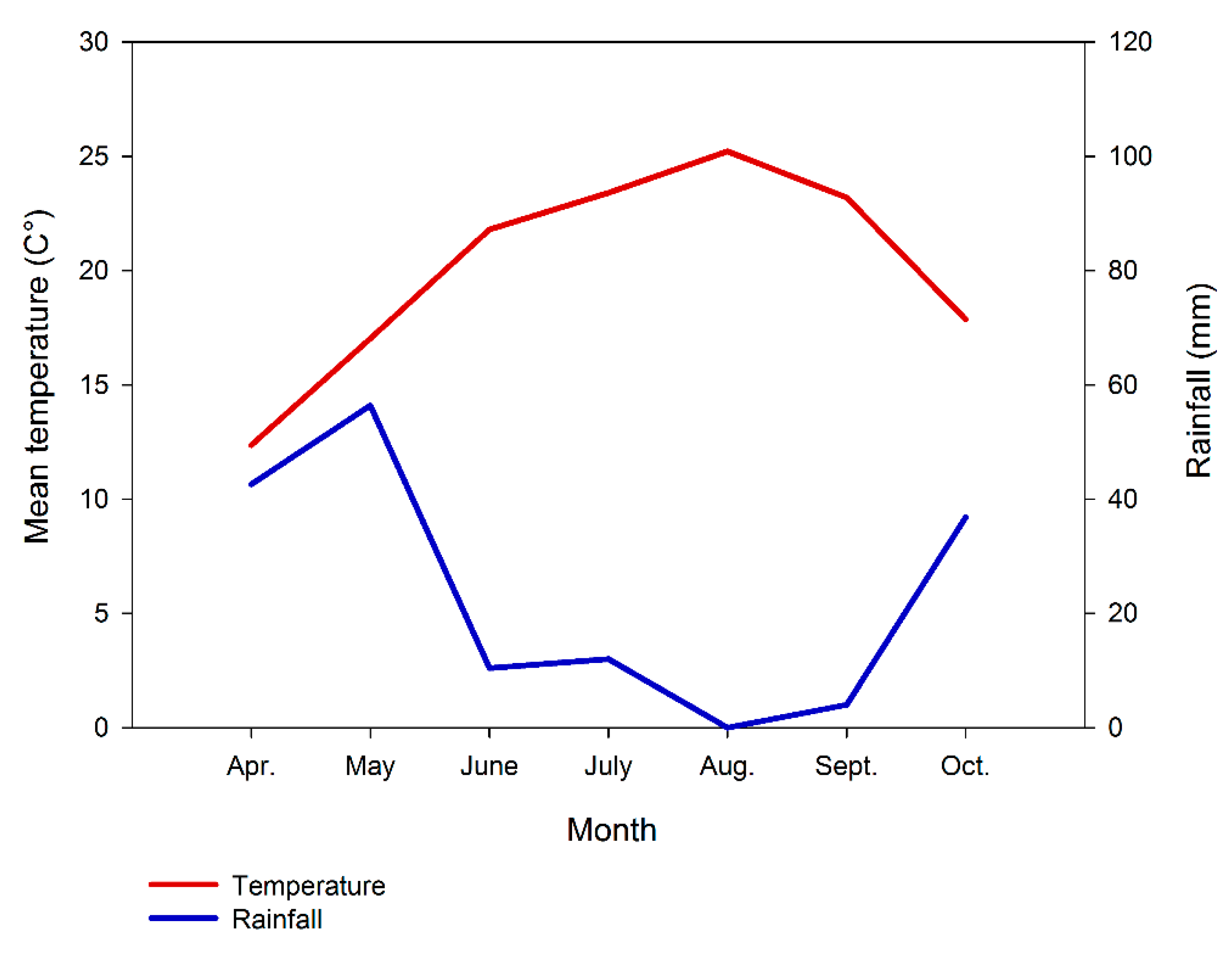
2.1. Study Area

The study area was located at Alberese (municipality of Grosseto) in the Maremma Regional Park (42°37′03″ N, 11°06′47″ E) (Tuscany, Italy). More specifically, it is a roughly 90-year-old, mixed oak forest, with 15–25 m tall trees, composed mainly of Quercus pubescens Willd. (roughly 60%), Quercus cerris L. (roughly 20%), Quercus suber L. (roughly 20%), and with sporadic (less than 1%) Quercus ilex L. individuals. The Mediterranean undergrowth, typical of this environment, is very scarce in the study area, having the forest long been used for pasture. Many of the oaks occurring in the area showed symptoms of decline, such as exudates on the trunk, bark cankers, dead branches and twigs, and beetle exit holes. The study area has a typical Mediterranean climate with hot, dry summers (Figure 1).

2.2. Plant Sample Collection

Tree sampling was performed according to the specific composition of the population. To this purpose, 10 trees were selected: six Q. pubescens, two Q. cerris, and two Q. suber. Samplings were carried out once a month from April 2015 to October 2015 on the same selected oak trees. During each sampling, four current-year twigs (2–4 cm in diameter) were collected from each of these trees at the height of 2.50 m around the crown, one from each cardinal point.

2.3. Fungal Isolation from Plants

Plant samples were taken to the laboratory within 12 h from collection. They were sterilized by immersion in 10% H2O2 for three minutes, then they were washed twice with sterilized distilled water, and dried on sterile tissue paper. A 2-cm-long tissue piece was removed from each twig, excluding the outer bark. From each piece 15 wood samples were removed (roughly 5 mm long and 1 mm thick) and placed, in groups of five, in three Petri dishes (90 mm in diameter) containing Potato Dextrose Agar (PDA) medium with 1 g of agar. Thus, for each sampled twig three Petri dishes were obtained. The plates were incubated at 20 °C in darkness for a week. Fungal colonies growing from the wood samples were isolated and subcultured on Oak Leaf Agar (OLA) [16,17]. Whole colonies were observed with an optical microscope and identified by macro and micro-morphological analyses, using the keys provided by Booth [18], Gams [19], Carmichael [20], Sutton [21], and Von Arx [22]. The isolation frequency of each fungus species per each month was calculated using the following formula:
Fi = (Ni/Nt) × 100
where Fi is the fungus species frequency, Ni is the number of times the species was isolated and Nt is the total number of wood samples placed in PDA.

2.4. Molecular Identification of Isolates

DNA-based identification was necessary for discriminating among related taxa whose micro-morphological characteristics alone proved inconclusive for species determination. Hyphal-tip-derived, fresh cultures were incubated under darkness for one week on MEA medium. Genomic DNA was extracted [23] and the rDNA-ITS region was PCR-amplified using the universal primers ITS1 (5’-TCCGTAGGTGAACCTGCGG) and ITS4 (5-TCCTCCGCTTATTGATATGC) [24]. PCR cycling conditions and subsequent amplicon sequencing were as in Moricca et al. [15]. Sequences were processed in the GenBank database [25], with a BLAST search for the highest identities that was used for the identification of taxa at the species level, considering a minimum threshold of 98% [26].

2.5. Insect Collection

The study area was surveyed every two weeks, from April to October 2015, to monitor xylophagous insect presence. Different sampling methods were employed with the aim to collect living, possibly just-emerged, insects to be utilized for fungal isolation. Sweep nets were used directly on host plants or on flowers, since many adult xylophagous beetles have a flower-visiting behaviour. Some twilight or nocturnal flying beetles were collected by attracting them with light onto a vertical white sheet. Attacked twigs, branches or stems were debarked to find adult insects and, finally, oak branches and twigs were sampled and the eggs reared in the laboratory until the adults emerged. All sampled specimens were taken to the laboratory within six hours from collection in sterile plastic containers.
In addition, to have information about insect frequency in the area during the study period, suspended bait traps were also employed. Five Lindgren funnel traps and five bottle traps were located in the study area at least 100 m from each other. Lindgren funnel traps are generally used for trapping saproxylic beetles, which are attracted by their shape and their black color. In addition, in our study they were lured with ethanol, which is known to attract saproxylic beetles. Bottle traps were laced with a mixture of red wine, banana and sugar, which lures beetles. Both kinds of traps were hung on tree branches at about 3–4 m of height. Every two weeks their bait was changed and they were checked, with insects captured being collected separately for date and trap. All sampled specimens were taken to the laboratory to be identified.

2.6. Fungal Isolation from Insects

Living insects collected (excluding trapped ones) were analyzed in the laboratory. After their identification, the insects were left to walk on PDA Petri dishes, partially following the methodology used by Sabbatini Peverieri [27]. Then, each beetle was surface-washed by vortexing for 1 min in 300 μL of sterile distilled water with 1% of Tween-80 detergent. The resulting solutions were used to inoculate the PDA medium. All the Petri dishes were incubated at 20 °C in darkness for five days. Emerging fungal colonies were subcultured in a pure OLA medium. After one week of growth, the colonies were observed under an optical microscope and identified by analysing their macro- and micro-morphological characters, and by the DNA-based method described above.

2.7. Data Analysis

The significance of the data was determined with ANOVA, after the percent data were arcsin transformed. The differences in fungal isolation frequency were examined for significance using the Duncan’s New Multiple Range Test.

3. Results

In the study area throughout the June–September 2015 period, rainfall was below 11 mm, and in August there was no rain. As regards temperatures, the means reached their maximum value in August, exceeding 25 °C. In addition, from June to September temperatures were always over 20 °C (Figure 1).

3.1. Recovered Fungal Taxa

A number of fungi, belonging to 21 different genera, were isolated from the oak samples collected during the study period (April 2015–October 2015). Of these, 13 were identified to the species level by coupling both conventional and molecular identification. Recovered taxa included common, ubiquitous contaminants, such as: Alternaria sp., Aspergillus sp. and Cladosporium sp. Five harmful species of the Botryosphaeriaceae family, frequently associated with woody hosts [28], heavily colonized the sampled plant material. An array of more or less common microbial inhabitants of Mediterranean oak forests, namely Camarosporium sp., Candida sp., Cephalosporium sp., Fusarium solani (Mart.) Sacc., Gliocladium sp., Gonatorrhodiella sp., Pestalotiopsis versicolor (Speg.) Steyaert, Pollularia sp., Rhizoctonia solani J.G. Kühn, Rhizopus sp., Trichoderma viride Pers., Ulocladium consortiale (Thüm.) E.G. Simmons, Verticillium dahliae Kleb., were also isolated from the oak tree tissues. The charcoal canker agent Biscogniauxia mediterranea (De Not.) Kuntze was too found infecting oaks, though its occurrence in the stand resulted almost negligible (only 1.0% and 3.5% isolation frequency in September and October, respectively).
Following this initial screening of the endophytic assemblage, we narrowed our investigation to botryosphaeriaceous fungi, owing to: (i) the overwhelming prevalence (higher isolation frequencies) of this group of fungi in the stand; (ii) their prominent role in the onset of the decline of tree species worldwide, especially on those trees already weakened by environmental stress factors [28]. The five botryosphaeriaceous species isolated in this study were: Botryosphaeria dothidea (Moug.) Ces. and De Not, Diplodia corticola A.J.L. Phillips, A. Alves and J. Luque, Diplodia seriata De Not., Dothiorella sarmentorum (Fr.) A.J.L. Phillips, A. Alves and J. Luque, and Neofusicoccum parvum (Pennycook and Samuels) Crous, Slippers and A.J.L. Phillips (Table 1). These fungi were found throughout the whole study period; however, their isolation frequency turned out significantly different among months (Table 2), increasing gradually during the growing season (Table 1). Higher values were recorded from June onwards for D. sarmentorum, from July onwards for B. dothidea and from August onwards for D. corticola, D. seriata and N. parvum. More specifically, the highest isolation frequency (%) for each fungal species, with exception of N. parvum, was recorded in October (Table 1).

3.2. Recovered Insect Species

During the study period, we collected various xylophagous and non-xylophagous insect species; however, non-xylophagous taxa, or those which feed only on decaying deadwood, were excluded. Therefore, the xylophagous species belonging to the following taxa were considered: Buprestidae, Cerambycidae, and Curculionidae Scolytinae. Only xylophagous species feeding on oaks were taken into consideration: the Cerambycidae family was the most numerous, with 14 species, compared with Buprestidae (eight species) and Scolytinae (four species) (Table 3).

3.3. Fungus-Insect Associations

Botryosphaeriaceous fungi were isolated from collected beetles. Fungal propagules of B. dothidea, D. seriata and D. corticola were found on five insect species: two Buprestidae, A. thalassophila and C. fasciatus, and three Cerambycidae, C. welensii, C. sartor, and P. kaehleri (Table 3). D. seriata was the predominant fungal species, being associated with three different insect species. With the exception of P. kaehleri, whose proportion of specimens bearing fungal propagules (D. seriata) was 8.69%, all the other insects revealed percentages ranging from 25% (C. sartor/Diplodia seriata), to 37.5% (C. welensii/D. corticola), with A. thalassophila/B. dothidea and C. fasciatus/D. seriata which presented intermediate values (33.33%). C. fasciatus larvae bore galleries in live branches (under 8 cm in diameter) of stressed oaks. Subsequent branch death occurs once larvae reach maturity, because at that time they bore annular galleries under the bark of the branches [29]. Generally, C. welensii colonizes oaks in a state of physiological decline [30]. However, its attacks to young plants in good vegetative condition are becoming more and more frequent [30,31]. A. thalassophila, C. sartor, and P. kaehleri feed on various broadleaved trees; nevertheless, these are not considered oak pests, as they bore galleries on the wood of dead trees or on the deadwood of living trees, particularly dead branches [32,33].
These five insect species were captured from June to August. During June–July, they were the most frequent xylophagous beetles, representing more than 43% of all species collected with suspended bait traps. In June only A. thalassophila and P. kaehleri were captured (Table 4). While the former was collected only in that month, P. kaehleri captures increased during the growing season, reaching its peak in August. Considering the whole study period, P. kaehleri was the most frequent species, representing almost 11% of all collected beetle species. Anyway, buprestid beetles deserve special mentions, because traps used in our study are not particularly attractive to some of these species; therefore, they would be more numerous if other trapping methods were used.
However, not all the species collected were taken into consideration, because some of them were captured only with traps; therefore, they were not used for fungal isolation. This was particularly important for bark beetles, which were collected only with suspended bait traps. The four bark-beetle species trapped are considered key species in oak decline and they were rather frequent in the study area (Table 4), being more than 15% of all the collected specimens. This was particularly true in September, when about 62% of captures consisted of these four bark-beetle species.

4. Discussion

In our study area, xylophagous beetles associated with declining oaks were found. Specifically, the two wood-boring beetles, C. welensii and C. fasciatus, were rather frequent in summer catches. Other wood-boring beetles feeding on oaks were also collected, but, mostly, they are not considered pests, being species that feed only on deadwood. In contrast, the bark beetles caught during the study could play a key role in oak decline, since both their aggressiveness and their oviposition behaviour enhance their function as fungal vectors. In fact, adults carrying fungal propagules bore galleries under the oak bark, directly infecting the colonized trees. In addition, two of the bark beetles caught, S. intricatus and X. dispar, have already been associated with fungal pathogens inhabiting the woody tissues [34]. However, since the bark beetles in our study were captured only with suspended bait traps, they were not used for fungal isolation, consequently their role as fungal vectors in the study area was not investigated.
Botryosphaeriaceae fungi were isolated from five beetle species, C. welensii and C. fasciatus included. Previous studies demonstrated that C. welensii emergence holes are entry ways for fungal pathogens inside trees [35]; however, information about its role as a fungal vector is not available. Here, D. corticola was isolated from specimens of this insect species. D. seriata, instead, was found on C. fasciatus adults. This species has already been associated with Botryosphaeriaceae fungi, Tiberi et al. [36] having isolated D. mutila from adults of this buprestid. These two beetles, differing from bark beetles, lay eggs without penetrating the oak bark; therefore, their importance in the spread of fungal infection may appear less important. Nonetheless, they do bring fungal propagules into direct contact with oak trees, since the egg-laying activity of C. welensii includes probing the bark with its ovipositor, as well as laying eggs into bark crevices and pruning wounds [37]. Thus, propagules are highly likely to come into contact with suitable entry sites. As regards C. fasciatus, females usually lay eggs near buds of young branches, although, they also oviposit around wounds [29]. In that case, they would play the same important role as C. welensii. In addition, these two wood-boring beetles are attracted by stressed trees, particularly drought-stressed ones, which are their preferred hosts, carrying with them propagules of Botryosphaeriaceae fungi, which likewise prefer physiologically-impaired trees. It appears evident that the development of Botryosphaeriaceae-related diseases is increasingly more likely, and potentially more severe when beetles and fungi co-occur on the same host [38].
An extremely hot and dry summer occurred during our study period. August had no rains and temperatures reached their highest values, and this could have affected the biology of both fungi and beetles. D. corticola and D. seriata were isolated more frequently from August to October, just after the driest period. It is known that members of the Botryosphaeriaceae family, especially some Diplodia species [39], are thermophilic or thermotolerant; thus, these fungi become more aggressive when temperatures rise, a fact that coincides also with a greater drought stress to trees [15]. High temperatures also favour C. welensii and C. fasciatus, as they are thermophilic species too [40].
According to previous studies focusing on other species, co-occurrence of insects and fungi shows a strong seasonality [41]. Fungal isolation frequency is known to be season-dependent [42]. This because the physiological status of the tissues influences fungal growth and sporulation, being linked to availability of carbon for the fungi. It is, however, also true that prolonged summer droughts lead to plant carbon starvation and reduced ability to counteract the attack by biotic stressors like insects and fungi [6,43]. Consequently, drought-stressed trees may become more suitable to these biotic agents, increasing their population abundance [44,45]. Accordingly, C. welensii and C. fasciatus emerged in the study area from July to August, exactly during the driest and hottest period of the year and when propagule pressure of the fungi in the stand was substantially high. The final outcome is insect and pathogen outbreaks often causing extended tree mortality [46].
The botryosphaeriaceous fungi isolated in this study are well known endophytes and latent pathogens, with a cosmopolitan host range and wide geographical distribution [28,47]. These microorganisms are able to aggressively attack the host plants when these undergo physiological stress and to induce a variety of disease symptoms [28]. Although some of these species (e.g., D. corticola and N. parvum) have in recent years come strongly to the fore in several regions of the world [48,49,50], their infection biology, in part because of their sometimes inconspicuous occurrence, has long been neglected [51,52]. As a matter of fact, the life history strategies of many of these taxa remain, even today, partially unexplored. Few studies have investigated, for example, to what extent these opportunistic fungi are transmitted in the woods by insect vectors [36,53]. This paper aims to partly fill this gap.

5. Conclusions

Several lines of reasoning suggest that the investigated xylophagous insects may well have a role in the dispersal of fungal species: (i) the thermotolerance or thermophily of members of both groups of parasites. These traits increase the chances of transportation of fungi by insects, being both insect population density and propagule pressure of fungi higher during hotter years; (ii) their synchronicity in their occurrence and activity, coinciding with the drought of summer months; and last but not least, (iii) the isolation of fungal propagules from the body of some of the insect species. From an epidemiological perspective, it is also worth noting that beetles, besides increasing fungal dispersal and propagule pressure, bring fungi to stressed oaks precisely during the time when these are most susceptible.
However, while it is evident that insects are effectively carrying fungal propagules and that environmental stress is the first driver of tree weakening, the causal interconnections between environmental variables and the fungus-insect-tree tripartite interactions are difficult to prove. That’s because many factors may contribute to generate a more complex framework, which escapes analyses of temporal and spatial co-occurrence. Tree decline, for instance, is usually a long-term process, during which fungi may take advantage of impaired tree defenses and at the same time affect tree’s response to environmental stress. Furthermore, fungi may be spreading from last year’s growth inside the tree twigs, blurring the temporal aspects. To clarify this aspect, it would be interesting to repeat this research to ascertain whether the fungus-insect associations found here are stable and repeated over the years. If these harmful interactions were confirmed, they would provide a more plausible explanation for the extensive mortality of some Mediterranean forest stands whose etiology seemed uncertain. In fact, a single factor of damage (fungi or insects) alone did not explain in many cases the extent and gravity of the observed decline/dieback phenomena. In this connection, it would also deserve investigating whether other microorganisms (bacterial agents), responsible for the more recent and emerging AOD (Acute Oak Decline) syndrome [54], may also be involved.

Author Contributions

A.R., R.T., S.M. and T.P. designed the experiments and analysed the data. A.P., F.C., B.G. and M.B. contributed with field experiments and data collection. S.M. and T.P. wrote the manuscript.

Conflicts of Interest

The authors declare no conflict of interest.

References

  1. Brasier, C.M.; Scott, J.K. European oak declines and global warming: A theoretical assessment with special reference to the activity of Phytophthora cinnamomi. EPPO Bull. 1994, 24, 221–232. [Google Scholar] [CrossRef]
  2. Ragazzi, A.; Vagniluca, S.; Moricca, S. European expansion of oak decline: Involved microorganisms and methodological approaches. Phytopathol. Mediterr. 1995, 34, 207–226. [Google Scholar]
  3. Thomas, F.M.; Blanck, R.; Hartmann, G. Abiotic and biotic factors and their interactions as causes of oak decline in Central Europe. For. Pathol. 2002, 32, 277–307. [Google Scholar] [CrossRef]
  4. Sturrock, R.N.; Frankel, S.J.; Brown, A.V.; Hennon, P.E.; Kliejunas, J.T.; Lewise, K.J.; Worrallf, J.J.; Woods, A.J. Climate change and forest diseases. Plant Pathol. 2011, 60, 49–133. [Google Scholar] [CrossRef]
  5. Diffenbaugh, N.S.; Pal, J.S.; Giorgi, F.; Gao, X. Heat stress intensification in the Mediterranean climate change hotspot. Geophys. Res. Lett. 2007, 34, 1–6. [Google Scholar] [CrossRef]
  6. Allen, C.D.; Macalady, A.K.; Chenchouni, H.; Bachelet, D.; McDowell, N.; Vennetier, M.; Kitzberger, T.; Rigling, A.; Breshears, D.D.; Hogg, E.H.T.; et al. A global overview of drought and heat-induced tree mortality reveals emerging climate change risks for forests. For. Ecol. Manag. 2010, 259, 660–684. [Google Scholar] [CrossRef]
  7. Moricca, S.; Linaldeddu, B.T.; Ginetti, B.; Scanu, B.; Franceschini, A.; Ragazzi, A. Endemic and emerging pathogens threatening cork oak trees: Management options for conserving a unique forest ecosystem. Plant Dis. 2016, 100, 2184–2193. [Google Scholar] [CrossRef]
  8. Tiberi, R.; Branco, M.; Bracalini, M.; Croci, F.; Panzavolta, T. Cork oak pests: A review of insect damage and management. Ann. For. Sci. 2016, 73, 219–232. [Google Scholar] [CrossRef]
  9. Manion, P.D. Tree Disease Concepts, 2nd ed.; Prentice-Hall: Englewood Cliffs, NJ, USA, 1991. [Google Scholar]
  10. Halmschlager, E. Endophytic fungi and oak decline. In Recent Advances in Studies on Oak Decline, Proceedings of An International Congress, Selva di Fasano, Brindisi, Italy, 13–18 September 1992; Luisi, N., Lerario, P., Vannini, A., Eds.; Dipartimento di Patologia Vegetale, Università Degli Studi: Bari, Italy, 1993; pp. 77–83. [Google Scholar]
  11. Kowalski, T.; Kehr, R.D. Endophytic fungi colonization of branch bases in several forest tree species. Sydowia 1992, 44, 137–168. [Google Scholar]
  12. Halmschlager, E.; Butin, H.; Donaubauer, E. Endophytische pilze in blättern und zweigen von Quercus petraea. Eur. J. For. Pathol. 1993, 23, 51–63. [Google Scholar] [CrossRef]
  13. Ragazzi, A.; Moricca, S.; Capretti, P.; Dellavalle, I.; Mancini, F.; Turco, E. Endophytic fungi in Quercus cerris: Isolation frequency in relation to phenological phase, tree health and the organ affected. Phytopathol. Mediterr. 2001, 40, 165–171. [Google Scholar] [CrossRef]
  14. Ragazzi, A.; Moricca, S.; Capretti, P.; Dellavalle, I.; Turco, E. Differences in composition of endophytic mycobiota in twigs and leaves of healthy and declining Quercus species in Italy. For. Pathol. 2003, 33, 31–38. [Google Scholar] [CrossRef]
  15. Moricca, S.; Ginetti, B.; Ragazzi, A. Species- and organ-specificity in endophytes colonizing healthy and declining Mediterranean oaks. Phytopathol. Mediterr. 2012, 51, 587–598. [Google Scholar] [CrossRef]
  16. Cohen, S.D. Technique for large scale isolation of Discula umbrinella and other foliar endophytic fungi from Quercus species. Mycologia 1999, 91, 917–922. [Google Scholar] [CrossRef]
  17. Moricca, S.; Ragazzi, A. The holomorph Apiognomonia quercina/Discula quercina as a pathogen/endophyte in oak. In Endophytes of Forest Trees; Pirttilä, A.M., Frank, A.C., Eds.; Springer: Dordrecht, The Netherlands, 2011; pp. 47–66. [Google Scholar]
  18. Booth, C. The Genus Fusarium; Commonwealth Mycological Institute: Kew, UK, 1971. [Google Scholar]
  19. Gams, W. Cephalosporium-Artige Schimmelpilze (Hyphomycetes); Gustav Fischer Verlag: Stuttgart, Germany, 1971. [Google Scholar]
  20. Charmichael, J.W.; Kendrich, W.B.; Conner, I.L.; Sigler, L. Genera of Hyphomycetes; The University of Alberta Press: Edmonton, AB, Canada, 1980. [Google Scholar]
  21. Sutton, B.C. The Coelomycetes Fungi Imperfecti with Pycnidia, Acervuli and Stromata; Commonwealth Mycological Institute: Kew, UK, 1980. [Google Scholar]
  22. Von Arx, J.A. Plant Pathogenic Fungi; Cramer, J., Ed.; Beihefte zur Nova Hedwigia: Berlin, Germany, 1987. [Google Scholar]
  23. Moricca, S.; Raddi, P.; Borja, I.; Vendramin, G.G. Differentiation of Seiridium species associated with virulent cankers on cypress in the Mediterranean region by PCR-SSCP. Plant Pathol. 2000, 49, 774–781. [Google Scholar] [CrossRef]
  24. White, T.J.; Bruns, T.; Lee, S.; Taylor, J. Amplified and direct sequencing of fungal ribosomal RNA genes for phylogenies. In PCR Protocols: A Guide to Methods and Applications; Innis, M.A., Gelfand, D.H., Sninsky, J.J., White, T.J., Eds.; Academic: San Diego, CA, USA, 1990; pp. 315–322. [Google Scholar]
  25. Benson, D.A.; Cavanaugh, M.; Clark, K.; Karsch-Mizrachi, I.; Lipman, D.J.; Ostell, J.; Sayers, E.W. GenBank. Nucleic Acids Res. 2013, 41, D36–D42. [Google Scholar] [CrossRef] [PubMed]
  26. Sánchez Márquez, S.; Bills, G.F.; Zabalgogeazcoa, I. Diversity and structure of the fungal endophytic assemblages from two sympatric coastal grasses. Fungal Divers. 2008, 33, 87–100. [Google Scholar]
  27. Sabbatini Peverieri, G.; Villari, C.; Tiberi, R.; Capretti, P. Occurrence of fungal root rot diseases on pine trees in Tuscany and its relationship with Tomicus destruens. J. Plant Pathol. 2005, 87, 304. [Google Scholar]
  28. Slippers, B.; Wingfield, M.J. Botryosphaeriaceae as endophytes and latent pathogens of woody plants: Diversity, ecology and impact. Fungal Biol. Rev. 2007, 21, 90–106. [Google Scholar] [CrossRef]
  29. Jurc, M.; Bojović, S.; Komjanc, B.; Krč, J. Xylophagous entomofauna in branches of oaks (Quercus spp.) and its significance for oak health in the Karst region of Slovenia. Biologia 2009, 64, 130–138. [Google Scholar] [CrossRef]
  30. Torres-Vila, L.M.; Sanchez-González, Á.; Ponce-Escudero, F.; Martín-Vertedor, D.; Ferrero-García, J.J. Assessing mass trapping efficiency and population density of Cerambyx welensii Küster by mark-recapture in dehesa open woodlands. Eur. J. For. Res. 2012, 131, 1103–1116. [Google Scholar] [CrossRef]
  31. Sallé, A.; Nageleisen, L.; Lieutier, F. Bark and wood boring insects involved in oak declines in Europe: Current knowledge and future prospects in a context of climate change. For. Ecol. Manag. 2014, 328, 79–93. [Google Scholar] [CrossRef]
  32. Curletti, G. I Buprestidi d’Italia; Museo Civico di Scienze Naturali: Brescia, Italy, 1994. [Google Scholar]
  33. Contarini, E. Elenco faunistico commentato (check-list) dei Cerambicidi (Coleoptera Xylophytophaga) del Parco Naturale della Vena del Gesso romagnola. Quad. Stud. Nat. Romagna 2014, 40, 39–65. [Google Scholar]
  34. Tiberi, R.; Panzavolta, T.; Bracalini, M.; Ragazzi, A.; Ginetti, B.; Moricca, S. Interactions between insects and fungal pathogens of forest and ornamental trees. Ital. J. Mycol. 2016, 45, 54–65. [Google Scholar] [CrossRef]
  35. Martín, J.; Cabezas, J.; Buyolo, T.; Patón, D. The relationship between Cerambyx spp. damage and subsequent Biscogniauxia mediterranum infection on Quercus suber forests. For. Ecol. Manag. 2005, 216, 166–174. [Google Scholar] [CrossRef]
  36. Tiberi, R.; Ragazzi, A.; Marianelli, L.; Peverieri Sabbatini, P.; Roversi, P.F. Insects and fungi involved in oak decline in Italy. In IOBC/WPRS Bulletin, Working Group “Integrated Protection in Oak Forests”, Proceedings of the Meeting at Oeiras, Lisbonne, Portugal, 1–4 October 2001; Villemant, C., Sousa, E., Eds.; IOBC: Zürich, Switzerland, 2002; Volume 25, pp. 67–74. [Google Scholar]
  37. Torres-Vila, L.M.; Mendiola-Diaz, F.J.; Conejo-Rodríguez, Y.; Sánchez-González, Á. Reproductive traits and number of matings in males and females of Cerambyx welensii (Coleoptera: Cerambycidae) an emergent pest of oaks. Butt. Entomol. Res. 2016, 106, 292–303. [Google Scholar] [CrossRef] [PubMed]
  38. Belhoucine, L.; Bouhraoua, R.T.; Meijer, M.; Houbraken, J.; Harrak, M.J.; Samson, R.A.; Equihua-Martinez, A.; Pujade-Villar, J. Mycobiota associated with Platypus cylindrus (Coleoptera: Curculionidae, Platypodidae) in cork oak stands of North West Algeria, Africa. Afr. J. Microbiol. Res. 2011, 5, 4411–4423. [Google Scholar] [CrossRef]
  39. Schumacher, J.; Heydeck, P.; Dahms, C. Increasing endangerment of forests by pathogenic thermophilic fungi-demonstrated by the example of the microfungus Diplodia pinea (DESM.) Kickx on Pinus. In Julius-Kühn-Archiv, Proceedings of the 57th German Plant Protection Conference, Berlin, Germany, 6–9 September 2010; Julius Kühn Institut, Bundesforschungsinstitut für Kulturpflanzen: Quedlinburg, Germany, 2010; Volume 428, p. 372. [Google Scholar]
  40. Cárdenas, A.M.; Gallardo, P. The effect of temperature on the preimaginal development of the jewel beetle, Coraebus florentinus (Coleoptera: Buprestidae). Eur. J. Entomol. 2012, 109, 21–28. [Google Scholar] [CrossRef]
  41. Brown, N.; Jeger, M.; Kirk, S.; Williams, D.; Xu, X.; Pautasso, M.; Denman, S. Acute Oak Decline and Agrilus biguttatus: The co-occurrence of stem bleeding and D-shaped emergence holes in Great Britain. Forests 2017, 8, 87. [Google Scholar] [CrossRef]
  42. Ragazzi, A.; Moricca, S.; Capretti, P.; Dellavalle, I. Endophytic presence of Discula quercina on declining Quercus cerris. J. Phytopathol. 1999, 147, 437–440. [Google Scholar] [CrossRef]
  43. Ragazzi, A.; Moricca, S.; Dellavalle, I. Water Stress and the Development of Cankers by Diplodia mutila on Quercus robur. J. Phytopathol. 1999, 147, 425–428. [Google Scholar] [CrossRef]
  44. Desprez-Loustau, M.L.; Marcais, B.; Nageleisen, L.M.; Piou, D.; Vannini, A. Interactive effects of drought and pathogens in forest trees. Ann. For. Sci. 2006, 63, 597–612. [Google Scholar] [CrossRef]
  45. Haavik, L.J.; Billings, S.A.; Guldin, J.M.; Stephen, F.M. Emergent insects, pathogens and drought shape changing patterns in oak decline in North America and Europe. For. Ecol. Manag. 2015, 354, 190–205. [Google Scholar] [CrossRef]
  46. Jactel, H.; Petit, J.; Desprez-Loustau, M.L.; Delzon, S.; Piou, D.; Battisti, A.; Koricheva, J. Drought effects on damage by forest insects and pathogens: A meta-analysis. Glob. Chang. Biol. 2012, 18, 267–276. [Google Scholar] [CrossRef]
  47. Sieber, T.N. Endophytic fungi in forest trees: Are they mutualists? Fungal Biol. Rev. 2007, 21, 75–89. [Google Scholar] [CrossRef]
  48. Sakalidis, M.L.; Slippers, B.; Wingfield, B.D.; Hardy, G.E.St.J.; Burgess, T.I. The challenge of understanding the origin, pathways and extent of fungal invasions: Global populations of the Neofusicoccum parvum-N. ribis species complex. Divers. Distrib. 2013, 19, 873–883. [Google Scholar] [CrossRef]
  49. Dreaden, T.J.; Shin, K.; Smith, J.A. First report of Diplodia corticola causing branch cankers on live oak (Quercus virginiana) in Florida. Plant Dis. 2011, 95, 1027. [Google Scholar] [CrossRef]
  50. Lynch, S.C.; Eskalen, A.; Zambino, P.; Scott, T. First report of bot canker caused by Diplodia corticola on coast live oak (Quercus agrifolia) in California. Plant Dis. 2010, 94, 1510. [Google Scholar] [CrossRef]
  51. Alves, A.; Correia, A.; Luque, J.; Phillips, A.J.L. Botryosphaeria corticola sp. nov. on Quercus species, with notes and description of Botryosphaeria stevensii and its anamorph Diplodia mutila. Mycologia 2004, 96, 598–613. [Google Scholar] [CrossRef] [PubMed]
  52. Alves, A.; Linaldeddu, B.T.; Deidda, A.; Scanu, B.; Phillips, A.J.L. The complex of Diplodia species associated with Fraxinus and some other woody hosts in Italy and Portugal. Fungal Divers. 2014, 67, 143–156. [Google Scholar] [CrossRef]
  53. Hanso, M.; Drenkhan, R. Diplodia pinea is a new pathogen on Austrian pine (Pinus nigra) in Estonia. Plant Pathol. 2009, 58, 797. [Google Scholar] [CrossRef]
  54. Denman, S.; Plummer, S.; Kirk, S.; Peace, A.; McDonald, J.E. Isolation studies reveal a shift in the cultivable microbiome of oak affected with Acute Oak Decline. Syst. Appl. Microbiol. 2016, 39, 484–490. [Google Scholar] [CrossRef] [PubMed]
Figure 1. Meteorological data from the Alberese (Grosseto-Italy) meteorological weather station in 2015. Data supplied by the Hydrological Regional Sector (SIR) of Tuscany.
Forests 08 00228 g001
Table 1. Percentage isolation frequencies of fungi of the Botryosphaeriaceae family recovered from oak samples in the studied woodland in Marina di Alberese (Grosseto-Italy). Values in columns followed by the same letter do not differ significantly (p ≤ 0.01, Duncan’s New Multiple Range Test); isolation frequencies below the 1% threshold are not reported. Standard deviation is in parentheses.
Month of CollectionFrequency of Isolation (%) (600 Monthly Samplings)
Fungus Species
Botryosphaeria dothideaDiplodia corticolaDiplodia seriataDothiorella sarmentorumNeofusicoccum parvum
April1.75 (0.65)3.37 a (3.24)3.25 a (1.35)1.37 a (0.20)1.25 a (0)
May3.75 b (2.25)4.50 a (3.59)3.50 a (1.80)1.89 a (0.33)1.50 a (1.10)
June6.90 c (1.12)4.75 a (1.98)3.78 a (1.00)3.12 b (0.78)1.87 a (0.90)
July8.78 d (0.72)4.90 a (3.59)4.25 a (2.10)3.12 b (1.23)2.00 a (0.20)
August9.00 d (2.20)13.89 b (0.65)6.75 b (3.08)3.25 b (0.50)3.00 b (1.25)
September10.25 d (0.12)15.25 bc (5.82)7.65 b (1.80)3.50 b (1.30)3.25 b (2.00)
October12.50 e (0.10)16.00 c (1.67)11.97 c (0.89)8.97 c (5.95)3.50 b (1.35)
Table 2. Analysis of variance (1-way ANOVA) on isolation frequency (percent data arcsin transformed).
VariabilitydfDeviationVarianceF
Total131575.21
Between pathogens4915.07228.7657.04 **
Between months6648.11108.0126.93 **
Error312.034.01
** Significant at p < 0.01.
Table 3. Xylophagous Coleoptera feeding on oaks collected in the studied woodland in Marina di Alberese (Grosseto-Italy) and their association with botryosphaeriaceous fungi.
Coleoptera Species CollectedBotryosphaeriaceae Species IsolatedNumber of Insects TestedIsolation Frequency (%)
Buprestidae
Acmaeoderella adspersula (Illiger, 1803) 30
Acmaeoderella flavofasciata (Piller and Mitterpacher, 1783) 80
Anthaxia millefolii polychloros Abeille de Perrin, 1894 150
Anthaxia scutellaris Gene, 1839 150
Anthaxia thalassophila Abeille de Perrin, 1900Botryosphaeria dothidea633.33
Anthaxia umbellatarum (Fabricius, 1787) 110
Coraebus fasciatus (Villers, 1789) Diplodia seriata1233.33
Latipalpis plana (Olivier, 1790) 30
Cerambycidae
Callimus abdominalis (Olivier, 1795) * 0NA
Cerambyx welensii (Küster, 1845)Diplodia corticola837.50
Chlorophorus sartor (Müller, 1766)Diplodia seriata425.00
Chlorophorus glabromaculatus (Goeze, 1777) * 0NA
Deilus fugax (Olivier, 1790) 10
Deroplia genei (Aragona, 1830) * 0NA
Niphona picticornis Mulsant, 1839 * 0NA
Phymatodes testaceus (Linnaeus, 1758) 130
Pseudosphegesthes cinerea (Laporte and Gory, 1836) * 0NA
Purpuricenus kaehleri (Linnaeus, 1758)Diplodia seriata238.69
Stenopterus rufus (Linnaeus, 1767) 90
Stictoleptura cordigera (Fuessly, 1775) 20
Stictoleptura rufa (Brulle, 1832) * 0NA
Trichoferus holosericeus (Rossi, 1790) 120
Curculionidae: Scolytinae
Xyleborus dispar (Fabricius, 1792) * 0NA
Scolytus intricatus (Ratzeburg, 1837) * 0NA
Xyleborinus saxesenii Ratzeburg, 1837 * 0NA
Xyleborus monographus (Fabricius, 1792) * 0NA
* Insect species which could not be analyzed for fungus association. NA = not assigned.
Table 4. Catching frequencies (%) of xylophagous beetles resulted associated with Botryosphaeriaceae species in the oak forest in Marina di Alberese (Grosseto-Italy). Percentages were calculated on the total number of xylophagous beetles collected with suspended bait traps.
Insect SpeciesMonth of Collection
AprilMayJuneJulyAugustSeptemberOctoberTotal
Xylophagous Beetles Associated with Fungi
Anthaxia thalassophila0013.0400004.83
Coraebus fasciatus00018.990004.53
Cerambyx welensii0006.3314.71004.53
Chlorophorus sartor0002.535.88001.81
Purpuricenus kaehleri005.8015.1923.530010.88
Total frequency0017.3943.0444.120026.59
Bark-beetles00027.8527.9462.50015.41
Total number of xylophagous beetles4261387968160331

