Article

Early Detection and Identification of the Main Fungal Pathogens for Resistance Evaluation of New Genotypes of Forest Trees

by
Konstantin A. Shestibratov
1,
Oleg Yu. Baranov
2,
Natalya M. Subbotina
1,
Vadim G. Lebedev
1,
Stanislav V. Panteleev
2,
Konstantin V. Krutovsky
3,4,5,6,* and
Vladimir E. Padutov
2
1
Forest Biotechnology Group, Branch of Shemyakin and Ovchinnikov Institute of Bioorganic Chemistry, Russian Academy of Sciences, 6 Prospect Nauki, Pushchino, Moscow 142290, Russia
2
Laboratory for Genetics and Biotechnology, Forest Research Institute, National Academy of Sciences of Belarus, 71 Proletarskaya Str., Gomel 246001, Belarus
3
Department of Forest Genetics and Forest Tree Breeding, George-August University of Göttingen, Büsgenweg 2, 37077 Göttingen, Germany
4
Laboratory of Population Genetics, Vavilov Institute of General Genetics, Russian Academy of Sciences, Gubkina Str. 3, Moscow 119991, Russia
5
Laboratory of Forest Genomics, Genome Research and Education Center, Institute of Fundamental Biology and Biotechnology, Siberian Federal University, Akademgorodok, 50a/2, Krasnoyarsk 660036, Russia
6
Department of Ecosystem Sciences and Management, Texas A&M University, College Station, TX 77843-2138, USA
*
Author to whom correspondence should be addressed.
Forests 2018, 9(12), 732; https://doi.org/10.3390/f9120732
Submission received: 31 October 2018 / Revised: 19 November 2018 / Accepted: 19 November 2018 / Published: 23 November 2018

Abstract

:
The growing importance of forest plantations increases the demand for phytopathogen resistant forest trees. This study describes an effective method for early detection and identification of the main fungal phytopathogens in planting material of silver birch (Betula pendula) and downy birch (B. pubescens), based on the estimation of the size of the internal transcribed spacers (ITS1 and ITS2) in the 18S-5.8S-28S rDNA gene cluster, which are species-specific for most micromycetes. The electrophoretic assay of the ITS1 and ITS2 loci has allowed us to identify predominant phytopathogenic fungal species in downy and silver birch in planta. This new molecular genetic method can be used to screen birch and other forest trees for different fungal pathogens to evaluate disease resistance. This information can be useful in breeding new genotypes of forest trees, including transgenic clones with modified wood composition.

1. Introduction

Fungal diseases are a serious problem in forestry, and can be the cause of epiphytotics leading to the death of forests: as examples, American chestnut, butternut, and American elm [1]. This is particularly relevant for forest plantations, which have less diversity and stability than natural forests and, therefore, are more susceptible to diseases. Modeling has indicated that short rotation in forest plantations accelerates both the virulence evolution in root-rot pathogenic fungi and the development of epiphytotics [2]. Moreover, global climate change may also promote distribution of forest pathogens. It has been shown that the expected changes in temperature and precipitation will favor the spread of beech bark disease in the forests of North America [3]. Thus, special attention is needed to assess disease resistance of new genotypes of forest trees, including transgenic lines. Lignin manipulation is one of the main objectives in forest biotechnology. Its content in wood directly correlates with the efficiency of the pulping process, and affects waste management. However, lignin plays an important role in plant defense against pests and other phytopathogens [4]. Thus, a change in the composition and/or content of lignin can reduce plant resistance to phytopathogens. Generally, in addition to the biotechnologically generated desirable traits (intended effects), the appearance of unintended effects that can negatively affect agronomic performance is possible [5]. The detection of such effects can be done by comparing transgenic genotypes with related conventional counterparts [6]. Testing whether transgenic genotypes that have lower lignin content are less resistant to phytopathogens would mean detection of these phytopathogens in transformed and untransformed clones.
Birch species (Betula L.) are among the most widespread forest trees, and have great importance in forestry, forest formation, and soil improvement. They also have an important ecological role as pioneer species after clear-cuts and forest fires [7]. They are a fast growing species that provide high quality timber for industrial purposes. Downy birch (B. pubescens Ehrh.) and silver birch (B. pendula Roth) are commercially important forest species in Europe [8]. Their natural area includes North Africa, Western Asia, and Central Asia, as well as the entire Europe and Northern Eurasia (excluding the Iberian Peninsula). In Northern Europe, these species are the most important deciduous trees in plantation forestry [9]. Intensively managed forest plantations are characterized by a limited number of clones of the same species, which increases the risk of pathogen attacks. In this regard, the methods of diagnosing and identification of various phytopathogens based on DNA analysis have a great potential value [10]. Molecular diagnostic methods have been developed for detection of various pathogens in oak [11], plane trees [12], pines [13], and other forest species, but not in birch. Moreover, these methods were designed to identify pathogens of only one particular species or genus in a single analysis.
The traditional method of phytopathogen detection is based on visual inspection of disease symptoms and is often unreliable, performed in the late stages of the disease, and requires qualified personnel [14], especially for tree species. Molecular diagnostics, based on the detection of pathogen DNA using PCR methods, allows assessing the resistance of a new genotype quickly, with high accuracy, and at the early stages of disease development [15]. For several reasons, the ribosomal DNA (rDNA) loci encoding 5S, 5.8S, 18S, and 28S ribosomal RNAs (rRNAs) are widely used marker regions for the detection and identification of micromycetes [16]. The rDNA loci encoding 5.8S, 18S, and 28S rRNAs form a cluster of the 18S–5.8S–28S loci with two internal transcribed spacers (ITS1 and ITS2) between the 18S–5.8S, and 5.8S–28S loci, respectively. There are at least 50 copies of this cluster per genome, and this multiplicity enhances the sensitivity of the PCR analysis (i.e., the probability of pathogen detection at its low concentration in plant tissue). The ITS loci are relatively conserved within a species [16], but highly divergent between species, which facilitates taxonomic identification of the pathogen causing infection. These loci are well studied and their nucleotide sequences are well-represented in sequence databases, such as NCBI GenBank (https://www.ncbi.nlm.nih.gov), DNA Data Bank of Japan (DDBJ, NIG) (http://www.ddbj.nig.ac.jp), European Molecular Biology Laboratory (EMBL, EBI) (http://www.embl.de), Barcode of Life Data System (BOLD) (http://www.boldsystems.org), and DOE JGI Fungi Portal (https://genome.jgi.doe.gov/programs/fungi/index.jsf), which are very important for pathogen identification. Large scale molecular genetic studies of different fungi have revealed conserved rDNA regions and allowed the development of sets of universal primers for PCR amplification of ribosomal genes and intergenic spacers across different species [17]. DNA-based methods have been proposed for the identification of fungal species based on electrophoretic assay of the PCR amplified marker regions without preliminary sequencing of samples, including the identification of micromycetes [18]. The ITS region has been suggested as a universal marker for DNA barcoding of fungi [18]. However, most of the proposed protocols are not universally applicable, and have limitations in different phytopathological assays. For example, the use of an intergenic spacer (IGS) located between tandemly repeated copies of the rDNA gene clusters as a genetic marker may be limited for studying pathogenic basidiomycetes, because of the high sequence variation within species in this region, and the challenges of amplifying DNA regions larger than 3 kilobase pairs (Kbp), particularly from decayed tissues [19]. The application of single-strand conformation polymorphism (SSCP) analysis does not directly generate nucleotide sequence data, which reduces their compatibility with nucleotide sequence databases [20]. Moreover, the likelihood of methodological mistakes and artifacts becomes greater when complex procedures are required for sample preparation and electrophoretic mobility analysis [21]. Finally, the sequencing of DNA markers involves a relatively high analytical cost and special laboratory equipment.
In the present study, we have developed the ITS1 and ITS2 genetic markers, which can be used without sequencing. Their species-specific variation in size makes them highly informative and sufficient for identification of the main pathogenic species of birches, using gel electrophoresis following PCR amplification. It is very important that there is almost no intraspecific size variation of the ITS markers in micromycetes that could be similar to the interspecific size variation, which almost excludes false positive results.
Amplicon size analysis was carried out by denaturing polyacrylamide gel electrophoresis, which allowed species identification using both the application of standard DNA samples, and information about the amplicon sizes from nucleotide sequence databases.

2. Materials and Methods

2.1. Plant Material

Samples from silver and downy birch plantings with different infection symptoms were collected during 2017, in the fields and greenhouses at the Korenevskaya Experimental Forest Enterprise of the Forest Research Institute of the National Academy of Sciences of Belarus (Belarus), in the forest enterprises of Gomel Region (Belarus), and in the Moscow Region (Russia).

2.2. Phytopathological Analysis

During the phytopathological assay of the birch planting material, the main diseases that caused the highest losses of yield during commercial cultivation were determined. The determination of disease type was carried out, based on the symptoms defined by the generally accepted system of phytopathological assays (http://www.forestpathology.org/index.html).

2.3. Species-Specific Molecular Genetic Identification of Phytopathogens

Specific phytopathogenic micromycetes were identified using molecular genetic methods for fungal identification in planta [22]. For the pathogen diagnosis, samples of plant tissue were collected at the initial infection stage, which simplified the diagnosis by minimizing the content of saprotrophic microflora. All of the plant samples (e.g., leaf disc cuttings, and stem or root fragments) were fixed in sterile polypropylene tubes with 70% ethanol and stored at −18 °C. During sample preparation, the analyzed fragments of plant material were removed from the tubes, washed thoroughly with running water, and pieces that exhibited a particular infection type were taken for further analysis. They were washed thoroughly with distilled water and cut with a razor blade into 3–8 mm pieces under sterile conditions, so that a junction between healthy and infected tissues was located in the middle of each piece. The samples were then placed in Eppendorf centrifuge tubes for subsequent DNA isolation.

2.4. DNA Isolation and PCR Amplification

The total DNA was extracted from the samples according to a modified cetyltrimethyl ammonium bromide (CTAB) protocol [23]. PCR was carried out using 2× DreamTaq™ Green PCR Master Mix (Thermo Fisher Scientific, Waltham, MA, USA) with combinations of ITS1–ITS2 (amplifying partial 18S rRNA, ITS1, and partial 5.8S rRNA loci) or ITS3–ITS4 (amplifying partial 5.8S rRNA, ITS2, and partial 26S rRNA loci) PCR primer pairs for the amplification of the fungal rDNA species-specific genetic markers [16]. The forward primers were labeled with a fluorescent dye. The primer sequences are shown in Table 1. The amplification reaction mixture (25 μL) contained 1 μL (0.5–50 ng) of DNA template, 12.5 μL of 2× DreamTaq™ Green PCR Master Mix, 1 μL of 5 μM Dye-labelled (e.g., with FAM-dye) forward primer, 1 μL of 5 μM reverse primer, and 9.5 μL nuclease-free water. The DNA reaction mixtures were amplified in a PCR thermocycler (TProfessional Basic Thermocycle) (Biometra GmbH, Göttingen, Germany) by algorithm: 1 cycle at 95 °C for 3 min, followed by 35 cycles of 20 s at 95 °C, 20 s at 60 °C, and 20 s at 72 °C. The reaction was ended with final extension at 72 °C for 4 min before holding the sample at 4 °C for analysis.

2.5. Gel Electrophoresis

For high resolution gel electrophoresis and amplicon fragment analysis the PCR products were diluted to 1 ng/μL in deionized water, and 1 μL of the diluted PCR product was mixed with 18 μL of formamide and 1 μL of GeneScan™ 500 LIZ™ dye Size Standard (Thermo Fisher Scientific, Waltham, MA, USA) used as internal molecular weight markers. The mix was heated to 95 °C for 5 min to denature the products into single DNA strands and then cooled immediately on ice for 2 min. The denatured PCR products were then loaded into an ABI Prism 310 Genetic Analyzer (Thermo Fisher Scientific, Waltham, MA, USA) and electrophoretically separated in POP-4 polymer, according to the manufacturer’s manual. The fragment calls and analysis were performed using the GeneMapper v. 4.0 software (Thermo Fisher Scientific, MA, USA). In addition, all alternatively sized amplicon variants were sequenced. Initial species identification based on the amplicon sequences was carried out using an on-line BLAST tool (https://blast.ncbi.nlm.nih.gov/Blast.cgi). The subsequent species identification was based on the determination of species-specific fragments with a unique size that represented particular fungal species in the ABI-generated electrophoregram, with multiple peaks representing amplicon sequences in the PCR amplified sample (Figure 2).

3. Results and Discussion

The traditional phytopathological assay has determined a list of fungal infections in the birch planting material, such as leaf (leaf spots, powdery mildew, and rust leaves), shoot (necrosis and cancer pathologies), root, and vascular system (rot and wilting) diseases (Figure 1; [24]). Various leaf diseases were most common, with a predominance of powdery mildew (23.2%). In addition, for precise genetic identification of fungal pathogens, we used molecular genetic markers representing the nucleotide sequences of the ITS1 and ITS2 regions. They have indicated that multiple species of micromycetes were present in more than 80% of the infected plant tissue samples, although typically one or several fungal species predominated. Dominant phytopathogenic micromycetes species were detected both alone and in association with other fungal species, suggesting the key role of these fungi in pathogenesis. The presence of other minor micromycetes showed no particular pattern in different plant’s samples, either alone or as part of associations with other microbes, and, after species identification, they appeared to represent a group of secondary pathogens and saprophytic fungi. Table 2 presents a list of the main 12 phytopathogenic fungal species, based on the ITS1 and ITS2 markers.
The analysis showed that the causative agents of powdery mildew were two pathogens: Phyllactinia guttata and Erysiphe ornate, mainly the first one. Melampsoridium betulinum was the most common pathogen (18.9%). This fungus causes birch rust, which is harmful in nurseries and also decreases seedling growth during the next spring after planting [25]. The disease was most severe in downy birch [26]. There were clear genetic differences in susceptibility to rust among birch clones [27], and an effective diagnostic method for detecting resistance to this pathogen should be useful in breeding. Leaf spots on birch are caused by a number of fungi [28]. In our study, the pathogens were Ophiognomonia intermedia, Sphaerulina betulae, and Alternaria alternate, in approximately equal proportions. The analysis of the amplicon nucleotide sequences amplified by the ITS1 and ITS4 primer pair (which included the rDNA region representing partially 18S rRNA, ITS1, 5.8S rRNA, ITS2, and partially 26S rRNA loci) has showed that all the revealed phytopathogens possessed a species-specific unique nucleotide sequence corresponding to the marker locus. The sizes of diagnostic loci in the same pathogen were identical to the samples from different geographic regions. The amplicon size variation was mainly due to polymorphism in the ITS1 and ITS2 loci (Table 3). The 5.8S rRNA gene and partial sequences of the 18S and 26SrRNA genes varied only in a few cases. In general, the main interspecies differences were due to nucleotide substitutions [29].
Typical computer-generated capillary gel electrophoregrams, derived for infected birch samples, are presented in Figure 2. In the absence of fungal infection, with only the genomic DNA of silver or downy birch present as a template, only a single electrophoretic peak corresponding to the amplicon DNA fragment of the host plant should be present, as the birch ITS regions have similar annealing sites for the ITS1, ITS2, ITS3, and ITS4 primers. Thus, it is either a 299 bp long fragment, when the ITS1–ITS2 primer pair is used (Figure 2), or a 411 bp long fragment, when the ITS3–ITS4 primer pair is used. These fragments can be used as an additional internal control of the PCR reaction, and their absence may indicate a PCR or DNA isolation failure (or other technical errors in the protocol). If a single pathogen, or multiple pathogens, are present, DNA fragments of more than one size should be amplified. One of them should correspond to the host DNA, while others would indicate a phytopathogenic or saprophytic infection. Species identification of phytopathogens is based on the amplicon sizes (Table 3). To improve the resolution of the method, the electrophoretic fragment analysis can be performed with PCR products amplified by both primer pairs—ITS1–ITS2 and ITS3–ITS4. It is also possible to multiplex the amplicon analysis using primers labeled by spectrally different fluorescent dyes.
The ITS regions of the rDNA were used for identification of fungal pathogen in forest trees [30], including resistance evaluation [31,32], but only by sequencing DNA from pure microbial cultures. Alternatively, the 16S rRNA terminal restriction fragment length polymorphism (T-RFLP) method was used for profiling bacterial communities [33], but it required restriction enzyme treatment. We combined these two techniques, and developed a method that allows fast and efficient detection and identification of fungal phytopathogens in plant samples without using pure cultures. We confirmed that the nucleotide structure of pathogen diagnostic loci was conservative, regardless of the geographic origin of the samples, and, therefore, the size of the amplified diagnostic loci can be reliably used for fungal species identification. This method allows studying mycobiomes of different plants, by comparing their species compositions. In addition, we plan to use this method for the evaluation of disease resistance of transgenic aspen and birch clones with a modified wood composition [34].

4. Conclusions

We proposed a relatively simple method for molecular genetic detection and identification of phytopathogens, and demonstrated its efficiency on the main species of phytopathogenic micromycetes. The method is based on the PCR fragment analysis of the ITS1 and ITS2 loci, which allows for the identification of micromycetes without the need to sequence the amplicons. The proposed molecular genetic method is faster (processing time is approximately 4–5 h), does not require designing species-specific PCR primers, and is less expensive than direct sequencing. The obtained results are more reliable than those based on species-specific PCR, as cross-amplification is not a problem for this method. It is also applicable for early assessment of disease resistance in new genotypes of forest trees developed for short-rotation plantations, including both nontransgenic and transgenic clones. Moreover, this analysis allows detection and identification of not only distinct species, but also their associations, thereby enabling metagenomic analyses. Although it was tested on birch tree species, the developed PCR primers can be used to amplify pathogenic DNA isolated from any other forest tree species.

Author Contributions

Investigation, O.Y.B. and S.V.P.; writing—original draft preparation, O.Y.B.; writing—review and editing, K.A.S., K.V.K. and V.G.L.; visualization, N.M.S.; supervision, V.E.P.; project administration, K.A.S.

Funding

This research was carried out within the state program of The Federal Agency of Scientific Organizations of Russian Federation (theme “Modification of the wood structure and the phenotype of aspen plants by super expression of xyloglucanes gene sp-Xeg and inhibition of expression 4-Coumarate: CoA Ligase gene” No. 01201352438).

Conflicts of Interest

The authors declare no conflict of interest. The funders had no role in the design of the study; in the collection, analyses, or interpretation of data; in the writing of the manuscript, or in the decision to publish the results.

References

  1. Schlarbaum, S.E.; Hebard, F.; Spaine, P.C.; Kamalay, J.C. Three American tragedies: Chestnut blight, butternut canker, and Dutch elm disease. In Proceedings of the Exotic Pests of Eastern Forests Conference, Nashville, TN, USA, 8–10 April 1997; pp. 45–54. [Google Scholar]
  2. Soularue, J.-P.; Robin, C.; Desprez-Loustau, M.-L.; Dutech, C. Short rotations in forest plantations accelerate virulence evolution in root-rot pathogenic fungi. Forests 2017, 8, 205. [Google Scholar] [CrossRef]
  3. Stephanson, C.A.; Ribarik Coe, N. Impacts of beech bark disease and climate change on American beech. Forests 2017, 8, 155. [Google Scholar] [CrossRef]
  4. Liu, Q.; Luo, L.; Zheng, L. Lignins: Biosynthesis and biological functions in plants. Int. J. Mol. Sci. 2018, 19, 335. [Google Scholar] [CrossRef] [PubMed]
  5. Rischer, H.; Oksman-Caldentey, K.-M. Unintended effects in genetically modified crops: Revealed by metabolomics? Trends Biotechnol. 2006, 24, 102–104. [Google Scholar] [CrossRef] [PubMed]
  6. Ladics, G.S.; Bartholomaeus, A.; Bregitzer, P.; Doerrer, N.G.; Gray, A.; Holzhauser, T.; Jordan, M.; Keese, P.; Kok, E.; Macdonald, P.; et al. Genetic basis and detection of unintended effects in genetically modified crop plants. Transgenic Res. 2015, 24, 587–603. [Google Scholar] [CrossRef] [PubMed] [Green Version]
  7. Den Herder, M.; Kouki, J.; Ruusila, V. The effects of timber harvest, forest fire, and herbivores on regeneration of deciduous trees in boreal pine-dominated forests. Can. J. For. Res. 2009, 39, 712–722. [Google Scholar] [CrossRef]
  8. Hynynen, J.; Niemiströ, P.; Viherä-Aarnio, A.; Brunner, A.; Hein, S.; Velling, P. Silviculture of birch (Betulapendula Roth and Betulapubescens Ehrh.) in northern Europe. Forestry 2010, 83, 103–119. [Google Scholar] [CrossRef]
  9. West, P.W. Choosing the species and site. In Growing Forest Plantations; West, P.W., Ed.; Springer International Publishing: Basel, Switzerland, 2014; pp. 45–54. ISBN 978-3-319-01826-3. [Google Scholar]
  10. Shcherbakova, L.A. Advanced methods of plant pathogen diagnostics. In Comprehensive and Molecular Phytopathology, 1st ed.; Dyakov, Y.T., Dzhavakhiya, V., Korpela, T., Eds.; Elsevier: Amsterdam, The Netherlands, 2007; pp. 75–116. ISBN 978-0-444-52132-3. [Google Scholar]
  11. Bilodeau, G.J.; Pelletier, G.; Pelletier, F.; Levesque, C.A.; Hamelin, R.C. Multiplex real-time polymerase chain reaction (PCR) for detection of Phytophthoraramorum, the causal agent of sudden oak death. Can. J. Plant Pathol. 2009, 31, 195–210. [Google Scholar] [CrossRef]
  12. Pilotti, M.; Lumia, V.; Di Lernia, G.; Brunetti, A. Development of real-time PCR for in wood-detection of Ceratocystisplatani, the agent of canker stain of Platanus spp. Eur. J. Plant Pathol. 2012, 134, 61–79. [Google Scholar] [CrossRef]
  13. Luchi, N.; Oliveira Longa, C.M.; Danti, R.; Capretti, P.; Maresi, G. Diplodiasapinea: The main fungal species involved in the colonization of pine shoots in Italy. For. Pathol. 2014, 44, 372–381. [Google Scholar] [CrossRef]
  14. Liu, B.; Zhang, Y.; He, D.; Li, Y. Identification of apple leaf diseases based on deep convolutional neural networks. Symmetry 2018, 10, 11. [Google Scholar] [CrossRef]
  15. Bakonyi, J.; Nagy, Z.Á.; Rsek, T.É. PCR-based DNA markers for identifying hybrids within Phytophthoraalni. J. Phytopathol. 2006, 154, 168–177. [Google Scholar] [CrossRef]
  16. White, T.J.; Bruns, T.D.; Lee, S.B.; Taylor, J.W. Amplification and direct sequencing of fungal ribosomal RNA genes for phylogenetics. In PCR Protocols: A Guide to Methods and Applications; Innis, M.A., Gelfand, D.H., Sninsky, J.J., White, T.J., Eds.; Academic Press: San Diego, CA, USA, 1990; pp. 315–322. ISBN 978-0123721815. [Google Scholar]
  17. Elder, J.F., Jr.; Turner, B.J. Concerted evolution of repetitive DNA sequences in eukaryotes. Q. Rev. Biol. 1995, 70, 297–320. [Google Scholar] [CrossRef] [PubMed]
  18. Schoch, C.L.; Seifert, K.A.; Huhndorf, S.; Robert, V.; Spouge, J.L.; Levesque, C.A.; Chen, W. Nuclear ribosomal internal transcribed spacer (ITS) region as a universal DNA barcode marker for Fungi. Proc. Natl. Acad. Sci. USA. 2012, 109, 6241–6246. [Google Scholar] [CrossRef] [PubMed] [Green Version]
  19. Arteau, M.; Labrie, S.; Roy, D. Terminal-restriction fragment length polymorphism and automated ribosomal intergenic spacer analysis profiling of fungal communities in Camembert cheese. Int. Dairy J. 2010, 20, 545–554. [Google Scholar] [CrossRef]
  20. Callon, C.; Delbes, C.; Duthoit, F.; Montel, M.C. Application of SSCP-PCR fingerprinting to profile the yeast community in raw milk Salers cheeses. Syst. Appl. Microbiol. 2006, 29, 172–180. [Google Scholar] [CrossRef] [PubMed]
  21. Gori, K.; Ryssel, M.; Arneborg, N.; Jespersen, L. Isolation and identification of the microbiota of Danish farmhouse and industrially produced surface-ripened cheeses. Microb. Ecol. 2013, 65, 602–615. [Google Scholar] [CrossRef] [PubMed]
  22. Padutov, V.E.; Baranov, O.Y.; Voropaev, E.V. Molecular Genetic Analysis Methods; Unipol: Minsk, Belarus, 2007; p. 176. ISBN 978-985-6768-12-8. [Google Scholar]
  23. Rogers, S.O.; Bendich, A.J. Extraction of total cellular DNA from plants, algae and fungi. In Plant Molecular Biology Manual; Gelvin, S.B., Schilperoort, R.A., Eds.; Springer: Heidelberg, Netherlands, 1994; pp. 1–8. ISBN 978-94-011-7654-5. [Google Scholar]
  24. Panteleev, S.V. Molecular genetic diagnosis and identification of fungal pathogens of planting material of tree species in forest nurseries of Belarus. Ph.D. Thesis, Forest Institute, Gomel, Republic of Belarus, 2013. (In Russian). [Google Scholar]
  25. Poteri, M. Screening of Birch, Betula spp., for Rust Resistance to Melampsoridiumbetulinum; Research Papers; Finnish Forest Research Institute: Helsinki, Finland, 1998; Volume 689, p. 44. [Google Scholar]
  26. Jalkanen, R. Synthesis and new observations on needle pathogens of larch in Northern Finland. Forests 2016, 7, 25. [Google Scholar] [CrossRef]
  27. Organisation for Economic Co-operation and Development (OECD). Consensus document on the biology of European White Birch (Betulapendula Roth). In Series on Harmonisation of Regulatory Oversight in Biotechnology; No. 28; OECD Environment Directorate: Paris, France, 2003; p. 46. [Google Scholar]
  28. Phillips, D.H.; Burdekin, D.A. Diseases of ash (Fraxinus spp.), birch (Betula spp.) and alder (Alnus spp.). In Diseases of Forest and Ornamental Trees; Phillips, D.H., Burdekin, D.A., Eds.; Palgrave Macmillan: London, UK, 1992; pp. 284–298. ISBN 978-1-349-10955-5. [Google Scholar]
  29. Baranov, O.Y.; Panteleev, S.V. Molecular genetic assay of rDNA loci of main forest tree species pathogens in Belarus. Challenge For. 2012, 72, 220–223. (In Russian) [Google Scholar]
  30. Broders, K.; Munck, I.; Wyka, S.; Iriarte, G.; Beaudoin, E. Characterization of fungal pathogens associated with white pine needle damage (WPND) in Northeastern North America. Forests 2015, 6, 4088–4104. [Google Scholar] [CrossRef]
  31. Hauptman, T.; Ogris, N.; de Groot, M.; Piškur, B.; Jurc, D. Individual resistance of Fraxinus angustifolia clones to ash dieback. For. Pathol. 2016, 46, 269–280. [Google Scholar] [CrossRef]
  32. Cleary, M.R.; Blomquist, M.; Vetukuri, R.R.; Bohlenius, H.; Witzell, J. Susceptibility of common tree species in Sweden to Phytophthora cactorum, P. cambivora and P. plurivora. For. Pathol. 2017, 47, e12329. [Google Scholar] [CrossRef]
  33. Ding, T.; Palmer, M.W.; Melcher, U. Community terminal restriction fragment length polymorphisms reveal insights into the diversity and dynamics of leaf endophytic bacteria. BMC Microbiol. 2013, 13, 1. [Google Scholar] [CrossRef] [PubMed]
  34. Kovalitskaya, Y.A.; Dayanova, L.K.; Azarova, A.B.; Shestibratov, K.A. RNA interference-mediated down-regulation of 4-coumarate: coenzyme A ligase in Populus tremula alters lignification and plant growth. Int. J. Envir. Sci. Educat. 2016, 11, 12259–12271. [Google Scholar]
Figure 1. Infectious diseases identified in downy and silver birch based on visual phytopathological inspection (%).
Forests 09 00732 g001
Figure 2. The example of two computer-generated electrophoregrams with multiple peaks representing species-specific fungal and host plant (B. pendula) amplicon DNA fragments from the two PCR-amplified samples of DNA isolated from infected silver birch leaf (A) and shoot (B) tissues, amplified using the ITS1–ITS2 primer pair combination, and separated in the capillary gel electrophoresis using an ABI Prism 310 Genetic Analyzer. Brown peaks represent the DNA fragments of the GeneScan™ 500 LIZ™ dye Size Standard.
Forests 09 00732 g002
Table 1. Primer sequences used for the PCR amplification of the fungal ITS1 and ITS2 loci.
LocusPrimerPrimer Sequence (5′–3′)
ITS1ITS1
ITS2
FAM-TCCGTAGGTGAACCTGCGG
GCTGCGTTCTTCATCGATGC
ITS2ITS3
ITS4
FAM-GCATCGATGAAGAACGCAGC
TCCTCCGCTTATTGATATGC
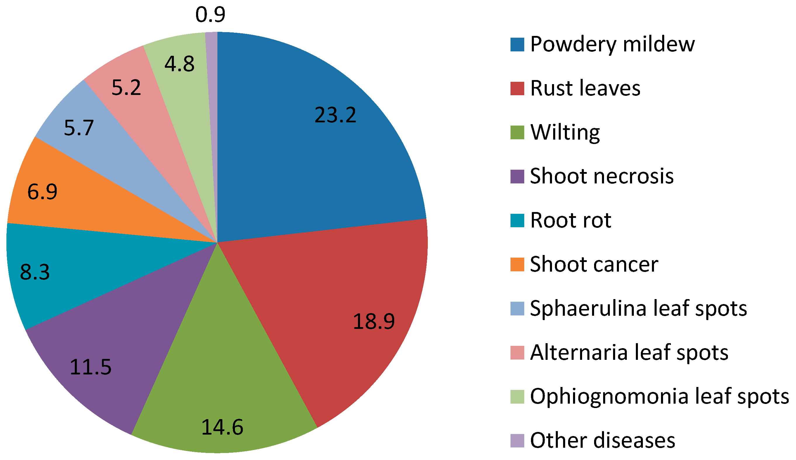
Table 2. The occurrence of the main phytopathogenic fungal species and diseases in silver and downy birch.
PhytopathogenDisease TypeOccurrence, %
Phyllactinia guttata (Wallr.) Lev.Powdery mildew13.2
Erysiphe ornate (U. Braun) U. Braun & S. Takam.Powdery mildew10.0
Melampsoridium betulinum (Pers.) Kleb.Rust leaves18.9
Fusarium avenaceum (Fr.) Sacc.Wilting13.2
Nectria sp. (Fr.) Fr.Shoot necrosis4.1
Melanconium bicolor Nees.Shoot necrosis3.7
Phytophthora cactorum (Leb. & Cohn) SchroeterShoot necrosis3.5
Pythium sp. PringsheimRoot rot7.6
Botryosphaeria dothidea (Moug. & Fr.) Ces. & DeNot.Shoot cancer6.2
Ophiognomonia intermedia (Rehm) SogonovOphiognomonia leaf spots4.8
Sphaerulina betulae (Pass.) Quaedvlieg, Verkley & CrousSphaerulina leaf spots5.7
Alternaria alternata (Fr.) Keissl.Alternaria leaf spots4.9
Other speciesOther diseases0.9
Table 3. The list of the main phytopathogenic fungal species identified in silver and downy birch and the sizes of their species-specific diagnostic ITS amplicons obtained using the ITS1–ITS2 and ITS3–ITS4 primer pair combinations.
Phytopathogenic SpeciesITS1–ITS2, bpITS3–ITS4, bp
Sphaerulina betulae (Pass.) Quaedvlieg, Verkley & Crous225231
Ophiognomonia intermedia (Rehm) Sogonov268351
Alternaria alternata (Fr.) Keissl.244346
Phyllactinia guttata (Wallr.) Lev.314364
Botryosphaeria dothidea (Moug. & Fr.) Ces. & DeNot.259344
Erysiphe ornate (U. Braun) U. Braun & S. Takam.298362
Melampsoridium betulinum (Pers.) Kleb.328406
Pythium sp. Pringsheim298633
Phytophthora cactorum (Leb. & Cohn) Schroeter295602
Fusarium avenaceum (Fr.) Sacc.233355
Melanconium bicolor Nees.270349
Nectria sp. (Fr.) Fr.217348

