Article

Investigating Seed Dormancy in Pinus bungeana Zucc. ex Endl.: Understanding the Contributions of Enclosing Tissues and Temperature on Germination

1
College of Landscape Architecture, Nanjing Forestry University, Nanjing 210037, China
2
Collaborative Innovation Center of Sustainable Forestry in Southern China, Nanjing Forestry University, Nanjing 210037, China
3
Southern Tree Seed Inspection Center, National Forestry Administration, Nanjing 210037, China
4
College of Forestry, Nanjing Forestry University, Nanjing 210037, China
*
Author to whom correspondence should be addressed.
Forests 2018, 9(7), 401; https://doi.org/10.3390/f9070401
Submission received: 21 May 2018 / Revised: 28 June 2018 / Accepted: 3 July 2018 / Published: 4 July 2018
(This article belongs to the Section Forest Ecophysiology and Biology)

Abstract

:
Pinus bungeana Zucc. ex Endl. is an endangered conifer tree species that is endemic to Western Central China. With the aim to confirm whether the Pinus bungeana seeds were dormant or not, an isolated embryo was cultured and seed coat interference with imbibition was examined by the water uptake test; the contribution of enclosing structures to germination inhibition and the effect of different temperatures (10 °C, 15 °C, 20 °C, 25 °C, and 30 °C) on seed germination were investigated by the germination test, then total germination percentage (TGP) and mean germination time (MGT) were calculated. Results showed that the Pinus bungeana seeds were non-dormant, seeds without any prechilling treatment germinated readily and achieved high germination (approximately 90%) at favorable temperatures (15 and 20 °C). At 25 °C, seed germination was inhibited and intact seeds exhibited 5% germination, but an interference with imbibition was not responsible for this result. In the seed tissue removal experiment, cracking the seed coat did not improve germination effectively, removing the seed coat and leaving the nucellar membrane either intact or having a quarter of it removed both elicited greater germination (34.7% and 40%, respectively). Meanwhile, removing the seed coat and removing either half or all of the nucellar membrane both promoted higher germination (approximately 80%), with germination rates that were nearly the same as that for the isolated embryos (86%). Germination inhibition was mainly induced by the enclosing structures and the nucellar membrane played an important role in inhibiting germination.

1. Introduction

Pinus bungeana Zucc. ex Endl. is an evergreen conifer species within the family Pinaceae and is naturally distributed in Western Central China, geographically ranging from 29°55′ to 38°25′ N latitude and from 103°36′ to 115°17′ E latitude, across warm temperate, northern subtropic, and mid-subtropic climatic zones [1,2]. It is known as a favorite ornamental plant in China and is also widely used as an afforestation tree species due to its adaptability to the dry and cold climates. In addition, its cones and pollen are valuable medicinal herbs, its turpentine is an important chemical raw material, and its edible seeds are known as pine nuts.
Seed germination is a complex physiological process that is affected by internal (dormancy, maturity, and genetic) and external factors (temperature, light, oxygen, and moisture) [3]. Each has a specific effect on the germination process, either acting alone or working together with each other [4]. Seed dormancy occurs during seed development, making the mature seeds fail to germinate under conditions that are normally favorable for germination. Dormancy evolved in both gymnosperms and angiosperms, and it is an adaptability promoting germination when circumstances are more ideal for seedlings and plant establishment [5]. There are several types of seed dormancy that can usually be classified into two categories: internal (or embryo) dormancy and physical (or seed coat-imposed) dormancy. The internal dormancy is due to underdeveloped embryos or the presence of metabolic inhibitors, while the physical dormancy is conferred by the tissues surrounding the embryo, which impose dormancy by blocking the water absorption or gas exchange, exerting mechanical restraint on the radicle emergence, playing the role of a barrier against the escape of chemical inhibitors from the embryo, or containing inhibitors themselves, or through the combination of any one of these factors [4,6].
Pinus bungeana is propagated from seeds, however, the seeds develop strong dormancy [2,7,8,9,10,11,12], which may be a challenge for seed reproduction and limit the use of Pinus bungeana as a plantation tree. So far, although there is already some information on the analysis of dormancy in Pinus bungeana seeds, only partial explanations are available and sometimes the explanations are apparently contradictory. For example, some researchers [8,9,10] have considered that seed coat played a dominating role in seed dormancy while the embryo was non-dormant, while other researchers [11] have pointed out that the embryo showed dormancy and the germination of embryo was regulated by the endosperm. As reported by some investigators, the presence of inhibitory substances in the seed tissues was mainly responsible for the seeds’ dormancy, but they held a different view on the content of inhibitory substances of each seed tissue. Wang et al. [9] reached the conclusion that the inhibitors in the seed coat were the most important factor affecting dormancy, followed by the inhibitors in the nucellar membrane, with the least important inhibitors in the megagametophyte, while Dong et al. [12] obtained the opposite results, where they deemed that the content of inhibitors contained in each seed tissue were: megagametophyte > nucellar membrane > seed coat. Furthermore, other studies have indicated that the water-impermeable seed coat and nucellar membrane had significant interference on the water absorption of the seeds, and the permeability barrier was one of the most important causes of seed dormancy [9,12].
Under laboratory conditions, seeds of some species like Tilia americana L., Pinus parviflora Siebold & Zucc., and Pinus lambertiana Douglas, which germinate slowly and steadily at 25 °C (a temperature generally used in seed germination), usually require over one month for all viable seeds to germinate. This prolonged germination pattern may indicate that the seeds are dormant and require pretreatment such as stratification or gibberellin exposure to germinate, or that this temperature is beyond the optimum temperature range for some species like the Western Australian perennial species of Acacia blakelyi Maiden, Acacia pulchella R.Br., Allocasuarina humilis (Otto & A.Dietr.) L.A.S.Johnson, Beaufortia elegans Schauer, Conostylis neocymosa Hopper, Kennedia prostrate R.Br., Melaleuca acerosa Schau., and Xanthorrhoea drummondii Harv., which have optimum germination percentages between 15 and 20 °C, and Leptospermum spinescens Endl., which has an optimum at 10 °C [4,13,14].
Temperature plays a vital role in the process of seed germination, and most species need a suitable range or alternating mode to achieve the maximum germination. Temperature influences the percentage and speed of germination, which acts directly on the water imbibition and physiological metabolic reaction that regulates this process [15]. In addition, temperature regulates seed germination by removing primary and/or secondary dormancy. Moreover, the period of germination may be revised completely in response to temperature [4].
Previous findings of low levels of germination at 25 °C are usually explained by the presence of dormancy [2,7,8,9,10,11,12], and little is known about the suitable conditions for the germination of Pinus bungeana seeds, especially the effect of temperature on germination. The failure of many seeds to germinate may not be due to the seeds needing pretreatment but may be that the seeds merely require specific temperature regimes to germinate [16]. For example, either no germination or extremely low germination was observed at 25 °C and 30 °C, while high final germination was obtained when seeds were incubated at lower temperatures of 15 °C and 20 °C for species such as Pinacia oleracea L. [17,18], Ornithopus pinnatus (Mill.) Druce [19], and Myrsine parvifolia A. DC. [20], and coniferous species of Pinus halepensis Miller and Pinus brutia Tenore [21].
Based on these studies, we hypothesized that the Pinus bungeana seeds may not be dormant but that a lower or higher temperature would be favored by seed germination of this species. Then, specific experiments were conducted in the following to verify the hypothesis. Because the dormancy of Pinus bungeana seeds has not been documented clearly, and the germination characteristics have not been fully explored, this study set out to investigate and describe the seed dormancy and germination characteristics in Pinus bungeana and provide valuable information for future plantation programs. Specifically, the aims of the present study were to: (1) investigate the specific seed tissues involved in inhibiting germination and their relative importance at 25 °C; (2) determine whether the seeds are dormant or not according to the basic elements required for germination (moisture and temperature); and (3) test the effect of temperature on germination and determine the suitable temperature range for seed germination.

2. Materials and Methods

2.1. Seed Material

Freshly matured seeds of Pinus bungeana were collected in October 2015 from a Pinus bungeana natural forest in Xiaolong Mountain, in Gansu Province, Northeast China (34°0′~34°40′ N, 105°30~106°30′ E, 700~2500 m above sea level). Seeds were extracted from the cones after collection, and then transported to the laboratory of Nanjing Forestry University. The damaged and insect-infected seeds were abandoned and the empty ones were excluded by using the floating method [22,23]. Seed moisture content was approximately 9.9%, which was determined by drying 50 seeds in triplicate at 103 ± 2 °C for 17 h in an oven following the International Seed Testing Association [24]. These fresh seeds were then stored at 5 °C for three days before the experiments were commenced.

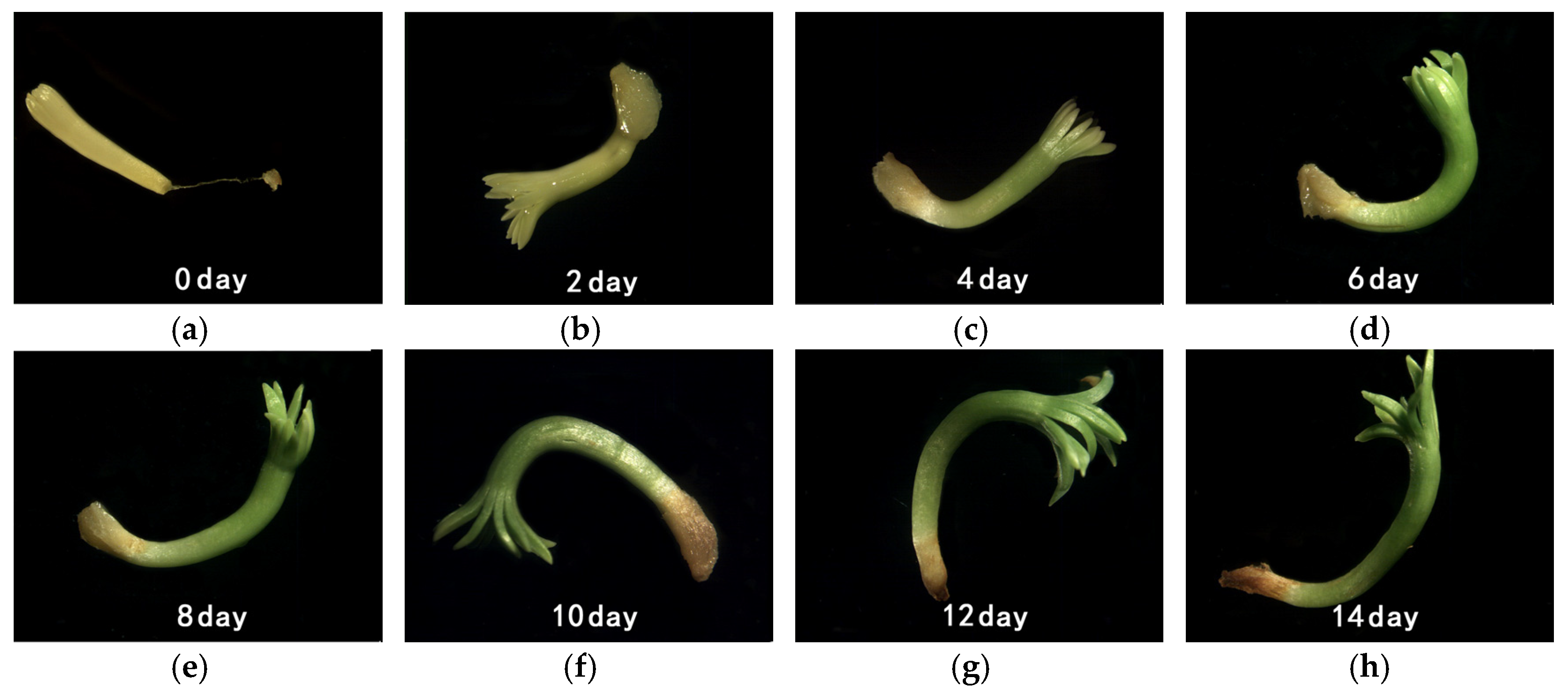
2.2. In Vitro Embryo Culture

Three replicates of 50 seeds were surface sterilized in 75% ethanol for 5 min, rinsed three times with sterile distilled water, and the seed coats were removed using forceps. The decoated seeds were dissected using a single sided razorblade, and the embryos were excised from the seeds and incubated on moist absorbent cotton at 25 °C with an 8-h photoperiod. Cultured embryos were observed at 12:00 p.m. every day for embryo growth and photographed using a stereomicroscope (SZX16, OLYMPUS Co., Tokyo, Japan) every two days over the 14-day incubation period. Radicle elongated to 2 mm and geotropic curvature were used as germination criterion.

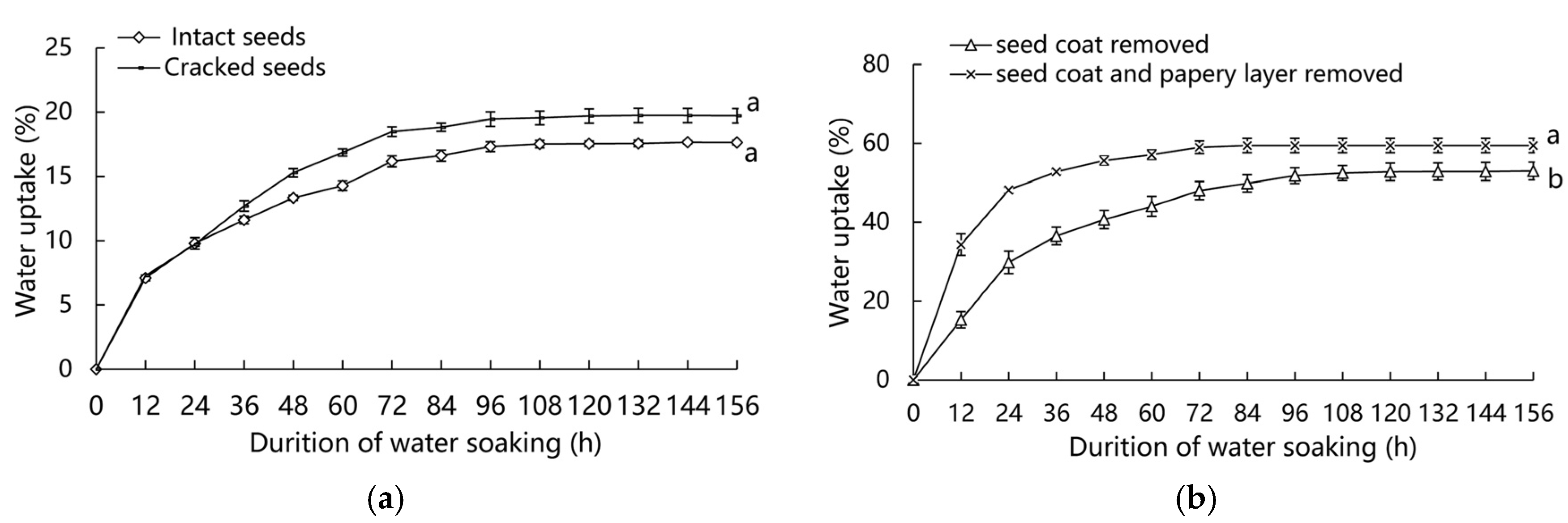
2.3. Water Uptake Test

Three replicates of 50 seeds were weighed and thereafter immersed in distilled water under ambient conditions (25 °C). Before the experiments, seeds were preliminarily treated as follows: (1) intact seeds, cracked seeds; and (2) seeds with seed coat removed while the nucellar membrane was left on the megagametophyte (seed coat removed), seeds with both the seed coat and nucellar membrane completely removed (seed coat and nucellar membrane removed). Seed weight (g) was measured every 12 h until seeds reached a constant weight. Before weighing, the seed surfaces were dried using filter paper. The increase in seed fresh weight (IFW) (%) over imbibition period was calculated to determine water uptake with the following equation:
IFW ( % ) = ( W t W 0 ) W 0 × 100
where Wt is weight of imbibed seeds at time t and W0 represents the initial weight of the seeds before imbibing.

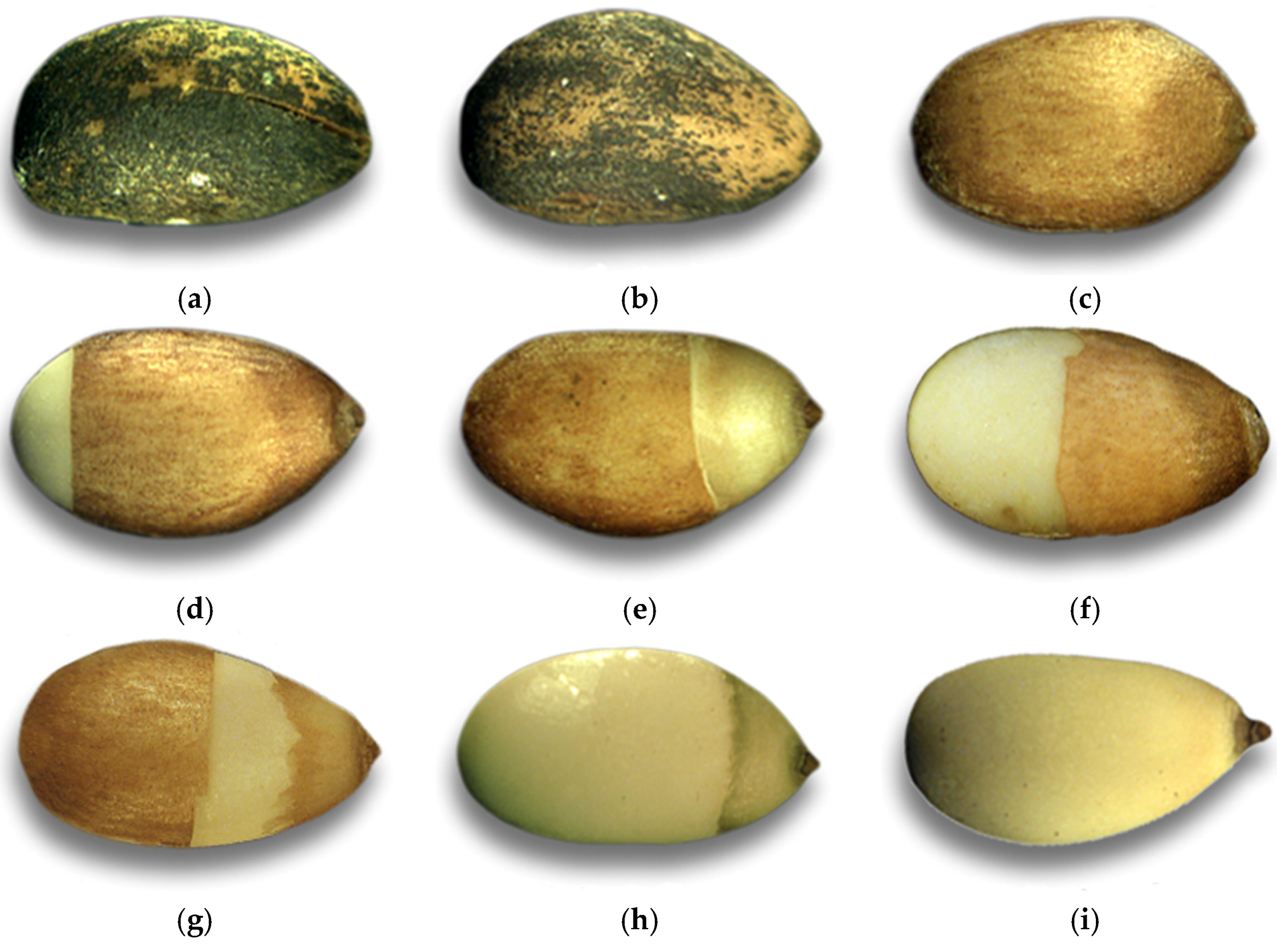
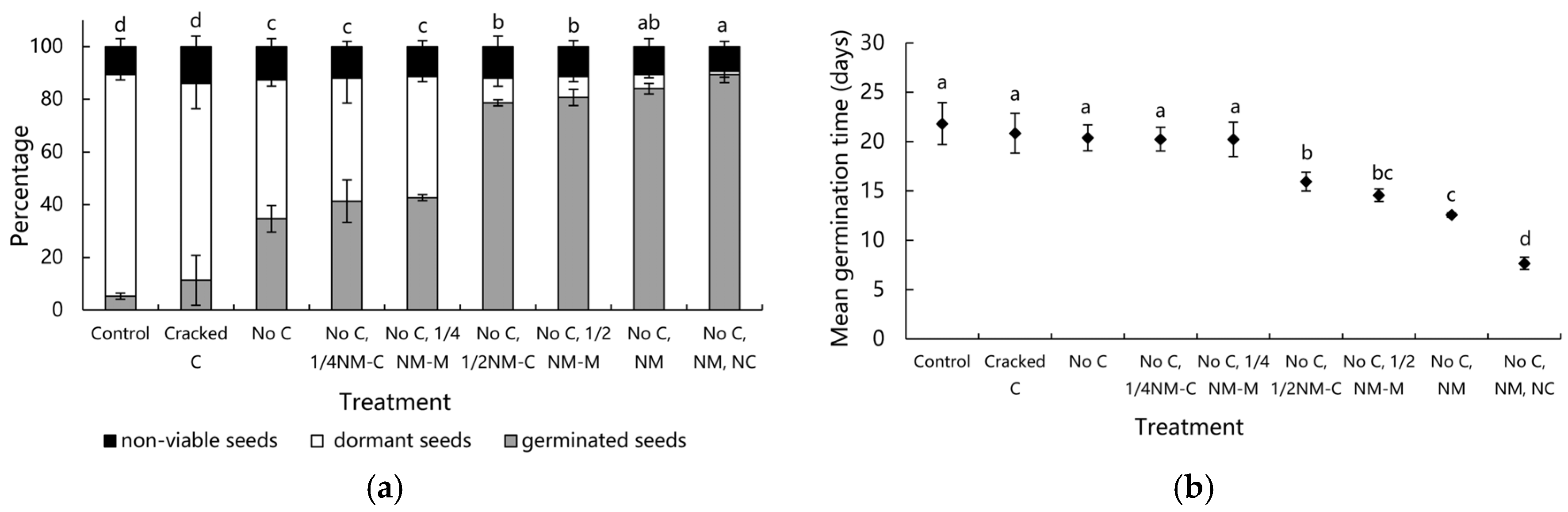
2.4. Determining Seed Tissues That Inhibit Germination

The major enclosing structures of the embryo in Pinus bungeana seeds are the megagametophyte, nucellar cap surrounding the micropylar end, seed coat, and nucellar membrane. To investigate the contribution of these tissues in the dormancy of Pinus bungeana seeds, the effects of sequentially removing these tissues were studied.
Dissections were performed under laminar flow using forceps and tweezers (previously sterilized in 75% ethanol). Prior to removing the tissues, seeds were imbibed in sterile distilled water for 96 h at 25 °C. The seeds were surface sterilized in 75% ethanol for 5 min and then rinsed three times with sterile distilled water.
Experiments included the following treatments: (1) intact seed coat (Control; Figure 1a); (2) seed coat cracked gently at the micropylar end (Cracked C; Figure 1b); (3) seed coat removed, nucellar membrane left intact (No C; Figure 1c); (4) seed coat and whole nucellar membrane removed, nucellar cap left intact (No C, NM; Figure 1h); and (5) seed coat, nucellar membrane, and nucellar cap removed (No C, NM, NC; Figure 1i). To investigate the further contribution of the nucellar membrane, experiments were performed altering the integrity of the nucellar membrane as follows; (6) seed coat removed and about a quarter of the nucellar membrane at the chalazal end ripped off laterally (No C, 1/4 NM-C; Figure 1d); (7) seed coat removed and about a quarter of the nucellar membrane in the micropylar end ripped off laterally (No C, 1/4 NM-M; Figure 1e); (8) seed coat removed and half of the nucellar membrane at the chalazal end ripped off laterally (No C, 1/2 NM-C; Figure 1f); and (9) seed coat removed and about half of the nucellar membrane in the micropylar end ripped off laterally (No C,1/2 NM-M; Figure 1g).
After preliminary treatments, the remainder of the seeds were incubated on moist absorbent cotton at 25 °C (as mentioned in the introduction, the dormancy of Pinus bungeana seeds is usually conducted at this temperature) and an 8-h photoperiod. The seeds were moistened with distilled water when necessary. A seed was considered to be germinated with the emergence of the radicle (radicle ≥ 2 mm). Three replicates of 50 seeds were used for each treatment. Germination percentage was recorded at 12:00 p.m. every day over the 30-day incubation period. Viability was assessed in the non-germinated seeds at the end of each experiment with a positive tetrazolium test [24].
At the end of the incubation time, the following parameters were assessed: total germination percentage (TGP) and mean germination time (MGT), with calculations based on the following equations:
TGP ( % ) = ( n N ) × 100
where n is germinated seeds at the end of the experiment and N is the total number of seeds originally placed on moist absorbent cotton to germinate.
MGT ( d a y s ) = ( n i × t ) n
where ni is the number of germinated seeds at day t and n is the total number of germinated seeds at the end of the experiment.

2.5. Effect of Temperature on Germination

Tests were conducted under five constant temperature regimes (10 °C, 15 °C, 20 °C, 25 °C, and 30 °C) with an 8-h photoperiod. Three replicates of 100 seeds were incubated on moist absorbent cotton. Before the germination test, seed lots were imbibed in distilled water for 96 h. Germination percentage was recorded at 12:00 p.m. every day over the 30-day incubation period. A seed was considered to be germinated with the emergence of radicle (radicle ≥ 2 mm). At the end of the incubation time, TGP values were calculated as described in the previous section.

2.6. Statistical Analyses

The data were analyzed by SPSS 19.0 (IBM, Armonk, NY, USA) and Excel (Office 2013 Pro Plus, Microsoft Corporation, Redmond, WA, USA) software. Statistical analysis was performed using a one-way analysis of variance (ANOVA) followed by Duncan’s Multiple Range Test (DMRT). Values were expressed as means ± SD (standard deviation) for three replicates in each of the independent experiments; p-values < 0.05 were considered significant.

3. Results

3.1. In Vitro Embryo Culture

Isolated embryos placed on moist absorbent cotton germinated readily when non-embryonic seed tissues were removed, exhibiting 86% germination and showing great variation in morphology and size (Figure 2). The embryonic axes elongated and the cotyledons expanded and turned green after 14 days in culture. Thus, the embryos themselves were non-dormant and the dormancy in seeds was imposed by the enclosing structures that wrapped the embryos.

3.2. Water Uptake Test

It took about 120 h for the intact seeds and cracked seeds to achieve a near saturation level of water uptake. Imbibition of cracked seeds varied only slightly from that of the intact seeds. At the end of the imbibition time, intact seeds took up 17.7% of initial weight in water and cracked seeds took up 19.7%. No significant differences were observed after 156 h among the two seed treatments. The imbibition rate was just slightly higher in cracked seeds (Figure 3a).
When both the seed coat and nucellar membrane were removed, the seeds absorbed water more rapidly than when only the seed coat was removed. The water uptake of seeds that had both their seed coat and nucellar membrane removed increased to 48.2% in 24 h, and then increased to a saturated level of 53.0% at the time of 84 h. However, the water uptake of seeds that had their seed coat removed only increased to 29.8% in 24 h, and it would take at least 120 h for them to achieve a saturated level of water uptake. Significant differences were measured after 156 h among the two treatments (Figure 3b).

3.3. Determining Seed Tissues That Inhibit Germination

To determine which structures contributed to preventing germination in dormant Pinus bungeana seeds, different seed tissues were sequentially removed and the total germination percentage and mean germination time were evaluated. Large differences were measured among treatments for TGP (Figure 4a) and MGT (Figure 4b). In contrast to isolated embryos excised from seeds, only 5.3% of intact seeds germinated after incubation for 30 days at 25 °C, and no significant difference was observed in germination percentage between the cracked seeds and intact seeds (11.3% and 5.3%, respectively). Furthermore, the removal of the seed coat and either leaving the nucellar membrane wrapping the megagametophyte intact or having a quarter of it removed (whether at the chalazal end or in the micropylar end) all promoted germination (approximately 34.7% and 40%, respectively). Higher germinations were induced when the seed coat and half of the nucellar membrane were removed rather than when the seed coat and a quarter of the nucellar membrane were removed (approximately 40% vs. 80%, respectively). Removal of the seed coat and nucellar membrane, with the nucellar cap removed or not, both permitted high germination (84% and 89.3%, respectively), which was nearly the same as that for the isolated embryos. Meanwhile, the MGT was increased significantly if the nucellar cap sheathing the micropylar end of the megagametophyte was left intact. The smallest MGT (fastest germination) was obtained by treatments with the seed coat, the whole nucellar membrane, and the nucellar cap all removed (7.7 days), followed by the treatments with seed coat removed and with the whole nucellar membrane removed or half of the nucellar membrane removed seeds (between 12.6 and 16.0 days), while the MGTs of the rest of the treatments were all around 20 days and almost the same as that for the control (21.8 days).

3.4. Effect of Temperature on Germination

Seeds incubated at various temperatures displayed significant differences in germination (Figure 5) (p < 0.05). The time-course of germination under different temperatures showed that out of the tested temperatures, 20 °C yielded the fastest germination rate. The germination percentage at 20 °C was the highest (92.3%), and 15 °C was also effective for a high germination response (87.7%), while incubation at higher temperatures (25 °C and 30 °C) resulted in lower total germination (5.2% and 0.7%, respectively) and no seeds germinated at 10 °C.

4. Discussion

4.1. Germination of Isolated Embryos

Isolated embryo cultures suggested that the embryos themselves were non-dormant and that the dormancy of Pinus bungeana seeds seemed to be mainly regulated by the tissues that enclosed the embryos. Previous studies published on the subject of conifer seed dormancy have also supported this result; in seeds of coniferous species such as Picea glauca (Moench) Voss [25], Pseudotsuga menziesii (Mirb.) Franco [26], Chamaecyparis nootkatensis (D. Don) Spach [27], Pinus sylvestris L. [28], Picea abies Karst. [29], and Pinus taeda L. [30,31,32], their embryos often germinated when they were excised from the seeds and placed in water, which indicated that the conifer embryos were not inherently dormant and that the tissues wrapping the embryos played an inhibitory role and impeded their radicle emergence.

4.2. Water Imbibition of Seeds

Water uptake is a fundamental requirement to complete seed germination. Interference with water uptake has been demonstrated as one of the factors that contribute to the dormancy of seeds in several families such as Leguminosae, Cannaceae, Convolvulaceae, and Malvaceae, which are characterized by holding a hard testa [33]. In seeds of Pinus taeda [30,31], the seed coat prevents imbibition by restricting swelling of the megagametophyte and embryo while in some other conifer seeds such as Pinus monticola Douglas ex D. Don [34], Pinus lambertiana [34], Pinus sylvestris, and Picea abies [28,29], the thin nucellar membrane surrounding the megagametophyte has been found to act as an effective barrier against imbibition and germination. Our study showed that despite the presence of several membrane sclereids in the hard Pinus bungeana seed coat, the seeds with an intact seed coat were still able to uptake water, even though they did it at slower rates than those with the seed coat removed. In the conifer seeds, there are always portal-like structures in the seed coat for the entrance of water. One is the micropyle, and the other two structural openings are found at the chalazal end and at the ridge connecting the two halves of the seed. They make the water enter the seeds, acting either alone or working together with each other.
Although the seed coat did not create a completely impermeable barrier, similarity to what has been observed in Pinus taeda seeds [30,31], the seed coat could limit the swelling of the megagametophyte to a certain degree. This restriction might have persisted until the seeds germinated and weakened as the seed coats split. This was one of the reasons that the water uptake of the seeds with the seed coat removed was significantly higher than those with the seed coat left on. Furthermore, the considerable difference of water uptake between seeds with the seed coat removed and seeds with seed coat left on was somewhat misleading, as that the presence of the seed coat contributed much to the dry weight, but it imbibed less water than the megagametophyte and embryo. This exaggerated the difference in water absorption between the two treatments to some extent (although certain differences indeed exist). Even so, after a 156-h water soak, the moisture content levels of the megagametophytes and embryos within the intact seeds and cracked seeds were close to that of the seeds that had been stratificated for months (about 30%), which could germinate. Although the thin lipophilic nucellar membrane regulated imbibition more effectively than the hard seed coat, when the seed coat was removed, regardless of whether the nucellar membrane was removed or not, the moisture content levels were all >40% (data not shown), and the moisture content was likely to be adequate for the metabolic processes required for germination. Therefore, the coat-enhanced dormancy of Pinus bungeana seeds was not likely to be due to an interference with imbibition.

4.3. Identification of Seed Structures Involved in Restricting Germination

By removing the enclosing seed tissues sequentially, it might be concluded that the nucellar cap, the nucellar membrane, and the seed coat all contributed to the coat-imposed dormancy, and the nucellar membrane played the dominating role in the dormancy and germination of Pinus bungeana seeds. The seed coat of Pinus bungeana was hard and thick, and appeared to present a mechanical barrier to radicle elongation. Cracking the seed coat along the micropylar ridge relieved some of the physical constraint, whilst supplying an entryway for oxygen exchange and water uptake, but this did not greatly improve the seed germination. The germination percentage of the cracked seeds was closer to that of the intact seeds than those with the seed coat removed, suggesting that the seed coat might act as more than a mechanical barrier to germination. In addition, removing the seed coat further improved the permeability compared with the cracked seeds, and inhibitors that might be present in the seed coat and which could inhibit germination were no longer present with the removal of the seed coat, but only 34.7% germination was obtained, indicating that the seed coat was not the only factor that inhibited germination.
The nucellar membrane of conifer seeds is thin and fragile and likely does not present a mechanical barrier to germination. Prior to the studies by Tillman-Sutela and Kauppi on the water uptake in Pinus sylvestris and Picea abies [28,29], this tissue had received little attention. However, in our study, when the seed coat was removed while the nucellar membrane was left intact, germination rates of 34.7% were observed after 30 days in culture. Furthermore, it was found that seed germination improved gradually when the enlarging of the nucellar membrane was removed. When the whole nucellar membrane was removed, a germination rate of 84% was observed. Clearly, the nucellar membrane plays an important role in the germination inhibition of Pinus bungeana seeds. Simultaneously, the nucellar membrane and seed coat might have a synergistic effect on inhibiting seed germination. Furthermore, the removal of a quarter of the nucellar membrane did not improve germination greatly, which indicates that permeability might not be the only way that the nucellar membrane plays a role in impeding germination, and there might be inhibitors such as abscisic acid (ABA) present in the nucellar membrane.
The role of the nucellar cap in the dormancy of coniferous seeds has been studied in Pinus sylvestris, Picea abies, and Picea glauca, with the nucellar cap shown to act in a manner that physically constrains radicle protrusion through the micropylar opening of the megagametophyte [25,28,29]. For the Pinus bungeana seeds, the thick and waxy nucellar cap also has some effects on the embryo protruding from the megagametophyte, yet the inhibition was very limited, only slowing down the seed germination rate to a minor extent, and can be ruled out as a major factor in preventing seed germination. Furthermore, research on the dormancy mechanism of Pinus monticola seeds suggested that the megagametophyte seemed to prevent radicle extension to a greater extent than the nucellar cap, nucellar membrane, and the seed coat [34,35]. However, in our experiment, the embryo cultured with megagametophyte germinated quickly and had high germination (approximately 90%), which was nearly the same as that for the isolated embryos. It can be seen that the megagametophyte of the Pinus bungeana seed exhibited no impediment to the radicle elongation.

4.4. Responses of Germination to Temperature

At 25 °C, the intact Pinus bungeana seeds exhibited low germination (5%) in the germination test, whilst the sequential tissue removal experiments confirmed that germination was mainly prevented by tissues external to the embryo. This result was supported by earlier studies on Pinus bungeana seeds [9,12]. Although these previous findings of low levels of germination usually explained that the seeds were dormant, little was known about the seed germination conditions of this species. Seed germination depends on several environmental conditions, usually including moisture, oxygen, and temperature. The water absorption test revealed that the seed coat and nucellar membrane indeed slowed the rate of absorption, but they did not create a completely impermeable barrier, and the moisture content of intact seeds could meet the requirement for germination, so the interference with the imbibition of these two tissues might not be responsible for the inability to germinate. Furthermore, the interference with oxygen uptake could also be ruled out as a main factor to prevent germination. In our experiment, cracking the seed coat along the micropylar ridge provided a portal for gas transit, but seed germination was only marginally improved, whilst after having a quarter of the nucellar membrane removed, germination was 5% higher compared to when the nucellar membrane was left intact. That is, when the water and oxygen were available for germination, there were still many seeds that did not germinate and appeared dormant. Presumably, this might be caused by an unfavorable temperature. Accordingly, the effect of temperature on Pinus bungeana seeds germination was further examined.
Ghildiyal and Sharma [16] pointed out that the failure of many seeds to germinate may not be due to a need for pretreatment but may be that the seeds merely require a specific temperature to complete germination. Pooled germination data from the five temperatures showed that out of the tested temperatures, the fastest germination rate was obtained at 20 °C. At 15 °C, there was a slight decrease in rate and final germination when compared with 20 °C, while at 10 °C, 25 °C, and 30 °C significant inhibition of germination was observed. A dormant seed is one that does not have the capacity to germinate in a specified period of time under any combination of normal physical environmental factors that are otherwise favorable for its germination [33]. Obviously, at the favorable temperatures (15 °C, 20 °C), the seeds of Pinus bungeana were non-dormant, while the high temperatures (25 °C, 30 °C) inhibited the total germination and decreased the germination rate of Pinus bungeana seeds. In the work of Thanos and Skordilis [21], with seeds of Pinus halepensis and Pinus brutia, a temperature of 20 °C was equally advisable for the germination process, favoring germination and reducing the mean germination time. Moreover, seeds of Pinus halepensis displayed a wider range of temperature requirements than those of Pinus brutia, since the former also exhibited high germination at 15 °C. Nevertheless, it must be noted that the germination in these two conifer species and Pinus bungeana were restricted to a rather narrow range of temperatures, around 20 °C, while higher temperatures (>20 °C) produced a sharp decrease in germination or resulted in no germination occurring. This temperature variation is called thermos-inhibition, which is characterized by the absence of germination when the temperature is slightly above the optimum conditions [36]. Germination inhibition by high temperatures (25 °C–30 °C) has been observed in other species, such as Spinacia oleracea L. [17,18], Ornithopus pinnatus [19], and Myrsine parvifolia [20], and the seeds of these three species have also shown higher final germination at temperatures of 15 °C and 20 °C, while significantly lower germination occurred at 25 °C and 30 °C.
For the seeds of Pinus bungeana, 25 °C and 30 °C exceeded the suitable temperature range for germination, resulting in thermos-inhibition, whereas when the seeds that failed to germinate after a 30-day period at high temperatures (25 °C and 30 °C) were transferred to 20 °C (suitable germination temperature) and continued to incubate subsequently, they reached high germination (91% and 88%, respectively, data not shown). These results demonstrated that the inhibition of germination at high temperatures on Pinus bungeana seeds was reversible when seeds were returned to lower temperatures and allowed us to exclude the possibility that secondary dormancy was induced at high temperatures.

4.5. Roles Seed Structures Played in Germination at Different Temperatures

By analyzing the seed structures involved in restricting germination, we found that seed tissues (seed coat, nucellar membrane) played an important role in inhibiting seed germination at the temperature of 25 °C, but the intact Pinus bungeana seeds germinated readily at 15 °C and 20 °C, so how do these tissues contribute to germination at these different temperatures? In this respect, the germination of Arabidopsis thaliana L. seeds was also strongly dependent on the incubation temperature; seeds did not germinate at high temperature (20–27 °C) but germinated easily at a low temperature (13 °C) or when a fluridone (an inhibitor of ABA biosynthesis) treatment was given at high temperature [37]. Equally, the same behavior has been observed in Lactuca sativa Linn. seeds, which germinated at 10–22 °C but not at temperatures above 25–30 °C. This threshold was raised by the application of gibberellins, cytokinins, and ethylene, while ABA decreased the temperature range for germination [38]. These results indicated a very probable role of ABA in the failure of seeds to germinate at relatively high temperatures. We speculate that the same mechanism could operate in the Pinus bungeana seeds, and there might be inhibitors such as ABA present in the seed tissues that play a role in the germination inhibition at 25 °C.
It has been reported that in Arabidopsis thaliana seeds, the ABA content decreased faster at lower temperature of 13 °C, which was the optimal temperature for germination in comparison to the higher temperature of 27 °C [37]. The same variations in ABA contents were observed in lettuce seeds cultured in a similar temperature condition [38]. Consequently, when the Pinus bungeana seeds were incubated at 25 °C, this high temperature may have promoted ABA biosynthesis and prevented ABA inactivation through metabolism and/or conjugation, making seed tissues contain higher levels of ABA than that at 15 °C and 20 °C. Simultaneously, evidence exists that shows that oxygen concentration might determine the catabolic rate of germination inhibitors or ABA [39,40]. The availability of oxygen in imbibed seed was strongly dependent on temperature, and the low temperature increased the dissolved oxygen concentration in water [4,41]. When the Pinus bungeana seeds were cultured at 25 °C, the presence of seed tissues might have been instrumental for the absence of germination through imposing oxygen deprivation to the embryo, which, in turn, promoted ABA synthesis and/or impeded ABA inactivation. Moreover, it is known that the incubation temperature can affect embryo responsiveness to ABA [42,43,44], and the sensitivity of the embryo to ABA inhibition reduced at low temperature [43]. Probably, the sensitivity of embryos to ABA increased at a high temperature of 25 °C, which might also be responsible for the inability of the Pinus bungeana seeds to germinate. Taken together, the germination inhibition in Pinus bungeana seeds may be related to high levels of ABA, an increase in the sensitivity of the embryo to ABA, or a lower concentration of dissolved oxygen in the seed tissues at 25 °C.

5. Conclusions

In the range of temperatures analyzed in the study, the best effect was achieved for a temperature of 20 °C, followed by 15 °C. At these favorable temperatures, the seeds without any prechilling treatment achieved high germination, approximately 90%. Clearly, the seeds of Pinus bungeana were non-dormant, and did not require any pretreatment to germinate, while the failure of Pinus bungeana seeds to germinate at 25 °C was because the seeds merely required a lower temperature to complete the germination process. Thus, an interference with imbibition can be ruled out as a main cause for the failure of germination. At 25 °C, the isolated embryos germinated readily and obtained 86% germination, so the tissues surrounding the embryos could be considered to be responsible for the significant delay of germination when incubated at the temperature of 25 °C. Moreover, the nucellar membrane played an important role in germination inhibition, followed by the seed coat, while the nucellar cap had no effect on the total germination percentage but slowed down the germination rate to a minor extent, and the megagametophyte exhibited no impediment to the radicle elongation. Nevertheless, it should be noted that we did not investigate what particular roles the seed tissues, especially the nucellar membrane, played in inhibiting germination. In our study, the effects of seed tissues on water uptake were investigated, and we intend to study the effect of these tissues on oxygen exchange and the presence of chemical inhibitors in the near future. This can provide us with a thorough understanding of the specific mechanisms that led to the seeds failing to germinate at higher temperature conditions.

Author Contributions

C.G., Y.S., and F.S. participated in the discussion and experimental designs; Y.S. and F.S. prepared the experiment materials; C.G. performed the experiments, analyzed the data, and drafted the manuscript.

Funding

Financial support from a project funded by the Priority Academic Program Development of Jiangsu Higher Education Institutions (PAPD).

Conflicts of Interest

The authors declare that they have no conflicts of interest.

References

  1. Yan, Z.; Xuezhong, Z.; Xiaoan, W. A study on geographical distribution law of Pinus bungeana natural forests in china. Acta Bot. Boreal. Occident. Sin. 1995, 15, 161–166. [Google Scholar]
  2. Wang, X.; Liu, J.; X.; Wang, J.; Liu, C. Geographical variation of morphological characteristics of Pinus bungeana seeds and cones. J. Beijing For. Univ. 1998, 20, 25–31. [Google Scholar]
  3. Kleczewski, N.M.; Herms, D.A.; Bonello, P. Effects of soil type, fertilization and drought on carbon allocation to root growth and partitioning between secondary metabolism and ectomycorrhizae of Betula papyrifera. Tree Physiol. 2010, 30, 807–817. [Google Scholar] [CrossRef] [PubMed]
  4. Bewley, J.D.; Bradford, K.; Hilhorst, H.; Nonogaki, H. Seeds: Physiology of Development, Germination and Dormancy, 3rd ed.; Plenum Press: New York, NY, USA, 2014. [Google Scholar]
  5. Baskin, C.C.; Baskin, J.M. Seeds: Ecology, Biogeography, and Evolution of Dormancy and Germination; Academic Press: New York, NY, USA, 2001. [Google Scholar]
  6. Bewley, J.D. Seed germination and dormancy. Plant Cell 1997, 9, 1055–1066. [Google Scholar] [CrossRef] [PubMed]
  7. Zhang, C.-X.; Li, Q.; Kong, L. Induction, development and maturation of somatic embryos in Bunge’s pine (Pinus bungeana Zucc. ex Endl.). Plant Cell Tissue Org. Cult. 2007, 91, 273–280. [Google Scholar] [CrossRef]
  8. Xin, Z. Seed dormancy mechanism of six pine species. J. Southwest For. Coll. 2008, 28, 5–9. [Google Scholar]
  9. Wang, X.; Liu, J.; Wang, J. Variation of dormancy characteristic of different Pinus bungeana seed sources. Chin. J. Appl. Ecol. 2000, 11, 9–12. [Google Scholar]
  10. Shulan, Y. Seed dormancy and germination of lacebark pine (Pinus bungeana). Seed 1983, 1, 24–28. [Google Scholar]
  11. Dong, L.; Shao, C.; Zhang, Z. Characteristics of embryo dormancy and germination of Pinus bungeana. Sci. Silv. Sin. 2003, 39, 47–54. [Google Scholar]
  12. Lifen, D.; Bin, X.; CuiPing, C. Investigating seed dormancy in Pinus bungeana. Shaanxi For. Sci. Technol. 1987, 1, 15–17. [Google Scholar]
  13. Bell, D.T.; Plummer, J.A.; Taylor, S.K. Seed germination ecology in Southwestern Western Australia. Bot. Rev. 1993, 59, 24–73. [Google Scholar] [CrossRef]
  14. Bellairs, S.M.; Bell, D.T. Temperature effects on the seed germination of ten kwongan species from Eneabba, Western Australia. Aust. J. Bot. 1990, 38, 451–458. [Google Scholar] [CrossRef]
  15. Marcos Filho, J. Fisiologia De Sementes De Plantas Cultivadas, 2nd ed.; Abrates: Londrina, Brazil, 2015. [Google Scholar]
  16. Ghildiyal, S.K.; Sharma, C.M. Effect of seed size and temperature treatments on germination of various seed sources of Pinus wallichiana and Pinus roxburghii from Garhwal Mmalaya. Indian For. 2005, 131, 56–65. [Google Scholar]
  17. Leskovar, D.; Esensee, V.; Belefant-Miller, H. Pericarp, leachate, and carbohydrate involvement in thermoinhibition of germinating spinach seeds. J. Am. Soc. Hortic. Sci. 1999, 124, 301–306. [Google Scholar]
  18. Chitwood, J.; Shi, A.; Evans, M.; Rom, C.E.; Gbur, E.; Motes, D.; Chen, P.; Hensley, D. Effect of temperature on seed germination in spinach (Spinacia oleracea). HortScience 2016, 51, 1475–1478. [Google Scholar] [CrossRef]
  19. Lopes, R.R.; Souza, C.H.L.D.; Bertoncelli, P.; Franke, L.B. Overcoming dormancy and determining optimal temperature for Slender serradella seed germination. Rev. Bras. De Zootec. 2015, 44, 413–419. [Google Scholar] [CrossRef]
  20. Ribeiro, J.N.S.; Costa, C.S.B. The effect of temperature regulation on seed germination of the tropical tree Myrsine parvifolia A. DC near its southern limit. South Afr. J. Bot. 2015, 98, 128–133. [Google Scholar] [CrossRef]
  21. Thanos, C.A.; Skordilis, A. The effects of light, temperature and osmotic stress on the germination of Pinus halepensis and P. brutia seeds. Seed Sci. Technol. 1987, 15, 163–174. [Google Scholar]
  22. Boydak, M.; Dirik, H.; Tilki, F.; Calikoglu, M. Effects of water stress on germination in six provenances of Pinus brutia seeds from different bioclimatic zones in Turkey. Turk. J. Agric. For. 2003, 27, 91–97. [Google Scholar]
  23. Dangasuk, O.G.; Panetsos, K.P. Altitudinal and longitudinal variations in Pinus brutia (Ten.) of Crete Island, Greece: Some needle, cone and seed traits under natural habitats. New For. 2004, 27, 269–284. [Google Scholar] [CrossRef]
  24. ISTA. International Seed Testing Association; The International Seed Testing Association (ISTA): Bassersdorf, Switzerland, 2005. [Google Scholar]
  25. Downie, B.; Bewley, J. Dormancy in white spruce (Picea glauca [Moench.] Voss.) seeds is imposed by tissues surrounding the embryo. Seed Sci. Res. 1996, 6, 9–15. [Google Scholar] [CrossRef]
  26. Bianco, J.; Garello, G.; Le Page-Degivry, M.T. De novo ABA synthesis and expression of seed dormancy in a gymnosperm: Pseudotsuga menziesii. Plant Growth Regul. 1997, 21, 115–119. [Google Scholar] [CrossRef]
  27. Ren, C.; Kermode, A.R. Analyses to determine the role of the megagametophyte and other seed tissues in dormancy maintenance of yellow cedar (Chamaecyparis nootkatensis) seeds: Morphological, cellular and physiological changes following moist chilling and during germination. J. Exp. Bot. 1999, 50, 1403–1419. [Google Scholar]
  28. Tillman-Sutela, E.; Kauppi, A. The morphological background to imbibition in seeds of Pinus sylvestris L. of different provenances. Trees 1995, 9, 123–133. [Google Scholar] [CrossRef]
  29. Tillman-Sutela, E.; Kauppi, A. The significance of structure for imbibition in seeds of the Norway spruce, Picea abies (L.) Karst. Trees 1995, 9, 269–278. [Google Scholar] [CrossRef]
  30. Barnett, J. Seed coat influences dormancy of loblolly pine seeds. Can. J. For. Res. 1972, 2, 7–10. [Google Scholar] [CrossRef]
  31. Barnett, J.P. Delayed germination of southern pine seeds related to seed coat constraint. Can. J. For. Res. 1976, 6, 504–510. [Google Scholar] [CrossRef]
  32. Cooke, J.; Cooke, B.; Gifford, D. Loblolly pine seed dormancy: Constraints to germination. New For. 2002, 23, 239–256. [Google Scholar] [CrossRef]
  33. Baskin, J.M.; Baskin, C.C. A classification system for seed dormancy. Seed Sci. Res. 2004, 14, 1–16. [Google Scholar] [CrossRef]
  34. Hoff, R.J. Dormancy in Pinus monticola seed related to stratification time, seed coat, and genetics. Can. J. For. Res. 1987, 17, 294–298. [Google Scholar] [CrossRef]
  35. Feurtado, J.A.; Ren, C.; Ambrose, S.J.; Cutler, A.J.; Ross, A.R.S.; Abrams, S.R.; Kermode, A.R. The coat-enhanced dormancy mechanism of western white pine (Pinus monticola Dougl. ex D. Don) seeds is mediated by abscisic acid homeostasis and mechanical restraint. Seed Sci. Technol. 2008, 36, 283–300. [Google Scholar] [CrossRef]
  36. Pacheco Junior, F.; Silva, J.; Rondinelli da Silva Negreiros, J.; Rose Gomes da Silva, M.; Braga Farias, S. Germination and vigor of long-pepper seeds (Piper hispidinervum) as a function of temperature and light. Revis. Cienc. Agron. 2013, 44, 325–333. [Google Scholar] [CrossRef]
  37. Ali-Rachedi, S.; Bouinot, D.; Wagner, M.-H.; Bonnet, M.; Sotta, B.; Grappin, P.; Jullien, M. Changes in endogenous abscisic acid levels during dormancy release and maintenance of mature seeds: Studies with the Cape Verde Islands ecotype, the dormant model of Arabidopsis thaliana. Plant. 2004, 219, 479–488. [Google Scholar] [CrossRef] [PubMed]
  38. Yoshioka, T.; Endo, T.; Satoh, S. Restoration of seed germination at supraoptimal temperatures by fluridone, an inhibitor of abscisic acid biosynthesis. Plant Cell Physiol. 1998, 39, 307–312. [Google Scholar] [CrossRef]
  39. Barthe, P.; Garello, G.; Bianco-Trinchant, J.; le Page-Degivry, M.T. Oxygen availability and ABA metabolism in Fagus Sylvatica Seeds. Plant Growth Regulation. 2000, 30, 185–191. [Google Scholar] [CrossRef]
  40. Neill, S.J.; Horgan, R. Abscisic acid and related compounds. In Principles and Practice of Plant Hormone Analysis; Rivier, L., Crozier, A., Eds.; Academic Press: London, UK, 1987; pp. 111–167. [Google Scholar]
  41. Corbineau, F.; Côme, D. Control of seed germination and dormancy by the gaseous environment. In Seed Development and Germination; Kigel, J., Galili, G., Eds.; Dekker: New York, NY, USA, 1995; pp. 397–424. [Google Scholar]
  42. Poljakoff-Mayber, A.; Corbineau, F.; Côme, D. A possible mechanism of high temperature dormancy regulation in seeds of Avena sativa L. (cv. Moyencourt). Plant Growth Regul. 1990, 9, 147–156. [Google Scholar] [CrossRef]
  43. Walker-Simmons, M.K. Enhancement of ABA responsiveness in wheat embryos by high temperature. Plant Cell Environ. 1988, 11, 769–775. [Google Scholar] [CrossRef]
  44. Corbineau, F.; Côme, D.; Viémont, J.D.; Crabbé, J. Dormancy of cereal seeds as related to embryo sensitivity to ABA and water potential. In Dormancy in Plants: From Whole Plants Behaviour to Cellular Control; Viémont, J.D., Crabbé, J., Eds.; CAB International: Bringford, UK, 2000; pp. 183–191. [Google Scholar]
Figure 1. Seed treatments: (a) Intact seed (Control); (b) Seed coat cracked (Cracked C); (c) Seed coat removed (No C); (d) Both seed coat and a quarter of the nucellar membrane at the chalazal end removed (No C, 1/4 NM-C); (e) Both seed coat and a quarter of the nucellar membrane in the micropylar end removed (No C, 1/4 NM-M); (f) Both seed coat and half of the nucellar membrane at the chalazal end removed (No C, 1/2 NM-C); (g) Both seed coat and half of the nucellar membrane in the micropylar end removed (No C, 1/2 NM-M); (h) Both seed coat and whole nucellar membrane removed (No C, NM); (i) Seed coat, nucellar membrane, and nucellar cap all removed (No C, NM, NC).
Forests 09 00401 g001
Figure 2. Germination process of excised Pinus bungeana Zucc. ex Endl. embryos. (ah) Variations in morphology of the isolated embryos during a 14-day period.
Forests 09 00401 g002
Figure 3. Water absorption curves of seeds with different treatment. (a) Water absorption curves of the intact seeds and cracked seeds; (b) Water absorption curves of seeds that had their seed coat and nucellar membrane removed and seeds that had their seed coat removed. Vertical bars represent the standard deviation.
Forests 09 00401 g003
Figure 4. The contribution of enclosing tissues on seeds germination. (a) Percentage of germinated seeds (TGP), non-viable seeds, and dormant seeds; (b) Mean germination time (MGT) obtained after 30 days of incubation. Bars and rhomboids represent means ± SD from three replications of 50 seeds each. Values with the same letter in the same graph are not significantly different at p < 0.05.
Forests 09 00401 g004
Figure 5. Cumulative germination percentage for Pinus bungeana seeds incubated at five different temperatures.
Forests 09 00401 g005

