Next Issue
Volume 10, June
Previous Issue
Volume 10, April


Cancers, Volume 10, Issue 5 (May 2018) – 26 articles

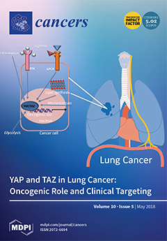
Cover Story (view full-size image): Lung cancer is the leading cause of cancer death worldwide. There is no current therapy able to efficiently treat this disease, as lung cancer is often diagnosed at an advanced stage. At the cellular level, acquired resistance to the current therapies is a major obstacle. The role of YAP and TAZ in promoting tumor formation, progression, and resistance to therapy in lung cancer is emerging. This makes YAP and TAZ rather promising druggable targets to design new therapeutic strategies for successfully treating lung cancer. View this paper
  • Issues are regarded as officially published after their release is announced to the table of contents alert mailing list.
  • You may sign up for e-mail alerts to receive table of contents of newly released issues.
  • PDF is the official format for papers published in both, html and pdf forms. To view the papers in pdf format, click on the "PDF Full-text" link, and use the free Adobe Reader to open them.
Order results
Result details
Section
Select all
Export citation of selected articles as:
12 pages, 1732 KiB  
Article
Eukaryotic Translation Initiation Factor 4A Down-Regulation Mediates Interleukin-24-Induced Apoptosis through Inhibition of Translation
by Xuelin Zhong, Leah Persaud, Hilal Muharam, Ashleigh Francis, Dibash Das, Bertal Huseyin Aktas and Moira Sauane
Cancers 2018, 10(5), 153; https://doi.org/10.3390/cancers10050153 - 22 May 2018
Cited by 10 | Viewed by 3578
Abstract
Dysregulated activity of helicase eIF4A drives transformation to and maintenance of cancer cell phenotype by reprogramming cellular translation. Interleukin 24 (IL-24) is a tumor-suppressing protein, which has the ability to inhibit angiogenesis, sensitize cancer cells to chemotherapy, and induce cancer cell-specific apoptosis. In [...] Read more.
Dysregulated activity of helicase eIF4A drives transformation to and maintenance of cancer cell phenotype by reprogramming cellular translation. Interleukin 24 (IL-24) is a tumor-suppressing protein, which has the ability to inhibit angiogenesis, sensitize cancer cells to chemotherapy, and induce cancer cell-specific apoptosis. In this study, we found that eIF4A is inhibited by IL-24. Consequently, selective reduction of translation was observed for mRNAs harboring strong secondary structures in their 5′-untranslated regions (5′UTRs). These mRNAs encode proteins, which function in cell survival and proliferation. Consistently, overexpression of eIF4A conferred cancer cells with resistance to IL-24-induced cell death. It has been established that inhibition of eIF4A triggers mitochondrial-mediated apoptosis. We showed that IL-24 induces eIF4A-dependent mitochondrial depolarization. We also showed that IL-24 induces Sigma 1 Receptor-dependent eIF4A down-regulation and mitochondrial depolarization. Thus, the progress of apoptosis triggered by IL-24 is characterized by a complex program of changes in regulation of several initiation factors, including the eIF4A. Full article
Show Figures

Figure 1

16 pages, 3342 KiB  
Article
Determining the Radiation Enhancement Effects of Gold Nanoparticles in Cells in a Combined Treatment with Cisplatin and Radiation at Therapeutic Megavoltage Energies
by Celina Yang, Kyle Bromma, Wonmo Sung, Jan Schuemann and Devika Chithrani
Cancers 2018, 10(5), 150; https://doi.org/10.3390/cancers10050150 - 22 May 2018
Cited by 34 | Viewed by 5347
Abstract
Combined use of chemotherapy and radiation therapy is commonly used in cancer treatment, but the toxic effects on normal tissue are a major limitation. This study assesses the potential to improve radiation therapy when combining gold nanoparticle (GNP) mediated radiation sensitization with chemoradiation [...] Read more.
Combined use of chemotherapy and radiation therapy is commonly used in cancer treatment, but the toxic effects on normal tissue are a major limitation. This study assesses the potential to improve radiation therapy when combining gold nanoparticle (GNP) mediated radiation sensitization with chemoradiation compared to chemoradiation alone. Incorporation of GNPs with 2 Gy, 6 MV (megavoltage) radiation resulted in a 19 ± 6% decrease in survival of MDA-MB-231 cells. Monte-Carlo simulations were performed to assess dosimetric differences in the presence of GNPs in radiation. The results show that physics dosimetry represents a small fraction of the observed effect. The survival fraction of the cells exposed to GNPs, cisplatin, and radiation was 0.16 ± 0.007, while cells treated with cisplatin and radiation only was 0.23 ± 0.011. The presence of GNPs resulted in a 30 ± 6% decrease in the survival, having an additive effect. The concentration of the GNPs and free drug used for this study was 0.3 and 435 nM, respectively. These concentrations are relatively lower and achievable in an in vivo setting. Hence, the results of our study would accelerate the incorporation of GNP-mediated chemoradiation into current cancer therapeutic protocols in the near future. Full article
(This article belongs to the Special Issue Nanotechnology and Cancer)
Show Figures

Figure 1

17 pages, 888 KiB  
Review
40 Years of Research Put p53 in Translation
by Virginie Marcel, Flora Nguyen Van Long and Jean-Jacques Diaz
Cancers 2018, 10(5), 152; https://doi.org/10.3390/cancers10050152 - 21 May 2018
Cited by 41 | Viewed by 6881
Abstract
Since its discovery in 1979, p53 has shown multiple facets. Initially the tumor suppressor p53 protein was considered as a stress sensor able to maintain the genome integrity by regulating transcription of genes involved in cell cycle arrest, apoptosis and DNA repair. However, [...] Read more.
Since its discovery in 1979, p53 has shown multiple facets. Initially the tumor suppressor p53 protein was considered as a stress sensor able to maintain the genome integrity by regulating transcription of genes involved in cell cycle arrest, apoptosis and DNA repair. However, it rapidly came into light that p53 regulates gene expression to control a wider range of biological processes allowing rapid cell adaptation to environmental context. Among them, those related to cancer have been extensively documented. In addition to its role as transcription factor, scattered studies reported that p53 regulates miRNA processing, modulates protein activity by direct interaction or exhibits RNA-binding activity, thus suggesting a role of p53 in regulating several layers of gene expression not restricted to transcription. After 40 years of research, it appears more and more clearly that p53 is strongly implicated in translational regulation as well as in the control of the production and activity of the translational machinery. Translation control of specific mRNAs could provide yet unsuspected capabilities to this well-known guardian of the genome. Full article
(This article belongs to the Special Issue p53 Signaling in Cancers)
Show Figures

Figure 1

21 pages, 892 KiB  
Review
Organoids Provide an Important Window on Inflammation in Cancer
by Kristi Baker
Cancers 2018, 10(5), 151; https://doi.org/10.3390/cancers10050151 - 21 May 2018
Cited by 28 | Viewed by 7493
Abstract
Inflammation is a primary driver of cancer initiation and progression. However, the complex and dynamic nature of an inflammatory response make this a very difficult process to study. Organoids are a new model system where complex multicellular structures of primary cells can be [...] Read more.
Inflammation is a primary driver of cancer initiation and progression. However, the complex and dynamic nature of an inflammatory response make this a very difficult process to study. Organoids are a new model system where complex multicellular structures of primary cells can be grown in a 3D matrix to recapitulate the biology of the parent tissue. This experimental model offers several distinct advantages over alternatives including the ability to be genetically engineered, implanted in vivo and reliably derived from a wide variety of normal and cancerous tissue from patients. Furthermore, long-term organoid cultures reproduce many features of their source tissue, including genetic and epigenetic alterations and drug sensitivity. Perhaps most significantly, cancer organoids can be cocultured in a variety of different systems with a patients’ own immune cells, uniquely permitting the study of autologous cancer-immune cell interactions. Experiments with such systems promise to shed light on the mechanisms governing inflammation-associated cancer while also providing prognostic information on an individual patient’s responsiveness to immunotherapeutic anti-cancer drugs. Thanks to their ability to capture important features of the complex relationship between a cancer and its microenvironment, organoids are poised to become an essential tool for unraveling the mechanisms by which inflammation promotes cancer. Full article
(This article belongs to the Special Issue Inflammation and Cancer)
Show Figures

Figure 1

10 pages, 4221 KiB  
Article
AZD1775 Increases Sensitivity to Olaparib and Gemcitabine in Cancer Cells with p53 Mutations
by Xiangbing Meng, Jianling Bi, Yujun Li, Shujie Yang, Yuping Zhang, Mary Li, Haitao Liu, Yiyang Li, Megan E. Mcdonald, Kristina W. Thiel, Kuo-Kuang Wen, Xinhao Wang, Meng Wu and Kimberly K. Leslie
Cancers 2018, 10(5), 149; https://doi.org/10.3390/cancers10050149 - 19 May 2018
Cited by 53 | Viewed by 6942
Abstract
Tumor suppressor p53 is responsible for enforcing cell cycle checkpoints at G1/S and G2/M in response to DNA damage, thereby allowing both normal and tumor cells to repair DNA before entering S and M. However, tumor cells with absent or mutated p53 are [...] Read more.
Tumor suppressor p53 is responsible for enforcing cell cycle checkpoints at G1/S and G2/M in response to DNA damage, thereby allowing both normal and tumor cells to repair DNA before entering S and M. However, tumor cells with absent or mutated p53 are able to activate alternative signaling pathways that maintain the G2/M checkpoint, which becomes uniquely critical for the survival of such tumor cells. We hypothesized that abrogation of the G2 checkpoint might preferentially sensitize p53-defective tumor cells to DNA-damaging agents and spare normal cells with intact p53 function. The tyrosine kinase WEE1 regulates cdc2 activity at the G2/M checkpoint and prevents entry into mitosis in response to DNA damage or stalled DNA replication. AZD1775 is a WEE1 inhibitor that overrides and opens the G2/M checkpoint by preventing WEE1-mediated phosphorylation of cdc2 at tyrosine 15. In this study, we assessed the effect of AZD1775 on endometrial and ovarian cancer cells in the presence of two DNA damaging agents, the PARP1 inhibitor, olaparib, and the chemotherapeutic agent, gemcitabine. We show that AZD1775 alone is effective as a therapeutic agent against some p53 mutated cell models. Moreover, the combination of AZD1775 with olaparib or gemcitabine is synergistic in cells with mutant p53 and constitutes a new approach that should be considered in the treatment of advanced and recurrent gynecologic cancer. Full article
(This article belongs to the Special Issue p53 Signaling in Cancers)
Show Figures

Figure 1

20 pages, 788 KiB  
Review
p53-Autophagy-Metastasis Link
by Tatiana V. Denisenko, Anastasia D. Pivnyuk and Boris Zhivotovsky
Cancers 2018, 10(5), 148; https://doi.org/10.3390/cancers10050148 - 18 May 2018
Cited by 38 | Viewed by 6653
Abstract
The tumor suppressor p53 as the “guardian of the genome” plays an essential role in numerous signaling pathways that control the cell cycle, cell death and in maintaining the integrity of the human genome. p53, depending on the intracellular localization, contributes to the [...] Read more.
The tumor suppressor p53 as the “guardian of the genome” plays an essential role in numerous signaling pathways that control the cell cycle, cell death and in maintaining the integrity of the human genome. p53, depending on the intracellular localization, contributes to the regulation of various cell death pathways, including apoptosis, autophagy and necroptosis. Accumulated evidence suggests that this function of p53 is closely involved in the process of cancer development. Here, present knowledge concerning a p53-autophagy-metastasis link, as well as therapeutic approaches that influence this link, are discussed. Full article
(This article belongs to the Special Issue p53 Signaling in Cancers)
Show Figures

Graphical abstract

10 pages, 521 KiB  
Review
Bone Marrow Defects and Platelet Function: A Focus on MDS and CLL
by Sarah Luu, Elizabeth E. Gardiner and Robert K. Andrews
Cancers 2018, 10(5), 147; https://doi.org/10.3390/cancers10050147 - 18 May 2018
Cited by 10 | Viewed by 4716
Abstract
The bloodstream typically contains >500 billion anucleate circulating platelets, derived from megakaryocytes in the bone marrow. This review will focus on two interesting aspects of bone marrow dysfunction and how this impacts on the quality of circulating platelets. In this regard, although megakaryocytes [...] Read more.
The bloodstream typically contains >500 billion anucleate circulating platelets, derived from megakaryocytes in the bone marrow. This review will focus on two interesting aspects of bone marrow dysfunction and how this impacts on the quality of circulating platelets. In this regard, although megakaryocytes are from the myeloid lineage leading to granulocytes (including neutrophils), erythrocytes, and megakaryocytes/platelets, recent evidence has shown that defects in the lymphoid lineage leading to B cells, T cells, and natural killer (NK) cells also result in abnormal circulating platelets. Current evidence is limited regarding whether this latter phenomenon might potentially arise from (a) some form of as-yet-undetected defect common to both lineages; (b) adverse interactions occurring between cells of different lineages within the bone marrow environment; and/or (c) unknown disease-related factor(s) affecting circulating platelet receptor expression/function after their release from megakaryocytes. Understanding the mechanisms underlying how both myeloid and lymphoid lineage bone marrow defects lead to dysfunction of circulating platelets is significant because of the potential diagnostic and predictive value of peripheral platelet analysis for bone marrow disease progression, the additional potential effects of new anti-cancer drugs on platelet function, and the critical role platelets play in regulation of bleeding risk, inflammation, and innate immunity. Full article
(This article belongs to the Special Issue The Role of Thrombosis and Haemostasis in Cancer)
Show Figures

Figure 1

16 pages, 3408 KiB  
Article
The Long Non-Coding RNA RP5-1024C24.1 and Its Associated-Gene MPPED2 Are Down-Regulated in Human Thyroid Neoplasias and Act as Tumour Suppressors
by Romina Sepe, Simona Pellecchia, Pierre Serra, Daniela D’Angelo, Antonella Federico, Maddalena Raia, Ricardo Cortez Cardoso Penha, Myriam Decaussin-Petrucci, Luigi Del Vecchio, Alfredo Fusco and Pierlorenzo Pallante
Cancers 2018, 10(5), 146; https://doi.org/10.3390/cancers10050146 - 18 May 2018
Cited by 18 | Viewed by 3611 | Correction
Abstract
Background: Well-differentiated papillary thyroid carcinoma (PTC) represents the thyroid neoplasia with the highest incidence. Long non-coding RNAs (lncRNAs) have been found deregulated in several human carcinomas, and hence, proposed as potential diagnostic and prognostic markers. Therefore, the aim of our study was [...] Read more.
Background: Well-differentiated papillary thyroid carcinoma (PTC) represents the thyroid neoplasia with the highest incidence. Long non-coding RNAs (lncRNAs) have been found deregulated in several human carcinomas, and hence, proposed as potential diagnostic and prognostic markers. Therefore, the aim of our study was to investigate their role in thyroid carcinogenesis. Methods: We analysed the lncRNA expression profile of 12 PTC and four normal thyroid tissues through a lncRNA microarray. Results: We identified 669 up- and 2470 down-regulated lncRNAs with a fold change >2. Among them, we focused on the down-regulated RP5-1024C24.1 located in an antisense position with respect to the MPPED2 gene which codes for a metallophosphoesterase with tumour suppressor activity. Both these genes are down-regulated in benign and malignant thyroid neoplasias. The restoration of RP5-1024C24.1 expression in thyroid carcinoma cell lines reduced cell proliferation and migration by modulating the PTEN/Akt pathway. Inhibition of thyroid carcinoma cell growth and cell migration ability was also achieved by the MPPED2 restoration. Interestingly, RP5-1024C24.1 over-expression is able to increase MPPED2 expression. Conclusions: Taken together, these results demonstrate that RP5-1024C24.1 and MPPED2 might be considered as novel tumour suppressor genes whose loss of expression contributes to thyroid carcinogenesis. Full article
Show Figures

Figure 1

11 pages, 509 KiB  
Review
[18F]FDG-PET/CT in Hodgkin Lymphoma: Current Usefulness and Perspectives
by Salim Kanoun, Cedric Rossi and Olivier Casasnovas
Cancers 2018, 10(5), 145; https://doi.org/10.3390/cancers10050145 - 18 May 2018
Cited by 21 | Viewed by 5410
Abstract
Functional imaging using 18-fluorodeoxyglycose ([18F]FDG) positron emission tomography combined with computed tomography (PET/CT) has become a major imaging modality in Hodgkin lymphoma. This imaging modality allows for a significant improvement in staging, increased sensitivity, which involves differentiating residual tumors from fibrosis [...] Read more.
Functional imaging using 18-fluorodeoxyglycose ([18F]FDG) positron emission tomography combined with computed tomography (PET/CT) has become a major imaging modality in Hodgkin lymphoma. This imaging modality allows for a significant improvement in staging, increased sensitivity, which involves differentiating residual tumors from fibrosis during assessment, and highly impacts treatment decisions into new PET-driven strategies. This review presents the main scientific data concerning the current applications of [18F]FDG-PET/CT in Hodgkin lymphoma at baseline, interim, and the end of treatment evaluation along with the main PET-driven trials for therapeutic decisions. The emergence of total metabolic tumor volume as a new functional prognostic factor will also be discussed. Full article
(This article belongs to the Special Issue Hodgkin's Lymphoma)
Show Figures

Figure 1

0 pages, 3106 KiB  
Article
Temozolomide Enhances Triple-Negative Breast Cancer Virotherapy In Vitro
by Rodolfo Garza-Morales, Roxana Gonzalez-Ramos, Akiko Chiba, Roberto Montes de Oca-Luna, Lacey R. McNally, Kelly M. McMasters and Jorge G. Gomez-Gutierrez
Cancers 2018, 10(5), 144; https://doi.org/10.3390/cancers10050144 - 17 May 2018
Cited by 27 | Viewed by 5855 | Correction
Abstract
Triple-negative breast cancer (TNBC) is one of the most aggressive types of cancer, and treatment is limited to chemotherapy and radiation. Oncolytic virotherapy may be a promising approach to treat TNBC. However, oncolytic adenovirus (OAd)-based mono-therapeutic clinical trials have resulted in modest outcomes. [...] Read more.
Triple-negative breast cancer (TNBC) is one of the most aggressive types of cancer, and treatment is limited to chemotherapy and radiation. Oncolytic virotherapy may be a promising approach to treat TNBC. However, oncolytic adenovirus (OAd)-based mono-therapeutic clinical trials have resulted in modest outcomes. The OAd potency could be increased by chemotherapy-induced autophagy, an intracellular degradation system that delivers cytoplasmic constituents to the lysosome. In this study, the ability of alkylating agent temozolomide (TMZ)-induced autophagy to increase OAd replication and oncolysis in TNBC cells was evaluated. Human TNBC MDA-MB-231 and HCC1937 cells and mouse 4T1 cells were infected with an OAd expressing the red fluorescent protein mCherry on the virus capsid (OAdmCherry) alone or in combination with TMZ. TNBC cells treated with OAdmCherry/TMZ displayed greater mCherry and adenovirus (Ad) early region 1A (E1A) expression and enhanced cancer-cell killing compared to OAdmCherry or TMZ alone. The combined therapy-mediated cell death was associated with virus replication and accumulation of the autophagy marker light chain 3 (LC3)-II. Overall, this study provides experimental evidence of TMZ’s ability to increase oncolytic virotherapy in both human and murine TNBC cells. Full article
(This article belongs to the Special Issue Oncolytic Virotherapy)
Show Figures

Figure 1

17 pages, 4169 KiB  
Review
DICER1 Syndrome: DICER1 Mutations in Rare Cancers
by Jake C. Robertson, Cheryl L. Jorcyk and Julia Thom Oxford
Cancers 2018, 10(5), 143; https://doi.org/10.3390/cancers10050143 - 15 May 2018
Cited by 103 | Viewed by 13018
Abstract
DICER1 syndrome is a rare genetic disorder that predisposes individuals to multiple cancer types. Through mutations of the gene encoding the endoribonuclease, Dicer, DICER1 syndrome disrupts the biogenesis and processing of miRNAs with subsequent disruption in control of gene expression. Since the first [...] Read more.
DICER1 syndrome is a rare genetic disorder that predisposes individuals to multiple cancer types. Through mutations of the gene encoding the endoribonuclease, Dicer, DICER1 syndrome disrupts the biogenesis and processing of miRNAs with subsequent disruption in control of gene expression. Since the first description of DICER1 syndrome, case reports have documented novel germline mutations of the DICER1 gene in patients with cancers as well as second site mutations that alter the function of the Dicer protein expressed. Here, we present a review of mutations in the DICER1 gene, the respective protein sequence changes, and clinical manifestations of DICER1 syndrome. Directions for future research are discussed. Full article
Show Figures

Figure 1

24 pages, 15664 KiB  
Review
A New Strategy to Control and Eradicate “Undruggable” Oncogenic K-RAS-Driven Pancreatic Cancer: Molecular Insights and Core Principles Learned from Developmental and Evolutionary Biology
by Robert E. Van Sciver, Michael P. Lee, Caroline Dasom Lee, Alex C. Lafever, Elizaveta Svyatova, Kevin Kanda, Amber L. Collier, Lauren L. Siewertsz van Reesema, Angela M. Tang-Tan, Vasilena Zheleva, Monicah N. Bwayi, Minglei Bian, Rebecca L. Schmidt, Lynn M. Matrisian, Gloria M. Petersen and Amy H. Tang
Cancers 2018, 10(5), 142; https://doi.org/10.3390/cancers10050142 - 14 May 2018
Cited by 14 | Viewed by 7646
Abstract
Oncogenic K-RAS mutations are found in virtually all pancreatic cancers, making K-RAS one of the most targeted oncoproteins for drug development in cancer therapies. Despite intense research efforts over the past three decades, oncogenic K-RAS has remained largely “undruggable”. Rather than targeting an [...] Read more.
Oncogenic K-RAS mutations are found in virtually all pancreatic cancers, making K-RAS one of the most targeted oncoproteins for drug development in cancer therapies. Despite intense research efforts over the past three decades, oncogenic K-RAS has remained largely “undruggable”. Rather than targeting an upstream component of the RAS signaling pathway (i.e., EGFR/HER2) and/or the midstream effector kinases (i.e., RAF/MEK/ERK/PI3K/mTOR), we propose an alternative strategy to control oncogenic K-RAS signal by targeting its most downstream signaling module, Seven-In-Absentia Homolog (SIAH). SIAH E3 ligase controls the signal output of oncogenic K-RAS hyperactivation that drives unchecked cell proliferation, uncontrolled tumor growth, and rapid cancer cell dissemination in human pancreatic cancer. Therefore, SIAH is an ideal therapeutic target as it is an extraordinarily conserved downstream signaling gatekeeper indispensable for proper RAS signaling. Guided by molecular insights and core principles obtained from developmental and evolutionary biology, we propose an anti-SIAH-centered anti-K-RAS strategy as a logical and alternative anticancer strategy to dampen uncontrolled K-RAS hyperactivation and halt tumor growth and metastasis in pancreatic cancer. The clinical utility of developing SIAH as both a tumor-specific and therapy-responsive biomarker, as well as a viable anti-K-RAS drug target, is logically simple and conceptually innovative. SIAH clearly constitutes a major tumor vulnerability and K-RAS signaling bottleneck in pancreatic ductal adenocarcinoma (PDAC). Given the high degree of evolutionary conservation in the K-RAS/SIAH signaling pathway, an anti-SIAH-based anti-PDAC therapy will synergize with covalent K-RAS inhibitors and direct K-RAS targeted initiatives to control and eradicate pancreatic cancer in the future. Full article
(This article belongs to the Special Issue Latest Development in Pancreatic Cancer)
Show Figures

Graphical abstract

14 pages, 928 KiB  
Review
Role of the Bone Microenvironment in the Development of Painful Complications of Skeletal Metastases
by Sun H. Park, Matthew R. Eber, D. Brooke Widner and Yusuke Shiozawa
Cancers 2018, 10(5), 141; https://doi.org/10.3390/cancers10050141 - 10 May 2018
Cited by 22 | Viewed by 5820
Abstract
Cancer-induced bone pain (CIBP) is the most common and painful complication in patients with bone metastases. It causes a significant reduction in patient quality of life. Available analgesic treatments for CIBP, such as opioids that target the central nervous system, come with severe [...] Read more.
Cancer-induced bone pain (CIBP) is the most common and painful complication in patients with bone metastases. It causes a significant reduction in patient quality of life. Available analgesic treatments for CIBP, such as opioids that target the central nervous system, come with severe side effects as well as the risk of abuse and addiction. Therefore, alternative treatments for CIBP are desperately needed. Although the exact mechanisms of CIBP have not been fully elucidated, recent studies using preclinical models have demonstrated the role of the bone marrow microenvironment (e.g., osteoclasts, osteoblasts, macrophages, mast cells, mesenchymal stem cells, and fibroblasts) in CIBP development. Several clinical trials have been performed based on these findings. CIBP is a complex and challenging condition that currently has no standard effective treatments other than opioids. Further studies are clearly warranted to better understand this painful condition and develop more effective and safer targeted therapies. Full article
(This article belongs to the Special Issue Targeting Bone Metastasis in Cancer)
Show Figures

Figure 1

14 pages, 3257 KiB  
Article
Toward the Discovery of a Novel Class of YAP–TEAD Interaction Inhibitors by Virtual Screening Approach Targeting YAP–TEAD Protein–Protein Interface
by Floriane Gibault, Mathilde Coevoet, Manon Sturbaut, Amaury Farce, Nicolas Renault, Frédéric Allemand, Jean-François Guichou, Anne-Sophie Drucbert, Catherine Foulon, Romain Magnez, Xavier Thuru, Matthieu Corvaisier, Guillemette Huet, Philippe Chavatte, Patricia Melnyk, Fabrice Bailly and Philippe Cotelle
Cancers 2018, 10(5), 140; https://doi.org/10.3390/cancers10050140 - 8 May 2018
Cited by 35 | Viewed by 8358
Abstract
Intrinsically disordered protein YAP (yes-associated protein) interacts with TEADs transcriptional factors family (transcriptional enhancer associated domain) creating three interfaces. Interface 3, between the Ω-loop of YAP and a shallow pocket of TEAD was identified as the most important TEAD zone for YAP-TEAD interaction. [...] Read more.
Intrinsically disordered protein YAP (yes-associated protein) interacts with TEADs transcriptional factors family (transcriptional enhancer associated domain) creating three interfaces. Interface 3, between the Ω-loop of YAP and a shallow pocket of TEAD was identified as the most important TEAD zone for YAP-TEAD interaction. Using the first X-ray structure of the hYAP50–71-hTEAD1209–426 complex (PDB 3KYS) published in 2010, a protein-protein interaction inhibitors-enriched library (175,000 chemical compounds) was screened against this hydrophobic pocket of TEAD. Four different chemical families have been identified and evaluated using biophysical techniques (thermal shift assay, microscale thermophoresis) and in cellulo assays (luciferase activity in transfected HEK293 cells, RTqPCR in MDA-MB231 cells). A first promising hit with micromolar inhibition in the luciferase gene reporter assay was discovered. This hit also decreased mRNA levels of TEAD target genes. Full article
Show Figures

Figure 1

14 pages, 514 KiB  
Review
Proline-Rich Protein Tyrosine Kinase 2 in Inflammation and Cancer
by Xiangdong Zhu, Yonghua Bao, Yongchen Guo and Wancai Yang
Cancers 2018, 10(5), 139; https://doi.org/10.3390/cancers10050139 - 8 May 2018
Cited by 27 | Viewed by 6972
Abstract
Focal adhesion kinase (FAK) and its homologous FAK-related proline-rich tyrosine kinase 2 (Pyk2) contain the same domain, exhibit high sequence homology and are defined as a distinct family of non-receptor tyrosine kinases. This group of kinases plays critical roles in cytoskeletal dynamics and [...] Read more.
Focal adhesion kinase (FAK) and its homologous FAK-related proline-rich tyrosine kinase 2 (Pyk2) contain the same domain, exhibit high sequence homology and are defined as a distinct family of non-receptor tyrosine kinases. This group of kinases plays critical roles in cytoskeletal dynamics and cell adhesion by regulating survival and growth signaling. This review summarizes the physiological and pathological functions of Pyk2 in inflammation and cancers. In particular, overexpression of Pyk2 in cancerous tissues is correlated with poor outcomes. Pyk2 stimulates multiple oncogenic signaling pathways, such as Wnt/β-catenin, PI3K/Akt, MAPK/ERK, and TGF-β/EGFR/VEGF, and facilitates carcinogenesis, migration, invasion, epithelial–mesenchymal transition and metastasis. Therefore, Pyk2 is a high-value therapeutic target and has clinical significance. Full article
Show Figures

Graphical abstract

14 pages, 4224 KiB  
Review
Nuclear Export Inhibition for Pancreatic Cancer Therapy
by Irfana Muqbil, Asfar S. Azmi and Ramzi M. Mohammad
Cancers 2018, 10(5), 138; https://doi.org/10.3390/cancers10050138 - 7 May 2018
Cited by 18 | Viewed by 5138
Abstract
Pancreatic cancer is a deadly disease that is resistant to most available therapeutics. Pancreatic cancer to date has no effective drugs that could enhance the survival of patients once their disease has metastasized. There is a need for the identification of novel actionable [...] Read more.
Pancreatic cancer is a deadly disease that is resistant to most available therapeutics. Pancreatic cancer to date has no effective drugs that could enhance the survival of patients once their disease has metastasized. There is a need for the identification of novel actionable drug targets in this unusually recalcitrant cancer. Nuclear protein transport is an important mechanism that regulates the function of several tumor suppressor proteins (TSPs) in a compartmentalization-dependent manner. High expression of the nuclear exporter chromosome maintenance region 1 (CRM1) or exportin 1 (XPO1), a common feature of several cancers including pancreatic cancer, results in excessive export of critical TSPs to the incorrect cellular compartment, leading to their functional inactivation. Small molecule inhibitors of XPO1 can block this export, retaining very important and functional TSPs in the nucleus and leading to the effective killing of the cancer cells. This review highlights the current knowledge on the role of XPO1 in pancreatic cancer and how this serves as a unique and clinically viable target in this devastating and by far incurable cancer. Full article
Show Figures

Figure 1

29 pages, 2367 KiB  
Review
YAP and TAZ in Lung Cancer: Oncogenic Role and Clinical Targeting
by Federica Lo Sardo, Sabrina Strano and Giovanni Blandino
Cancers 2018, 10(5), 137; https://doi.org/10.3390/cancers10050137 - 6 May 2018
Cited by 90 | Viewed by 11795
Abstract
Lung cancer is the leading cause of cancer death in the world and there is no current treatment able to efficiently treat the disease as the tumor is often diagnosed at an advanced stage. Moreover, cancer cells are often resistant or acquire resistance [...] Read more.
Lung cancer is the leading cause of cancer death in the world and there is no current treatment able to efficiently treat the disease as the tumor is often diagnosed at an advanced stage. Moreover, cancer cells are often resistant or acquire resistance to the treatment. Further knowledge of the mechanisms driving lung tumorigenesis, aggressiveness, metastasization, and resistance to treatments could provide new tools for detecting the disease at an earlier stage and for a better response to therapy. In this scenario, Yes Associated Protein (YAP) and Trascriptional Coactivator with PDZ-binding motif (TAZ), the final effectors of the Hippo signaling transduction pathway, are emerging as promising therapeutic targets. Here, we will discuss the most recent advances made in YAP and TAZ biology in lung cancer and, more importantly, on the newly discovered mechanisms of YAP and TAZ inhibition in lung cancer as well as their clinical implications. Full article
Show Figures

Figure 1

23 pages, 5914 KiB  
Article
Electron Nuclear Dynamics Simulations of Proton Cancer Therapy Reactions: Water Radiolysis and Proton- and Electron-Induced DNA Damage in Computational Prototypes
by Erico S. Teixeira, Karthik Uppulury, Austin J. Privett, Christopher Stopera, Patrick M. McLaurin and Jorge A. Morales
Cancers 2018, 10(5), 136; https://doi.org/10.3390/cancers10050136 - 6 May 2018
Cited by 15 | Viewed by 6112
Abstract
Proton cancer therapy (PCT) utilizes high-energy proton projectiles to obliterate cancerous tumors with low damage to healthy tissues and without the side effects of X-ray therapy. The healing action of the protons results from their damage on cancerous cell DNA. Despite established clinical [...] Read more.
Proton cancer therapy (PCT) utilizes high-energy proton projectiles to obliterate cancerous tumors with low damage to healthy tissues and without the side effects of X-ray therapy. The healing action of the protons results from their damage on cancerous cell DNA. Despite established clinical use, the chemical mechanisms of PCT reactions at the molecular level remain elusive. This situation prevents a rational design of PCT that can maximize its therapeutic power and minimize its side effects. The incomplete characterization of PCT reactions is partially due to the health risks associated with experimental/clinical techniques applied to human subjects. To overcome this situation, we are conducting time-dependent and non-adiabatic computer simulations of PCT reactions with the electron nuclear dynamics (END) method. Herein, we present a review of our previous and new END research on three fundamental types of PCT reactions: water radiolysis reactions, proton-induced DNA damage and electron-induced DNA damage. These studies are performed on the computational prototypes: proton + H2O clusters, proton + DNA/RNA bases and + cytosine nucleotide, and electron + cytosine nucleotide + H2O. These simulations provide chemical mechanisms and dynamical properties of the selected PCT reactions in comparison with available experimental and alternative computational results. Full article
(This article belongs to the Special Issue Proton and Carbon Ion Therapy)
Show Figures

Graphical abstract

15 pages, 703 KiB  
Review
The Guardian of the Genome Revisited: p53 Downregulates Genes Required for Telomere Maintenance, DNA Repair, and Centromere Structure
by Eléonore Toufektchan and Franck Toledo
Cancers 2018, 10(5), 135; https://doi.org/10.3390/cancers10050135 - 6 May 2018
Cited by 94 | Viewed by 9218
Abstract
The p53 protein has been extensively studied for its capacity to prevent proliferation of cells with a damaged genome. Surprisingly, however, our recent analysis of mice expressing a hyperactive mutant p53 that lacks the C-terminal domain revealed that increased p53 activity may alter [...] Read more.
The p53 protein has been extensively studied for its capacity to prevent proliferation of cells with a damaged genome. Surprisingly, however, our recent analysis of mice expressing a hyperactive mutant p53 that lacks the C-terminal domain revealed that increased p53 activity may alter genome maintenance. We showed that p53 downregulates genes essential for telomere metabolism, DNA repair, and centromere structure and that a sustained p53 activity leads to phenotypic traits associated with dyskeratosis congenita and Fanconi anemia. This downregulation is largely conserved in human cells, which suggests that our findings could be relevant to better understand processes involved in bone marrow failure as well as aging and tumor suppression. Full article
(This article belongs to the Special Issue p53 Signaling in Cancers)
Show Figures

Figure 1

13 pages, 1077 KiB  
Article
The Prevalence of CD146 Expression in Breast Cancer Subtypes and Its Relation to Outcome
by Ingeborg E. De Kruijff, Anna M. Timmermans, Michael A. Den Bakker, Anita M.A.C. Trapman-Jansen, Renée Foekens, Marion E. Meijer-Van Gelder, Esther Oomen-de Hoop, Marcel Smid, Antoinette Hollestelle, Carolien H.M. Van Deurzen, John A. Foekens, John W.M. Martens and Stefan Sleijfer
Cancers 2018, 10(5), 134; https://doi.org/10.3390/cancers10050134 - 5 May 2018
Cited by 19 | Viewed by 4182
Abstract
CD146, involved in epithelial-to-mesenchymal transition (EMT), might affect cancer aggressiveness. We here investigated the prevalence of CD146 expression in breast cancer subtypes, its relation to prognosis, the relation between CD146 and EMT and the outcome to tamoxifen. Primary breast cancer tissues from 1342 [...] Read more.
CD146, involved in epithelial-to-mesenchymal transition (EMT), might affect cancer aggressiveness. We here investigated the prevalence of CD146 expression in breast cancer subtypes, its relation to prognosis, the relation between CD146 and EMT and the outcome to tamoxifen. Primary breast cancer tissues from 1342 patients were available for this retrospective study and immunohistochemically stained for CD146. For survival analyses, pure prognosis was studied by only including lymph-node negative patients who did not receive (neo)adjuvant systemic treatment (n = 551). 11% of the tumors showed CD146 expression. CD146 expression was most prevalent in triple-negative cases (64%, p < 0.001). In univariable analysis, CD146 expression was a prognostic factor for both metastasis-free survival (MFS) (p = 0.020) and overall survival (OS) (p = 0.037), but not in multivariable analysis (including age, tumor size, grade, estrogen receptor (ER), progesterone receptor (PR), human epidermal growth factor receptor 2 (HER2) and Ki-67). No correlation between CD146 and EMT nor difference in outcome to first-line tamoxifen was seen. In this large series, our data showed that CD146 is present in primary breast cancer and is a pure prognostic factor for MFS and OS in breast cancer patients. We did not see an association between CD146 expression and EMT nor on outcome to tamoxifen. Full article
Show Figures

Figure 1

8 pages, 571 KiB  
Perspective
Translation Control by p53
by Justina Kasteri, Dibash Das, Xuelin Zhong, Leah Persaud, Ashleigh Francis, Hilal Muharam and Moira Sauane
Cancers 2018, 10(5), 133; https://doi.org/10.3390/cancers10050133 - 5 May 2018
Cited by 19 | Viewed by 5217
Abstract
The translation of mRNAs plays a critical role in the regulation of gene expression and therefore, in the regulation of cell proliferation, differentiation and apoptosis. Unrestricted initiation of translation causes malignant transformation and plays a key role in the maintenance and progression of [...] Read more.
The translation of mRNAs plays a critical role in the regulation of gene expression and therefore, in the regulation of cell proliferation, differentiation and apoptosis. Unrestricted initiation of translation causes malignant transformation and plays a key role in the maintenance and progression of cancers. Translation initiation is regulated by the ternary complex and the eukaryotic initiation factor 4F (eIF4F) complex. The p53 tumor suppressor protein is the most well studied mammalian transcription factor that mediates a variety of anti-proliferative processes. Post-transcriptional mechanisms of gene expression in general and those of translation in particular play a major role in shaping the protein composition of the cell. The p53 protein regulates transcription and controls eIF4F, the ternary complex and the synthesis of ribosomal components, including the down-regulation of rRNA genes. In summary, the induction of p53 regulates protein synthesis and translational control to inhibit cell growth. Full article
(This article belongs to the Special Issue p53 Signaling in Cancers)
Show Figures

Figure 1

4 pages, 148 KiB  
Editorial
Aptamers: Promising Tools for Cancer Diagnosis and Therapy
by Laura Cerchia
Cancers 2018, 10(5), 132; https://doi.org/10.3390/cancers10050132 - 3 May 2018
Cited by 18 | Viewed by 3881
Abstract
The most common approaches to cancer treatment have been, for decades, based on surgical excision, radio- and/or chemotherapy, which, in spite of their modest survival benefits, still encounter several limitations, in part due to their lack of specificity.[...] Full article
(This article belongs to the Special Issue Aptamers: Promising Tools for Cancer Diagnosis and Therapy)
10 pages, 661 KiB  
Review
Insights of Crosstalk between p53 Protein and the MKK3/MKK6/p38 MAPK Signaling Pathway in Cancer
by Lorenzo Stramucci, Angelina Pranteda and Gianluca Bossi
Cancers 2018, 10(5), 131; https://doi.org/10.3390/cancers10050131 - 3 May 2018
Cited by 85 | Viewed by 9169
Abstract
TP53 is universally recognized as a pivotal protein in cell-cycle fate and apoptotic induction and, unsurprisingly, it is one of the most commonly hijacked control mechanisms in cancer. Recently, the kinase MKK3 emerged as a potential therapeutic target in different types of solid [...] Read more.
TP53 is universally recognized as a pivotal protein in cell-cycle fate and apoptotic induction and, unsurprisingly, it is one of the most commonly hijacked control mechanisms in cancer. Recently, the kinase MKK3 emerged as a potential therapeutic target in different types of solid tumor being linked to mutant p53 gain-of-function. In this review, we summarize the delicate relationship among p53 mutational status, MKK3/MKK6 and the downstream activated master kinase p38MAPK, dissecting a finely-tuned crosstalk, in a potentially cell-context dependent scenario that urges towards a deeper characterization of the different molecular players involved in this signaling cascade and their interactions. Full article
(This article belongs to the Special Issue p53 Signaling in Cancers)
Show Figures

Figure 1

26 pages, 4189 KiB  
Article
The EBV-Encoded Oncoprotein, LMP1, Induces an Epithelial-to-Mesenchymal Transition (EMT) via Its CTAR1 Domain through Integrin-Mediated ERK-MAPK Signalling
by Mhairi A. Morris, Louise Laverick, Wenbin Wei, Alexandra M. Davis, Samantha O’Neill, Liam Wood, Jack Wright, Christopher W. Dawson and Lawrence S. Young
Cancers 2018, 10(5), 130; https://doi.org/10.3390/cancers10050130 - 1 May 2018
Cited by 31 | Viewed by 6119
Abstract
The Epstein–Barr virus (EBV)-encoded latent membrane protein 1 (LMP1) oncogene can induce profound effects on epithelial growth and differentiation including many of the features of the epithelial-to-mesenchymal transition (EMT). To better characterise these effects, we used the well-defined Madin Darby Canine Kidney (MDCK) [...] Read more.
The Epstein–Barr virus (EBV)-encoded latent membrane protein 1 (LMP1) oncogene can induce profound effects on epithelial growth and differentiation including many of the features of the epithelial-to-mesenchymal transition (EMT). To better characterise these effects, we used the well-defined Madin Darby Canine Kidney (MDCK) epithelial cell model and found that LMP1 expression in these cells induces EMT as defined by characteristic morphological changes accompanied by loss of E-cadherin, desmosomal cadherin and tight junction protein expression. The induction of the EMT phenotype required a functional CTAR1 domain of LMP1 and studies using pharmacological inhibitors revealed contributions from signalling pathways commonly induced by integrin–ligand interactions: extracellular signal-regulated kinases/mitogen-activated protein kinases (ERK-MAPK), PI3-Kinase and tyrosine kinases, but not transforming growth factor beta (TGFβ). More detailed analysis implicated the CTAR1-mediated induction of Slug and Twist in LMP1-induced EMT. A key role for β1 integrin signalling in LMP1-mediated ERK-MAPK and focal adhesion kianse (FAK) phosphorylation was observed, and β1 integrin activation was found to enhance LMP1-induced cell viability and survival. These findings support an important role for LMP1 in disease pathogenesis through transcriptional reprogramming that enhances tumour cell survival and leads to a more invasive, metastatic phenotype. Full article
(This article belongs to the Special Issue Epstein–Barr Virus Associated Cancers)
Show Figures

Figure 1

15 pages, 1656 KiB  
Article
EBV Positive Diffuse Large B Cell Lymphoma and Chronic Lymphocytic Leukemia Patients Exhibit Increased Anti-dUTPase Antibodies
by Marshall Williams and Maria Eugenia Ariza
Cancers 2018, 10(5), 129; https://doi.org/10.3390/cancers10050129 - 1 May 2018
Cited by 8 | Viewed by 4822
Abstract
The Epstein-Barr virus (EBV), which is a ubiquitous γ-herpesvirus, establishes a latent infection in more than 90% of the global adult population. EBV-associated malignancies have increased by 14.6% over the last 20 years, and account for approximately 1.5% of all cancers worldwide and [...] Read more.
The Epstein-Barr virus (EBV), which is a ubiquitous γ-herpesvirus, establishes a latent infection in more than 90% of the global adult population. EBV-associated malignancies have increased by 14.6% over the last 20 years, and account for approximately 1.5% of all cancers worldwide and 1.8% of all cancer deaths. However, the potential involvement/contribution of lytic proteins to the pathophysiology of EBV-associated cancers is not well understood. We have previously demonstrated that the EBV-deoxyuridine triphosphate nucleotidohydrolase (dUTPase) modulates innate and adaptive immune responses by engaging the Toll-Like Receptor 2 (TLR2), which leads to the modulation of downstream genes involved in oncogenesis, chronic inflammation, and in effector T-cell function. Furthermore, examination of serum samples from diffuse large B-cell lymphoma (DLBCL) and chronic lymphocytic leukemia patients revealed the presence of increased levels of anti-dUTPase antibodies in both cohorts compared to controls with the highest levels (3.67-fold increase) observed in DLBCL female cases and the lowest (2.12-fold increase) in DLBCL males. Using computer-generated algorithms, dUTPase amino acid sequence alignments, and functional studies of BLLF3 mutants, we identified a putative amino acid motif involved with TLR2 interaction. These findings suggest that the EBV-dUTPase: TLR2 interaction is a potential molecular target that could be used for developing novel therapeutics (small molecules/vaccines). Full article
(This article belongs to the Special Issue Epstein–Barr Virus Associated Cancers)
Show Figures

Figure 1

16 pages, 721 KiB  
Review
Targeting Epigenetic Aberrations in Pancreatic Cancer, a New Path to Improve Patient Outcomes?
by Brooke D. Paradise, Whitney Barham and Martín E. Fernandez-Zapico
Cancers 2018, 10(5), 128; https://doi.org/10.3390/cancers10050128 - 28 Apr 2018
Cited by 20 | Viewed by 6084
Abstract
Pancreatic cancer has one of the highest mortality rates among all types of cancers. The disease is highly aggressive and typically diagnosed in late stage making it difficult to treat. Currently, the vast majority of therapeutic regimens have only modest curative effects, and [...] Read more.
Pancreatic cancer has one of the highest mortality rates among all types of cancers. The disease is highly aggressive and typically diagnosed in late stage making it difficult to treat. Currently, the vast majority of therapeutic regimens have only modest curative effects, and most of them are in the surgical/neo-adjuvant setting. There is a great need for new and more effective treatment strategies in common clinical practice. Previously, pathogenesis of pancreatic cancer was attributed solely to genetic mutations; however, recent advancements in the field have demonstrated that aberrant activation of epigenetic pathways contributes significantly to the pathogenesis of the disease. The identification of these aberrant activated epigenetic pathways has revealed enticing targets for the use of epigenetic inhibitors to mitigate the phenotypic changes driven by these cascades. These pathways have been found to be responsible for overactivation of growth signaling pathways and silencing of tumor suppressors and other cell cycle checkpoints. Furthermore, new miRNA signatures have been uncovered in pancreatic ductal adenocarcinoma (PDAC) patients, further widening the window for therapeutic opportunity. There has been success in preclinical settings using both epigenetic inhibitors as well as miRNAs to slow disease progression and eliminate diseased tissues. In addition to their utility as anti-proliferative agents, the pharmacological inhibitors that target epigenetic regulators (referred to here as readers, writers, and erasers for their ability to recognize, deposit, and remove post-translational modifications) have the potential to reconfigure the epigenetic landscape of diseased cells and disrupt the cancerous phenotype. The potential to “reprogram” cancer cells to revert them to a healthy state presents great promise and merits further investigation. Full article
(This article belongs to the Special Issue Latest Development in Pancreatic Cancer)
Show Figures

Figure 1

Previous Issue
Next Issue
Back to TopTop