Article

Characterization of a Metagenome-Derived β-Glucosidase and Its Application in Conversion of Polydatin to Resveratrol

CAS Key Laboratory of Tropical Marine Bio-Resources and Ecology, South China Sea Institute of Oceanology, Chinese Academy of Sciences, Guangzhou 510301, China
*
Author to whom correspondence should be addressed.
Catalysts 2016, 6(3), 35; https://doi.org/10.3390/catal6030035
Submission received: 8 December 2015 / Revised: 25 January 2016 / Accepted: 19 February 2016 / Published: 1 March 2016
(This article belongs to the Special Issue Enzyme Catalysis)

Abstract

:
For the beneficial pharmacological properties of resveratrol, there is increasingly interest in enzymatic conversion of polydatin to resveratrol. The metagenomic technique provides an effective strategy for mining novel polydatin-hydrolysis enzymes from uncultured microorganisms. In this study, a metagenomic library of mangrove soil was constructed and a novel β-glucosidase gene MlBgl was isolated. The deduced amino acid sequences of MlBgl showed the highest identity of 64% with predicted β-glucosidase in the GenBank database. The gene was cloned and overexpressed in Escherichia coli BL21(DE3). Sodium dodecyl sulfate-polyacrylamide gel electrophoresis (SDS-PAGE) assay demonstrated the purified recombinant β-glucosidase r-MlBgl with a molecular weight approximately of 71 kDa. The optimal pH and temperature of purified recombinant r-MlBgl were 7.0 and 40 °C, respectively. r-MlBgl could hydrolyze polydatin effectively. The kcat and kcat/Km values for polydatin were 989 s−1 and 1476 mM−1·s−1, respectively. These properties suggest that -r-MlBgl has potential application in the enzymatic conversion of polydatin to resveratrol for further study.

1. Introduction

Resveratrol (3,4′,5-trihydroxystilbene), a non-flavonoid polyphenolic compound, reportedly has a wide range of pharmacological properties, including antitumor, antithrombosis, antiatherosclerosis, antioxidant, and antiinflammatory [1,2,3]. It was listed as one of the most promising drugs for anti-cancer and anti-cardiovascular disease [4]. Resveratrol has been developed as a dietary supplement and food additive due to its multiple biological and pharmacological activities. There is increasing demand for resveratrol in food and health products. Currently, resveratrol is mainly obtained from natural plants by solvent extraction. However, there is only a small concentration of resveratrol (about 0.2%) in plants and it is expensive to obtain large quantities by large-scale extraction. It was reported that polydatin (resveratrol-3-O-β-d-glucoside), the glycoside form of the precursor of resveratrol, is 10–15 times higher than resveratrol [5,6]. Therefore, to obtain resveratrol more effectively and economically, the conversion of polydatin to resveratrol by cleaving the glucoside has been proved to be a feasible procedure.
β-Glucosidases (β-d-glucopyranoside glucohydrolases, E.C. 3.2.1.21) catalyze the hydrolysis of β-glucosidic linkages of various oligosaccharides and glycosides to form glucose and a shorter/debranched oligosaccharide [7]. β-Glucosidases have many applications in biological processes, such as hydrolysis of cellulose to produce ethanol [8], synthesis of alkyl glucoside and gentiooligaccharide [9], improving the flavor in food processing [10], hydrolysis of isoflavone glycosides [11], and conversion of polydatin to resveratrol [12]. Among the above mentioned biotechnological processes, hydrolysis of polydatin is an application with high commercial value. Compared with acid and heating hydrolysis, microbial and enzymatic conversion of polydatin to resveratrol only requires mild conditions, simple procedures, and results in less pollution [13]. Many studies on the microbial conversion of polydatin to resveratrol with various microorganisms have been reported [5,6,14,15]. Although the yield of resveratrol was increased, the long fermentation time and low productivity greatly restricted its industrial application. Enzymatic conversion of polydatin has been suggested to be the most efficient technique. Recently, researchers investigated the conversion of polydatin to resveratrol by using β-glucosidase from Aspergillus oryzae and Lactobacillus kimchi [12,16,17]. However, the catalytic efficiency of these β-glucosidases was still not satisfied due to their low hydrolysis activity. Moreover, these β-glucosidases that hydrolyse polydatin were from cultured microorganisms and little attention had been paid to β-glucosidases from unculturable microorganisms. More than 99% of the microorganisms cannot be cultured using conventional methods, which leaves a great deal of industry-potential β-glucosidases unmined [18]. The metagenomic approach has been successfully employed in the isolation and identification of novel β-glucosidases from various samples [19,20,21,22]. Nevertheless, the hydrolysis activity of these β-glucosidases for polydatin has not been investigated until now. Therefore, screening novel β-glucosidases with high conversion efficiency for polydatin from the metagenomic library is urgently demanded.
In this study, a novel gene MlBgl was isolated from the mangrove soil metagenomic library by functional screening. The recombinant β-glucosidase r-MlBgl was purified and characterized. The conversion of polydatin to resveratrol with metagenome-derived β-glucosidase was investigated for the first time.

2. Results and Discussion

2.1. Construction of the Metagenomic Library and Screening for Clones with β-Glucosidase Activity

A fosmid library of 100,000 clones was constructed using the metagenomic DNA isolated from mangrove soil. Restriction analysis of randomly selected recombinant plasmids revealed a high level of diversity of the foreign DNA fragments. The insert sizes ranged from 20 to 55 kb, with an average size of 30 kb [23]. The fosmid library possessed a capacity of approximately 3 Gb insertion DNA. Seventeen clones with β-glucosidase activity were isolated from 10,000 clones by function-based strategy. The positive fosmid clones were surrounded by a black spot on the selective plates after incubation at 37 °C for 24 h, indicating the activity of β-glucosidase. The positive rate of the β-glucosidase activity clone in the fosmid library was approximately 17/10,000 which is higher than some metagenomic libraries [24,25]. After screening the β-glucosidase activity from the fosmid library, the positive fosmid clones were rescreened for hydrolytic activity toward polydatin. Finally, one independent clone fosSCSIO2 with the highest activity toward polydatin was selected for further study.

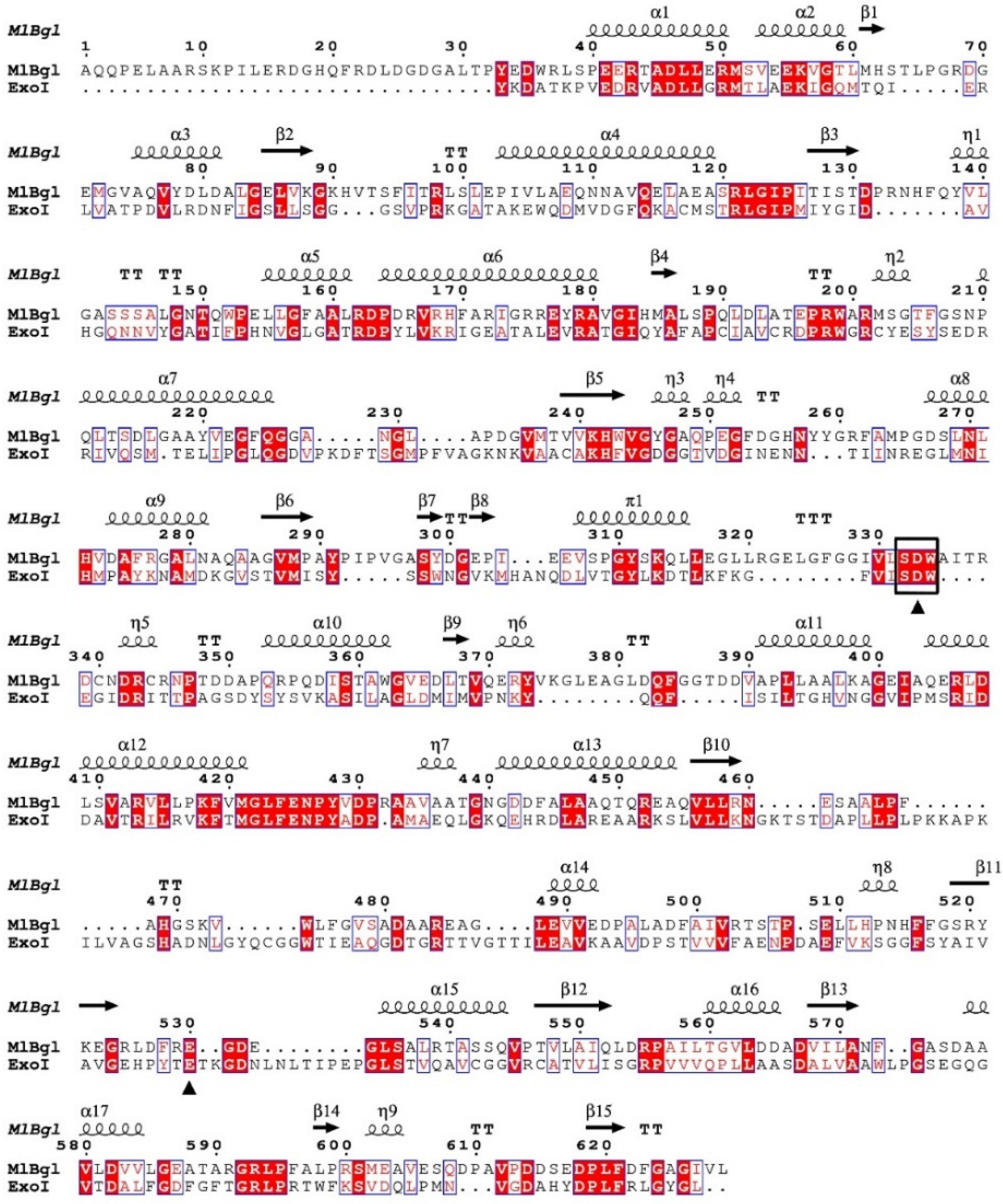
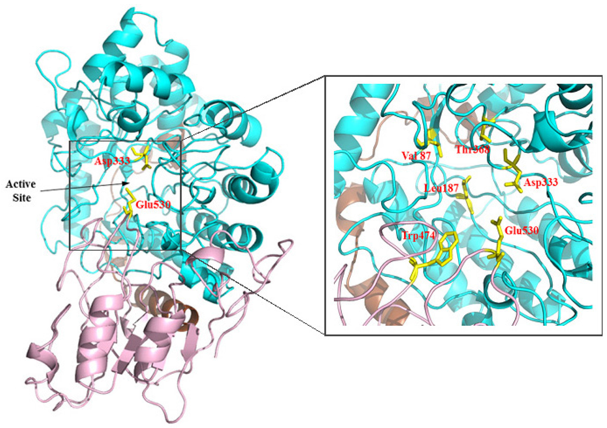
2.2. Sequence Analysis

The plasmid from fosmid clone fosSCSIO2 was extracted and subcloned. The positive subclone of plasmid pUC-MlBgl was sequenced. Sequence analysis showed that pUC-MlBgl contained an open reading frame (ORF) coding for β-glucosidase. The overall G + C content of the ORF was 59%. The deduced protein product consisted of 663 amino acids which contained a catalytic domain belonging to glycosyl hydrolase family 3 according to the CAZy database (http://prosite.expasy.org/). A signal peptide (residues 1–33) was found by the analysis of SignalP (version 4.1, http://www.cbs.dtu.dk/services/SignalP/). The predicted molecular mass of the protein was estimated to be 71.2 kDa. The deduced protein showed highest identity with a predicted β-glucosidase from Sphingomonas sp. LH128 (64% identity) in the GenBank database. The secondary structures of r-MlBgl and structure-based sequences alignment with β-d-glucan exohydrolase isoenzyme ExoI are shown in Figure 1. Multiple sequence alignment revealed that r-MlBgl shared the glycosyl hydrolase family 3 catalytic central conserved region Ser-Asp-Trp [26,27]. The conserved and similar amino acids are indicated by boxes and the predicted catalytic nucleophile residue Asp333 and acid/base residue Glu530 are indicated by filled triangles.
The 3D model structure of r-MlBgl was predicted based on the crystal structure of β-d-glucan exohydrolase isoenzyme ExoI by using the Swiss-Model server (Figure 2). The quality of the predicted model r-MlBgl was acceptable by checking using the Ramachandran plot. The structure of r-MlBgl contains two domain architecture, corresponding to the Glyco_hydro_3 (residues 117–449) and Glyco_hydro_3_C (residues 490–661) domain families. Domain 1 forms an (α/β)8 barrel and the second domain forms an (α/β)6 sheet. The active site cleft is formed by the interface of domains 1 and 2 in both monomers. β-Glucosidase has a retaining catalytic mechanism [28], the residue Asp functions as the catalytic nucleophile by attacking at the substrate anomeric center to form a covalent α-d-glucosyl enzyme intermediate, while the other residue Glu/His acts as an acid/base catalyst by protonating the glycosidic oxygen in the first step and deprotonating the nucleophilic water molecule in the second step [29]. Asp333 in domain 1 and Glu530 in domain 2 were well aligned with the catalytic residues (Asp285 and Glu491) in HvExoI [30], suggesting that Asp333 and Glu530 may act as the catalytic nucleophile and acid/base, respectively. According to the 3D model structure of HvExoI, the residues Val87, Leu187, Thr368, and Trp474 were considered important and may participate in substrate recognition and binding. However, due to limitations of current homology 3D model structures and modeling methods, the crucial residues of r-MlBgl for the catalysis are required for a detailed structural comparative analysis and further site directed mutation assay.

2.3. Enzyme Overexpression and Purification

The β-glucosidase gene MlBgl was overexpressed in Escherichia coli BL21(DE3) cells. The recombinant r-MlBgl with an N-terminal 6 × His-tagged protein was purified with Ni2+-NTA chromatography. SDS-PAGE analysis showed that r-MlBgl was purified to homogeneity, with a molecular mass approximately of 71 kDa which corresponded to the calculated mass (Figure 3).

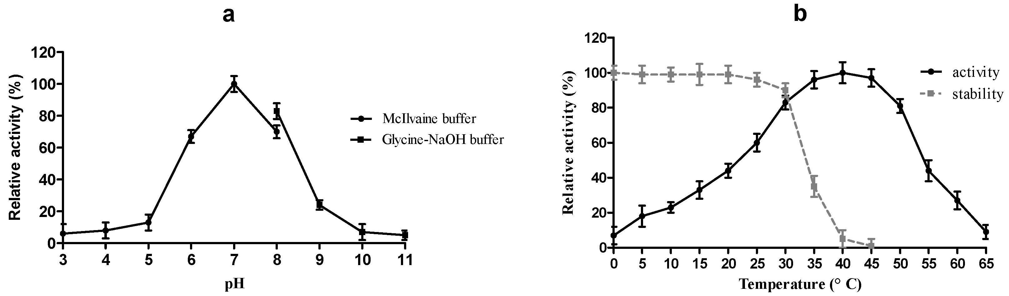
2.4. Physico-Chemical Characterization of r-MlBgl

Figure 4a shows that r-MlBgl was most active at pH 7.0. The optimal temperature of r-MlBgl was determined to be 40 °C (Figure 4b). The optimal pH and temperature of r-MlBgl was similar to β-glucosidase isolated from the metagenomic library of mangrove soil [20]. r-MlBgl maintained more than 50% activity at temperatures from 25 to 35 °C and approximately 10% activity at 0 °C, indicating it was a cold adapted enzyme. The cold adapted β-glucosidases have the advantage in reaction of requiring low temperature, such as increasing resveratrol in wine [31]. Also, they can offer economic benefits through energy savings in food processing or bioconversion [32]. The activity of r-MlBgl was stable at a temperature below 30 °C, but its activity decreased dramatically when the temperature climbed up to 35 °C after incubating for 1 h (Figure 4b). The cold-active enzymes were usually highly active at low temperature but thermolabile [33]. Compared with some cold-active β-glucosidases, r-MlBgl showed higher thermostability at 30 °C [32,34,35]. High hydrolytic efficiency and thermostability are both important properties in industrial application. Althought r-MlBgl exhibited higher activity at low temperature than thermophilic β-glucosidases [36,37], the lower thermostability limited its application. It was reported that the activity and thermostability of cold-active enzymes could be co-evolutionary [38], so direct evolution work of improving the activity and thermostability of r-MlBgl is now under study.
Activity of r-MlBgl in the presence of 1 mM and 10 mM concentration of different metal ions or chemical reagents is shown in Table 1. Most metal ions had no significant inhibition on the activity of r-MlBgl except Zn2+, Ba2+, and Cu2+ at 1 mM concentration. Whereas, Ca2+, Ni2+, Co2+, EDTA, and SDS at 10 mM were also potential inhibitors.

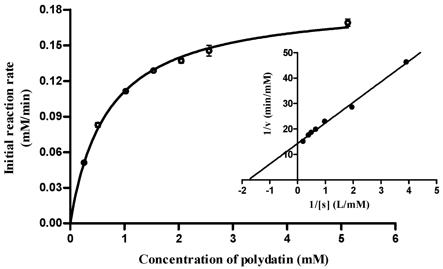
2.5. Substrate Specificity and Enzymatic Kinetics

To investigate the substrate specificity, activities of r-MlBgl towards different substrates were assayed (Table 2). r-MlBgl proved to be most active toward polydatin, and showed high activity on p-nitrophenyl-β-d-glucopyranoside (pNPG) and esculin, indicating that r-MlBgl could hydrolyze aryl β-glycosidic bonds effectively. r-MlBgl exhibited weak activities toward p-nitrophenyl-β-d-galactopyranoside and p-nitrophenyl-β-d-xylopyranoside but no detectable activity toward p-nitrophenyl-β-d-cellobioside. It was similar to the β-glucosidases BglA and BglJ from Aspergillus oryzae which could hydrolyze p-nitrophenyl-β-d-xylopyranoside slowly but with no detectable activity toward p-nitrophenyl-β-d-cellobioside [39]. For the oligosaccharides and polysaccharides, r-MlBgl showed no activities toward cellobioside, cellotriose, carboxymethyl cellulose, avicel, and starch, indicating that r-MlBgl could not hydrolyze the oligosaccharides and polysaccharides form by β-(1,4), α-(1,4), and α-(1,6) glycosidic bonds. The substrate specificity analysis suggested that r-MlBgl possessed strong substrate specificity. It could only hydrolyze aryl β-glycosides and had no hydrolysis activities toward oligosaccharides and polysaccharides. According to the classification by substrate specificity, r-MlBgl belongs to aryl β-glucosidase [40].
The kinetic parameters of r-MlBgl were determined using pNPG as the substrate. The initial rate of the reaction was measured under optimal conditions. The kinetic parameters of Km and Vmax were determined by plotting the substrate concentration vs. the initial velocity of each reaction and subjecting the data to non linear regression analysis. Results showed that the Km and Vmax values of r-MlBgl were 0.69 mM and 248.26 U·mg−1 respectively. The kcat and the kcat/Km were 581.52 s−1 and 842.78 mM−1·s−1, respectively. β-Glucosidases from different sources of microorganisms have variable kinetic parameters of Km and kcat values but usually lower than 1 mM and 1000 s−1, respectively [11,17,39]. The detected Km and kcat values of r-MlBgl toward this substrate were within the same range.

2.6. Enzymatic Conversion of Polydatin to Resveratrol

Enzymatic conversion of polydatin to resveratrol was determined by HPLC. The hydrolyzing parameters of r-MlBgl towards polydatin were investigated by Michaelis-Menten plots (Figure 5). The productivity of resveratrol was 1.28 mM/h. The Km, kcat and kcat/Km values for polydatin were 0.67 mM, 989 s−1 and 1476 mM−1·s−1, respectively. Due to the important role of resveratrol and its pharmacological properties, considerable scientific attention is being achieved in the enzymatic conversion of polydatin to resveratrol. It was reported that β-glucosidases from Lactobacillus kimchi and Aspergillus oryzae hydrolyze polydatin effectively. The comparison of productivity and kinetic parameters of r-MlBgl with other β-glucosidases toward polydatin are seen in Table 3. Compared with these reported β-glucosidases, r-MlBgl exhibited the highest productivity of resveratrol of the β-glucosidases from Aspergillus oryzae and Lactobacillus kimchi per unit of enzyme [12,17]. r-MlBgl also exhibited higher kcat and kcat/Km values than β-glucosidase from Lactobacillus kimchi, indicating it is able to hydrolyze polydatin more effectively.

3. Experimental Section

3.1. Strains, Vectors and Reagents

Escherichia coli EPI300-T1R and Copycontrol pCC2FOS (Epicentre, Madison, WI, USA) were used as host strain and vector for construction of the metagenomic library. pET22b (+) (Novagen, Madison, WI, USA) was used as the expression vector to produce the target protein in Escherichia coli BL21(DE3) (Novagen). DNA isolation and purification kits, Escherichia coli DH5α, pMD19-T vector, restriction endonucleases, DNA polymerase, dNTPs, T4 DNA ligase and isopropyl-β-d-1-thiogalactopyranoside (IPTG) were purchased from TaKaRa. Substrates p-nitrophenyl-β-d-glucopyranoside (pNPG), polydatin, and resveratrol standards were from Sigma-Aldrich. All other chemicals were of analytical grade unless otherwise stated.

3.2. Metagenomic Library Construction

The topsoil samples (0–10 cm) were collected from the Mangrove Reserve of Sanya City (18°15′16.32′′N, 109°30′28.10′′E), Hainan province of China. The samples were stored at −20 °C until the DNA extractions were performed. Extraction of the total genomic DNA from soil samples was performed according to the method reported with minor modifications [23]. The DNA fragments (35–48 kb) were obtained and purified by pulsed-field gel electrophoresis (PFGE). And then the metagenomic library was constructed following the instructions of the CopyControl Fosmid Library Production Kit.

3.3. Screening of β-Glucosidase Gene MlBgl

Recombinant Escherichia coli EPI300-T1R strains of the metagenomic library were replicated onto Luria-Bertani (LB) agar plates containing 0.1% esculin to detect β-glucosidase activity. The plates were incubated at 28 °C for 16 h and then placed at room temperature for 2–3 days. The clones that turned dark brown on the plate were selected as β-glucosidase positive recombinants. The fosmid DNA of positive clones were isolated and partially digested with Sau3AⅠ. DNA fragments of 1–5 kb were recovered and ligated into pUC19 with BamH І digested. And then transformed to Escherichia coli DH5α. The subcloned library was rescreened of β-glucosidase activities and the plasmid from positive clones was sequenced.

3.4. Sequence Analysis

The sequence similarities and conserved motif of MlBgl was analyzed by BLAST program (version 2.3, http://www.ncbi.nlm.nih.gov/BLAST). Multiple sequence alignment of MlBgl with similar β-glucosidase sequences was carried out using the ClustalX (Version 2.0, EMBL-EBI) and DNAMAN (Version 6.0, Lynnon Biosoft Corp., San Ramon, CA, USA) program. The three-dimensional (3D) model of r-MlBgl was generated by homology modelling using the Swiss-Model server (http://swissmodel.expasy.org). The crystal structure of β-d-glucan exohydrolase isoenzyme ExoI (PDB code: 1EX1) was served as template for modelling the structure of r-MlBgl. The coloring and labeling of the 3D structures were performed by using PyMOL software (version 1.8, www.pymol.org).

3.5. Gene Expression and Purification of Recombinant β-Glucosidase r-MlBgl

The β-glucosidase gene MlBgl was amplified by using the primer pair of GCATGCCATGGGCGCACAGCAACCGGAATTGGCAG (Nco І digestion site underlined) and CCTCCGCTCGAGCAGCACGATCCCGGCTCCAAAG (Xho І digestion site underlined). The PCR product was digested with restriction endonucleases Nco І and Xho І, then ligated into pET-22b (+) vector which digested with the same enzymes. The recombinant plasmid pET-MlBgl was transformed into Escherichia coli BL21(DE3) to express the target protein. The transformed Escherichia coli BL21(DE3) cells carrying pET-MlBgl were cultured in LB medium containing ampicillin (100 µg/mL) at 37 °C. After inducing in 1 mM IPTG at 22 °C for 16 h, cells were harvested, washed twice with Tris-HCl buffer (pH 7.6), and lysed by sonication on ice for 20 min at a pulse frequency of 4 s/4 s. The lysate was centrifuged at 10,000 g for 20 min at 4 °C. The supernatant was applied to a Ni2+-NTA agarose gel column for purification. The recombinant β-glucosidase r-MlBgl was eluted with 200 mM imidazole and then dialyzed in deionized double-distilled H2O at 4 °C for 24 h. The purified protein was detected with sodium dodecyl sulfate-polyacrylamide gel electrophoresis (SDS-PAGE). The protein concentration was determined by the Bradford method [42] with bovine serum albumin as standard.

3.6. Enzyme Activity Assay

β-Glucosidase activity was determined using pNPG as the substrate [43]. The reaction mixure contained 1 μL of appropriately diluted enzyme, 10 μL 25 mM pNPG, and 89 μL 0.2 M McIlvaine buffer (pH 7.0). After incubation at 40 °C for 5 min, the reaction was terminated with 50 μL 1 M Na2CO3. The amount of liberated p-nitrophenol was measured by detecting the absorption at 405 nm. The control was set by adding 50 μL 1 M Na2CO3 before addition of the enzyme. One unit of β-glucosidase activity (U) was defined as the amount of enzyme that produced 1 μmol p-nitrophenol per min under the optimal conditions. To determine the Km and Vmax values towards pNPG, a series of concentrations (0.25–2.5 mM) of pNPG in McIlvaine buffer (pH 7.0) were incubated at 40 °C for 5 min. The reaction starting and stopping conditions were the same as those used previously.

3.7. Physico-Chemical Characterization of r-MlBgl

The characterization of r-MlBgl was detected by using pNPG as the substrate. The optimal reaction pH was tested in varying pH buffer (pH 3.0–11.0) at 40 °C. The buffers used were 0.2 M McIlvaine buffer for pH 3.0–8.0 and 0.05 M glycine-NaOH buffer for pH 8.0–11.0. The optimal reaction temperature was determined in McIlvaine buffer (pH 7.0) at a temperature from 0 to 65 °C at intervals of 5 °C. The thermal stability of r-MlBgl was determined by measuring the residual activity after incubating enzyme at different temperatures from 0 °C to 45 °C for 1 h. To investigate the effects of different metal ions on enzyme activity, 1 mM and 10 mM (final concentration) of Na+, K+, Li+, NH4+, Mg2+, Fe3+, Zn2+, Ca2+, Cu2+, Ni2+, Ba2+, Co2+, EDTA, and SDS were individually added to the reaction system. All enzyme activities were determined in three independent experiments.

3.8. Substrate Specificity

Substrate specificity of r-MlBgl was determined by using pNPG, p-nitrophenyl-β-d-galactopyranoside, p-nitrophenyl-β-d-cellobioside and p-nitrophenyl-β-d-xylopyranoside at 2.5 mM final concentration. r-MlBgl activities on esculin, carboxymethylcellulose (CMC), xylan, avicel and starch as substrates at 1% concentration were also tested. The activities were estimated by assaying the reducing sugars via DNS (3,5-dinitrosalicylic acid) method [44]. The product from reaction of DNS and reducing sugar was brown on heating, and the amount of reducing sugar was determined by detecting the brown product at OD540 using the glucose curve as standard. Activities on cellobiose and cellotriose were determined by using the glucose assay kit. One unit of activity (U) was defined as the amount of enzyme that produced 1 μmol reducing sugar per min under the optimal conditions.

3.9. Enzymatic Conversion of Polydatin to Resveratrol

To determine the kinetic properties of recombinant r-MlBgl toward polydatin, 200 μL reaction mixtures containing 0.0125 U enzyme and a series of concentrations of polydatin (0.256–5.12 mM) in McIlvaine buffer (pH 7.0) were incubated at 40 °C for 5 min. The reaction was stopped by adding 800 μL methanol. For analysis of the amount of resveratrol produced, the reaction mixtures were diluted 10 times with methanol and filtered through a 0.22 μm membrane prior to high performance liquid chromatography (HPLC). An Agilent system equipped with a reverse phase column C 18 (4.6 × 250 mm, 5 μm) was used in this study. The sample was injected and eluted with a linear gradient of solvent A (0.1% formic acid in water) and solvent B (100% acetonitrile) from 95:5 to 20:80 for 45 min, and then from 20:80 to 95:5 for 5 min at a flow rate of 1 mL/min. The UV absorption was measured at 306 nm. The amount of polydatin and resveratrol in the samples were calculated using standard curves prepared from known concentrations of standards. All enzymatic conversions were determined in three independent experiments, and a reaction mixture with heat inactivated enzyme was used as a blank control. The kinetic parameters of Km and Vmax towards polydatin were subjected to nonlinear regression analysis with the GraphPad Prism 5.0 software (Version 5.0, GraphPad software Inc., San Diego, CA, USA).

3.10. Nucleotide Sequence Accession Number

The nucleotide sequences of the β-glucosidase gene MlBgl from the mangrove metagenomic library were deposited in the GenBank database under accession number KF424271.

4. Conclusions

A metagenomic library of mangrove soil was constructed and a novel GH3 β-glucosidase was isolated by functional screening. The recombinant r-MlBgl was overexpressed, purified, and characterized. r-MlBgl exhibited high relative activities at low temperature. It had strong substrate specificity that could only hydrolyze aryl β-glycosides. A very attractive characteristic of r-MlBgl was the high catalytic efficiency toward polydatin. The kcat, kcat/Km and productivity of r-MlBgl toward polydatin were higher than the β-glucosidases previously reported. These properties make it a good candidate in enzymatic conversion of polydatin to resveratrol. This study also highlights the utility of the metagenomic approach in discovering novel β-glucosidases for hydrolysis of polydatin.

Acknowledgments

This work was supported by grants from the Administration of Ocean and Fisheries of Guangdong Province (GD2012-D01-002); the National High Technology Research and Development Program of China (NO.2012AA092104); the “Strategic Priority Research Program” of the Chinese Academy of Sciences (NO.XDA10030400); and the Natural Science Foundation of Guangdong Province, China (Grant No.2015A030310270).

Author Contributions

Zhimao Mai performed the experiments and wrote the manuscript. Hongfei Su analyzed the data and contributed reagents/materials/analysis tools. Si Zhang conceived and designed the experiments.

Conflicts of Interest

The authors declare no conflict of interest.

References

  1. Bertelli, A.; Ferrara, F.; Diana, G.; Fulgenzi, A.; Corsi, M.; Ponti, W.; Ferrero, M.; Bertelli, A. Resveratrol, a natural stilbene in grapes and wine, enhances intraphagocytosis in human promonocytes: A co-factor in antiinflammatory and anticancer chemopreventive activity. Int. J. Tissue React. 1998, 21, 93–104. [Google Scholar]
  2. Fremont, L. Biological effects of resveratrol. Life Sci. 2000, 66, 663–673. [Google Scholar] [CrossRef]
  3. Jiang, L.; Gu, Y.; Ye, J.; Liu, F.; Zhao, Y.; Wang, C.; Xu, Y.; Cao, X.; Zhang, L.; Dong, W. Resveratrol prevents hepatic steatosis induced by hepatitis C virus core protein. Biotechnol. Lett. 2012, 34, 2205–2212. [Google Scholar] [CrossRef] [PubMed]
  4. Sánchez-Patán, F.; Monagas, M.; Moreno-Arribas, M.V.; Bartolomé, B. Determination of microbial phenolic acids in human faeces by UPLC-ESI-TQ MS. J. Agric. Food Chem. 2011, 59, 2241–2247. [Google Scholar] [CrossRef] [PubMed]
  5. Wang, H.; Liu, L.; Guo, Y.X.; Dong, Y.S.; Zhang, D.J.; Xiu, Z.L. Biotransformation of piceid in Polygonum cuspidatum to resveratrol by Aspergillus oryzae. Appl. Microbiol. Biotechnol. 2007, 75, 763–768. [Google Scholar] [CrossRef] [PubMed]
  6. Chong, Y.; Yan, A.; Yang, X.; Cai, Y.; Chen, J. An optimum fermentation model established by genetic algorithm for biotransformation from crude polydatin to resveratrol. Appl. Biochem. Biotechnol. 2012, 166, 446–457. [Google Scholar] [CrossRef] [PubMed]
  7. Voorhorst, W.; Eggen, R.; Luesink, E.J.; De Vos, W. Characterization of the celB gene coding for beta-glucosidase from the hyperthermophilic archaeon Pyrococcus furiosus and its expression and site-directed mutation in Escherichia coli. J. Bacteriol. 1995, 177, 7105–7111. [Google Scholar] [PubMed]
  8. Shewale, J. β-Glucosidase: Its role in cellulase synthesis and hydrolysis of cellulose. Int. J. Biochem. 1982, 14, 435–443. [Google Scholar] [CrossRef]
  9. Hu, Y.; Luan, H.; Zhou, K.; Ge, G.; Yang, S.; Yang, L. Purification and characterization of a novel glycosidase from the china white jade snail (Achatina fulica) showing transglycosylation activity. Enzym. Microb. Technol. 2008, 43, 35–42. [Google Scholar] [CrossRef]
  10. González-Pombo, P.; Fariña, L.; Carrau, F.; Batista-Viera, F.; Brena, B.M. A novel extracellular β-glucosidase from Issatchenkia terricola: Isolation, immobilization and application for aroma enhancement of white Muscat wine. Process Biochem. 2011, 46, 385–389. [Google Scholar] [CrossRef]
  11. Du, L.; Wang, Z.; Zhao, Y.; Huang, J.; Pang, H.; Wei, Y.; Lin, L.; Huang, R. A β-glucosidase from Novosphingobium sp. GX9 with high catalytic efficiency toward isoflavonoid glycoside hydrolysis and (+)-catechin transglycosylation. Appl. Microbiol. Biotechnol. 2014, 98, 7069–7079. [Google Scholar] [CrossRef] [PubMed]
  12. Chen, M.; Li, D.; Gao, Z.; Zhang, C. Enzymatic transformation of polydatin to resveratrol by piceid-β-d-glucosidase from Aspergillus oryzae. Bioprocess Biosyst. Eng. 2014, 37, 1411–1416. [Google Scholar] [CrossRef] [PubMed]
  13. Loughlin, W.A. Biotransformations in organic synthesis. Bioresour. Technol. 2000, 74, 49–62. [Google Scholar] [CrossRef]
  14. Tian, T.; Sun, Q.; Shen, J.; Zhang, T.; Gao, P.; Sun, Q. Microbial transformation of polydatin and emodin-8-β-d-glucoside of Polygonum cuspidatum Sieb. et Zucc into resveratrol and emodin respectively by Rhizopus microsporus. World J. Microbiol. Biotechnol. 2008, 24, 861–866. [Google Scholar] [CrossRef]
  15. Jin, S.; Luo, M.; Wang, W.; Zhao, C.J.; Gu, C.B.; Li, C.Y.; Zu, Y.G.; Fu, Y.J.; Guan, Y. Biotransformation of polydatin to resveratrol in Polygonum cuspidatum roots by highly immobilized edible Aspergillus niger and Yeast. Bioresour. Technol. 2013, 136, 766–770. [Google Scholar] [CrossRef] [PubMed]
  16. Zhang, C.; Li, D.; Yu, H.; Zhang, B.; Jin, F. Purification and characterization of piceid-β-d-glucosidase from Aspergillus oryzae. Process Biochem. 2007, 42, 83–88. [Google Scholar] [CrossRef]
  17. Ko, J.A.; Park, J.; Kwon, H.; Ryu, Y.; Jeong, H.; Park, S.; Kim, C.; Oh, H.; Park, C.; Lim, Y. Purification and functional characterization of the first stilbene glucoside-specific β-glucosidase isolated from Lactobacillus kimchi. Enzyme Microbiol. Technol. 2014, 67, 59–66. [Google Scholar] [CrossRef]
  18. Amann, R.I.; Ludwig, W.; Schleifer, K.H. Phylogenetic identification and in situ detection of individual microbial cells without cultivation. Microbiol. Rev. 1995, 59, 143–169. [Google Scholar] [PubMed]
  19. Jiang, C.; Li, S.X.; Luo, F.F.; Jin, K.; Wang, Q.; Hao, Z.Y.; Wu, L.L.; Zhao, G.C.; Ma, G.F.; Shen, P.H. Biochemical characterization of two novel β-glucosidase genes by metagenome expression cloning. Bioresour. Technol. 2011, 102, 3272–3278. [Google Scholar] [CrossRef] [PubMed]
  20. Li, G.; Jiang, Y.; Fan, X.J.; Liu, Y.H. Molecular cloning and characterization of a novel β-glucosidase with high hydrolyzing ability for soybean isoflavone glycosides and glucose-tolerance from soil metagenomic library. Bioresour. Technol. 2012, 123, 15–22. [Google Scholar] [CrossRef] [PubMed]
  21. Feng, Y.; Duan, C.J.; Liu, L.; Tang, J.L.; Feng, J.X. Properties of a metagenome-derived β-glucosidase from the contents of rabbit cecum. Biosci. Biotechnol. Biochem. 2009, 73, 1470–1473. [Google Scholar] [CrossRef] [PubMed]
  22. Nacke, H.; Engelhaupt, M.; Brady, S.; Fischer, C.; Tautzt, J.; Daniel, R. Identification and characterization of novel cellulolytic and hemicellulolytic genes and enzymes derived from German grassland soil metagenomes. Biotechnol. Lett. 2012, 34, 663–675. [Google Scholar] [CrossRef] [PubMed]
  23. Mai, Z.; Su, H.; Li, L.; Zhang, S. Construction of a mangrove soil metagenome library and identification of two novel β-glucosidase genes. Biotechnol. Bull. 2014, 6, 168–172. [Google Scholar]
  24. Kim, S.J.; Lee, C.M.; Han, B.R.; Kim, M.Y.; Yeo, Y.S.; Yoon, S.H.; Koo, B.S.; Jun, H.K. Characterization of a gene encoding cellulase from uncultured soil bacteria. FEMS Microbiol. Lett. 2008, 282, 44–51. [Google Scholar] [CrossRef] [PubMed]
  25. Jiang, C.; Hao, Z.Y.; Jin, K.; Li, S.X.; Che, Z.Q.; Ma, G.F.; Wu, B. Identification of a metagenome-derived β-glucosidase from bioreactor contents. J. Mol. Catal. B 2010, 63, 11–16. [Google Scholar] [CrossRef]
  26. Mao, X.; Hong, Y.; Shao, Z.; Zhao, Y.; Liu, Z. A novel cold-active and alkali-stable β-glucosidase gene isolated from the marine bacterium Martelella mediterrânea. Appl. Biochem. Biotechnol. 2010, 162, 2136–2148. [Google Scholar] [CrossRef] [PubMed]
  27. Li, Y.K.; Chir, J.; Tanaka, S.; Chen, F.Y. Identification of the general acid/base catalyst of a family 3β-glucosidase from Flavobacterium meningosepticum. Biochemistry 2002, 41, 2751–2759. [Google Scholar] [CrossRef] [PubMed]
  28. Vocadlo, D.J.; Mayer, C.; He, S.; Withers, S.G. Mechanism of action and identification of Asp242 as the catalytic nucleophile of Vibrio furnisii N-acetyl-β-d-glucosaminidase using 2-acetamido-2-deoxy-5-fluoro-α-l-idopyranosyl fluoride. Biochemistry 2000, 39, 117–126. [Google Scholar] [CrossRef] [PubMed]
  29. Zechel, D.L.; Withers, S.G. Glycosidase mechanisms: anatomy of a finely tuned catalyst. Acc. Chem. Res. 2000, 33, 11–18. [Google Scholar] [PubMed]
  30. Varghese, J.N.; Hrmova, M.; Fincher, G.B. Three-dimensional structure of a barley β-d-glucan exohydrolase, a family 3 glycosyl hydrolase. Structure 1999, 7, 179–190. [Google Scholar] [CrossRef]
  31. Todaro, A.; Palmeri, R.; Barbagallo, R.N.; Pifferi, P.G.; Spagna, G. Increase of trans-resveratrol in typical Sicilian wine using β-glucosidase from various sources. Food Chem. 2008, 107, 1570–1575. [Google Scholar] [CrossRef]
  32. Shipkowski, S.; Brenchley, J.E. Characterization of an unusual cold-active β-glucosidase belonging to family 3 of the glycoside hydrolases from the psychrophilic isolate Paenibacillus sp. strain C7. Appl. Environ. Microbiol. 2005, 71, 4225–4232. [Google Scholar] [CrossRef] [PubMed]
  33. Siddiqui, K.S.; Cavicchioli, R. Cold-adapted enzymes. Annu. Rev. Biochem. 2006, 75, 403–433. [Google Scholar] [CrossRef] [PubMed]
  34. Crespim, E.; Zanphorlin, L.M.; de Souza, F.H.; Diogo, J.A.; Gazolla, A.C.; Machado, C.B.; Figueiredo, F.; Sousa, A.S.; Nóbrega, F.; Pellizari, V.H. A novel cold-adapted and glucose-tolerant GH1 β-glucosidase from Exiguobacterium antarcticum B7. Int. J. Biol. Macromol. 2016, 82, 375–380. [Google Scholar] [CrossRef] [PubMed]
  35. Fan, H.X.; Miao, L.L.; Liu, Y.; Liu, H.C.; Liu, Z.P. Gene cloning and characterization of a cold-adapted β-glucosidase belonging to glycosyl hydrolase family 1 from a psychrotolerant bacterium Micrococcus antarcticus. Enzym. Microbiol. Technol. 2011, 49, 94–99. [Google Scholar] [CrossRef] [PubMed]
  36. Gu, N.Y.; Kim, J.L.; Kim, H.J.; You, D.J.; Kim, H.W.; Jeon, S.J. Gene cloning and enzymatic properties of hyperthermostable β-glycosidase from Thermus thermophilus HJ6. J. Biosci. Bioeng. 2009, 107, 21–26. [Google Scholar] [CrossRef] [PubMed]
  37. Parry, N.; Beever, D.; Owen, E.; Vandenberghe, I.; Van Beeumen, J.; Bhat, M. Biochemical characterization and mechanism of action of a thermostable β-glucosidase purified from Thermoascus aurantiacus. Biochem. J. 2001, 353, 117–127. [Google Scholar] [CrossRef] [PubMed]
  38. Zhang, N.; Suen, W.C.; Windsor, W.; Xiao, L.; Madison, V.; Zaks, A. Improving tolerance of Candida antarctica lipase B towards irreversible thermal inactivation through directed evolution. Protein Eng. 2003, 16, 599–605. [Google Scholar] [CrossRef] [PubMed]
  39. Kudo, K.; Watanabe, A.; Ujiie, S.; Shintani, T.; Gomi, K. Purification and enzymatic characterization of secretory glycoside hydrolase family 3 (GH3) aryl β-glucosidases screened from Aspergillus oryzae genome. J. Biosci. Bioeng. 2015, 120, 614–623. [Google Scholar] [CrossRef] [PubMed]
  40. Bhatia, Y.; Mishra, S.; Bisaria, V. Microbial β-glucosidases: Cloning, properties, and applications. Crit. Rev. Biotechnol. 2002, 22, 375–407. [Google Scholar] [CrossRef] [PubMed]
  41. Yeom, S.J.; Kim, B.N.; Kim, Y.S.; Oh, D.K. Hydrolysis of isoflavone glycosides by a thermostable β-glucosidase from Pyrococcus furiosus. J. Agric. Food Chem. 2012, 60, 1535–1541. [Google Scholar] [CrossRef] [PubMed]
  42. Bradford, M.M. A rapid and sensitive method for the quantitation of microgram quantities of protein utilizing the principle of protein-dye binding. Anal. Biochem. 1976, 72, 248–254. [Google Scholar] [CrossRef]
  43. Mai, Z.; Yang, J.; Tian, X.; Li, J.; Zhang, S. Gene cloning and characterization of a novel salt-tolerant and glucose-enhanced β-glucosidase from a marine Streptomycete. Appl. Biochem. Biotechnol. 2013, 169, 1512–1522. [Google Scholar] [CrossRef] [PubMed]
  44. Somogyi, M. Notes on sugar determination. J. Biol. Chem. 1952, 195, 19–23. [Google Scholar]
Figure 1. The secondary structures of r-MlBgl and structure-based sequences alignment with β-d-glucan exohydrolase isoenzyme ExoI. The secondary structures and their designations are shown: arrows and coils represent the β-strands and α-helices respectively. The glycosyl hydrolase family 3 catalytic central conserved region Ser-Asp-Trp is boxed. The catalytic nucleophile residue Asp333 and acid/base residue Glu530 are indicated by filled triangles. The structure-based sequences alignment was carried out by the Swiss-Model server and the secondary structure was assigned with ESPript 3.0 (http://espript.ibcp.fr/ESPript/cgi-bin/ESPript.cgi).
Catalysts 06 00035 g001
Figure 2. The three-dimensional model of r-MlBgl predicted by homology modelling based on the crystal structure of barley ExoI. The domain 1, the domain 2, and the linker are colored in cyan, lightpink, and wheat, respectively. The predicted nucleophile and acid/base catalyst active site residues of r-MlBgl (Asp333 and Glu530 respectively) are represented as sticks and shown in yellow. The catalytic residues and other conserved residues forming the substrate binding site are shown as sticks in the boxed panel. The picture was generated in PyMOL software(version 1.8, www.pymol.org).
Catalysts 06 00035 g002
Figure 3. A 10% SDS-PAGE analysis of the purified r-MlBgl. M protein marker; 1 recombinant Escherichia coli BL21(DE3) harboring pET-MlBgl induced with IPTG; 2 purified r-MlBgl.
Catalysts 06 00035 g003
Figure 4. Effects of pH and temperature on the activity of the purified r-MlBgl. (a) Effect of pH on r-MlBgl activity. The effect of pH on r-MlBgl activity was determined between pH 3.0–11.0 at 40 °C; (b) Effect of temperature on r-MlBgl activity and thermostability assay. The activity was determined in McIlvaine buffer (pH 7.0) at various temperatures (0 to 65 °C); Thermostability of r-MlBgl was assayed after pre-incubation at temperatures 0 to 45 °C for 1 h. The error bars represent the means ± SD (n = 3).
Catalysts 06 00035 g004
Figure 5. Michaelis-Menten plots for the reactions with substrate of polydatin. The inset shows the Lineweaver-Burk plots. Each data point represents the mean ± SD of three independent experiments.
Catalysts 06 00035 g005
Table 1. Effect of 1 or 10 mM metal ions and chemical reagents on the activity of r-MlBgl.
ReagentsRelative Activity (%) a
Concentration (1 mM)Concentration (10 mM)
None100.0 ± 2.6 b100.0 ± 4.2
Na+98.9 ± 5.481.53 ± 3.4
K+104.5 ± 2.585.6 ± 3.2
Li+91 ± 3.680 ± 4.1
NH4+102.4 ± 4.293.3 ± 4.8
Mg2+104.1 ± 4.388.1 ± 3.5
Fe3+96.5 ± 3.258.8 ± 3.6
Zn2+12.3 ± 2.45.7 ± 1.1
Ca2+58.4 ± 4.345.6 ± 2.9
Cu2+8.2 ± 2.35.9 ± 1.4
Ni2+82.4 ± 4.259.3 ± 3.8
Ba2+44.5 ± 3.423.4 ± 3.1
Co2+66.1 ± 3.544.7 ± 4.6
EDTA56.4 ± 4.725.9 ± 3.8
SDS76.4 ± 4.855.2 ± 3.1
a Assay was performed under optimum conditions; b Values represent the means ± SD (n = 3).
Table 2. Substrate specificity of r-BglNH.
SubstratesConfiguration of Glycoside LinkageSpecific Activity (U·mg−1) a
Aryl β-glycosides--
p-Nitrophenyl-β-d-glucopyranosideβ-Glucose248.26
p-Nitrophenyl-β-d-galactopyranosideβ-Galactose 28.79
p-Nitrophenyl-β-d-cellobiosideβ-CellobioseND
p-Nitrophenyl-β-d-xylopyranosideβ-Xylose18.87
Polydatinβ-Glucose422.21
Esculinβ-Glucose126.69
Oligosaccharides and polysaccharides--
Cellobioseβ-(1,4)-GlucoseND
Cellotrioseβ-(1,4)-GlucoseND
Carboxymethyl celluloseβ-(1,4)-GlucoseND
Avicelβ-(1,4)-GlucoseND
Starchα-(1,4) and α-(1,6)-GlucoseND
a Assay was determined at the optimum condition; ND, no detectable activity.
Table 3. A comparison of productivity and kinetic parameters of r-MlBgl for polydatin with other β-glucosidases.
Enzyme OriginAmountsTime (h)Productivity (mM/h)Km (mM)kcat (s−1)kcat/Km (mM−1·s−1)References
Lactobacillus kimchi3.28 μg/mL400.640.201.296.45[17]
Aspergillus oryzaeNR a2NR0.74NRNR[16]
Aspergillus oryzae5 U/mL45.12NRNRNR[12]
Metagenomic library0.13 U/mL11.28 b0.679891476This study
a NR, not reported; b The reaction mixture containing 0.125 U/mL r-MlBgl and 1 mg/mL polydatin was performed in 0.2 M McIlvaine buffer (pH 7.0) at 30 °C for 1 h. The productivity (mM/h) was defined as the increase in the concentration of resveratrol produced in 1 h [41].

