Article

New Acetylenic Amine Derivatives of 5,8-Quinolinediones: Synthesis, Crystal Structure and Antiproliferative Activity

1
Department of Organic Chemistry, School of Pharmacy with the Division of Laboratory Medicine in Sosnowiec, Medical University of Silesia in Katowice, 4 Jagiellońska Str., 41-200 Sosnowiec, Poland
2
Department of Solid State Physics, Institute of Physics, University of Silesia, 4 Uniwersytecka Str., 40-007 Katowice, Poland
3
Silesian Center for Education and Interdisciplinary Research, University of Silesia, 75 Pułku Piechoty 1, 41-500 Chorzów, Poland
4
Department of Cell Biology, School of Pharmacy with the Division of Laboratory Medicine in Sosnowiec, Medical University of Silesia in Katowice, 8 Jedności Str., 41-200 Sosnowiec, Poland
5
Department of Physics of Crystals, Institute of Physics, University of Silesia, 4 Uniwersytecka Str., 40-007 Katowice, Poland
6
Department of Biophysics and Molecular Physics, Institute of Physics, University of Silesia, 4 Uniwersytecka Str., 40-007 Katowice, Poland
*
Author to whom correspondence should be addressed.
Crystals 2017, 7(1), 15; https://doi.org/10.3390/cryst7010015
Submission received: 20 December 2016 / Revised: 2 January 2017 / Accepted: 4 January 2017 / Published: 7 January 2017
(This article belongs to the Section Crystal Engineering)

Abstract

:
Acetylenic amine derivatives of the 5,8-quinolinedione were synthesized and characterized by the 1H and 13C NMR, IR spectroscopy and MS spectra. Additionally, the 6- and 7-substituted allylamine-5,8-quinolinediones were synthesized for comparison purposes. The crystal structure was determined for the 6-chloro-7-propargylamine-5,8-quinolinedione and 7-chloro-6-propargylamine-5,8-quinolinedione. Additionally, the IR spectral analysis supplemented by the density functional theory (DFT) calculations were carried out. It was found that different positions of the propargylamine side chain had a distinct influence on crystal structure, formation of H-bonds and the carbonyl stretching IR bands. Correlation between the frequency separation Δν of the carbonyl IR bands and the position of the 6- and 7-substituents was found. The 7-substituted derivatives exhibited a higher frequency separation Δν. The observed correlation could provide an opportunity to use the IR spectroscopy to study substitution reactions. Cytotoxic activities against three human cancer cell lines for the 5,8-quinolinedione derivatives with different amine substituents, i.e., propargylamine, N-methylpropargylamine, 1,1-dimethylpropargylamine, allylamine and propylamine were also analysed with respect to their molecular structure.

Graphical Abstract

1. Introduction

The 5,8-quinolinedione derivatives were among the first compounds to be systematically modified in order to find products with higher biological activities, such as anticancer, anti-inflammatory or antibacterial [1,2,3,4,5,6,7,8,9]. For example, it was found that substitution of the electron-withdrawing groups at the 6- or 7-positions of the 5,8-quinolinedione led to an increase in the DNA degradation [8,9,10,11,12,13].
There are many reports on the synthesis, structure and biological activity of the amine derivatives of 5,8-quinolinedione, whereas studies on the alkyne amino analogues are very scarce [14]. Natural and synthetic acetylenic derivatives of the quinoline attract increasing attention since many of them display wide biological activity spectra [15,16,17,18,19,20,21,22]. According to the literature data, introduction of the alkyne group may significantly improve biological activity of these compounds [21,22].
In this study, we present synthesis and antiproliferative activity of the series of acetylenic amino derivatives of the 5,8-quinolinedione. Moreover, the structural properties of two acetylenic compounds, i.e., 6-chloro-7-propargylamine-5,8-quinolinedione and7-chloro-6-propargylamine-5,8-quinolinedione were determined by X-ray diffraction and IR spectroscopy.
Our attention was also focused on the carbonyl stretching bands in the infrared spectra of the propargylamine-substituted 5,8-quinolinedione, which are known as bands very sensitive to morphology. For para-quinones, one or two carbonyl bands can be observed. This feature can be influenced by many factors: mainly intra- and intermolecular interactions and conformational changes [23,24]. It can create the opportunity for additional structural investigations using IR carbonyl bands. For example, we recently found an interesting correlation between the frequency separation of carbonyl bands and the position of propylamine substituents on 5,8-quinolinediones [25]. Therefore, in this report we also aimed to check whether a similar correlation exists for the propargylamine-substituted 5,8-quinolinediones.

2. Results and Discussion
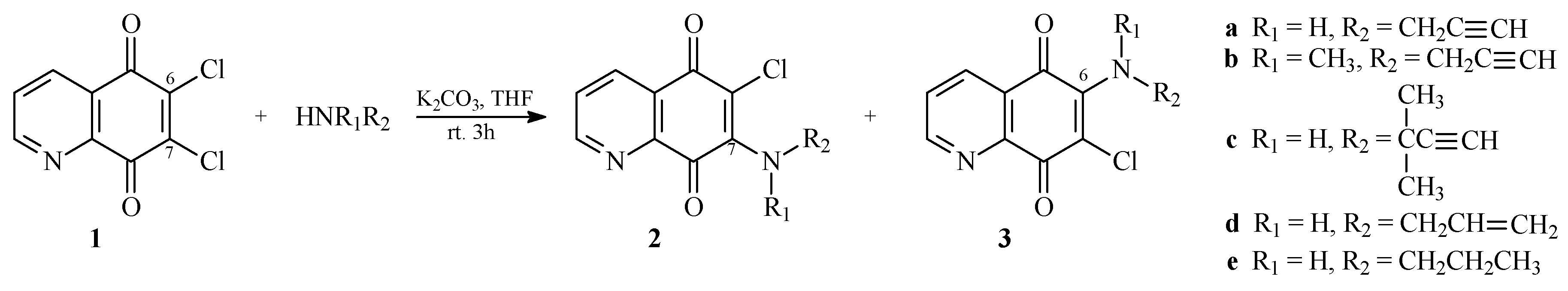
2.1. Chemistry

The 6,7-dichloro-5,8-quinolinedione 1 was prepared by the oxidation of the 8-hydroxyquinoline [12] and used as a starting compound for the synthesis of the acetylenic derivatives 23 using procedures described in the literature [8,9,25]. Treatment of compound 1 with the corresponding amine in tetrahydrofuran in the presence of potassium carbonate at room temperature gave a mixture of 7- and 6-aminosubstituted derivatives, 2ae and 3ae, respectively (Scheme 1).
The obtained mixtures were separated by column chromatography to afford pure products 2ae and 3ae with the 68%–58% and 16%–21% yields, respectively. The structures of all derivatives 23 were determined by the 1H, 13C NMR, IR and MS spectra.
For both isomeric compounds 2 and 3 the 1H NMR chemical shifts were similar, and therefore it was not possible to distinguish between 6- and 7-substituents based on the spectra (see Figure S1–S8 in supplement material). According to the literature data [8,12], such differentiation is possible when using the 13C NMR spectra. It was found that the isomers 2 and 3 showed different signal intensities of the C-5, C-8, C-6 and C-7 atoms. For the 6-aminosubstituted derivatives, the signal intensities of the C-5 and C-7 atoms were higher than those for the C-8 and C-6 atoms. For the 7-aminosubstituted derivatives, the signal intensities followed the opposite relation, i.e., they were higher for the C-8 and C-6 atoms. Therefore, it was confirmed that compounds 2 and 3 possessed the amine group at the C-7 and C-6 positions, respectively. Additionally, for derivatives 2a and 3a, the X-ray diffraction analysis confirmed also substitution of the propargylamine chain at the C-7 and C-6 positions, respectively.

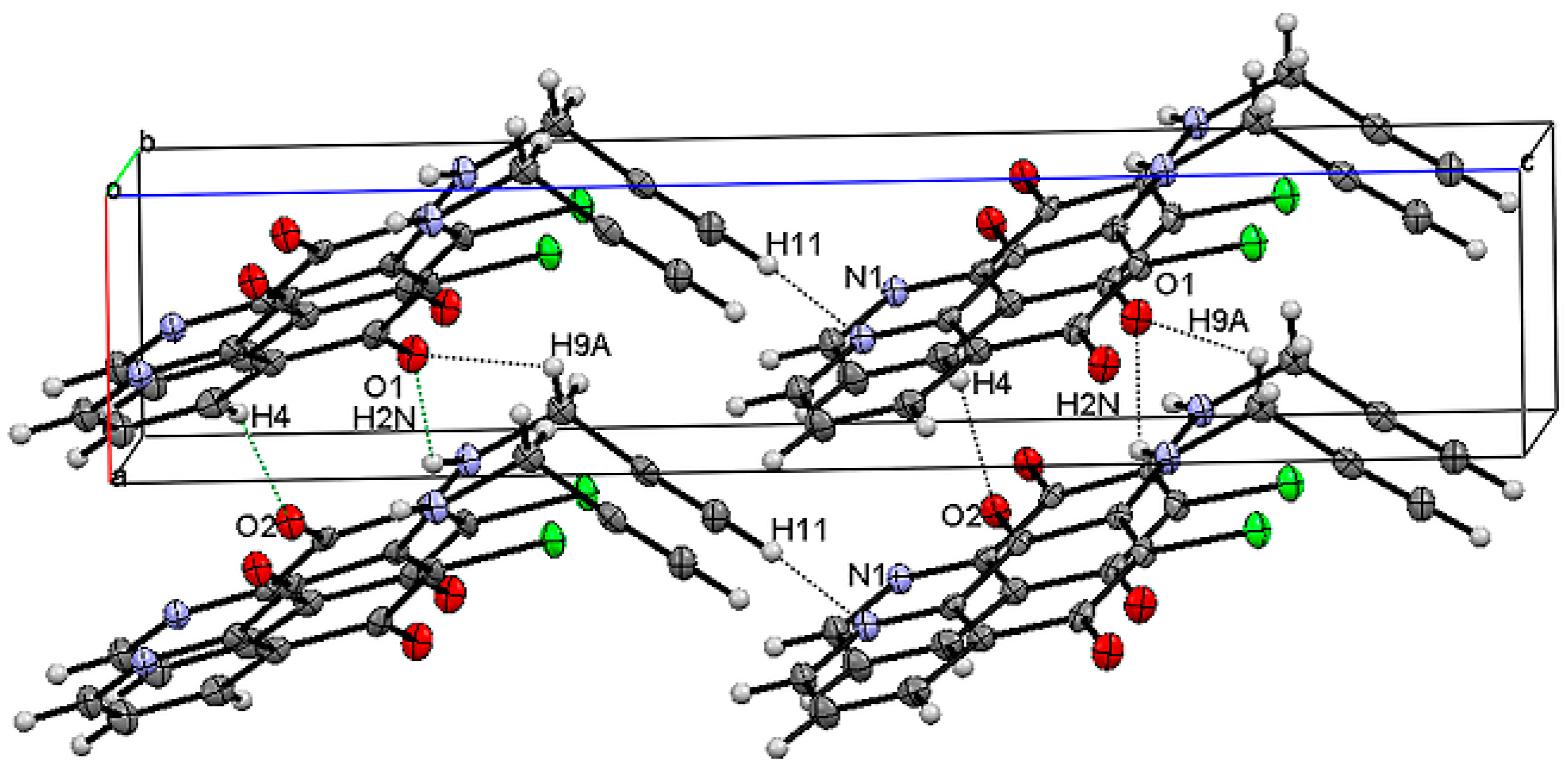
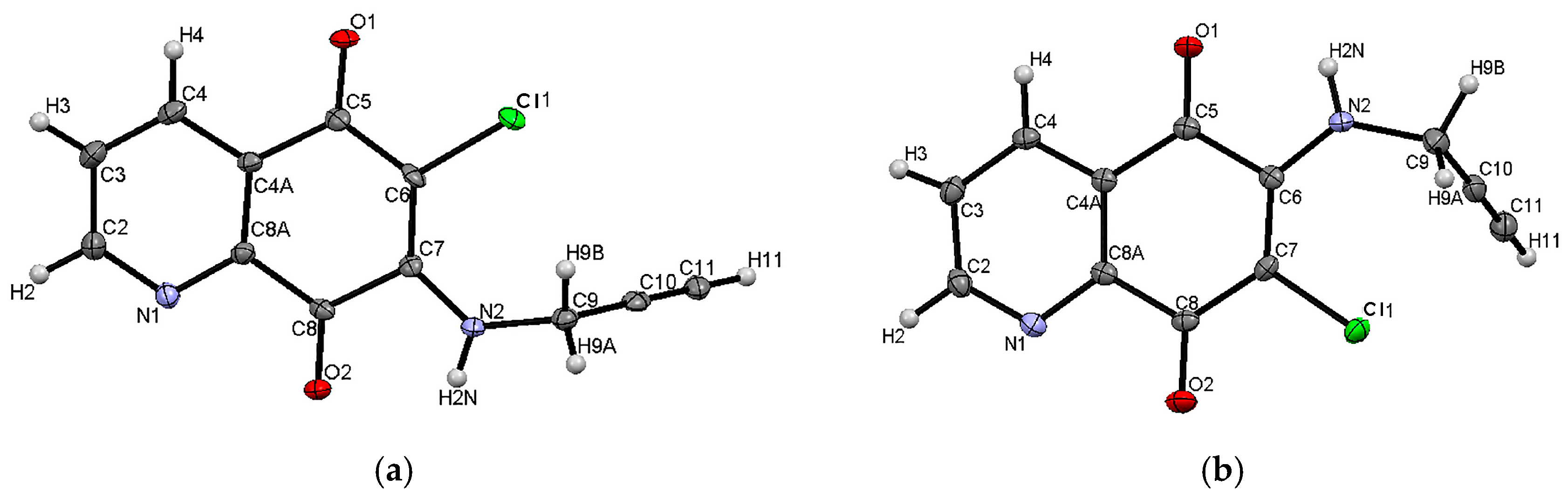
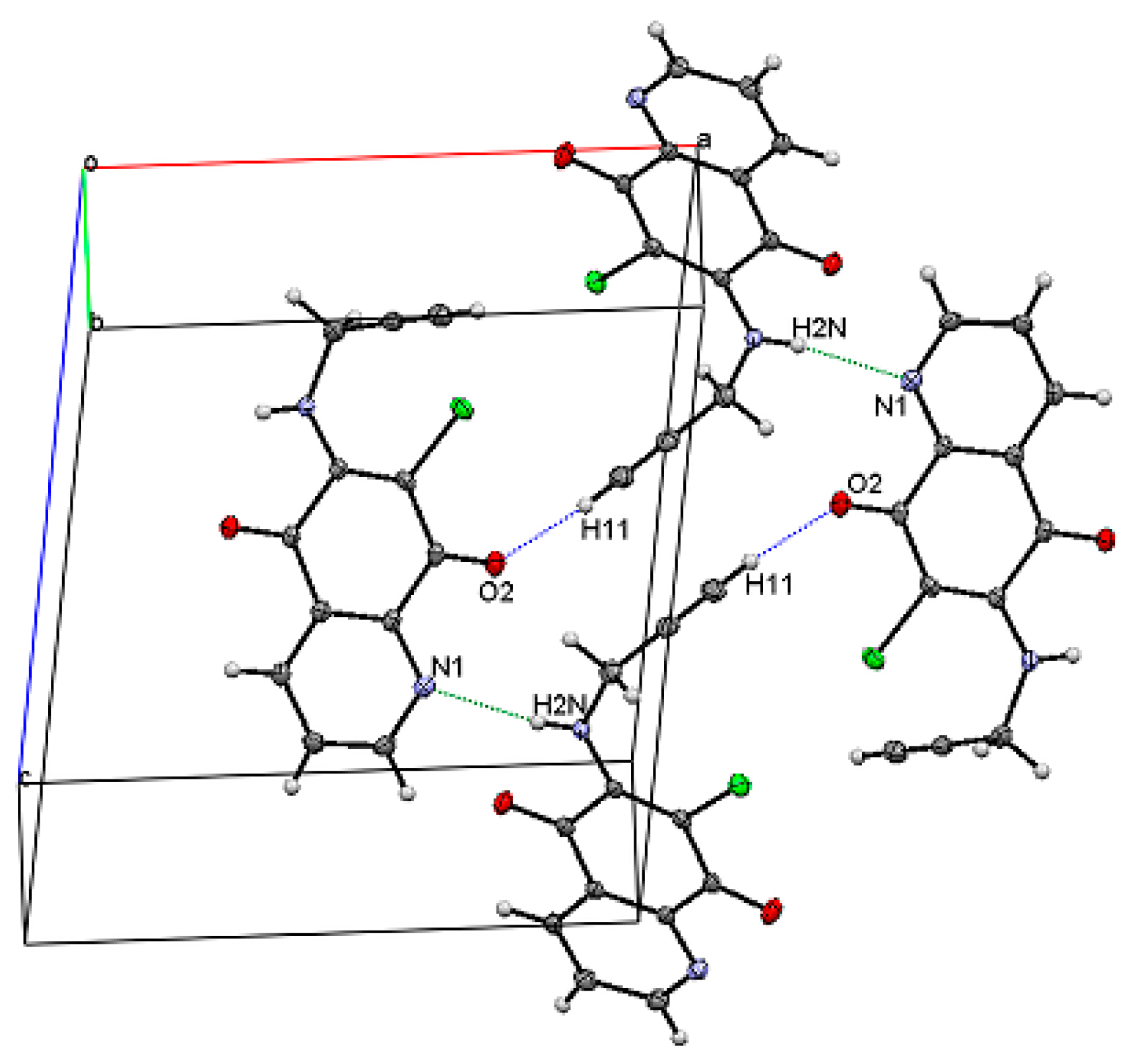
2.2. Crystal Structure and Formation of Hydrogen Bonds
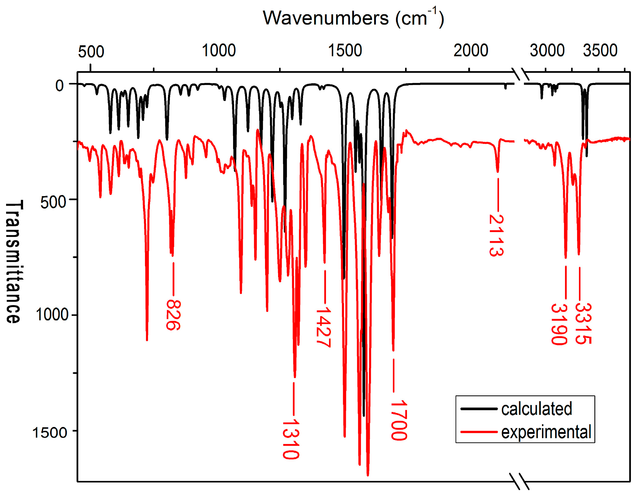
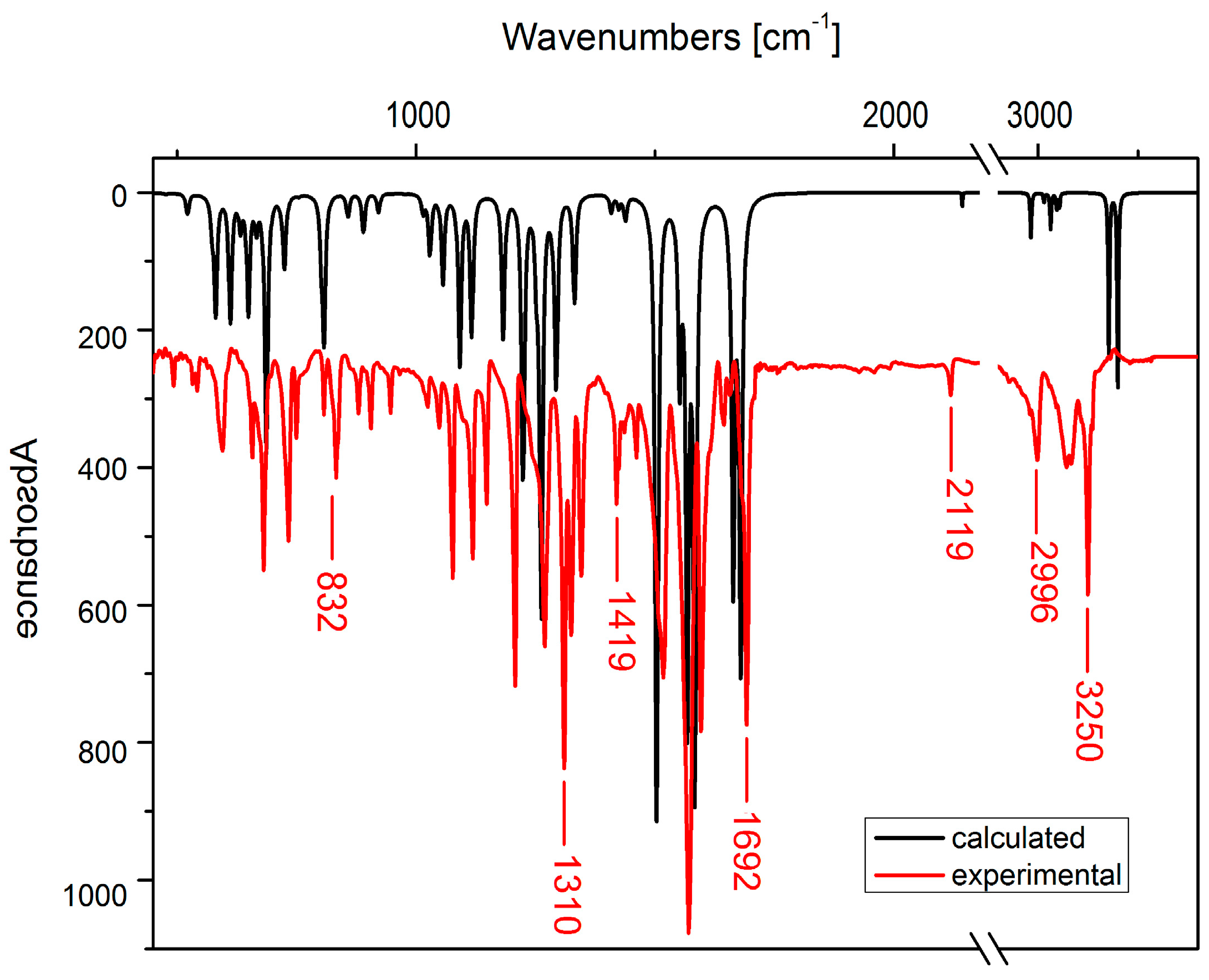
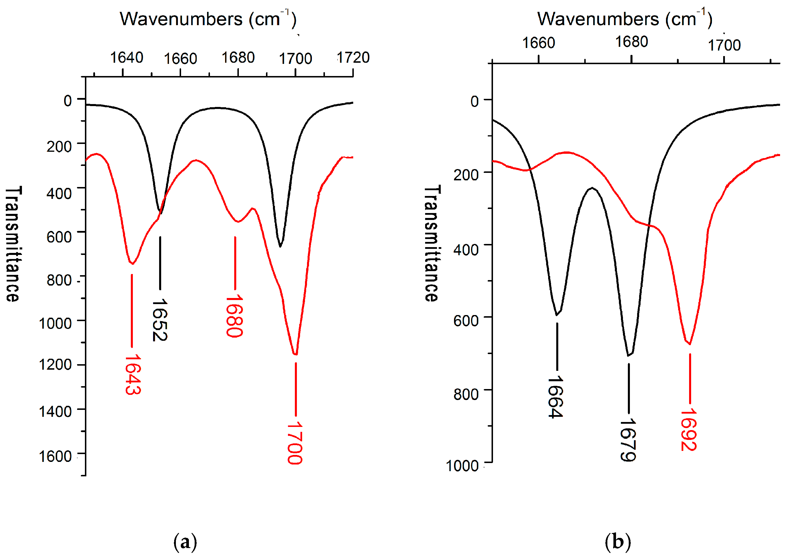
The 6-chloro-7-propargylamine-5,8-quinolinedione 2a and 7-chloro-6-propargylamine-5,8-quinolinedione 3a crystallized in two different monocyclic space groups, i.e., Pc and P21/n, respectively. Figure 1 shows molecular structures and atom numbers of the compounds 2a and 3a. In Table 1, the crystal parameters, experimental data and refinement details are shown.
The selected values of bond distances and angles are presented in Table S1 (supplement material). In terms of bond distances and angles, the geometry of molecules 2a and 3a shows typical values [23,25]. These are in good agreement with the calculated values. The observed discrepancies between experimental and calculated values are mainly due to the method of calculations. They were done for a single molecule in a vacuum, which means that intermolecular interactions were not taken into account.
The unit cell of 2a contains two molecules (Z = 2). The 5,8-quinolinedione rings accomplish a planar structure. In the unit cell these planes are arranged parallel to each other (see Figure S9 in supplement material). An angle between plane of rings and the propargylamine chain N2C9C10C11 is equal to 84.77°. This conformation is very similar to that which occurred for the corresponding angle in the crystal structure of the 6-chloro-7-propylamine-5,8-quinolinedione (89.77°) described earlier by Jastrzebska et al. [25]. Figure 2 depicts the hydrogen bonds found in the crystal structure of 2a. In Table 2 parameters of the hydrogen bonds for 2a are collected.
Both carbonyl groups of 2a participate in the formation of hydrogen bonds. The oxygen atom O1 forms the bifurcated hydrogen bond, which can be described as: N2–H2N···O1···H9A–C9 (Figure 2). Two other short hydrogen bonds C11–H11···N1 and C4–H4···O2 have also been found in 2a with the H···A distances equal to 2.373 and 2.300 Å, respectively (Table 3). According to the literature data [26,27], for the hydrogen bonds from the acidic C–H donors in the C≡C–H to the N acceptors, the mean H···N distance is reported to be 2.40 Å. The reason for the shorter H···N distance in 2a might be the higher basicity of the pyridyl N atom.
For the 7-chloro-6-propargylamine-5,8-quinolinedione 3a, the crystal unit cell contains four molecules (Z = 4, Table 1). The molecules form two layers with the 5,8-quinolinedione rings located inside the unit (see Figure S10 in supplement material). An angle between the 5,8-quinolinedione rings’ plane and the propargylamine chain is equal to 68.57° and is significantly smaller than that for 2a (84.77°). Simultaneously, this angle is very similar to the corresponding angle in the crystal structure of the 7-chloro-6-propylamine-5,8-quinolinedione 3e (68.57°), which was described earlier [25]. Figure 3 shows the unit cell and the hydrogen bonds identified in the crystal structure of 3a. All parameters of the H-bonds seen in Figure 3 are summarized in Table 2.
For 3a crystal structure, the inter- and intra-molecular hydrogen bonds C11–H11···O1 and N2–H2N···N1 are observed, respectively. The N···N distance between the donor and acceptor nitrogen nuclei for the 3a and 3e are equal to 2.957 Å and 3.151 Å, respectively [25]. This pronounced difference could be explained by the higher basicity of the N–H donor group from the propargylamine chain in comparison to that from the propylamine.

2.3. IR Spectra

Analysis of the IR spectral bands, especially in the frequency ranges of the carbonyl and amine stretching vibrations, have been performed using the calculated harmonic vibrational spectra. Comparison of the experimental and the density functional theory (DFT)-calculated spectra allowed also to obtain information about an impact of the H-bond formation on the vibrational bands, e.g., νstr(N–H), νstr(C=O) or νstr(C≡C–H).
In Figure 4 and Figure 5, the IR spectra for compounds 2a and 3a, both experimental and calculated, are presented. Assignments of the selected bands for all spectra are shown in Table 3.
As shown in Figure 4 and Figure 5, the calculated spectra well reproduce these experimental. This also gives good agreement between calculated and experimental frequencies, which can be seen in Table 3. The observed differences are mainly due to the fact that we are comparing the theoretical spectra of a single molecule in a vacuum with the experimental spectra of crystalline substance.
At lower wavenumbers, i.e., below 1300 cm−1, the observed bands are mainly assigned to the aromatic C–C and C–H vibrations. One can also observe the C–C and C≡C–H aliphatic bend vibrations near 580–590 cm−1 and 650–660 cm−1, respectively. For compound 2a the band at 1427 cm−1 is assigned to the C–H aliphatic stretching vibrations. As is seen in Figure 4a, its experimental and calculated band intensities show significant difference. The higher intensity of the experimental band is due to formation of the hydrogen bond C9–H9A···O1. According to literature data [26,27,28], the enhancement of the band intensity for the stretching vibrations of the X–H group (H-bond donor group) is associated with the exceptionally great variation of the electric dipole moment of X–H···Y. This enhancement of intensity is sometimes used to extract information on H-bond [28].
In Figure 6, the experimental and calculated IR spectra in the range of the carbonyl bands ~1600–1750 cm−1 are exposed. Each molecule of 2a and 3a possess two carbonyl groups in the para position. Stretching vibrations of two carbonyl groups are usually coupled into two vibrations located at different frequencies, i.e., asymmetric (out of phase) νas at higher frequency and symmetric (in phase) νs at lower frequency (see Table 3).
Analysis of the calculated spectra revealed the band νas is attributed mainly to the carbonyl vibration at the C-8 atom, whereas the νs band is attributed to the C=O vibrations at the C-5 atom. Furthermore, for the 7-substituted derivative, the N–H bending is involved in the νas carbonyl stretching, while for the 6-substituted derivative, the N–H bending is involved in the νs carbonyl vibrations. A very similar situation occurred for the 6- and 7-propylamine-substituted 5,8-quinolinedione derivatives described previously by Jastrzebska et al. [25]. As in this case, the C=O stretching and the N–H bending vibrations showed coupling effect if they were positioned in close proximity within the molecule. Moreover, there is a correlation between the frequency separation ∆ν = νas − νs of the carbonyl bands and the position of the substituent, i.e., the 7-substituted derivative shows higher value of ∆ν than the 6-substituted one. For the 7-propargylamine-substituted 5,8-quinolinedione the calculated and experimental separation values ∆ν are 57 cm−1 and 43 cm−1 versus 10 cm−1 and 15 cm−1 for the 6-substituted derivative, respectively (see Table 3). The similar situation occurred in the case of the 7- and 6-propylamine-substituted 5,8-quinolinediones described previously [25], for which the ∆ν were 59 cm−1 and 51 cm−1 versus 31 cm−1 and 7 cm−1 for the 7- and 6-substituted derivatives, respectively.
For the 7-propargylamino-5,8-quinolinedione 2a, the νas stretching band shows two peaks at 1700 and 1680 cm−1 (see Figure 5), while for the 6-substituted derivative 3a only single peak at 1692 cm−1 is observed. This effect can be due to the formation of the bifurcated H-bond N2–H2N···O1···H9A–C9 described in the previous subsection. The N–H group of the propargylamine chain is involved in both the bifurcated H-bond and the νas carbonyl stretching vibrations at the C-8 atom. It is also worth noting that the observed splitting into two peaks at 1700 and 1680 cm−1 for the νas stretching band of the 7-substituted derivative is probably not associated with the type of interaction with the D-H system, but originates rather from the νas distinctive characteristics.
The bifurcated H-bond also strongly influences the N–H stretching vibrations, giving two peaks at the 3315 and 3258 cm−1. For the 6-substituted propargylamine derivative, the bifurcated H-bond is absent giving only single band at 3250 cm−1 due to the N–H stretching vibrations.

2.4. Antiproliferative Activity

Compounds 1, 2ae and 3ae were tested for the antiproliferative activity in vitro against the three human cancer cell lines: melanoma (C-32), glioblastoma (SNB-19) and breast cancer (T47D). Results of the analysis have been summarized in Table 4.
It is seen that introduction of the alkynyl, allyl and propyl chains at the C-7 or C-6 position leads to an increase in the cytotoxic activity for the (C-32) and (SNB-19) cell lines in comparison to the 6,7-dichloro-5,8-quinolinedione 1. Furthermore, the acetylenic amine derivatives 2ac and 3ac show higher activity than the reference compound cisplatin against the C-32 and T47D cell lines. All amino derivatives of the 5,8-quinolinedione show high cytotoxic activity against the melanoma (C-32) cell line, with the IC50 varying in the range 0.58 to 0.75 µg/mL. Comparing the activity of compounds with alkane (2e and 3e), alkene (2d and 3d) and alkyne (2a and 3a) moiety, showed that the cytotoxic of derivatives depends on the type of bond in the substituent; the rank order of activity against the C-32 cell line, is as follows: propargyl > allyl > propyl. Moreover, for the other cell line (SNB-19 and T47D) the highest activity showed propargylamino compounds 2a and 3a. The activity of 3a and 2a against the glioblastoma (SNB-19) cell line for which the IC50 parameters have the lowest values 0.09 ± 0.01 µg/mL and 0.26 ± 0.02 µg/mL, respectively. These results suggested that the triple bond seems to be essential for anticancer activity.
For compounds with the acetylenic amine substituents, the cytotoxic activity against the melanoma (C-32) and the breast cancer (T47D) cell lines follows the order: N-methylpropargylamine < 1,1-dimethylpropargylamine < propargylamine. As one can see, expansion of the acetylenic amine chain by binding methyl groups gives a reduction of the cytotoxic activity.

3. Materials and Methods

3.1. General Techniques

Melting points were measured in the open capillary tubes on a Boetius melting point apparatus. NMR spectra (600/150 MHz) were registered on a Bruker Avance 600 spectrometer (Bruker, Billerica, MA, USA). The spectra were recorded for 1H and 13C NMR at room temperature. Chemical shifts were reported in ppm (ν) and J values in Hz. Multiplicity was designated as the singlet (s), doublet (d), triplet (t) and multiplet (m). High-resolution mass spectral analysis was carried out on a Bruker Impact II instrument (Bruker, Billerica, MA, USA) The infrared spectra (IR) were registered using the IRAffinity 1 spectrometer (Shimadzu, Japan) and the KBr pellet method for the sample preparation. All spectra were recorded in the range of 400–4000 cm−1 at room temperature. TLC was carried out on silica gel plates (Merck, Darmstadt, Germany) using a mixture of chloroform and ethanol as an eluent. The visualization was accomplished with UV light and iodine vapour. Column chromatography was performed on silica gel (Merck) with the mixture of chloroform and ethanol (40:1, v/v) as an eluent.

3.2. Chemistry

The 6,7-dichloro-5,8-quinolinedione 1 was synthesized from the 8-hydroxyquinoline according to the method described in the literature [12].
Synthesis of the 6-chloro-7-substituted-5,8-quinolinediones 2ae and 7-chloro-6-substituted-5,8-quinolinediones 3ae was as follows:
Synthesis was carried out using the procedure previously described by Kadela et al. [8,9,25]. Briefly, the 6,7-dichloro-5,8-quinolinedione 1 (0.1 g, 0.441 mmol) was dissolved in dry tetrahydrofuran (1 mL). Next, the potassium carbonate (0.061 g, 0.441 mmol) and corresponding amine (0.441 mmol) were added to the mixture. After 3 h of stirring at room temperature, the solvent was removed under reduced pressure. The residue was purified by the column chromatography (CHCl3/EtOH, 40:1 v/v) to give pure products 2 and 3.
6-chloro-7-propargylamine-5,8-quinolinedione 2a Yield 68%; mp 140–142 °C; 1H NMR (CDCl3, 600 MHz) δ 2.42 (t, J = 2.4 Hz, 1H, CH), 4.68 (dd, J = 2.4 Hz, 2H, CH2), 6.19 (t, 1H, NH), 7.68 (dd, J23 = 4.8 Hz, J34 = 7.8 Hz, 1H, H-3), 8.47 (dd, J24 = 1.8 Hz, J34 = 7.8 Hz, 1H, H-4), 8.96 (dd, J24 = 1.8 Hz, J23 = 4.8 Hz, 1H, H-2). 13C NMR (CDCl3, 150 MHz) δ ppm: 35.1 (CH2), 73.8 (C≡CH), 78.8 (C≡CH), 128.4 (C-6), 129.5 (C-3), 129.7 (C-4a), 134.8 (C-4), 143.9 (C-7), 145.9 (C-8a), 153.7 (C-2), 165.4 (C-8), 178.4 (C-5). IR (KBr) νmax (cm−1) 3315–3258 (N–H), 3190–3038 (C–H), 2113 (C≡C), 1700 (C=O), 1643 (C=O), 1599–1565 (C–H). HRMS (APCI) m/z 247.0265 (calcd for C12H8ClN2O2, 247.0274).
6-chloro-7-(N-methylpropargylamine)-5,8-quinolinedione 2b Yield 61%; mp 130–132 °C; 1H NMR (CDCl3, 600 MHz) δ 2.38 (t, J = 2.4 Hz, 1H, CH), 3.32 (s, 3H, CH3), 4.33 (d, J = 2.4 Hz, 2H, CH2), 7.65 (dd, J23 = 4.8 Hz, J34 = 7.8 Hz, 1H, H-3), 8.46 (dd, J24 = 1.8 Hz, J34 = 7.8 Hz, 1H, H-4), 8.97 (dd, J24 = 1.8 Hz, J23 = 4.8 Hz, 1H, H-2); 13C NMR (CDCl3, 150 MHz) δ 41.8 (CH2), 44.8 (CH3), 73.5 (C≡CH), 78.6 (C≡CH), 123.2 (C-6), 128.4 (C-3), 128.4 (C-4a), 134.6 (C-4), 147.1 (C-8a), 150.7 (C-7), 154.2 (C-2), 177.2 (C-8), 180.1 (C-5); IR (KBr) νmax (cm−1) 3360–3280 (N–H), 3036–2855 (C–H), 2117 (C≡C), 1692 (C=O), 1680 (C=O), 1588–1558 (C–H). HRMS (APCI) m/z 261.0420 (calcd for C13H10ClN2O2, 261.0431).
6-chloro-7-(1,1-dimethylpropargylamine)-5,8-quinolinedione 2c Yield 58%; mp 156–157 °C; 1H NMR (CDCl3, 600 MHz) δ 1.88 (s, 6H, CH3, CH3), 2.45 (t, J = 2.4 Hz, 1H, CH), 6.03 (s, 1H, NH), 7.66 (dd, J23 = 4.8 Hz, J34 = 7.8 Hz, 1H, H-3), 8.46 (dd, J24 = 1.8 Hz, J34 = 7.8 Hz, 1H, H-4), 8.95 (dd, J24 = 1.8 Hz, J23 = 4.8 Hz, 1H, H-2); 13C NMR (CDCl3, 150 MHz) δ 29.7 (CH3), 32.8 (CH3), 51.1 (NHC), 72.3 (C≡CH), 86.6 (C≡CH), 126.1 (C-6), 128.1 (C-3), 129.1 (C-4a), 134.6 (C-4), 145.6 (C-8a), 146.4 (C-7), 153.7 (C-2), 176.0 (C-8), 178.5 (C-5); IR (KBr) νmax (cm−1) 3367–3241 (N–H), 2926–2871 (C–H), 2113 (C≡C), 1700 (C=O), 1683 (C=O), 1653–1635 (C–H). HRMS (APCI) m/z 275.0577 (calcd for C14H12ClN2O2, 275.0587).
6-chloro-7-allylamine-5,8-quinolinedione 2d Yield 65%; mp 122–123 °C; 1H NMR (CDCl3, 600 MHz) δ 4.53 (dt, J = 1.2 Hz, J = 6.0 Hz, 2H, NHCH2), 5.30 (dt, J = 1.2 Hz, J = 9.0 Hz, 2H, CH=CH2), 6.00 (m, 1H, CH=CH2), 6.25 (s, 1H, NH), 7.66 (dd, J23 = 4.8 Hz, J34 = 7.8 Hz, 1H, H-3), 8.48 (dd, J24 = 1.8 Hz, J34 = 7.8 Hz, 1H, H-4), 8.93 (dd, J24 = 1.8 Hz, J23 = 4.8 Hz, 1H, H-2); 13C NMR (CDCl3, 150 MHz) δ 47.2 (NHCH2), 117,9 (CH=CH2), 128.2 (C-6), 128.3 (C-3), 129.7 (C-4a), 134.7 (CH=CH2), 135.6 (C-4), 144.4 (C-8a), 146.8 (C-7), 155.4 (C-2), 176.3 (C-8), 178.8 (C-5); IR (KBr) νmaxx (cm−1) 3304 (N–H), 2957–2854 (C–H), 1693 (C=O), 1680 (C=O), 1598–1560 (C–H); HRMS (APCI) m/z 261.0425 (calcd for C12H10ClN2O2, 249.0431).
7-chloro-6-propargylamine-5,8-quinolinedione 3a Yield 19%; mp 140–142 °C; 1H NMR (CDCl3, 600 MHz) δ 2.37 (t, J = 2.4 Hz, 1H, CH), 4.67 (dd, J = 2.4 Hz, 2H, CH2), 6.01 (t, 1H, NH), 7.62 (dd, J23 = 4.8 Hz, J34 = 7.8 Hz, 1H, H-3), 8.37 (dd, J24 = 1.8 Hz, J34 = 7.8 Hz, 1H, H-4), 9.03 (dd, J24 = 1.8 Hz, J23 = 4.8 Hz, 1H, H-2); 13C NMR (CDCl3, 150 MHz) δ 35.1 (CH2), 73.7 (C≡CH), 78.9 (C≡CH), 126.8 (C-6), 130.2 (C-3), 131.0 (C-4a), 133.7 (C-4), 143.1 (C-7), 148.0 (C-8a), 155.3 (C-2), 165.4 (C-8), 179.7 (C-5); IR (KBr) νmax (cm−1) 3271 (N–H), 3250–3058 (C–H), 2119 (C≡C), 1692 (C=O), 1682 (C=O), 1597–1569 (C–H); HRMS (APCI) m/z 247.0263 (calcd for C12H8ClN2O2, 247.0274).
7-chloro-6-(N-methylpropargylamine)-5,8-quinolinedione 3b Yield 21%; mp 126–127 °C; 1H NMR (CDCl3, 600 MHz) δ 2.39 (t, J = 2.4 Hz, 1H, CH), 3.31 (s, 3H, CH3), 4.29 (d, J = 2.4 Hz, 2H, CH2), 7.63 (dd, J23 = 4.8 Hz, J34 = 7.8 Hz, 1H, H-3), 8.38 (dd, J24 = 1.8 Hz, J34 = 7.8 Hz, 1H, H-4), 9.01 (dd, J24 = 1.8 Hz, J23 = 4.8 Hz, 1H, H-2); 13C NMR (CDCl3, 150 MHz) δ 41.7 (CH2), 44.7 (CH3), 73.4 (C≡CH), 78.8 (C≡CH), 125.3 (C-7), 127.2 (C-3), 129.7 (C-4a), 135.0 (C-4), 147.2 (C-8a), 149.5 (C-6), 155.3 (C-2), 176.6 (C-8), 181.3 (C-5); IR (KBr) νmax (cm−1) 3175 (N–H), 2927–2854 (C–H), 2107 (C≡C), 1674 (C=O), 1592–1520; HRMS (APCI) m/z 261.0422 (calcd for C13H10ClN2O2, 261.0431).
7-chloro-6-(1,1-dimethylpropargylamine)-5,8-quinolinedione 3c Yield 16%; mp 148–149 °C; 1H NMR (CDCl3, 600 MHz) δ 1.86 (s, 6H, CH3, CH3), 2.44 (t, J = 2.4 Hz, 1H, CH), 5.85 (s, 1H, NH), 7.61 (dd, J23 = 4.8 Hz, J34 = 7.8 Hz, 1H, H-3), 8.39 (dd, J24 = 1.8 Hz, J34 = 7.8 Hz, 1H, H-4), 9.02 (dd, J24 = 1.8 Hz, J23 = 4.8 Hz, 1H, H-2); 13C NMR (CDCl3, 150 MHz) δ 29.5 (CH3), 32.7 (CH3), 51.3 (NHC), 72.2 (C≡CH), 86.5 (C≡CH), 127.4 (C-7), 128.2 (C-3), 129.0 (C-4a), 134.4 (C-4), 145.5 (C-8a), 147.5 (C-6), 153.5 (C-2), 177.1 (C-8), 182.1 (C-5); IR (KBr) νmax (cm−1) 3316 (N–H), 3154–2963 (C–H), 2100 (C≡C), 1685 (C=O), 1653 (C=O), 15989–1560 (C–H); HRMS (APCI) m/z 275.0575 (calcd for C14H12ClN2O2, 275.0587).
7-chloro-6-allylamine-5,8-quinolinedione 3d Yield 20%, mp 139–141 °C. 1H NMR (CDCl3, 600 MHz) δ 4.51 (dt, J = 1.2 Hz, J = 6.0 Hz, 2H, NHCH2), 5.29 (dt, J = 1.2 Hz, J = 9.0 Hz, 2H, CH=CH2), 5.99 (m, 1H, CH=CH2), 6.15 (s, 1H, NH), 7.59 (dd, J23 = 4.8 Hz, J34 = 7.8 Hz, 1H, H-3), 8.67 (dd, J24 = 1.8 Hz, J34 = 7.8 Hz, 1H, H-4), 9.01 (dd, J24 = 1.8 Hz, J23 = 4.8 Hz, 1H, H-2); 13C NMR (CDCl3, 150 MHz) δ 47.2 (NHCH2), 117,8 CH=CH2), 126.5 (C-7), 126.8 (C-3), 129.7 (C-4a), 134.8 (CH=CH2), 134.6 (C-4), 147.4 (C-8a), 148.4 (C-6), 155.3 (C-2), 178.3 (C-8), 180.0 (C-5); IR (KBr) νmax (cm−1) 3321 (N–H), 3080–2926 (C–H), 1683 (C=O), 1647 (C=O), 1602–1559 (C–H); HRMS (APCI) m/z 261.0424 (calcd for C12H10ClN2O2, 249.0431).
6-chloro-7-propylamine-5,8-quinolinedione 2e and 6-chloro-7-propylamine-5,8-quinolinedione 3e: the spectral data were previously described in the literature [25].

3.3. X-ray Diffraction

The single crystal X-ray experiment was carried out for the following two compounds: 6-chloro-7-propargylamine-5,8-quinolinedione 2a and 7-chloro-6-propargylamine-5,8-quinolinedione 3a, at 100.0(1) K. Single crystals of both compounds were preselected under microscope. The crystals were installed on a glass capillary and cooled down by Cryostream Cooler (Oxford Cryosystems Ltd, Oxford, UK). Data sets were collected using an Oxford Diffraction κ diffractometer with a Sapphire3 CCD detector (Oxford Diffraction Ltd., Yarnton, UK). For the integration of the collected data, the CrysAlis RED software (version 1.171.32.29, Agilent Technologies) was applied. The crystal structures were solved using direct methods with the SHELXS-97 software. The solutions were refined using SHELXL-97, SHELXS-2014, and SHELXL-2014/6 programs [29].
The supplementary crystallographic data for 2a and 3a were deposited at the Cambridge Crystallographic Data Centre (CCDC) as CCDC-1047971 and CCDC-104797. These data can be obtained free of charge from the CCDC via www.ccdc.cam.ac.uk/data_request/cif.

3.4. Density Functional Theory (DFT) Analysis

Harmonic vibrational spectra were calculated by the DFT method implemented in the Gaussian09 software package [30]. Details have been described in the earlier work [25]. Briefly, the ground state molecular structure was optimized in silico using the B3LYP exchange-correlation functional with the 6-31+G(d,p) basis set. The initial molecular structures of compounds 2a and 3a were taken from the X-ray crystallographic data. The obtained harmonic frequencies were scaled by a factor of 0.964 in accordance with [31]. Calculated vibrational modes were also analyzed using the GaussView 5.0 visualization software (Gaussian, Inc., Wallingford, CT, USA). The effect of the position of 6- and 7-propargylamine chain on carbonyl vibrations was observed by taking into account the displacement vectors.

3.5. Antiproliferative Assay In Vitro

3.5.1. Cell Culture

All compounds 1, 2ae and 3ae were screened for antiproliferative activity using three cultured cell lines: SNB-19 (human glioblastoma, DSMZ-German Collection of Microorganisms and Cell Cultures, Braunschweig, Germany), C-32 (human amelanotic melanoma, ATCC-American Type Culture Collection, Manassas, VA, USA) and T47D (human ductal breast epithelial tumor cell line, ATCC, Manassas, VA, USA). The cultured cells were kept at 37 °C in the 5% CO2 atmosphere. The cells were seeded (1 × 10⁴ cells/well/100 µL Dulbecco’s modified Eagle’s medium (DMEM) supplemented with 10% fetal calf serum (FCS), streptomycin and penicillin) using the 96-well plates (Corning Inc., Corning, NY, USA).

3.5.2. Analysis of Antiproliferative Activity

The cytotoxic activities of tested compounds were determined using the Cell Proliferation Reagent WST-1 assay (Roche Diagnostics, Mannheim, Germany). The entire procedure was previously described in detail in an earlier work [8]. Cells were exposed to tested compounds for 24 h at indicated concentrations (in the rank of 0.1–100 µg/mL of dimethyl sulfoxide (DMSO)), and their viabilities were quantified using a cell proliferation assay. The WST-1-formazan was detected using a microplate reader at 450 nm with the reference wavelength of 600 nm. Results were expressed as a mean value of at least three independent experiments performed in triplicate. The cytotoxic activity of the tested compound was compared to the cisplatin. The experiments were repeated in triplicate for each concentration of the compound. The IC50 parameter describes the concentration of compound (in μg/mL) that inhibits the proliferation rate of the tumor cells by 50% as compared to the control untreated cells. Calculation of the IC50 was performed using the GraphPad Prism 6 software (GraphPad Software, San Diego, CA, USA).

4. Conclusions

New 6- and 7-propargylamine-substituted 5,8-quinolinediones were synthesized and examined using the X-ray diffraction and IR spectroscopy supplemented by the density functional theory (DFT) calculations. Different positions of the propargylamine chain influenced crystal structure and formation of H-bonds. It was found that the H-bond distinctly affected the νas stretching band of the carbonyl groups only for the 7-propargylamine-substituted 5,8-quinolinedione.
Substantial changes in the frequency separation Δν of the carbonyl stretching bands for different positions of the propargylamine chain were found. Higher frequency separation Δν corresponds to the 7-substituted derivative. Correlation between the Δν and position of substituent may provide an opportunity to use the IR spectroscopy to study substitution reaction.
Cytotoxic activities against three human cancer cell lines for the 5,8-quinolinedione derivatives with different amine substituents, i.e., propargylamine, N-methylpropargylamine, 1,1-dimethylpropargylamine, allylamine and propylamine were analyzed with respect to their molecular structure. It was found that introduction of the acetylenic, allyl and propylamine chains at the C-7 or C-6 position led to an increase in the cytotoxic activity for the melanoma and glioblastoma cell lines in comparison to the starting compound 6,7-dichloro-5,8-quinolinedione 1. Furthermore, for the melanoma (C-32) and breast cancer (T47D) cell lines, the acetylenic amine derivatives showed higher activity than the reference compound cisplatin. The low IC50 values for the 7- and 6-substituted propargylamine derivatives against the glioblastoma (SNB-19) cell line were observed.

Supplementary Materials

The following are available online at www.mdpi.com/2073-4352/7/1/15/s1. Table S1: Selected geometric parameters given by X-ray diffraction experiment and theoretical calculations for compounds 2a and 3a. Figure S1: 6-chloro-7-propargylamine-5,8-quinolinedione 2a, (a) 1H NMR spectrum, (b) 13C NMR spectrum, (c) IR spectrum. Figure S2: 6-chloro-7-(N-methylpropargylamine)-5,8-quinolinedione 2b, (a) 1H NMR spectrum, (b) 13C NMR spectrum, (c) IR spectrum. Figure S3: 6-chloro-7-(1,1-dimethylpropargylamine)-5,8-quinolinedione 2c, (a) 1H NMR spectrum, (b) 13C NMR spectrum, (c) IR spectrum. Figure S4: 6-chloro-7-allylamine-5,8-quinolinedione 2d, (a) 1H NMR spectrum, (b) 13C NMR spectrum, (c) IR spectrum. Figure S5: 7-chloro-6-propargylamine-5,8-quinolinedione 3a, (a) 1H NMR spectrum, (b) 13C NMR spectrum, (c) IR spectrum. Figure S6: 7-chloro-6-(N-methylpropargylamine)-5,8-quinolinedione 3b, (a) 1H NMR spectrum, (b) 13C NMR spectrum, (c) IR spectrum. Figure S7: 7-chloro-6-(1,1-dimethylpropargylamine)-5,8-quinolinedione 3c, (a) 1H NMR spectrum, (b) 13C NMR spectrum, (c) IR spectrum. Figure S8: 7-chloro-6-allylamine-5,8-quinolinedione 3d, (a) 1H NMR spectrum, (b) 13C NMR spectrum, (c) IR spectrum. Figure S9: The crystal packing of 6-chloro-7-propargylamine-5,8-quinolinedione 2a. View along axis “b”. Figure S10: The crystal packing of 7-chloro-6-propargylamine-5,8-quinolinedione 3a. View along axis “b”.

Acknowledgments

This work was supported by the Medical University of Silesia in Katowice, Poland. Grant No. KNW-1-006/K/6/O and KNW-2-008/N/6/N.

Author Contributions

Monika Kadela-Tomanek and Stanisław Boryczka developed the concept of the work. Monika Kadela-Tomanek carried out the synthetic work and interpreted the results. Ewa Bębenek and Elwira Chrobak contributed to the synthesis and purification all new compounds. Maria Jastrzębska and Dorota Tarnawska participated in IR spectra interpretation. Małgorzata Latocha conducted a study of the biological activity. Joachim Kusz performed the X-ray analysis. Monika Kadela-Tomanek wrote the paper. All authors have given approval to the final version of the manuscript.

Conflicts of Interest

The authors declare no conflict of interest.

References

  1. Bringmann, G.; Reichert, Y.; Kane, V.V. The total synthesis of streptonigrin and related antitumor antibiotic natural products. Tetrahedron 2004, 60, 3539–3574. [Google Scholar] [CrossRef]
  2. Boger, D.L.; Yasuda, M.; Mitscher, L.A.; Drake, S.D.; Kitos, P.A.; Thompson, S.C. Streptonigrin and lavendamycin partial structures. Probes for the minimum, potent pharmacophore of streptonigrin, lavendamycin, and synthetic quinoline-5,8-diones. J. Med. Chem. 1987, 30, 1913–1928. [Google Scholar] [CrossRef]
  3. Fryatt, T.; Goroski, D.T.; Nilson, Z.D.; Moody, C.J.; Beall, H. Novel quinolinequinone antitumor agents: Structure-metabolism studies with NAD(P)H:quinone oxidoreductase (NQO1). Bioorg. Med. Chem. Lett. 1999, 9, 2195–2198. [Google Scholar] [CrossRef]
  4. Leteurtre, F.; Kohlhagen, G.; Pommier, Y. Streptonigrin induced topoisomerase II sites exhibit base preferences in the middle of the enzym stragger. Biochem. Biophys. Res. Commun. 1994, 203, 1259–1267. [Google Scholar] [CrossRef] [PubMed]
  5. Kim, Y.-S.; Park, S.-Y.; Lee, H.-J.; Suh, M.-E.; Schollmeyer, D.; Lee, C.-O. Synthesis and cytotoxicity of 6,11-dihydro-pyrido- and 6,11-dihydro-benzo[2,3-b]phenazine-6,11-dione derivatives. Bioorg. Med. Chem. 2003, 11, 1709–1714. [Google Scholar] [CrossRef]
  6. Alfadhli, A.; Mack, A.; Harper, L.; Berk, S.; Ritchie, C.; Barklis, E. Analysis of quinolinequinone reactivity, cytotoxicity, and anti-HIV-1 properties. Bioorg. Med. Chem. 2016, 24, 5618–5625. [Google Scholar] [CrossRef] [PubMed]
  7. Ryu, C.-K.; Song, A.L.; Hong, J.A. Synthesis and antifungal evaluation of 7-arylamino-5,8-dioxo-5,8-dihydroisoquinoline-4-carboxylates. Bioorg. Med. Chem. Lett. 2013, 23, 2065–2068. [Google Scholar] [CrossRef] [PubMed]
  8. Kadela, M.; Jastrzębska, M.; Bębenek, E.; Chrobak, E.; Latocha, M.; Kusz, J.; Książek, M.; Boryczka, S. Synthesis, structure and cytotoxic activity of mono- and dialkoxy derivatives of 5,8-quinolinedione. Molecules 2016, 21, 156. [Google Scholar] [CrossRef] [PubMed]
  9. Kadela-Tomanek, M.; Jastrzębska, M.; Pawełczak, B.; Bębenek, E.; Chrobak, E.; Latocha, M.; Książek, M.; Kusz, J.; Boryczka, S. Alkynyloxy derivatives of 5,8-quinolinedione: Synthesis, in vitro cytotoxicity studies and computational molecular modeling with NAD(P)H:quinone oxidoreductase 1. Eur. J. Med. Chem. 2017, 126, 969–982. [Google Scholar] [CrossRef] [PubMed]
  10. Rhee, H.K.; Park, H.J.; Lee, S.K.; Lee, C.-O.; Choo, H.-Y. Synthesis, cytotoxicity and DNA topoisomerase II inhibitory activity of benzofuroquinolinediones. Bioorg. Med. Chem. 2007, 15, 1651–1658. [Google Scholar] [CrossRef] [PubMed]
  11. Ryu, C.-K.; Jeong, H.J.; Lee, S.K.; You, H.-J.; Chio, K.U.; Shim, J.-Y.; Heo, Y.-H.; Lee, C.-O. Effects of 6-arylamine-5,8-quinolinediones and 6-chloro 7-arylo-5,8-isoquinolinediones on NAD(P)H:quinone oxidoreductase (NQO1) activity and their cytotoxic potential. Arch. Pharm. Res. 2001, 24, 390–396. [Google Scholar] [CrossRef] [PubMed]
  12. Mulchin, B.J.; Newton, C.G.; Baty, J.W.; Grasso, C.H.; Martin, W.J.; Walton, M.C.; Dangerfield, E.M.; Plunkett, C.H.; Berridge, M.V.; Harper, J.L.; et al. The anti-cancer, anti-inflammatory and tuberculostatic activities of a series of 6,7-substituted-5,8-quinolinequinones. Bioorg. Med. Chem. 2010, 18, 3238–3251. [Google Scholar] [CrossRef] [PubMed]
  13. Yoon, E.Y.; Choi, H.Y.; Shin, K.J.; Yoo, K.H.; Chi, D.Y.; Kim, D.J. The regioselectivity in the reaction of 6,7-dihaloquinoline-5,8-diones with amine nucleophiles in various solvents. Tetrahedron Lett. 2000, 41, 7475–7480. [Google Scholar] [CrossRef]
  14. Yin, H.; Kong, F.; Wang, S.; Yao, Z.-Y. Assembly of pentacyclic pyrido[4,3,2-mn]acridin-8-ones via a domino reaction initiated by Au(I)-catalyzed 6-endo-dig cycloisomerization of N-propargylaminoquinones. Tetrahedron Lett. 2012, 53, 7078–7082. [Google Scholar] [CrossRef]
  15. Mól, W.; Matyja, M.; Filip, B.; Wietrzyk, J.; Boryczka, S. Synthesis and antiproliferative activity in vitro of novel (2-butynyl)thioquinolines. Bioorg. Med. Chem. 2008, 16, 8136–8141. [Google Scholar] [CrossRef] [PubMed]
  16. Mól, W.; Naczyński, A.; Boryczka, S.; Wietrzyk, J.; Opolski, A. Synthesis and antiproliferative activity in vitro of diacetylenic thioquinolines. Pharmazie 2006, 61, 742–746. [Google Scholar] [PubMed]
  17. Boryczka, S.; Bębenek, E.; Wietrzyk, J.; Kempińska, K.; Jastrzębska, M.; Kusz, J.; Nowak, M. Synthesis, structure and cytotoxic activity of new acetylenic derivatives of betulin. Molecules 2013, 18, 4526–4543. [Google Scholar] [CrossRef] [PubMed]
  18. Marciniec, K.; Latocha, M.; Boryczka, S.; Kurczab, R. Synthesis, molecular docking study, and evaluation of the antiproliferative action of a new group of propargylthio- and propargylselenoquinolines. Med. Chem. Res. 2014, 23, 3468–3477. [Google Scholar] [CrossRef]
  19. Siddiq, A.; Dembitsky, V. Acetylenic anticancer agents. Anti-Cancer Agents Med. Chem. 2008, 8, 132–170. [Google Scholar] [CrossRef]
  20. Dembitsky, V.M.; Levitsky, D.O.; Gloriozovac, T.A.; Poroikovc, V.V. Acetylenic aquatic anticancer agents and related compounds. Nat. Prod. Commun. 2006, 1, 773–812. [Google Scholar]
  21. Kuklev, D.V.; Dembitsky, V.M. Epoxy acetylenic lipids: their analogues and derivatives. Prog. Lipid Res. 2014, 56, 67–91. [Google Scholar] [CrossRef] [PubMed]
  22. Kuklev, D.V.; Domb, A.J.; Dembitsky, V.M. Bioactive acetylenic metabolites. Phytomedicine 2013, 20, 1145–1159. [Google Scholar] [CrossRef] [PubMed]
  23. Sun, S.; Tang, H.; Wu, P. Interpretation of carbonyl band splitting phenomenon of a novel thermotropic liquid crystalline polymer without conventional mesogens: Combination method of spectral analysis and molecular simulation. J. Phys. Chem. B 2010, 114, 3439–3448. [Google Scholar] [CrossRef] [PubMed]
  24. Socrates, G. Infrared Characteristic Group Frequencies; John Wiley & Sons Inc.: New York, NY, USA, 1994. [Google Scholar]
  25. Jastrzebska, M.; Boryczka, S.; Kadela, M.; Wrzalik, R.; Kusz, J.; Nowak, M. Synthesis, crystal structure and infrared spectra of new 6- and 7-propylamine-5,8-quinolinediones. J. Mol. Struct. 2014, 1067, 160–168. [Google Scholar] [CrossRef]
  26. Desiraju, G.R.; Steiner, T. The Weak Hydrogen Bond; Oxford University Press: London, UK, 2006. [Google Scholar]
  27. Steiner, T. The hydrogen bond in the solid state. Angew. Chem. 2002, 41, 48–79. [Google Scholar] [CrossRef]
  28. Marechal, Y. The Hydrogen Bond and the Water Molecule; Elsevier: London, UK, 2007. [Google Scholar]
  29. Sheldrick, G.M. A short history of SHELX. Acta Cryst. A 2008, 64, 112–122. [Google Scholar] [CrossRef] [PubMed]
  30. Frisch, M.J.; Trucks, G.W.; Schlegel, H.B.; Scuseria, G.E.; Robb, M.A.; Cheeseman, J.R.; Scalmani, G.; Barone, V.; Mennucci, B.; Petersson, G.A.; et al. Gaussian 09, Revision A. 02; Gaussian Inc.: Wallingford, CT, USA, 2009. [Google Scholar]
  31. NIST Computational Chemistry Comparison and Benchmark Database, NIST Standard Reference Database Number 101, Release 16a, August 2013, Editor: Russell D. Johnson III. Available online: http://cccbdb.nist.gov/ (accessed on 11 November 2015).
Scheme 1. Synthesis of the 6-chloro-7-substituted 5,8-quinolinediones 2ae and 7-chloro-6-substituted 5,8-quinolinediones 3ae.
Crystals 07 00015 sch001
Figure 1. Molecular structures with atom numbering of (a) 6-chloro-7-propargylamine-5,8-quinolinedione 2a; (b) 7-chloro-6-propargylamine-5,8-quinolinedione 3a.
Crystals 07 00015 g001
Figure 2. Crystal structure and hydrogen bonds for 6-chloro-7-propargylamine-5,8-quinolinedione 2a.
Crystals 07 00015 g002
Figure 3. Crystal structure and hydrogen bonds in of 7-chloro-6-propargylamine-5,8-quinolinedione 3a.
Crystals 07 00015 g003
Figure 4. Experimental (red line) and calculated (black line) IR spectra for 6-chloro-7-propargylamine-5,8-quinolinedione 2a (450–3500) cm−1. See Table 3 for band assignments.
Crystals 07 00015 g004
Figure 5. Experimental (red line) and calculated (black line) IR spectra for 7-chloro-6-propargylamine-5,8-quinolinedione 3a (450–3500) cm−1. See Table 3 for band assignments.
Crystals 07 00015 g005
Figure 6. Experimental (red line) and calculated (black line) IR spectra showing carbonyl bands for (a) 6-chloro-7-propargylamine-5,8-quinolinedione 2a; and (b) 7-chloro-6-propargyl-5,8-quinolinedione 3a.
Crystals 07 00015 g006
Table 1. Crystal parameters, data collection and refinement details for compounds 2a and 3a.
Parameter2a3a
Chemical formulaC12H7ClN2O2C12H7ClN2O2
Mr246.65
Crystal system, space groupMonoclinic, PcMonoclinic, P21/n
Temperature (K)100
a, b, c (Å)4.0250 (12), 6.5937 (6), 19.3214 (13)11.1550 (3), 7.9114 (2), 12.0047 (3)
β (°)90.673 (12)97.554 (3)
V3)512.75 (16)1050.24 (5)
Z24
Radiation typeMo Kα
µ (mm−1)0.360.35
Crystal size (mm)0.38 × 0.05 × 0.040.56 × 0.22 × 0.03
DiffractometerOxford Diffraction diffractometer with Sapphire3 detector
Absorption correctionMulti-scan CrysAlis RED, Oxford Diffraction Ltd., Version 1.171.32.29 Empirical absorption correction using spherical harmonics, implemented in SCALE3 ABSPACK scaling algorithm.
Tmin, Tmax0.875, 0.9840.911, 1.000
No. of measured, independent and observed [I > 2σ(I)] reflections3745,
1261,
1144
7654,
1995,
1657
Rint0.0360.026
(sin θ/λ)max−1)0.6090.610
R[F2 > 2σ(F2)], wR(F2), S0.032, 0.078, 1.000.027, 0.072, 1.03
No. of reflections12611995
No. of parameters164160
No. of restraints2-
H-atom treatmentH atoms treated by a mixture of independent and constrained refinement
(Δ)max, (Δ)min (e Å−3)0.61, −0.250.30, −0.20
Absolute structureRefined as an inversion twin.-
Absolute structure parameter0.95 (14)-
Table 2. Parameters (Å, Degree) of the hydrogen bonds for compounds 2a and 3a.
D–H···AD–HH···AD···A<D–H···A
6-chloro-7-propargylamine-5,8-quinolinedione 2a
C4–H4···O20.95 (1)2.373 (4)3.219 (4)148.1
N2–H2N···O10.83 (1)2.403 (4)3.015 (4)131.2
C9–H9A···O10.83 (2)2.629 (2)3.207 (2)127.9
C11–H11···N10.95 (1)2.300 (2)3.158 (3)149.9
7-chloro-6-propargylamine-5,8-quinolinedione 3a
N2–H2N···N10.85 (1)2.181 (1)2.957 (1)152.5
C11–H11···O20.95 (1)2.338 (2)3.282 (1)172.2
Table 3. Experimental and calculated vibrational frequencies (cm−1) and band assignments for studied compounds 2a and 3a.
ExperimentalCalculatedAssignment
6-chloro-7-propargylamine-5,8-quinolinedione 2a
581579C–C aliphatic bend
652650C≡C–H bend
748–696723–689C–C ring stretch, C–H ring stretch
826–818803C–Cl bend
1153–11391175–1123C–C ring bend, C–H ring bend
11201220HN–C ring bend, C–H ring bend
1283–12501272C–C ring stretch
1332–13101299C–C ring bend
13531332C–H aliphatic bend
14271423C–H aliphatic stretch
15061504N–H bend
1599–15651534–1550C–H ring bend, C–H aliphatic stretch
16431652C=O sym stretch
1700
1680
1695C=O asym stretch, N–H bend
21132145C≡C stretch
29572964C–H aliphatic stretch
3085–30383109–3064C–H ring stretch
31903354C≡CH stretch
7-chloro-6-propargylamine-5,8-quinolinedione 3a
595611–580C–C aliphatic bend
657657C≡C–H bend
749–681725–687C–C ring stretch, C–H ring stretch
832–807806C–Cl bend
1147–10761182–1091C–C ring bend, C–H ring bend
12071224HN–C ring bend, C–H ring bend
12691261C–C ring stretch
1324–13101293C–C ring bend
13461331C–H aliphatic bend
14191407C–H aliphatic stretch
14611438C–H ring bend, C–C ring bend
15171503N–H bend
1597–15691584–1551C–H ring bend, C–H aliphatic stretch
16821664C=O sym stretch, N–H bend
16921679C=O asym stretch
21192143C≡C stretch
29962965C–H aliphatic stretch
3168–30583108–3030C–H ring stretch
32503354C≡CH stretch
32713399N–H stretch
Table 4. Cytotoxic activity of 6,7-dichloro-5,8-quinolinedione 1, amine derivatives of 5,8-quinolinedione 23 and cisplatin as a reference compound.
CompoundCytotoxic Activity IC50 (µg/mL)
C-32SNB-19T47D
142.48 ± 2.022.77 ± 0.078.26 ± 0.32
2a0.61 ± 0.020.26 ± 0.028.50 ± 0.54
2b0.75 ± 0.050.97 ± 0.019.22 ± 0.77
2c0.67 ± 0.010.98 ± 0.019.14 ± 0.77
2d0.63 ± 0.020.88 ± 0.059.03 ± 0.10
2e0.64 ± 0.030.50 ± 0.048.54 ± 0.41
3a0.58 ± 0.030.09 ± 0.011.01 ± 0.05
3b0.67 ± 0.050.44 ± 0.018.48 ± 0.55
3c0.65 ± 0.050.92 ± 0.074.57 ± 0.62
3d0.61 ± 0.030.79 ± 0.037.07 ± 0.28
3e0.64 ± 0.010.28 ± 0.053.05 ± 5.65
cisplatin1.51 ± 0.490.79 ± 0.0762.65 ± 2.70

