
Specific Internal Structure of Diamonds from Zarnitsa Kimberlite Pipe


Abstract
:1. Introduction
2. Results
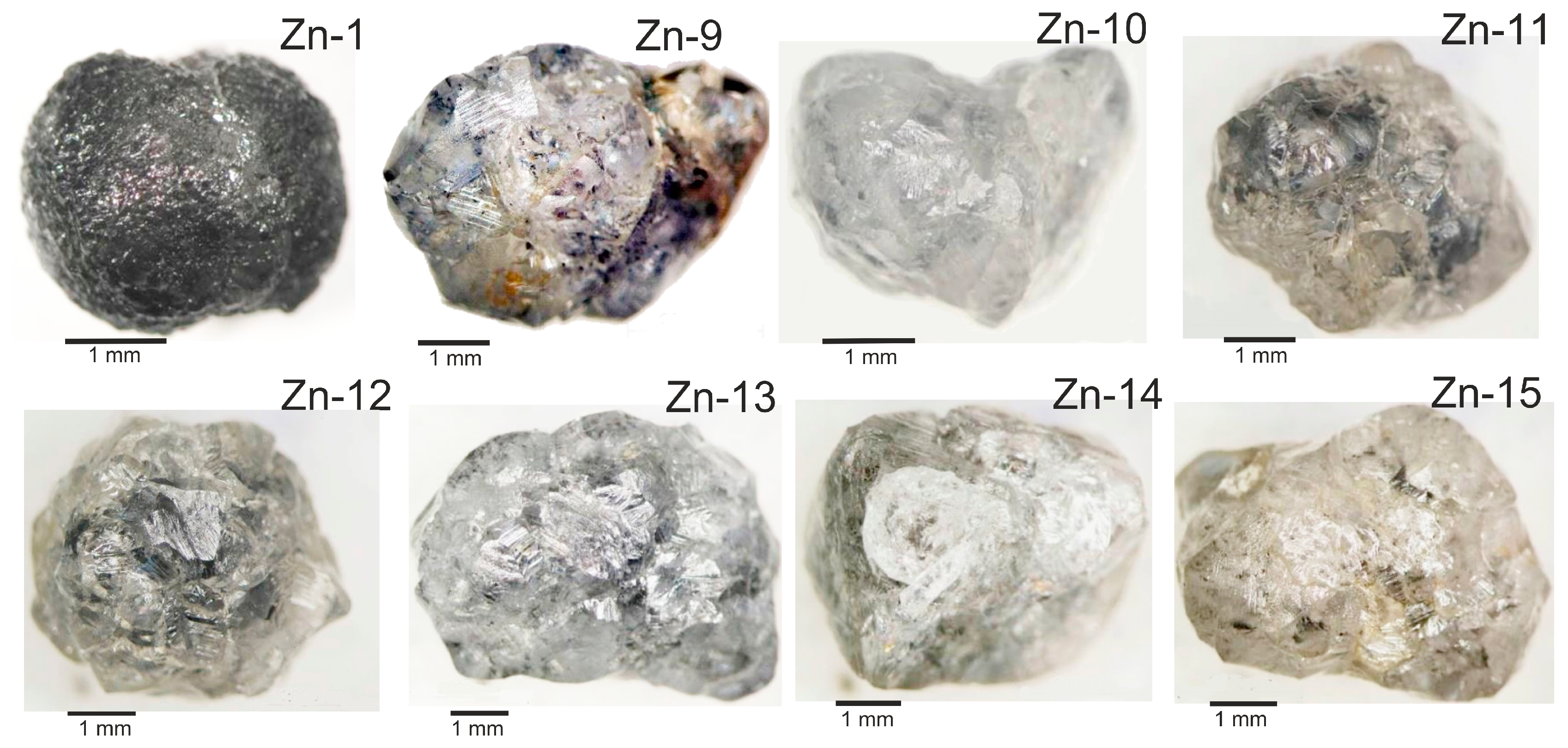
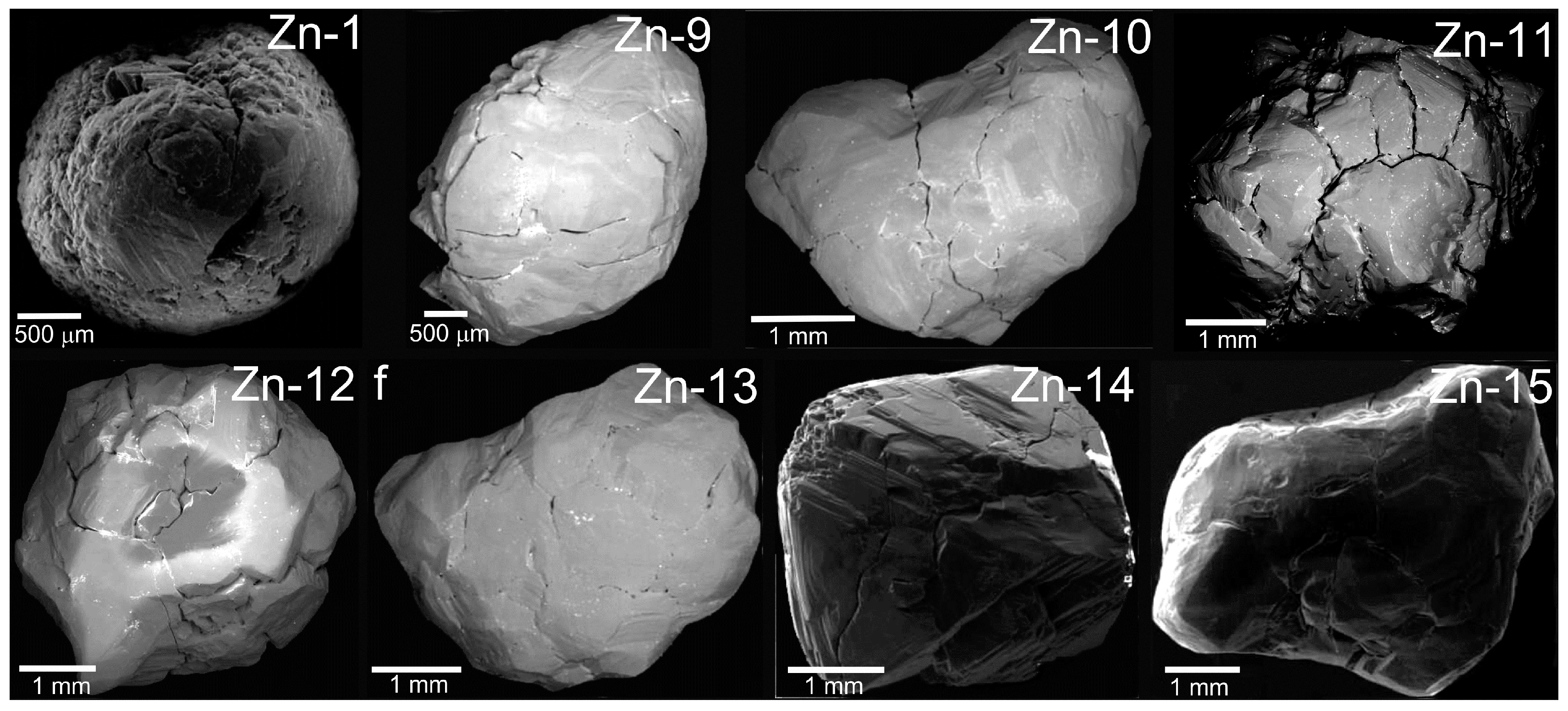
2.1. Morphology
2.2. Internal Structure
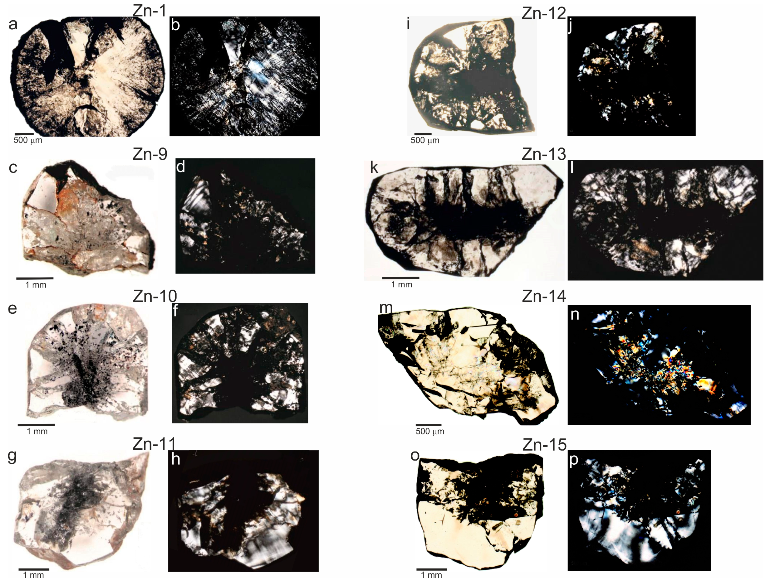
2.2.1. Optical Microscopy
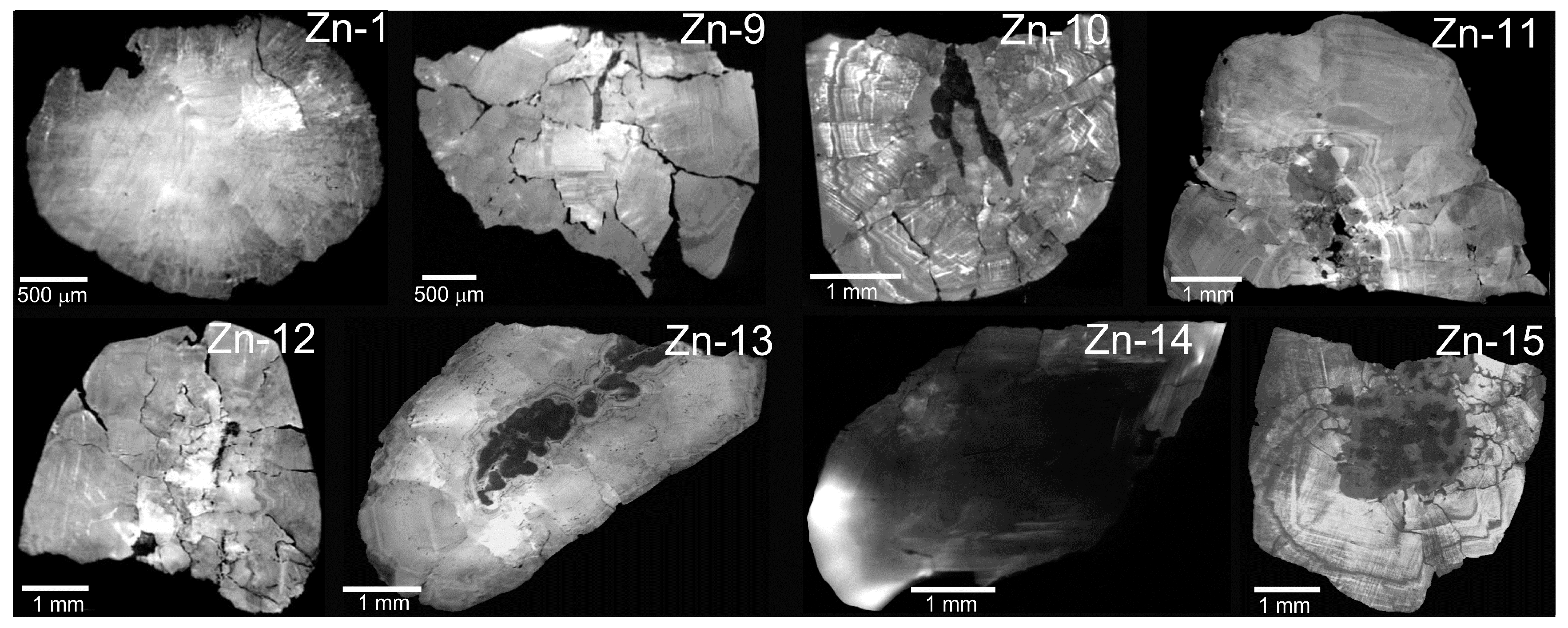
2.2.2. Cathodoluminescence Imagery
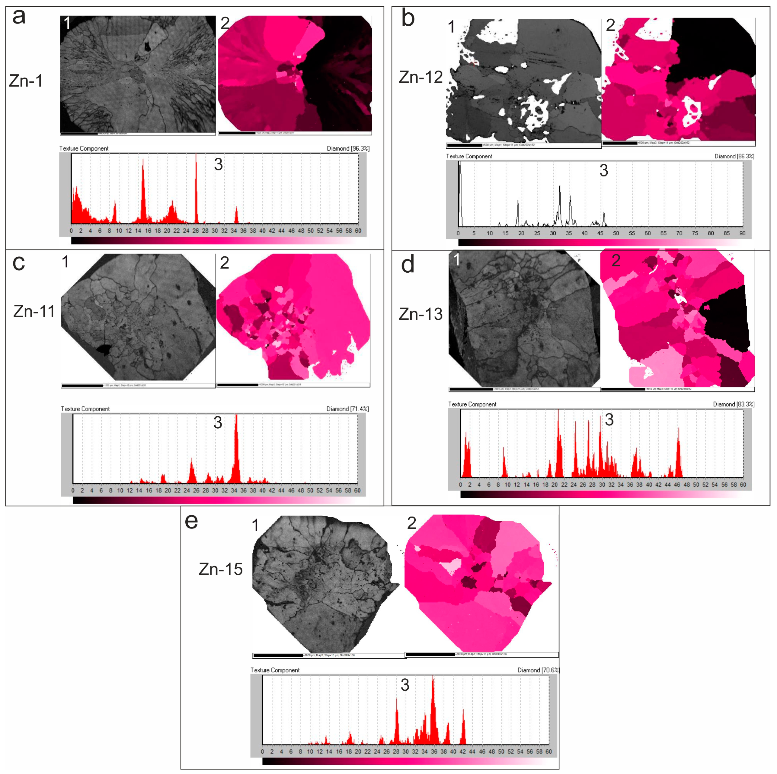
2.2.3. Electron Backscatter Diffraction (EBSD)
3. Discussion
4. Methods
5. Conclusions
Acknowledgments
Author Contributions
Conflicts of Interest
References
- Sarsadskikh, N.N.; Popugayeva, L.A. New data about manifestation ultramafic-alkaline magmatism within Siberian Platform. Razvedka I Okhrana Nedr. 1955, 5, 11–20. (In Russian) [Google Scholar]
- Kostrovitskiy, S.I.; Spetsius, Z.V.; Yakovlev, D.A.; Fon-der-Flaas, G.S.; Suvorova, L.F.; Bogush, I.N. Atlas of the Primary Diamond Deposits of the Yakutian Kimberlite Province; NIGP SC “ALROSA” (PAO): Mirny, Russia, 2015; p. 480. (In Russian) [Google Scholar]
- Ragozin, A.L.; Zedgenizov, D.A.; Kuper, K.E.; Shatsky, V.S. Radial mosaic internal structure of rounded diamond crystals from alluvial placers of Siberian Platform. Mineral. Petrol. 2016, 110, 861–875. [Google Scholar] [CrossRef]
- Ragozin, A.L.; Shatsky, V.S.; Rylov, G.M.; Goryainov, S.V. Coesite inclusions in rounded diamonds from placers of the northeastern siberian platform. Dokl. Earth Sci. 2002, 384, 385–389. [Google Scholar]
- Ragozin, A.L.; Shatskii, V.S.; Zedgenizov, D.A. New data on the growth environment of diamonds of the variety V from placers of the northeastern Siberian Platform. Dokl. Earth Sci. 2009, 425, 436–440. [Google Scholar] [CrossRef]
- Afanasyev, V.P.; Agashev, A.M.; Orihashi, Y.; Pokhilenko, N.P.; Sobolev, N.V. Paleozoic U–Pb age of rutile inclusions in diamonds of the V–VII variety from placers of the northeast Siberian Platform. Dokl. Earth Sci. 2009, 428, 1151–1155. [Google Scholar] [CrossRef]
- Smith, E.M.; Kopylova, M.G.; Frezzotti, M.L.; Afanasiev, V.P. Fluid inclusions in Ebelyakh diamonds: Evidence of CO2 liberation in eclogite and the effect of H2O on diamond habit. Lithos 2015, 216–217, 106–117. [Google Scholar] [CrossRef]
- Orlov, Y.L. The Mineralogy of Diamond; John Wiley: New York, NY, USA, 1977; p. 233. [Google Scholar]
- Shtukenberg, A.G.; Punin, Y.O.; Gunn, E.; Kahr, B. Spherulites. Chem. Rev. 2012, 112, 1805–1838. [Google Scholar] [CrossRef] [PubMed]
- Bartoshinskii, Z.V.; Kvasnitsa, V.N. Crystallomorphology of a Diamond from Kimberlite; Naukova Dumka: Kiev, Ukraine, 1991; p. 172. (In Russian) [Google Scholar]
- Frank, F.C.; Puttick, K.E.; Wilks, E.M. Etch pits and trigons on diamond: I. Philos. Mag. 1958, 3, 1262–1272. [Google Scholar] [CrossRef]
- Khokhryakov, A.F.; Pal’yanov, Y.N. The evolution of diamond morphology in the process of dissolution: Experimental data. Am. Mineral. 2007, 92, 909–917. [Google Scholar] [CrossRef]
- Khokhryakov, A.F.; Pal’yanov, Y.N. The dissolution forms of diamond crystals in CaCO3 melt at 7 GPa. Russ. Geol. Geophys. 2000, 41, 705–711. [Google Scholar]
- Khokhryakov, A.F.; Pal’yanov, Y.N. Evolution of diamond morphology in the processes of mantle dissolution. Lithos 2004, 73, S57. [Google Scholar]
- Khokhryakov, A.F.; Palyanov, Y.N. Effect of crystal defects on diamond morphology during dissolution in the mantle. Am. Mineral. 2015, 100, 1528–1532. [Google Scholar] [CrossRef]
- Khokhryakov, A.F.; Pal’yanov, Y.N.; Sobolev, N.V. Evolution of crystal morphology of natural diamond in dissolution processes: Experimental data. Dokl. Earth Sci. 2001, 381, 884–888. [Google Scholar]
- Khokhryakov, A.F.; Pal’yanov, Y.N. Influence of the fluid composition on diamond dissolution forms in carbonate melts. Am. Mineral. 2010, 95, 1508–1514. [Google Scholar] [CrossRef]
- Howell, D. Strain-induced birefringence in natural diamond: A review. Eur. J. Mineral. 2012, 24, 575–585. [Google Scholar] [CrossRef]
- Howell, D.; Wood, I.G.; Nestola, F.; Nimis, P.; Nasdala, L. Inclusions under remnant pressure in diamond: A multi-technique approach. Eur. J. Mineral. 2012, 24, 563–573. [Google Scholar] [CrossRef]
- Lang, A. Causes of birefringence in diamond. Nature 1967, 213, 248–251. [Google Scholar] [CrossRef]
- Tolansky, S. Birefringence of diamond. Nature 1966, 211, 158–160. [Google Scholar] [CrossRef]
- Frank, F.C. On the x-ray diffraction spikes of diamond. Proc. R. Soc. Lond. A Math. Phys. Sci. 1956, 237, 168–174. [Google Scholar] [CrossRef]
- Moore, M.; Lang, A.R. On the internal structure of natural diamonds of cubic habit. Philos. Mag. 1972, 26, 1313–1325. [Google Scholar] [CrossRef]
- Götze, J.; Kempe, U. Physical principles of cathodoluminescence (CL) and its applications in geosciences. In Cathodoluminescence and Its Application in the Planetary Sciences; Gucsik, A., Ed.; Springer: Berlin/Heildelberg, Germany, 2009; pp. 1–22. [Google Scholar]
- Lang, A.R. Topographic methods for studying defects in diamonds. Diam. Relat. Mater. 1993, 2, 106–114. [Google Scholar] [CrossRef]
- Sobolev, N.V. Deep Seated Inclusions in Kimberlites and the Problem of the Composition of the Upper Mantle; AGU: Washington, DC, USA, 1977; p. 279. [Google Scholar]
- Meyer, H.O.A. Genesis of diamond—A mantle saga. Am. Miner. 1985, 70, 344–355. [Google Scholar]
- Meyer, H.O.A. Inclusions in diamond. In Mantle Xenoliths; Nixon, P.H., Ed.; Wiley and Sons: New York, NY, USA, 1987; pp. 501–523. [Google Scholar]
- Harris, J.W. Diamond geology. In The Properties of Natural and Synthetic Diamond; Field, J.E., Ed.; Academic Press: London, UK, 1992; pp. 345–393. [Google Scholar]
- Agrosì, G.; Nestola, F.; Tempesta, G.; Bruno, M.; Scandale, E.; Harris, J. X-ray topographic study of a diamond from Udachnaya: Implications for the genetic nature of inclusions. Lithos 2016, 248, 153–159. [Google Scholar] [CrossRef]
- Orlov, Y.L. Diamond Morphology; AS USSR: Moscow, Russia, 1963; p. 235. [Google Scholar]
- Khokhryakov, A.F.; Pal'yanov, Y.N.; Sobolev, N.V. Crystal morphology as an indicator of redox conditions of natural diamond dissolution at the mantle PT parameters. Dokl. Earth Sci. 2002, 385, 534–537. [Google Scholar]
- Orlov, Y.L.; Bulienkov, N.A.; Martovitsky, V.P. The spherocrystals of diamond—New type of natural single crystals having a fibrous structure. Dokl. Akad. Nauk SSSR 1980, 252, 703–707. [Google Scholar]
- Moore, M.; Lang, A.R. On the origin of the rounded dodecahedral habit of natural diamond. J. Cryst. Growth 1974, 26, 133–139. [Google Scholar] [CrossRef]
- Shubnikov, A.V. About geometric selection law in the formation of a crystalline aggregate. Dokl. Akad. Nauk SSSR 1946, 51, 679–681. [Google Scholar]
- Humphreys, F.J. Review grain and subgrain characterisation by electron backscatter diffraction. J. Mater. Sci. 2001, 36, 3833–3854. [Google Scholar] [CrossRef]
© 2017 by the authors. Licensee MDPI, Basel, Switzerland. This article is an open access article distributed under the terms and conditions of the Creative Commons Attribution (CC BY) license (http://creativecommons.org/licenses/by/4.0/).
Share and Cite
Ragozin, A.; Zedgenizov, D.; Kuper, K.; Palyanov, Y. Specific Internal Structure of Diamonds from Zarnitsa Kimberlite Pipe. Crystals 2017, 7, 133. https://doi.org/10.3390/cryst7050133
Ragozin A, Zedgenizov D, Kuper K, Palyanov Y. Specific Internal Structure of Diamonds from Zarnitsa Kimberlite Pipe. Crystals. 2017; 7(5):133. https://doi.org/10.3390/cryst7050133
Chicago/Turabian StyleRagozin, Alexey, Dmitry Zedgenizov, Konstantin Kuper, and Yuri Palyanov. 2017. "Specific Internal Structure of Diamonds from Zarnitsa Kimberlite Pipe" Crystals 7, no. 5: 133. https://doi.org/10.3390/cryst7050133
APA StyleRagozin, A., Zedgenizov, D., Kuper, K., & Palyanov, Y. (2017). Specific Internal Structure of Diamonds from Zarnitsa Kimberlite Pipe. Crystals, 7(5), 133. https://doi.org/10.3390/cryst7050133