Enhancement of Rhodamine B Degradation by Ag Nanoclusters-Loaded g-C3N4 Nanosheets

 ,
,  , and
, and 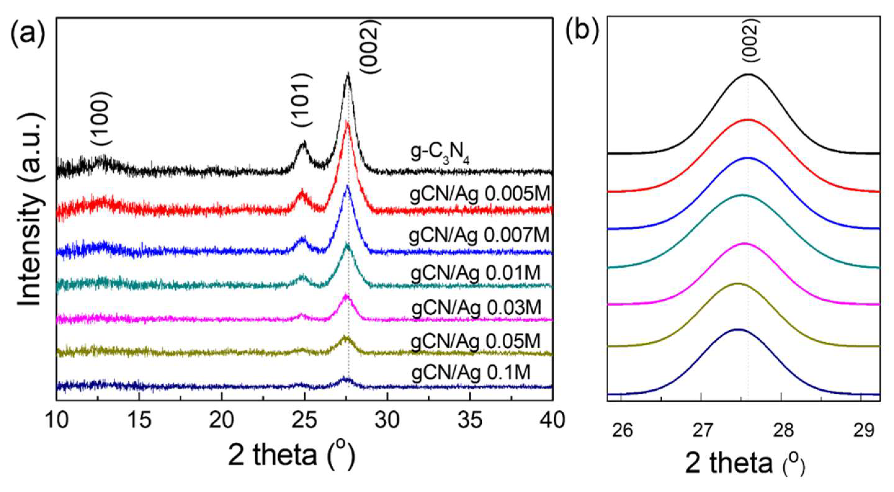
Abstract
:1. Introduction
2. Experiments
2.1. Materials
2.2. Synthesis of g-C3N4
2.3. Synthesis of Ag Nanoclusters-Loaded g-C3N4
2.4. Characterizations
2.5. Investigation of Photocatalytic Activity
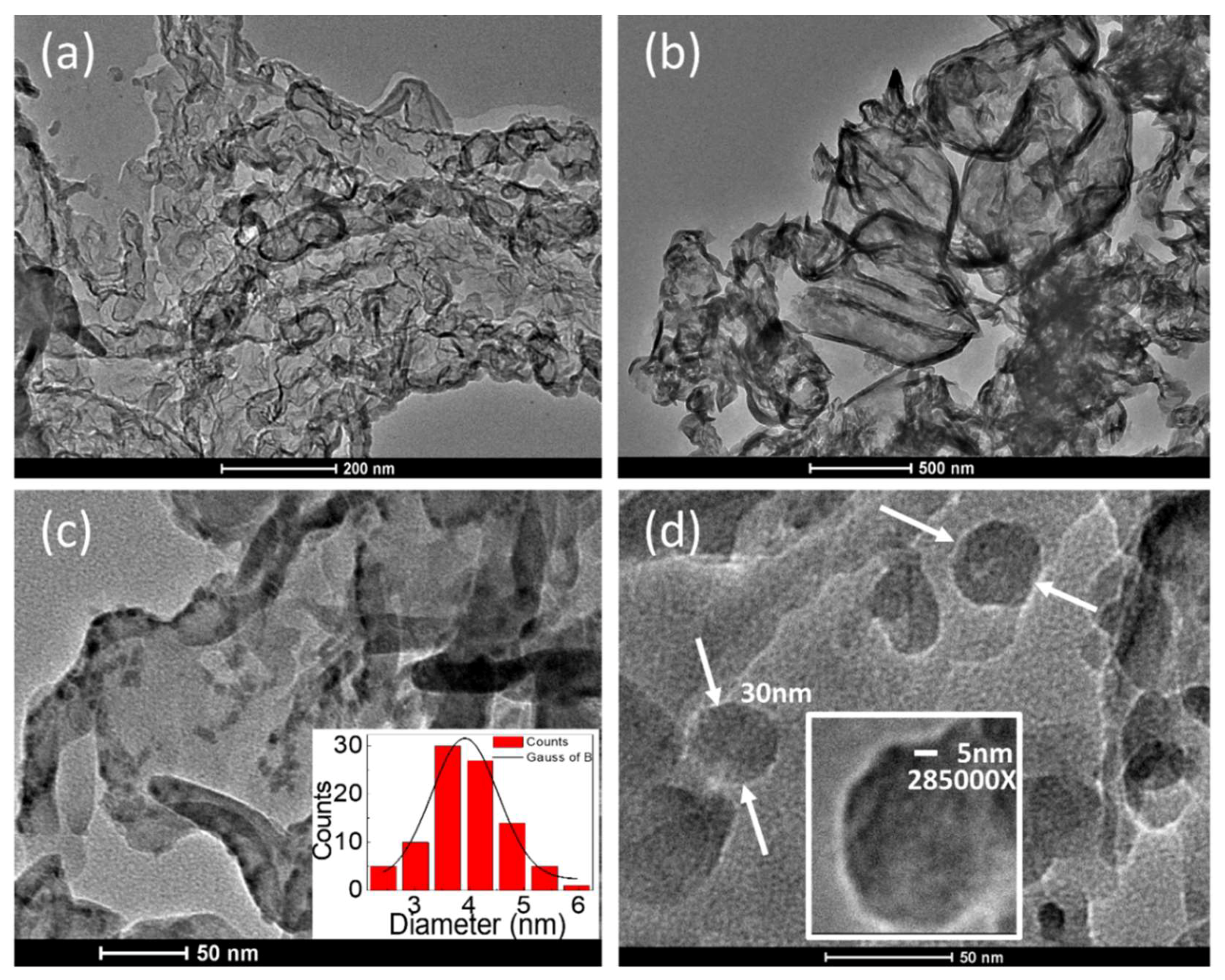
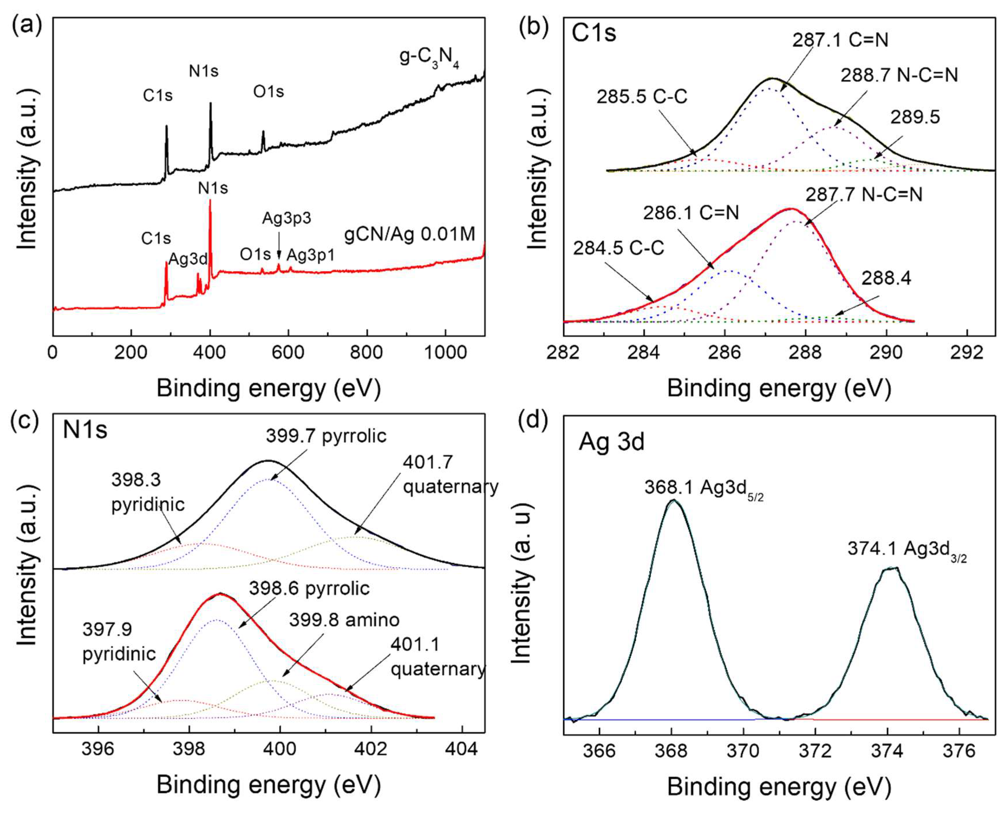
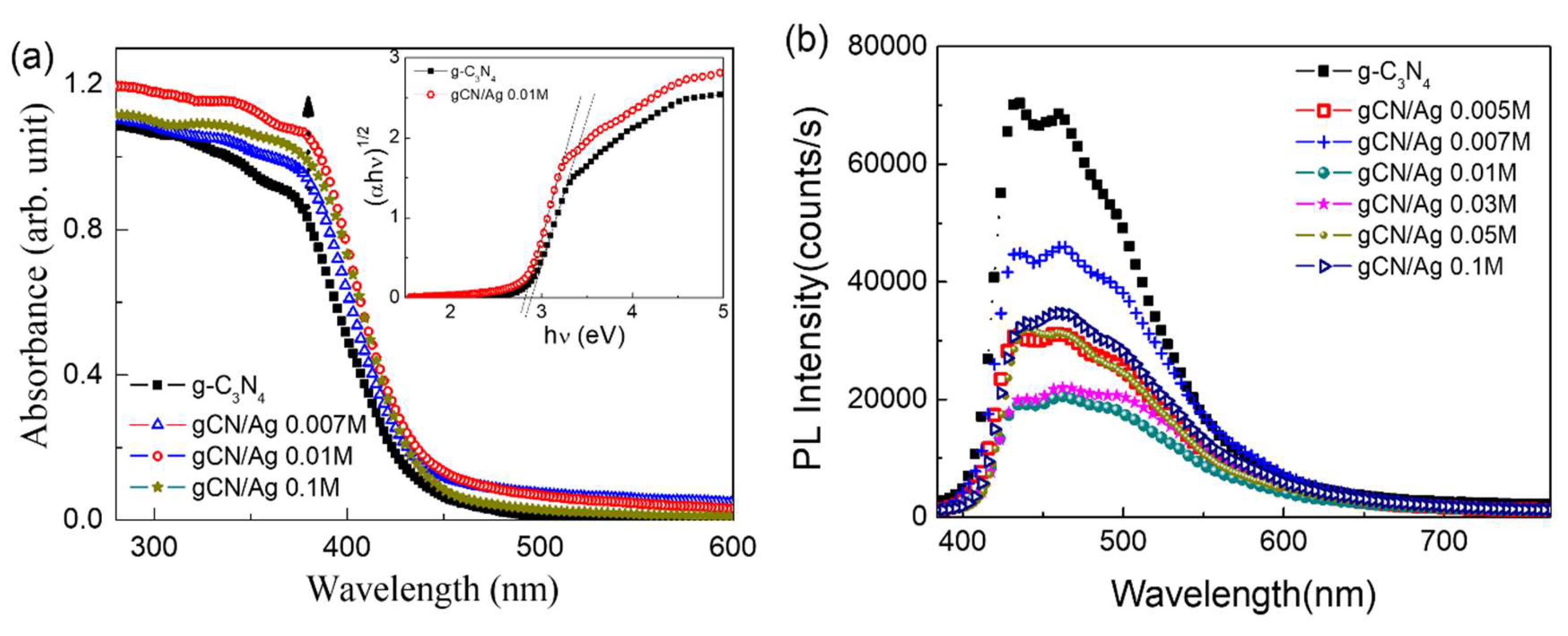
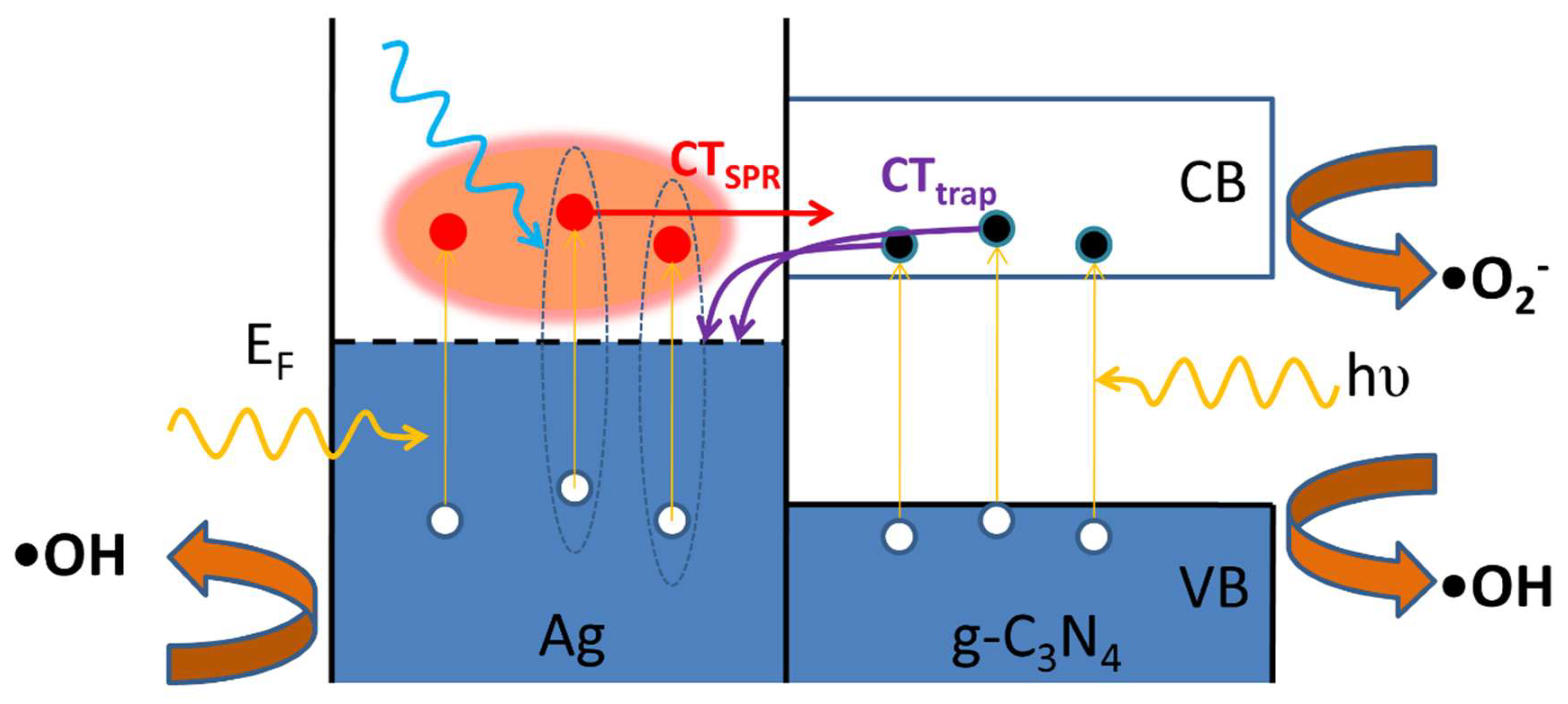
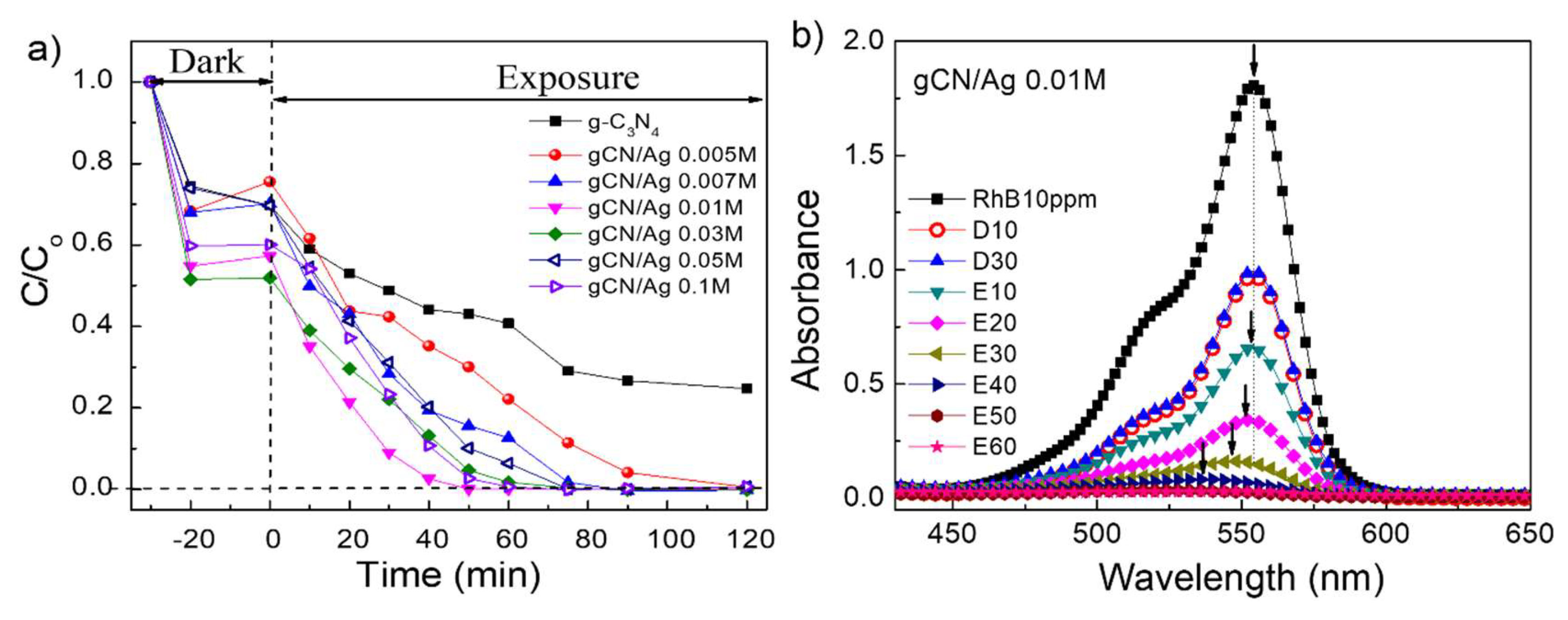
3. Results and Discussion
4. Conclusions
Author Contributions
Acknowledgments
Conflicts of Interest
References
- Zhang, Y.; Pan, Q.; Chai, G.; Liang, M.; Dong, G.; Zhang, Q.; Qiu, J. Synthesis and Luminescence Mechanism of Multicolor-Emitting g-C3N4 Nanopowders by Low Temperature Thermal Condensation of Melamine. Sci. Rep. 2013, 3, 1943. [Google Scholar] [CrossRef] [PubMed]
- Ye, S.; Wang, R.; Wu, M.Z.; Yuan, Y.P. A Review on g-C3N4 for Photocatalytic Water Splitting and CO2 Reduction. Appl. Surf. Sci. 2015, 358, 15–27. [Google Scholar] [CrossRef]
- Le, S.; Jiang, T.; Zhao, Q.; Liu, X.; Li, Y.; Fang, B.; Gong, A.M. Cu-Doped Mesoporous Graphitic Carbon Nitride for Enhanced Visible-Light Driven Photocatalysis. RSC Adv. 2016, 6, 38811–38819. [Google Scholar] [CrossRef]
- Yuan, Y.; Zhang, L.; Xing, J.; Utama, M.I.B.; Lu, X.; Du, K.; Li, Y.; Hu, X.; Wang, S.; Genc, A.; et al. High-Yield Synthesis and Optical Properties of g-C3N4. Nanoscale 2015, 7, 12343–12350. [Google Scholar] [CrossRef] [PubMed]
- Zhang, Y.; Schnepp, Z.; Cao, J.; Ouyang, S.; Li, Y.; Ye, J.; Liu, S. Biopolymer-Activated Graphitic Carbon Nitride Towards a Sustainable Photocathode Material. Sci. Rep. 2013, 3, 2163. [Google Scholar] [CrossRef] [PubMed]
- Pawar, R.C.; Kang, S.; Park, J.H.; Kim, J.H.; Ahn, S.; Lee, C.S. Room-Temperature Synthesis of Nanoporous 1D Microrods of Graphitic Carbon Nitride (g-C3N4) with Highly Enhanced Photocatalytic Activity and Stability. Sci. Rep. 2016, 6, 31147. [Google Scholar] [CrossRef] [PubMed]
- Yan, H.; Tian, X.; Pang, Y.; Feng, B.; Duan, K.; Zhou, Z.; Weng, J.; Wang, J. Heterostructured g-C3N4/Ag/TiO2 Nanocomposites for Enhancing the Photoelectric Conversion Efficiency of Spiro-Ometad-Based Solid-State Dye-Sensitized Solar Cells. RSC Adv. 2016, 6, 102444–102452. [Google Scholar] [CrossRef]
- Zhang, S.; Hang, N.T.; Zhang, Z.; Yue, H.; Yang, W. Preparation of G-C3n4/Graphene Composite for Detecting NO2 at Room Temperature. Nanomaterials 2017, 7, 12. [Google Scholar] [CrossRef] [PubMed]
- Jin, J.; Liang, Q.; Ding, C.; Li, Z.; Xu, S. Simultaneous Synthesis-Immobilization of Ag Nanoparticles Functionalized 2D g-C3N4 Nanosheets with Improved Photocatalytic Activity. J. Alloys Compd. 2017, 691, 763–771. [Google Scholar] [CrossRef]
- Fagan, R.; McCormack, D.E.; Hinder, S.J.; Pillai, S.C. Photocatalytic Properties of G-C3N4–TiO2 Heterojunctions under UV and Visible Light Conditions. Mater. Lett. 2016, 9, 286. [Google Scholar] [CrossRef] [PubMed]
- Oanh, L.T.M.; Do, D.B.; Hang, L.T.; Lai, N.D.; Phuong, N.T.; Thang, D.V.; Hung, N.M.; Minh, N.V. Influence of Annealing Temperature on Physical Properties and Photocatalytic Ability of g-C3N4 Nanosheets Synthesized through Urea Polymerization in Ar Atmostphere. Phys. B Condens. Matter 2018, 532, 48–53. [Google Scholar] [CrossRef]
- Li, Q.; Xu, D.; Ou, X.; Yan, F. Nitrogen-Doped Graphitic Porous Carbon Nanosheets Derived from in Situ Formed g-C3N4 Templates for the Oxygen Reduction Reaction. Chem. Asian J. 2017, 12, 1816–1823. [Google Scholar] [CrossRef] [PubMed]
- Liu, Y.; Zhang, H.; Lu, Y.; Wu, J.; Xin, B. A Simple Method to Prepare g-C3N4/Ag-Polypyrrole Composites with Enhanced Visible-Light Photocatalytic Activity. Catal. Commun. 2016, 87, 41–44. [Google Scholar] [CrossRef]
- Xu, J.; Wang, G.; Fan, J.; Liu, B.; Cao, S.; Yu, J. g-C3N4 Modified TiO2 Nanosheets with Enhanced Photoelectric Conversion Efficiency in Dye-Sensitized Solar Cells. J. Power. Sources 2015, 274, 77–84. [Google Scholar] [CrossRef]
- Ong, W.J.; Tan, L.L.; Chai, S.P.; Yong, S.T. Heterojunction Engineering of Graphitic Carbon Nitride (g-C3N4) Via Pt Loading with Improved Daylight-Induced Photocatalytic Reduction of Carbon Dioxide to Methane. Dalton Trans. 2015, 44, 1249–1257. [Google Scholar] [CrossRef] [PubMed]
- Xue, J.; Ma, S.; Zhou, Y.; Zhang, Z.; He, M. Facile Photochemical Synthesis of Au/Pt/g-C3N4 with Plasmon-Enhanced Photocatalytic Activity for Antibiotic Degradation. ACS Appl. Mater. Interfaces 2015, 7, 9630–9637. [Google Scholar] [CrossRef] [PubMed]
- Zhuang, J.; Lai, W.; Xu, M.; Zhou, Q.; Tang, D. Plasmonic Aunp/g-C3N4 Nanohybrid-Based Photoelectrochemical Sensing Platform for Ultrasensitive Monitoring of Polynucleotide Kinase Activity Accompanying Dnazyme-Catalyzed Precipitation Amplification. ACS Appl. Mater. Interfaces 2015, 7, 8330–8338. [Google Scholar] [CrossRef] [PubMed]
- Wang, J.; Liu, R.; Zhang, C.; Han, G.; Zhao, J.; Liu, B.; Jiang, C.; Zhang, Z. Synthesis of g-C3N4 Nanosheets/Au@Ag Nanoparticles Hybrids as Sers Probe for Cancer Cells Diagnostics. RSC Adv. 2015, 5, 86803–86810. [Google Scholar] [CrossRef]
- Konda, S.K.; Amiri, M.; Chen, A. Photoassisted Deposition of Palladium Nanoparticles on Carbon Nitride for Efficient Oxygen Reduction. J. Phys. Chem. C 2016, 120, 14467–14473. [Google Scholar] [CrossRef]
- Wang, H.-H.; Zhang, B.; Li, X.-H.; Antonietti, M.; Chen, J.-S. Activating Pd Nanoparticles on Sol–Gel Prepared Porous G-C3N4/SiO2 via Enlarging the Schottky Barrier for Efficient Dehydrogenation of Formic Acid. Inorg. Chem. Front. 2016, 3, 1124–1129. [Google Scholar] [CrossRef]
- Tian, K.; Liu, W.-J.; Jiang, H. Comparative Investigation on Photoreactivity and Mechanism of Biogenic and Chemosythetic Ag/C3N4 Composites under Visible Light Irradiation. ACS Sustain. Chem. Eng. 2015, 3, 269–276. [Google Scholar] [CrossRef]
- Jiang, D.; Zhang, Y.; Chu, H.; Liu, J.; Wan, J.; Chen, M. N-Doped Graphene Quantum Dots as an Effective Photocatalyst for the Photochemical Synthesis of Silver Deposited Porous Graphitic C3N4 Nanocomposites for Nonenzymatic Electrochemical H2O2 Sensing. RSC Adv. 2014, 4, 16163–16171. [Google Scholar] [CrossRef]
- Bing, W.; Chen, Z.; Sun, H.; Shi, P.; Gao, N.; Ren, J.; Qu, X. Visible-Light-Driven Enhanced Antibacterial and Biofilm Elimination Activity of Graphitic Carbon Nitride by Embedded Ag Nanoparticles. Nano Res. 2015, 8, 1648–1658. [Google Scholar] [CrossRef]
- Wang, X.; Zhao, Z.; Ou, D.; Tu, B.; Cui, D.; Wei, X.; Cheng, M. Highly Active Ag Clusters Stabilized on TiO2 Nanocrystals for Catalytic Reduction of P-Nitrophenol. Appl. Surf. Sci. 2016, 385, 445–452. [Google Scholar] [CrossRef]
- Panigrahi, S.; Basu, S.; Praharaj, S.; Pande, S.; Jana, S.; Pal, A.; Ghosh, S.K.; Pal, T. Synthesis and Size-Selective Catalysis by Supported Gold Nanoparticles: Study on Heterogeneous and Homogeneous Catalytic Process. J. Phys. Chem. C 2007, 111, 4596–4605. [Google Scholar] [CrossRef]
- Valden, M.; Lai, X.; Goodman, D.W. Onset of Catalytic Activity of Gold Clusters on Titania with the Appearance of Nonmetallic Properties. Science 1998, 281, 1647–1650. [Google Scholar] [CrossRef] [PubMed]
- Yi, J.; Liao, K.; Zhang, C.; Zhang, T.; Li, F.; Zhou, H. Facile in Situ Preparation of Graphitic-C3N4@Carbon Paper as an Efficient Metal-Free Cathode for Nonaqueous Li-O2 Battery. ACS Appl. Mater. Inter. 2015, 7, 10823–10827. [Google Scholar] [CrossRef] [PubMed]
- Su, F.Y.; Zhang, W.D. Carbonyl-Grafted g-C3N4 Porous Nanosheets for Efficient Photocatalytic Hydrogen Evolution. Chem. Asian J. 2017, 12, 515–523. [Google Scholar] [CrossRef] [PubMed]
- Ong, W.J.; Putri, L.K.; Tan, L.L.; Chai, S.P.; Yong, S.T. Heterostructured AgX/G-C3N4 (X=Cl and Br) Nanocomposites via a Sonication-Assisted Deposition-Precipitation Approach: Emerging Role of Halide Ions in the Synergistic Photocatalytic Reduction of Carbon Dioxide. Appl. Catal. B Environ. 2016, 180, 530–543. [Google Scholar] [CrossRef]
- Feng, L.; Yang, L.; Huang, Z.; Luo, J.; Li, M.; Wang, D.; Chen, Y. Enhancing Electrocatalytic Oxygen Reduction on Nitrogen-Doped Graphene by Active Sites Implantation. Sci. Rep. 2013, 3, 3306. [Google Scholar] [CrossRef] [PubMed]
- Kundu, S.; Nagaiah, T.C.; Xia, W.; Wang, Y.; Dommele, S.V.; Bitter, J.H.; Santa, M.; Grundmeier, G.; Bron, M.; Schuhmann, W.; et al. Electrocatalytic Activity and Stability of Nitrogen-Containing Carbon Nanotubes in the Oxygen Reduction Reaction. J. Phys. Chem. C 2009, 113, 14302–14310. [Google Scholar] [CrossRef]
- Shao, Y.; Zhang, S.; Engelhard, M.H.; Li, G.; Shao, G.; Wang, Y.; Liu, J.; Aksay, I.A.; Lin, Y. Nitrogen-Doped Graphene and Its Electrochemical Applications. J. Mater. Chem. 2010, 20, 7491–7496. [Google Scholar] [CrossRef]
- Wang, Y.; Shao, Y.; Matson, D.W.; Li, J.; Lin, Y. Nitrogen-Doped Graphene and Its Application in Electrochemical Biosensing. ACS Nano 2010, 4, 1790–1798. [Google Scholar] [CrossRef] [PubMed]
- Lu, M.; Pei, Z.; Weng, S.; Feng, W.; Fang, Z.; Zheng, Z.; Huang, M.; Liu, P. Constructing Atomic Layer g-C3N4–CdS Nanoheterojunctions with Efficiently Enhanced Visible Light Photocatalytic Activity. Phys. Chem. Chem. Phys. 2014, 16, 21280–21288. [Google Scholar] [CrossRef] [PubMed]
- Chen, Y.; Huang, W.; He, D.; Situ, Y.; Huang, H. Construction of Heterostructured g-C3N4/Ag/TiO2 Microspheres with Enhanced Photocatalysis Performance under Visible-Light Irradiation. ACS Appl. Mater. Interfaces 2014, 6, 14405–14414. [Google Scholar] [CrossRef] [PubMed]
- Mott, D.; Thuy, N.T.; Aoki, Y.; Maenosono, S. Aqueous Synthesis and Characterization of Ag and Ag-Au Nanoparticles: Addressing Challenges in Size, Monodispersity and Structure. Philos. Trans. Ser. A Math. Phys. Eng. Sci. 2010, 368, 4275–4292. [Google Scholar] [CrossRef] [PubMed]
- Charle, K.P.; Konig, L.; Nepijko, S.; Rabin, I.; Schulze, W. The Surface Plasmon Resonance of Free and Emberred Ag-Clusters in the Size Range of 1.5 < D < 30 nm. Cryst. Res. Technol. 1998, 33, 1085–1096. [Google Scholar]
- Zhang, S.; Zhang, B.P.; Li, S.; Huang, Z.; Yang, C.; Wang, H. Enhanced Photocatalytic Activity in Ag-Nanoparticle-Dispersed BaTiO3 Composite Thin Films: Role of Charge Transfer. J. Adv. Ceram. 2017, 6, 1–10. [Google Scholar] [CrossRef]
- Kaur, R.; Pal, B. Plasmonic Coinage Metal–TiO2 Hybrid Nanocatalysts for Highly Efficient Photocatalytic Oxidation under Sunlight Irradiation. New J. Chem. 2015, 39, 5966–5976. [Google Scholar] [CrossRef]
- Sridharan, K.; Jang, E.; Park, J.H.; Kim, J.H.; Lee, J.H.; Park, T.J. Silver Quantum Cluster (Ag9)-Grafted Graphitic Carbon Nitride Nanosheets for Photocatalytic Hydrogen Generation and Dye Degradation. Chem. Eur. J. 2015, 21, 9126–9132. [Google Scholar] [CrossRef] [PubMed]
- Gao, L.; Li, Z.; Liu, J. Facile Synthesis of Ag3VO4/B-AgVO3 Nanowires with Efficient Visible-Light Photocatalytic Activity. RSC Adv. 2017, 7, 27515–27521. [Google Scholar] [CrossRef]


© 2018 by the authors. Licensee MDPI, Basel, Switzerland. This article is an open access article distributed under the terms and conditions of the Creative Commons Attribution (CC BY) license (http://creativecommons.org/licenses/by/4.0/).
Share and Cite
Le, T.M.O.; Lam, T.H.; Pham, T.N.; Ngo, T.C.; Lai, N.D.; Do, D.B.; Nguyen, V.M. Enhancement of Rhodamine B Degradation by Ag Nanoclusters-Loaded g-C3N4 Nanosheets. Polymers 2018, 10, 633. https://doi.org/10.3390/polym10060633
Le TMO, Lam TH, Pham TN, Ngo TC, Lai ND, Do DB, Nguyen VM. Enhancement of Rhodamine B Degradation by Ag Nanoclusters-Loaded g-C3N4 Nanosheets. Polymers. 2018; 10(6):633. https://doi.org/10.3390/polym10060633
Chicago/Turabian StyleLe, Thi Mai Oanh, Thi Hang Lam, Thi Nhung Pham, Tuan Cuong Ngo, Ngoc Diep Lai, Danh Bich Do, and Van Minh Nguyen. 2018. "Enhancement of Rhodamine B Degradation by Ag Nanoclusters-Loaded g-C3N4 Nanosheets" Polymers 10, no. 6: 633. https://doi.org/10.3390/polym10060633
APA StyleLe, T. M. O., Lam, T. H., Pham, T. N., Ngo, T. C., Lai, N. D., Do, D. B., & Nguyen, V. M. (2018). Enhancement of Rhodamine B Degradation by Ag Nanoclusters-Loaded g-C3N4 Nanosheets. Polymers, 10(6), 633. https://doi.org/10.3390/polym10060633