Article

Spectroscopic and Structural Analyses of Opuntia Robusta Mucilage and Its Potential as an Edible Coating

by
Aurea Bernardino-Nicanor
1,
José Luis Montañez-Soto
2,
Eloy Conde-Barajas
1,
María de la Luz Xochilt Negrete-Rodríguez
1,
Gerardo Teniente-Martínez
1,
Enaim Aída Vargas-León
1,
José Mayolo Simitrio Juárez-Goiz
1,
Gerardo Acosta-García
1 and
Leopoldo González-Cruz
1,*
1
Tecnológico Nacional de México-Celaya, Antonio García Cubas Pte #600 esq. Av. Tecnológico. Celaya, Gto. Mexico, Celaya C.P. 38010, Mexico
2
Centro Interdisciplinario de Investigación para el Desarrollo Integral Regional del Instituto Politécnico Nacional, Unidad Michoacán, Justo Sierra No. 28 Jiquilpan, Mich C.P. 59510, Mexico
*
Author to whom correspondence should be addressed.
Coatings 2018, 8(12), 466; https://doi.org/10.3390/coatings8120466
Submission received: 28 November 2018 / Revised: 12 December 2018 / Accepted: 12 December 2018 / Published: 15 December 2018
(This article belongs to the Special Issue Advances in Organic and Polymeric Coatings )

Abstract

:
Mucilage extracted from the parenchymatous and chlorenchymatous tissues of Opuntia robusta were obtained using water or ethanol as the extraction solvent. The changes in the different tissues by using different extraction solvents were evaluated via scanning electron microscopy (SEM) and Fourier transform infrared (FT-IR) and Raman spectroscopy; in addition, the effect of mucilage coating on the various quality characteristics of tomato (Lycopersicum sculentum) was evaluated. The SEM results showed that the mucilage extracted from the parenchyma had a higher aggregation level that the mucilage extracted from the chlorenchyma. The presence of three characteristic bands of pectic substances in the FT-IR spectra between 1050 and 1120 cm−1 indicated that the mucilage extracted from the parenchymatous tissue had a higher content of pectic compounds than the mucilage extracted from the chlorenchymatous tissue. It was also observed in the Raman spectra that the level of pectic substances in the mucilage extracted from the parenchymatous was higher than that in the mucilage extracted from the chlorenchymatous tissue. The mucilage extracted from the parenchymatous tissue was more effective as an edible coating than the mucilage extracted from the chlorenchymatous tissue. Tomatoes covered with mucilage showed significantly enhanced firmness and reduced weight loss. The uncoated tomatoes showed higher lycopene content than the coated tomatoes on the 21st day. This study showed that the Opuntia robusta tissue and extraction solvent influence mucilage characteristics and that Opuntia robusta mucilage is a promising edible coating.

Graphical Abstract

1. Introduction

The tomato (Lycopersicum esculentum) is a climacteric fruit with a relatively short postharvest life during which softening and textural changes occur. In addition, many components of biochemical pathways involved in pigmentation, carbohydrate metabolism and ethylene biosynthesis are also modified [1]. Postharvest practices can have a significant effect on tomato sugar content; however, physical characteristics are the most important factor for the final consumer, and fresh tomato quality is determined by appearance, color, firmness and flavor. On the other hand during maturation, the weight loss of the tomato is increased by the moisture effect, solute movement, and water loss [2]. For this reason, diverse studies have been focusing on the control of tomato ripening through of the use of modified atmospheres, temperature and humidity control, hypobaric storage and edible coatings [3,4].
The use of edible coatings has been considered an alternative in response to the increasing demand for fresh and minimally processed tomatoes, enhancing their shelf life. The application of edible coatings in tomatoes is a promising technology since these coatings can act as a vehicle for the incorporation of fungicides and antimicrobial agents [3,5], reduce the rate of color change, and inhibit ethylene production [6]; they can also act as barriers to water loss and gas exchange, enable the controlled release of bioactive compounds, and enhance the antioxidant activity and total phenolic contents [7].
In the development of edible coatings, a broad range of edible biopolymers has been used, such as mucilage, starches, cellulose derivatives, pectin, carrageenan, chitosan/chitin, sucrose, gums, sodium alginate, proteins (animal or plant-based) and lipids [8]. The use of edible coatings has diverse advantages compared with synthetic polymers; edible coatings endow food products with a natural appearance, reduce the environmental impact, and are nontoxic, economical and easily available in the environment [9]. In addition, edible coatings offer biocompatibility and can be used as additive carriers of colorants, flavors, antioxidants or antimicrobials.
The plants of the Opuntia genus have high concentration of polysaccharides, principally mucilages that contain arabinose, galactose, galacturonic acid, rhamnose, and xylose [10], on the other hand, Rocchetti et al., [11] reported the presence of high content of β-glucans in the Opuntia ficus-indica cladodes, that are capable of forming gels in water, for this reason, the mucilaginous compounds of Opuntia robusta have been used in the development of foodstuffs [12]; in addition, it has been observed that the mucilage from nopal also has the ability to form edible coatings [13] and has been used to develop an edible coating that increases the shelf-life of strawberry [14].
However, few studies have been conducted using Raman, FT-IR and scanning electron microscopy (SEM) methods to compare mucilage extracted from the parenchyma and that from the chlorenchyma of Opuntia robusta or to examine the effect of modifying the extraction solvent. For this reason, in this study, FT-Raman, FT-IR spectroscopy and scanning electron microscopy (SEM) were used to determine the differences between mucilage extracted from the parenchyma and mucilage extracted from the chlorenchyma of Opuntia robusta and the effect of the extraction solvent on the structure of the mucilage. The suitability of Opuntia robusta mucilage as an edible coating to extend the shelf life of tomato was also evaluated.

2. Materials and Methods

2.1. Vegetative Material

Opuntia robusta is a shrubby to tree-like cactus originating in central Mexico that can attain a height of 3 m, is a source of fruits as well as young cladodes used for animal feed. Young cladodes from Opuntia robusta Wendl var. robusta were obtained from Tulancingo, Hidalgo, Mexico. The cladodes were harvested manually at 12:00 p.m. and selected according to size to normalize the state of maturity: 35 cm (large), 30 cm (width) and 4.5 cm (thickness). The samples were stored at 4 °C in a refrigerator (LG, Model GR-452SH, LG electronics, Mexico, Mexico) for up to 24 h until used.

2.2. Obtention of Mucilage

Chlorenchymatous tissue and spongy parenchymatous tissue were removed from the cladodes using a knife, and both tissues were subsequently cut into approximately 2 cm cubes, packed in PVC bags and stored in the freezer (LG Model GR-452SH) until used.
One hundred g of parenchymatous or chlorenchymatous tissue were placed in a flask (500 mL) containing a stirrer and 100 mL of ethanol or water. The mixture was stirred frequently (2 h; 50 °C) and then ground in a high-speed blender for 5 min. The mixture was allowed to stand for 1 h and filtered using Whatman (Maidstone, UK) No. 40 filter paper to obtain a fully clarified mucilaginous extract. The mucilaginous extract obtained from Opuntia robusta was dried (50 °C) until the complete elimination of moisture in a forced convection drying-oven (Binder, Model FD115-UL, Tuttlingen, Germany). Dried mucilage was milled into a fine powder and sieved through a size 40 mesh (425 μm). The mucilage powder was packaged in 25-g glass bottles and stored at 25 °C until used.

2.3. Scanning Electron Microscopy

Samples of Opuntia robusta mucilage powder were examined using a JEOL (JEOL, type EX-1200, Tokyo, Japan) scanning electron microscope fitted with a Kevex Si(Li) X-ray detector (Kevex inc, Newark, DE, USA). The analyses were performed under vacuum at an accelerating voltage of 15 kV. The samples were mounted on double-sided carbon tape and covered with approximately 10 nm of gold using a Denton sputter coater (Denton Vaccum LLC, Moorestown, NJ, USA).

2.4. FT-IR Spectroscopy

The FT-IR spectra of the Opuntia robusta mucilage were acquired on a Perkin Elmer FT-IR spectrophotometer (Perkin Elmer, Inc., Waltham, MA, USA) using potassium bromide (KBr) discs prepared from powered samples mixed with dry KBr. The spectra were recorded (16 scans) in transparent mode at a resolution of 4000 to 400 cm−1.

2.5. Raman Spectroscopy

The Raman measurements were performed on a Perkin-Elmer (Perkin Elmer, Inc., Waltham, MA, USA) 2000R NIR FT-Raman Spectrometer equipped with a Nd:YAG laser emitting at a wavelength of 1064 nm and an InGaAs detector. For these analyses, the 180° backscattering refractive geometry was used. The spectrometer was managed using Perkin-Elmer Spectrum software (Version 3.02.00 [2000]). The spectral data for rice bean starch were obtained at a wavenumber resolution of 4 cm−1 and at a nominal laser power of 500 mW. For each spectrum, 20 scans were accumulated to ensure an acceptable signal-to-noise ratio. All Raman spectra were collected at room temperature.

2.6. Edible Coating Preparation

Six different treatments were prepared, which differed in the solvent (water or ethanol) and cladode tissue (parenchyma or chlorenchyma) used, for the reconstitution of the mucilage powder (Table 1). For the preparation of the edible coatings, 50 g of the mucilage powder were placed in a flask (500 mL), and then water or ethanol was added to obtain a solution of mucilage with 12% soluble solids; this procedure was carried out for each cladode tissue.

Edible Coating Application

Prior to the application of edible coatings, the tomatoes were washed with distilled water. The coatings were applied uniformly by brushing on various tomatoes (26 pieces), three times. The tomatoes were stored at 20 °C. A control lot of tomatoes (26 pieces) was used (without edible coating).

2.7. Weight Loss During Postharvest Storage

Weight loss during postharvest storage was determined by subtracting the sample weight from their previous recorded weight and was presented as % of weight loss compared to the initial weight. Weight loss was calculated using the Equation (1).
weight   loss   ( % ) = Initial   weight   ( g ) final   weight   ( g ) Initial   weight   ( g ) × 100

2.8. Texture Measurement

A penetration test was performed on the skin of the whole fruit using a TA.XT2i texture analyzer (Stable Micro Systems, Surrey, UK) with a 2 mm diameter cylindrical probe. The samples were penetrated to a depth of 6 mm. The speed of the probe was 1.0 mm·s−1 during the pretest and penetration. The tomatoes were placed in a way that the probe penetrated their equatorial zone.

2.9. Determination of Lycopene

Fifty g of tomato were ground to a homogeneous puree using a hand-held mixer (Braun Inc., Lynnfield, MA, USA). The puree was mixed with 50 mL of a hexane-acetone-ethanol mixture (50:25:25) and placed in an orbital shaker (Eberbach Corp., Ann Arbor, MI, USA) (200 rpm) for 15 min. Thereafter, 3 mL of distilled water were added, and the sample was shaken for another 15 min and then vacuum-filtered. The combined filtrates were mixed with 50 mL of hexane in a separatory funnel, and 200 mL of double-distilled water were then added; the mixture was allowed to stand for 15 min to enable phase separation. The water–acetone–ethanol phase was discarded; the hexane phase was collected into an amber screw-capped vial and concentrated to dryness with high purity nitrogen (99.99%).
Lycopene was quantified using an external standard, and the absorbance was taken at 503 nm using a spectrophotometer (Thermo Fisher Scientific Inc., Waltham, MA, USA); hexane was used as solvent blank. The results were expressed as mg·kg−1 of sampler.

2.10. Statistical Analysis

Quantitative data are expressed as the mean ± standard deviation, and the results were statically analyzed with ANOVA and Tukey’s test at the 95% confidence level. SAS software (Statistical Analysis System V. 8.0) was used for the data analysis, and all experimental determinations were performed in triplicate.

3. Results and Discussion

3.1. Characterization of Mucilage

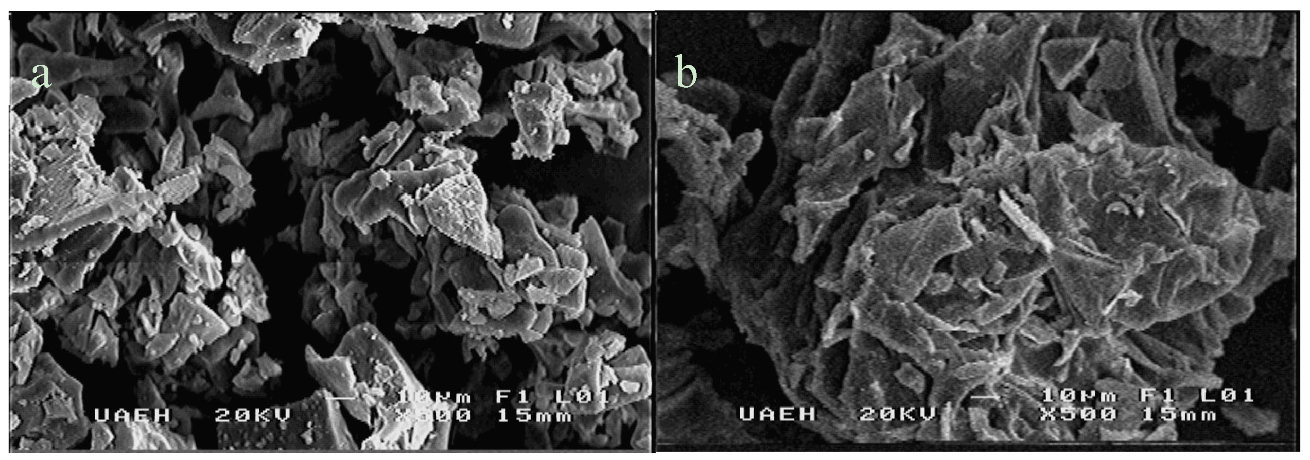
3.1.1. Morphology of Opuntia Mucilage

Scanning electron microphotographs (SEM) of the mucilage obtained from Opuntia robusta are shown in Figure 1. A higher aggregation level of small particles was observed in the mucilage extracted from the parenchyma (Figure 1b) than in the mucilage extracted from the chlorenchyma (Figure 1a). The particles of the mucilage extracted from the chlorenchyma of Opuntia robusta are mostly seen as aggregates of irregular shapes and dimensions and are fibrous in nature. Superimposed fibers have been observed in chia mucilage and increase the complexity of the aggregates [15]; apparently, a similar behavior can be observed in the mucilage extracted from the parenchyma of Opuntia robusta. On the other hand, the high aggregation observed in the mucilage obtained from Opuntia robusta is in concordance with those reported by du Toit et al. [16], indicating that Opuntia robusta has a higher fiber content that other Opuntia species.

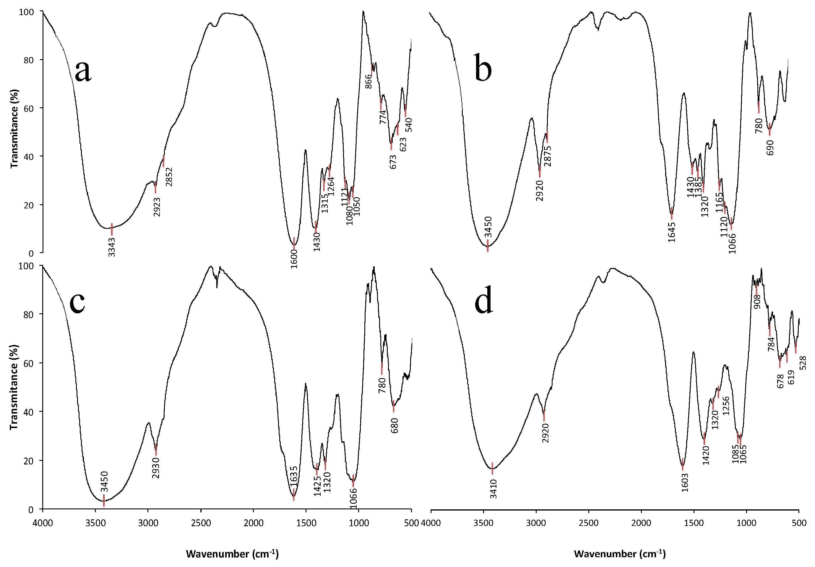
3.1.2. FT-IR Analysis of Structural Changes in Mucilage Due to the Extraction Process and the Source

The FT-IR spectra of the mucilage extracted using either water or ethanol are shown in Figure 2. It can be observed that the spectra of the mucilage present a broad, strong signal at approximately 3400 cm−1 due to the stretching vibration of O–H bonds; it can be observed that in both parenchyma and chlorenchyma tissues, this band was smoother in the spectrum of mucilage extracted with ethanol (Figure 2a,c) than in the spectrum of mucilage extracted with water (Figure 2b,d). Apparently, this difference is due to the formation of more hydrogen bonds in the mucilage by the interaction with ethanol. The best defined band was observed at approximately 2920 cm−1 for the ethanol-extracted mucilages of both tissues (parenchyma and chlorenchyma), and this band was assigned to the stretching vibration of C–H bonds from pyranose groups [17] or C–H group of the methyl ester of galacturonic acid [18].
However, some authors have indicated that this band can be attributed to proteins [19,20]; in addition, the water-extracted mucilages of both tissues show that the best defined band near 2870 cm−1 overlaps with the stretching vibration of the C–H bond from glucose units and with the –CH2 symmetric stretching vibration of proteins derived from carboxylic acids, indicating the presence of protein residues on the mucilage [19,21].
The broad bands observed between 1600 and 1640 cm−1 correspond to the scissor vibrations of –OH due to bound water [17] or the stretching vibration of the carboxyl group –COOH [19,22,23]. In mucilage extracted with water or ethanol, the presence of a free carboxyl group was observed, represented by the band at approximately 1430 cm−1 [22]. The best defined band for the mucilage extracted with ethanol was observed at approximately 1320 cm−1, indicating the presence of o–acetyl groups; other authors have attributed this band to the glycosidic linkage [24]. The three characteristic bands of pectic fractions [22] were observed only in the mucilage extracted from the parenchyma (1120, 1080, and 1050 cm−1); in the mucilage extracted from the chlorenchyma, an overlap was observed at approximately 1085 and 1065 cm−1.

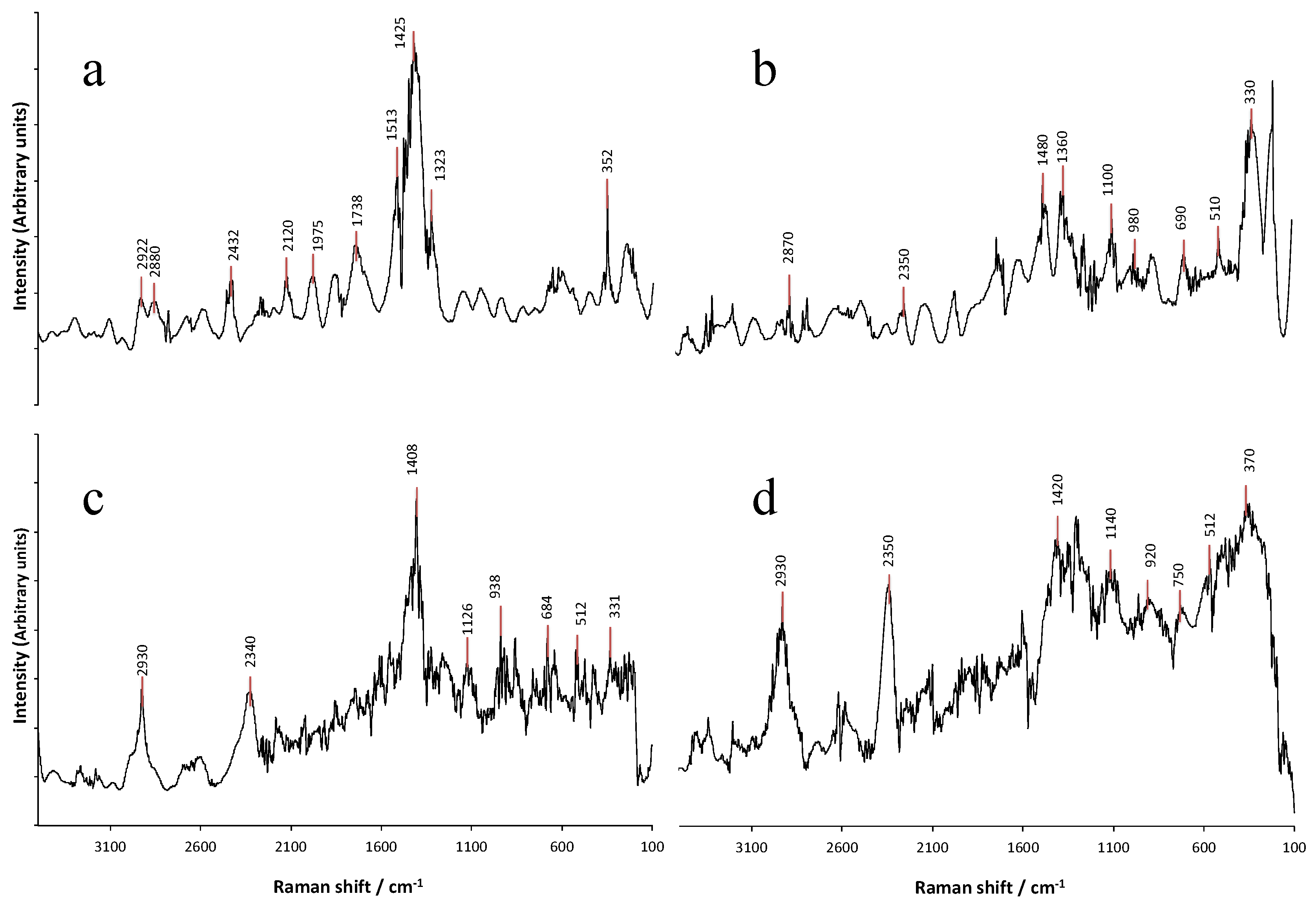
3.1.3. Raman Spectroscopy Analysis of Structural Changes in the Mucilage Due to the Extraction Process and the Source

The Raman spectra of the mucilage samples from Opuntia robusta are presented in Figure 3. The principal differences observed in the spectra of mucilage extracted with ethanol or water occurred in the principal bands associated with mucilage; on the other hand, differences in relative intensity were also observed between the patterns of Raman spectra of the mucilage extracted with ethanol (Figure 3b,d) and those of the mucilage extracted with water (Figure 3a,c) and between the spectra of the mucilage extracted from the chlorenchyma and that extracted from the parenchyma. In the mucilage extracted from the parenchyma, a well-defined band at 2930 cm−1 was observed in the sample in comparison with the mucilage extracted from the chlorenchyma, which presented a change in position; in addition, overlapping bands were observed (Figure 3a).
Some authors have indicated that the 2930 cm−1 band is related to the C–H stretching band of pectin, while the overlapping band at 2880 cm−1 is related to hemicellulose [25]; however, other authors have reported that this band is present in the mucilage of chia seeds [15].
The vibrational bands between 1400 and 1470 cm−1 are related to the elongation of the CH2 groups of xylose, galactose and arabinose, and the CH3 group of rhamnose; all of these bands were present for the Opuntia mucilage [10], and a higher intensity of these bands was observed in the water-extracted mucilage from the chlorenchyma than in the mucilage extracted from the parenchyma or extracted with ethanol; the intensity depends on the degree of crystallinity.
Additionally, the bands observed at 1350, 1270 and 1076 cm−1 are assigned as C–O–H related modes and probably correspond to residual galacturonic acid; however, some authors have indicated that the band at approximately 1330 cm−1 is related to the CH2 vibration of α-glucose and that the band at approximately 370 cm−1 is assigned to the skeletal vibrational mode of δs(C–C) [15].

3.2. Mucilage Used as Edible Coating

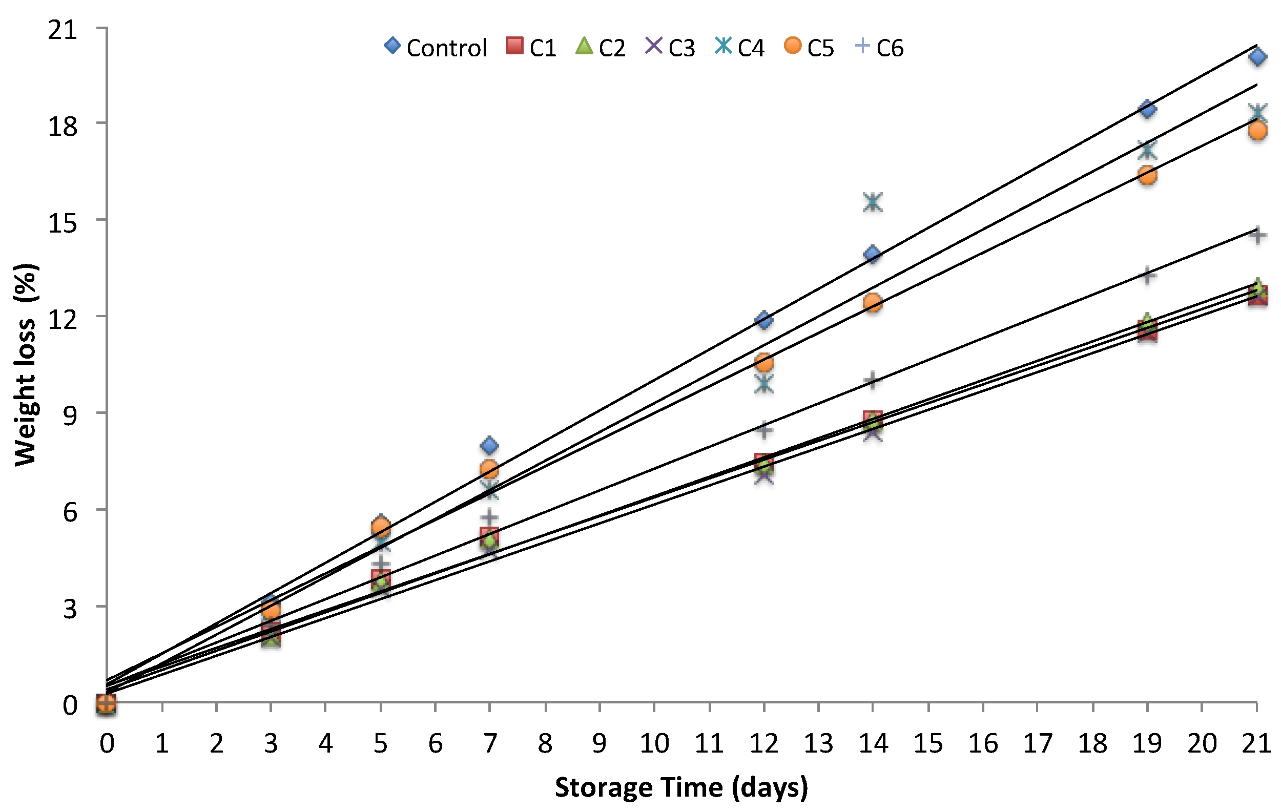
3.2.1. Weight Loss of Tomatoes

The tomatoes coated with parenchyma as an edible coating tended to have smaller weight loss than the control tomatoes (Figure 4). No significant difference in weight loss was observed in the three lots of tomatoes covered with parenchyma (C1, C2 and C3); however, the weight loss of the coated tomatoes was 7 wt.% lower than that of the control tomatoes (Figure 4). On the other hand, for the tomatoes coated with chlorenchyma, the differences between the three mucilage coatings tested (C4, C5 and C6) and the control (Figure 4) were significant. The tomatoes coated with chlorenchyma as an edible coating showed higher weight loss than the tomatoes coated with parenchyma; however, smaller weight loss was observed compared to that of the uncoated tomatoes (Figure 4).
The differences between the weight loss of the tomatoes coated with mucilage extracted from the parenchyma and that of tomatoes coated with mucilage extracted from the chlorenchyma could be due to the different compositions of the mucilage sources; however, it can be inferred that either mucilage source (parenchyma or chlorenchyma) formed a semipermeable layer that reduced respiration and transpiration on the tomato surface. It has been observed that coatings confer a physical barrier against O2, CO2, moisture and solute movement, consequently reducing water loss and weight loss [26,27,28].
Our results are in agreement with the finding of Del-Valle et al. [14], where the mucilage coating from Opuntia ficus-indica was effective in extending the shelf-life and other postharvest parameters of quality of strawberry; in other studies, the weight loss of breba fig was strongly influenced by Opuntia ficus-indica mucilage coating [29].

3.2.2. Effect on the Firmness of Tomatoes

Tomato firmness decreased significantly during storage in all coated and uncoated tomatoes (Figure 5); however, the coated tomatoes showed resistance against the loss of firmness and maintained texture during the storage period, particularly those tomatoes coated by mucilage extracted from the nopal parenchyma (Figure 5). The reduction in firmness of the uncoated sample was 62% compared to 47% for the tomatoes coated with mucilage extracted from the parenchyma and 60% for the tomatoes coated with mucilage extracted from the chlorenchyma (Figure 5). Similar firmness reduction trends were observed by Allegra [29], who showed that the loss of firmness in tomatoes coated with Opuntia ficus indica mucilage was significantly delayed during the storage period.
Surface coatings have been found to retain higher firmness, and the positive effect is attributed to the restriction in metabolic activities associated with cell wall-degrading enzymes or to the calcium content in species of the Opuntia genus [12,30], since it has been observed that calcium treatment maintains the firmness of peach, fig and strawberry [31,32,33].

3.2.3. Effect on the Lycopene Content of Tomatoes

The lycopene contents of the tomatoes coated and uncoated with mucilage during the red stage on the 21st day are shown in Table 2. In general, the results are in concordance with those of other studies on lycopene content in tomatoes that indicated that the lycopene content of stored tomatoes is in the range of 78.6 and 324 mg·kg−1 of tomato on a fresh weight basis and between 1710 and 4550 mg·kg−1 of tomato on a dry weight basis [34,35], however the extraction method was determinant to the extractable amount of lycopene and purity, reaching in some cases around purity level over 95% [36]. The lycopene content of the uncoated tomatoes on the 21st day of storage was 20% and 6% higher than those of the tomatoes coated with mucilage extracted from the parenchyma and the chlorenchyma, respectively.
Our results are in agreement with those of previous reports suggesting that the formation of lycopene depends on the rate of respiration during storage [37,38]; apparently, the mucilage provided a thick barrier against ethylene production and gas exchange between the inner and outer environments and therefore delayed the ripening of the fruit during storage [2].

4. Conclusions

Differences in morphology between the mucilage extracted from the parenchymatous tissue and the mucilage extracted from the chlorenchymatous tissue were detected. The FT-IR and Raman spectroscopy data showed that the mucilage extracted from the parenchymatous tissue had an apparently higher level of pectic substances than the mucilage extracted from the chlorenchymatous tissue; in addition, changes in the FT-IR and Raman spectra patterns were observed due to the extraction solvent. The scanning electron microscopy (SEM) showed higher aggregation level in the mucilage extracted from the parenchyma than in the mucilage extracted from chlorenchyma. Tomatoes covered with mucilage showed significantly enhanced firmness and reduced weight loss, while the uncoated tomatoes showed higher lycopene content than the coated tomatoes on the 21st day. Finally, this study showed that Opuntia robusta mucilage is a promising edible coating and that tissue and extraction solvent influence mucilage characteristics.

Author Contributions

Conceptualization, A.B.-N. and G.-C.L.; Formal Analysis, E.C.-B. and M.d.l.L.X.N.-R.; Funding Acquisition, A.B.-N. and G.-C.L.; Investigation, J.L.M.-S. and G.A.-G.; Methodology, E.A.V.-L.; Resources, G.T.-M. and J.M.S.J.-G.; Validation, G.T.-M. and J.M.S.J.-G.; Writing–Original Draft, A.B.-N. and G.-C.L.; Writing–Review & Editing, G.A.-G.; Supervision, A.B.-N. and G.-C.L.

Funding

This research was funded by National Technologic of Mexico (TecNM, No. 6405.18-P).

Acknowledgments

The authors would like to acknowledge the Autonomous University of Hidalgo State for technical services during this study.

Conflicts of Interest

The authors declare no conflict of interest.

References

  1. Alexander, L.; Grierson, D. Ethylene biosynthesis and action in tomato: A model for climacteric fruit ripening. J. Exp. Bot. 2002, 53, 2039–2055. [Google Scholar] [CrossRef] [PubMed]
  2. Ali, A.; Maqbool, M.; Ramachandran, S.; Alderson, P.G. Gum arabic as a novel edible coating for enhancing shelf-life and improving postharvest quality of tomato (Solanum lycopersicum L.) fruit. Postharvest Biol. Technol. 2010, 58, 42–47. [Google Scholar] [CrossRef]
  3. Rodríguez-Garcia, I.; Cruz-Valenzuela, M.R.; Silva-Espinoza, B.A.; Gonzalez-Aguilar, G.A.; Moctezuma, E.; Gutierrez-Pacheco, M.M.; Tapia-Rodríguz, M.R.; Ortega-Ramírez, L.A.; Ayala-Zavala, J.F. Oregano (Lippia graveolens) essential oil added within pectin edible coatings prevents fungal decay and increases the antioxidant capacity of treated tomatoes. J. Sci. Food Agric. 2016, 96, 3772–3778. [Google Scholar] [CrossRef] [PubMed]
  4. Ahmed, M.F.; Islam, M.Z.; Sarker, M.S.H.; Hasan, S.K.; Mizan, R. Development and performance study of controlled atmosphere for fresh tomato. World J. Eng. Tech. 2016, 4, 168–175. [Google Scholar] [CrossRef]
  5. Vodnar, D.C.; Pop, O.L.; Dulf, F.V.; Socaciu, C. Antimicrobial efficiency of edible films in food industry. Notulae Botanicae Horti Agrobotanici Cluj-Napoca 2015, 43, 302–312. [Google Scholar] [CrossRef]
  6. Zapata, P.J.; Guillén, F.; Martínez-Romero, D.; Castillo, S.; Valero, D.; Serrano, M. Use of alginate or zein as edible coatings to delay postharvest ripening process and to maintain tomato (Solanum lycopersicon Mill) quality. J. Sci. Food Agric. 2008, 88, 1287–1293. [Google Scholar] [CrossRef]
  7. Limchoowong, N.; Sricharoen, P.; Techawongstien, S.; Chanthai, S. An iodine supplementation of tomato fruits coated with an edible film of the iodide-doped chitosan. Food Chem. 2016, 200, 223–229. [Google Scholar] [CrossRef]
  8. Saha, A.; Tyagi, S.; Gupta, R.K.; Tyagi, Y.K. Natural gums of plant origin as edible coatings for food industry applications. Crit. Rev. Biotechnol. 2017, 37, 959–973. [Google Scholar] [CrossRef]
  9. Sánchez-Ortega, I.; García-Almendárez, B.E.; Santos-López, E.M.; Reyes-González, L.R.; Regalado, C. Characterization and antimicrobial effect of starch-based edible coating suspensions. Food Hydrocoll. 2016, 52, 906–913. [Google Scholar] [CrossRef]
  10. Trachtenberg, S.; Mayer, A.M. Composition and properties of Opuntia ficus-indica mucilage. Phytochemistry 1981, 20, 2665–2668. [Google Scholar] [CrossRef]
  11. Rocchetti, G.; Pellizzoni, M.; Montesano, D.; Lucini, L. Italian Opuntia ficus-indica cladodes as rich source of bioactive compounds with health-promoting properties. Foods 2018, 7, 24. [Google Scholar] [CrossRef] [PubMed]
  12. Bernardino-Nicanor, A.; Hinojosa-Hernández, E.N.; Juárez-Goiz, J.M.S.; Montañez-Soto, J.L.; Ramírez-Ortiz, M.E.; González-Cruz, L. Quality of Opuntia robusta and its use in development of mayonnaise-like product. J. Food Sci. Technol. 2015, 52, 343–350. [Google Scholar] [CrossRef] [PubMed]
  13. Espino-Díaz, M.; De Jesús Ornelas-Paz, J.; Martínez-Téllez, M.A.; Santillán, C.; Barbosa-Cánovas, G.V.; Zamudio-Flores, P.B.; Olivas, G.I. Development and characterization of edible films based on mucilage of Opuntia ficus-Indica (L.). J. Food Sci. 2010, 75, E347–E352. [Google Scholar] [CrossRef] [PubMed]
  14. Del-Valle, V.; Hernández-Muñoz, P.; Guarda, A.; Galotto, M.J. Development of a cactus-mucilage edible coating (Opuntia ficus indica) and its application to extend strawberry (Fragaria ananassa) shelf-life. Food Chem. 2005, 91, 751–756. [Google Scholar] [CrossRef]
  15. de la Paz Salgado-Cruz, M.; Calderón-Domínguez, G.; Chanona-Pérez, J.; Farrera-Rebollo, R.R.; Méndez-Méndez, J.V.; Díaz-Ramírez, M. Chia (Salvia hispanica L.) seed mucilage release characterisation. A microstructural and image analysis study. Ind. Crop. Prod. 2013, 51, 453–462. [Google Scholar] [CrossRef]
  16. du Toit, A.; de Wit, M.; Hugo, A. Cultivar and harvest month influence the nutrient content of Opuntia spp. cactus pear cladode mucilage extracts. Molecules 2018, 23, 916–928. [Google Scholar] [CrossRef] [PubMed]
  17. Ishurd, O.; Zgheel, F.; Elghazoun, M.; Elmabruk, M.; Kermagi, A.; Kennedy, J.F.; Knill, C.J. A novel (1→ 4)-α-d-glucan isolated from the fruits of Opuntia ficus indica (L.) Miller. Carbohydr. Polym. 2010, 82, 848–853. [Google Scholar] [CrossRef]
  18. Bayar, N.; Kriaa, M.; Kammoun, R. Extraction and characterization of three polysaccharides extracted from Opuntia ficus indica cladodes. Int. J. Biol. Macromol. 2016, 92, 441–450. [Google Scholar] [CrossRef]
  19. Contreras-Padilla, M.; Rodríguez-García, M.E.; Gutiérrez-Cortez, E.; del Carmen Valderrama-Bravo, M.; Rojas-Molina, J.I.; Rivera-Muñoz, E.M. Physicochemical and rheological characterization of Opuntia ficus mucilage at three different maturity stages of cladode. Eur. Polym. J. 2016, 78, 226–234. [Google Scholar] [CrossRef]
  20. Lian, X.; Wang, C.; Zhang, K.; Li, L. The retrogradation properties of glutinous rice and buckwheat starches as observed with FT-IR, 13C NMR and DSC. Int. J. Biol. Macromol. 2014, 64, 288–293. [Google Scholar] [CrossRef]
  21. Díaz, K.M.; Reyes, T.F.; Cabrera, L.P.; Sánchez, M.D.; García, M.A.; de Posada Piñán, E. Characterization of laser-treated Opuntia using FT-IR spectroscopy and thermal analysis. Appl. Phys. A 2013, 112, 221–224. [Google Scholar] [CrossRef]
  22. Lefsih, K.; Delattre, C.; Pierre, G.; Michaud, P.; Aminabhavi, T.M.; Dahmoune, F.; Madani, K. Extraction, characterization and gelling behavior enhancement of pectins from the cladodes of Opuntia ficus indica. Int. J. Biol. Macromol. 2016, 82, 645–652. [Google Scholar] [CrossRef] [PubMed]
  23. Jadhav, M.V.; Mahajan, Y.S. Assessment of feasibility of natural coagulants in turbidity removal and modeling of coagulation process. Desalin. Water Treat. 2014, 52, 5812–5821. [Google Scholar] [CrossRef]
  24. Kalegowda, P.; Chauhan, A.S.; Urs, S.M.N. Opuntia dillenii (Ker-Gawl) Haw cladode mucilage: Physico-chemical, rheological and functional behavior. Carbohydr. Polym. 2017, 157, 1057–1064. [Google Scholar] [CrossRef] [PubMed]
  25. Chylińska, M.; Szymańska-Chargot, M.; Zdunek, A. FT-IR and FT-Raman characterization of non-cellulosic polysaccharides fractions isolated from plant cell wall. Carbohydr. Polym. 2016, 154, 48–54. [Google Scholar] [CrossRef] [PubMed]
  26. Ruelas-Chacon, X.; Contreras-Esquivel, J.C.; Montañez, J.; Aguilera-Carbo, A.F.; Reyes-Vega, M.L.; Peralta-Rodriguez, R.D.; Sanchéz-Brambila, G. Guar gum as an edible coating for enhancing shelf-life and improving postharvest quality of roma tomato (Solanum lycopersicum L.). J. Food Qual. 2017, 2017, 8608304. [Google Scholar] [CrossRef]
  27. Cipolatti, E.P.; Kupski, L.; Rocha, M.D.; Oliveira, M.D.S.; Buffon, J.G.; Furlong, E.B. Application of protein-phenolic based coating on tomatoes (Lycopersicum esculentum). Food Sci. Technol. 2012, 32, 594–598. [Google Scholar] [CrossRef]
  28. Clifford, S.C.; Arndt, S.K.; Popp, M.; Jones, H.G. Mucilages and polysaccharides in Ziziphus species (Rhamnaceae): Localization, composition and physiological roles during drought-stress. J. Exp. Bot. 2002, 53, 131–138. [Google Scholar] [CrossRef]
  29. Allegra, A.; Sortino, G.; Inglese, P.; Settanni, L.; Todaro, A.; Gallotta, A. The effectiveness of Opuntia ficus-indica mucilage edible coating on post-harvest maintenance of ‘Dottato’fig (Ficus carica L.) fruit. Food Packag. Shelf Life 2017, 12, 135–141. [Google Scholar] [CrossRef]
  30. Sepúlveda, E.; Sáenz, C.; Aliaga, E.; Aceituno, C. Extraction and characterization of mucilage in Opuntia spp. J. Arid. Environ. 2007, 68, 534–545. [Google Scholar] [CrossRef]
  31. Irfan, P.K.; Vanjakshi, V.; Prakash, M.K.; Ravi, R.; Kudachikar, V.B. Calcium chloride extends the keeping quality of fig fruit (Ficus carica L.) during storage and shelf-life. Postharvest Biol. Technol. 2013, 82, 70–75. [Google Scholar] [CrossRef]
  32. Manganaris, G.A.; Vasilakakis, M.; Diamantidis, G.; Mignani, I. The effect of postharvest calcium application on tissue calcium concentration, quality attributes, incidence of flesh browning and cell wall physicochemical aspects of peach fruits. Food Chem. 2007, 100, 1385–1392. [Google Scholar] [CrossRef]
  33. García, J.M.; Herrera, S.; Morilla, A. Effects of postharvest dips in calcium chloride on strawberry. J. Agric. Food. Chem. 1996, 44, 30–33. [Google Scholar] [CrossRef]
  34. George, B.; Kaur, C.; Khurdiya, D.S.; Kapoor, H.C. Antioxidants in tomato (Lycopersium esculentum) as a function of genotype. Food Chem. 2004, 84, 45–51. [Google Scholar] [CrossRef]
  35. Fattore, M.; Montesano, D.; Pagano, E.; Teta, R.; Borrelli, F.; Mangoni, A.; Seccia, S.; Albrizio, S. Carotenoid and flavonoid profile and antioxidant activity in “Pomodorino Vesuviano” tomatoes. J. Food Compos. Anal. 2016, 53, 61–68. [Google Scholar] [CrossRef]
  36. Montesano, D.; Fallarino, F.; Cossignani, L.; Bosi, A.; Simonetti, M.S.; Puccetti, P.; Damiani, P. Innovative extraction procedure for obtaining high pure lycopene from tomato. Eur. Food Res. Technol. 2008, 226, 327. [Google Scholar] [CrossRef]
  37. Abebe, Z.; Tola, Y.B.; Mohammed, A. Effects of edible coating materials and stages of maturity at harvest on storage life and quality of tomato (Lycopersicon Esculentum Mill.) fruits. Afr. J. Agric. Res. 2017, 12, 550–565. [Google Scholar] [CrossRef]
  38. Javanmardi, J.; Kubota, C. Variation of lycopene, antioxidant activity, total soluble solids and weight loss of tomato during postharvest storage. Postharvest Biol. Technol. 2006, 41, 151–155. [Google Scholar] [CrossRef]
Figure 1. SEM micrograph of Opuntia robusta mucilage: (a) extracted from the chlorenchymatous tissue; (b) extracted from the parenchymatous tissue.
Coatings 08 00466 g001
Figure 2. FT-IR patterns of Opuntia robusta mucilage: (a) extracted from the parenchyma with ethanol; (b) extracted from the parenchyma with water; (c) extracted from the chlorenchyma with ethanol; (d) extracted from the chlorenchyma with water.
Coatings 08 00466 g002
Figure 3. FT-Raman patterns of Opuntia robusta mucilage: (a) extracted from the chlorenchyma with water; (b) extracted from the chlorenchyma with ethanol; (c) extracted from the parenchyma with water; (d) extracted from the parenchyma with ethanol.
Coatings 08 00466 g003
Figure 4. Effect of mucilage coating on weight loss of tomatoes during the 21-day storage period (20 ± 2 °C): (C1) edible coating sample prepared with parenchyma mucilage + water; (C2) edible coating sample prepared with parenchyma mucilage + ethanol; (C3) edible coating sample prepared with parenchyma mucilage + water:ethanol (50:50 vol.%); (C4) edible coating sample prepared with chlorenchyma mucilage + water; (C5) edible coating sample prepared with chlorenchyma mucilage + ethanol; (C6) edible coating sample prepared with chlorenchyma mucilage + water:ethanol (50:50 vol.%).
Coatings 08 00466 g004
Figure 5. Effect of mucilage coating on the firmness of tomatoes during the 21-day storage period (20 ± 2 °C): (C1) edible coating sample prepared with parenchyma mucilage + water; (C2) edible coating sample prepared with parenchyma mucilage + ethanol; (C3) edible coating sample prepared with parenchyma mucilage + water:ethanol (50:50 vol.%); (C4) edible coating sample prepared with chlorenchyma mucilage + water; (C5) edible coating sample prepared with chlorenchyma mucilage + ethanol; (C6) edible coating sample prepared with chlorenchyma mucilage + water:ethanol (50:50 vol.%).
Coatings 08 00466 g005
Table 1. Formulation of mucilage-based edible coatings.
Edible Coating Sample% Soluble SolidsSolventCladode Tissue
C112Waterparenchyma
C212Ethanolparenchyma
C312Mix *parenchyma
C412Waterchlorenchyma
C512Ethanolchlorenchyma
C612Mix *chlorenchyma
* Water:Ethanol 50:50 vol.% (relation obtained in previous experiments; data not showed).
Table 2. Effect of coating on the lycopene content of tomato on the 21st day of storage.
SampleLycopene Content (mg·100 g)
Fresh Weight BasisDry Weight Basis
Control168 ± 123685 ± 250
C1132 ± 9.02714 ± 184
C2137 ± 112854 ± 218
C3143 ± 133069 ± 284
C4163 ± 143456 ± 303
C5160 ± 183385 ± 299
C6152 ± 123096 ± 263

