
Exopolysaccharides of Fungal Origin: Properties and Pharmaceutical Applications


1
National Institute for Chemical-Pharmaceutical Research and Development-ICCF Bucharest, Vitan Avenue 112, District 3, 031299 Bucharest, Romania
2
Institute for Research in Circular Economy and Environment “Ernest Lupan”, 400609 Cluj-Napoca, Romania
3
Faculty of Industrial Engineering, Robotics and Product Management, Technical University of Cluj-Napoca, 400641 Cluj-Napoca, Romania
4
Industrial Engineering and Management Department, Faculty of Engineering, Lucian Blaga University of Sibiu, 550024 Sibiu, Romania
*
Author to whom correspondence should be addressed.
Processes 2023, 11(2), 335; https://doi.org/10.3390/pr11020335
Submission received: 17 December 2022
/
Revised: 14 January 2023
/
Accepted: 17 January 2023
/
Published: 20 January 2023
(This article belongs to the Special Issue Circular Economy and Efficient Use of Resources (Volume II))
Abstract
:Fungal exopolysaccharides (EPSs) represent an important group of bioactive compounds secreted by fungi. These biopolymers can be utilized individually or in combination with different bioactive substances for a broad range of pharmaceutical field applications, due to their various biological activities, such as antioxidant, antimicrobial, anti-inflammatory, antiviral, anti-diabetic, and anticoagulant effects. The paper presents an up-to-date review of the main fungal polysaccharides (pullulan, schizophyllan, scleroglucan, botryosphaeran, lentinan, grifolan, and lasiodiplodan), highlighting their structures, producing strains, and useful properties in a double position, as controlled release (rate and selectively targeting) drug carriers, but mostly as active immunomodulating and antitumor compounds in cancer therapy.
1. Introduction
Polysaccharides are the class of carbohydrates that are the most frequently encountered biopolymers in nature, found in different organisms ranging from bacteria to eukarya and playing essential roles in sustaining living organisms [1].
Fungi are eukaryotic organisms with a large spectrum of distribution in the biosphere and an important biotechnological potential [2]. Fungal polysaccharides are represented by biopolymers that are either part of the cell wall or form intracellular inclusions as energy reserves, or are secreted extracellularly (EPSs), making up a mechanism to protect the cells or for attachment to other surfaces [3].
Due to their significant biological activities, such as antitumor, immunomodulatory, antimicrobial, hypocholesterolemic, and hypoglycemic activities, polysaccharides synthesized by many species of fungi are used or are potentially useful in various biomedical applications and the pharmaceutical industries [4].
A high number of fungi have been used for their capacity to synthesize polysaccharides in submerged culture systems [5]. As a basic structure, fungal polysaccharides are glucans.
Glucans are polymers consisting of glucose monomers, known as a heterogeneous class of polysaccharides. Glucans are biologically active, depending on their molecular structure and molecular weight, and are closely related to various sources and production, purification, and isolation activities that may affect their characteristics.
Glucans are divided into two large classes: α- and β-glycoside bound glucans, with different characteristics and functions. In general, some α-glucans are amorphous and soluble in hot water, forming an energy reserve, and those present in the fungal walls are water-insoluble and microfibrilar, playing a structural role. Most β-glucans are insoluble in water and almost all solvents, mostly crystalline, and can form microfibrils.
Alpha glucans are represented by glucans with glycosyl units joined by α-1,4 and α-1,6 bonds, and glucans in which the glycosyl units are bound solely through α-1,3 bonds, or α-1,3 and α-1,4 bonds; in contrast, the classes of beta glucans are more complex, with more various structures, consisting in β-1,3 joined glucose units with variable amounts of β-1,6 joined branches [6].
The most common fungal β-glucans described in publications are those of (1→3)- and (1→3; 1→6)-β-D-glucan types. The biologically active ones appear to be branched (1→3)-β-D-glucans isolated from the fungal mycelia, but also possibly produced exo-cellularly by cultivation of fungi on specific culture media [7].
For the production of mycelial biomass and extracellular polysaccharides, significant attention has been paid to the use of submerged cultures of fungi, using various carbon and nitrogen sources, including cheaper fermentation substrates (agro-industrial waste), and beyond, as it also allows higher yields and good results to be obtained under controlled and optimized conditions that are industrially scalable [8,9]. The use of these substrates in biotechnological processes contributes to the reduction and valorization of agricultural residues as a result of the microbial conversion of low-value by-products into value-added products. Submerged fermentation can be performed in shaking flasks or stirred tank reactors to provide oxygenation and moderate mycelial shearing for hyphae separation. Regarding the physical conditions maintained during fermentation processes, generally, the pH of the culture medium ranges between 3.0 and 6.5, the temperature interval is between 22 °C and 30 °C, and the incubation period is 4 to 15 days for exopolysaccharide production [5].
Various microorganisms are important sources of α- and β-glucans, e.g., Aureobasidium pullulans, Schizophyllum commune, Sclerotium rolfsii, Botryosphaeria rhodina, Lentinus edodes, Grifola frondosa, and Lasiodiplodia theobromae being commonly used in microbial fermentation processes to produce bioactive compounds. Therefore, this review article focuses on some important fungal exopolysaccharides, including their pharmaceutical properties and applications.
2. Methodology
This paper summarizes aspects related to the main fungal polysaccharides, their chemical structures, pharmaceutical properties, and potential applications in the pharmaceutical industry. To ensure an efficient coverage of this research field, different open access databases were accessed to present an up-to-date review of the literature, using keywords such as microbial strains, fungi, submerged fermentations, exopolysaccharides, chemical structures, properties, applications, medicine, and pharmaceutical industry. A total of 137 bibliographic sources were used in this review paper to approach the research topic.
3. Results on Pharmaceutical Properties and Applications of Fungal Polysaccharides
Biopolymers are represented by α- and β-glucans, formed from repeating glucose units, but also of varying weights. These compounds exhibit a number of unique properties, given by the differences present in their bonding, chemical structure, branching, and substitutions [10]. Some of the commonly used fungal polysaccharides in the biomedical field include pullulan, schizophyllan, scleroglucan, botryospheran, as well as lentinan, grifolan and lasiodiplodan. These bioactive compounds offer a wide range of structural and physicochemical properties, making them suitable for a variety of applications [11].
3.1. Pullulan
Pullulan is a biopolymer obtained from the fermentation broth of the polymorphic fungus Aureobasidium pullulans as an extracellular polysaccharide [12]. So far, different microorganisms producing pullulan have been found: Tremella mesenterica, Cytaria harioti, Cytaria darwinii, Cryphonectria parasitica, Teloschistes flavicans, and Rhodototula bacarum, but Aureobasidium pullulans remains the main microorganism used to produce it [13,14].
The production cost of pullulan is mostly determined by the raw materials used as carbon sources in the biosynthesis processes. Different types of sugar substrates have been reported for pullulan production by submerged fermentation, such as glucose, sucrose, fructose, maltose, galactose, mannose, lactose, or agro-based wastes [15].
Agricultural wastes used as a starting point in carbon generation (e.g., sugarcane molasses residues, starch waste, olive oil waste, coconut by-products, rice hull hydrolysate) in fermentation media show the ability to make pullulan production more efficient, as well as the ability to be a promising substrate from an ecological and economic point of view [16,17], taking into account the fact that the development of the biotechnology industry is based on expanding the base of raw materials with new and cheaper substrates.
Moreover, nitrogen availability is an important factor in polysaccharide biosynthesis. Several nitrogen sources have been investigated for pullulan production, including peptone, malt extract, yeast extract, tryptone, (NH4)2SO4, NH4Cl, NaNO3, or NaNO2 [18]. It was shown that a 10:1 carbon/nitrogen ratio is the most favorable condition for obtaining pullulan [19].
Regarding the temperature and pH for polysaccharide production by the A. pullulans fungus, the optimum temperature was found to be 26 °C, and the optimum initial pH of the culture medium was in the interval range of 6.0–6.5. [20]. The fermentation period can be completed after 120 h of cultivation, depending on the strains and growth conditions, in order to achieve high pullulan production [16].
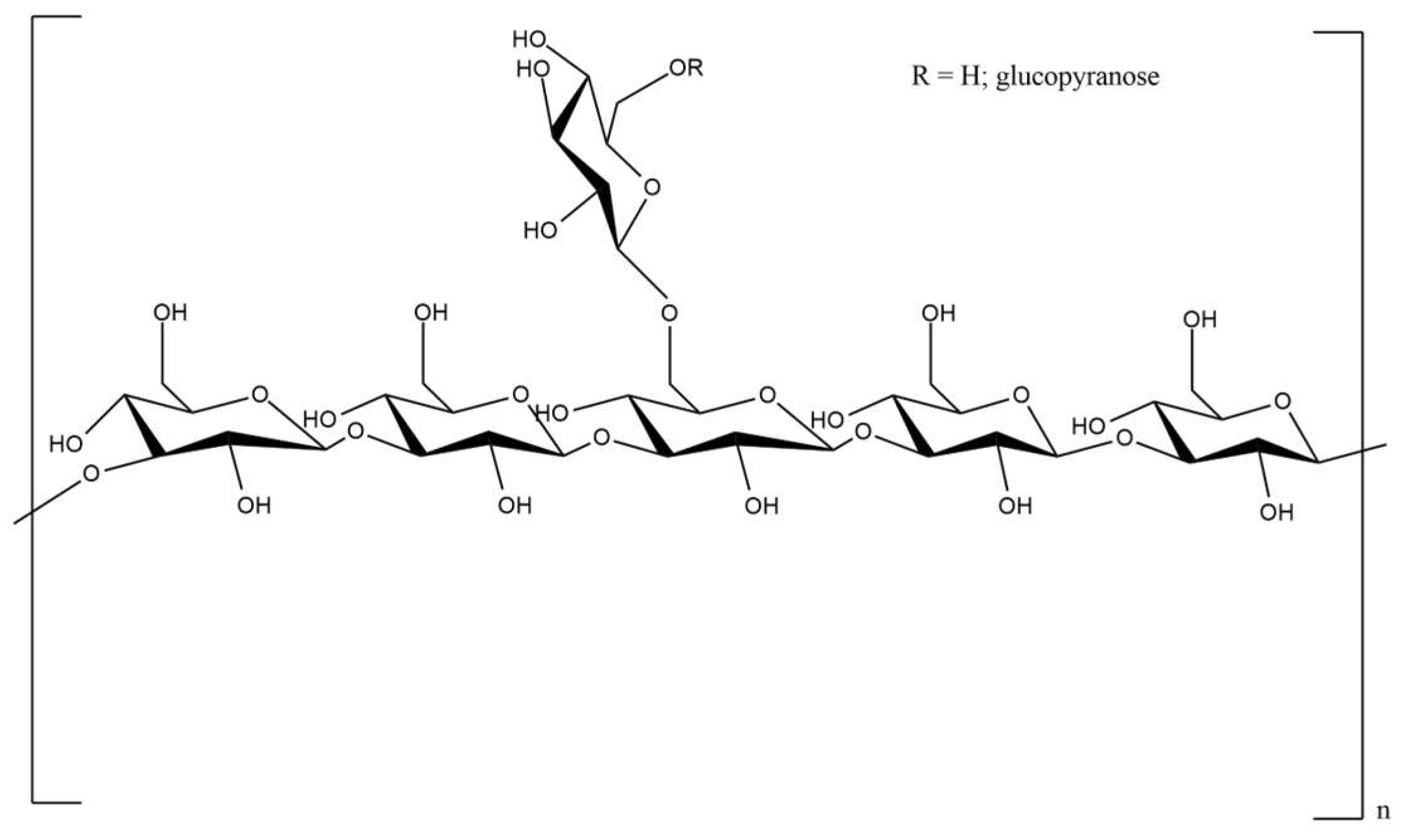
Pullulan is an α-glucan, which consists of α-(1,6) repeated maltotriose units via an α-(1,4) glyosidic bond [21,22]. The presence of α-(1→6) and α-(1→4) glyosidic linkages endows particular physico-chemical properties to pullulan [23]. The structure of pullulan is shown in Figure 1.
It is an odorless, highly water-soluble, tasteless, non-ionic, non-hygroscopic and non-reducing, non-mutagenic, and non-toxic edible natural polymer [12,24,25], with an average molecular charge (Mw) close to 362–480 kDa [12], properties that are extremely useful in different industrial applications [26].
Due to the fact that it presents a pharmaceutical substrate, in current uses, it relies on its unique film-forming and binding characteristics and is widely used for coating and granulating tablets or for oral care and wound care products [27,28].
Pullulan and its derivatives are also essential in a variety of pharmacological and biological applications [29], which include targeted drug delivery, gene targeting [30], wound healing [31], tissue engineering [32], and vaccination [33].
Studied as a drug carrier in the pharmaceutical field, pullulan is special because of its neutral nature, in which nine hydroxyl groups are present per repeating unit, making it suitable in the field of chemical derivatization. Thermosensitive and pH-sensitive microspheres have been made by grafting and crosslinking on the backbone of ether succinic carboxyl groups as well as poly-(N-isopropylacrylamides)-coacrylamides [27]. Pullulan proved to have an exceptional role as a carrier for anticancer drugs to target various body parts such as the liver, lungs, spleen, and brain, as well as for the sustained or prolonged release of specific cytotoxic molecules to the specific diseased site [12,34].
Novel drug-loaded nanoparticles by chemical grafting of lovastatin onto pullulan were developed by Wu et al. [35], who encapsulated doxorubicin, revealing inhibitory effects on triple-negative breast cancer cells, presented as a potential drug delivery system suitable for future clinical cancer treatment. Moreover, Wu et al. [36] studied the synergistic effect of pullulan nanoparticles loaded with 10-hydroxycaptothecin and methotrexate (MHNP) against drug-resistant tumors. MHNPs have a better ability to destroy HeLa cells compared to individual drug-loaded nanoparticles, being a potential carrier with an effective anti-tumor effect in tumor-targeted drug delivery.
Thus, it can be used to generate nanoconjugates based on different compounds. An example of this can be seen in thiolate pullulan nanoparticles that have been coupled with antibodies in order to remove portions of chromatin, but also to avoid AND degradation. Because chromatin degradation often takes place in the liver in this situation, pullulan acts as the ideal carrier [37]. Additionally, pullulan was partially oxidized (carboxylated) in order to produce silver-containing nanoparticles that act as antimicrobials [38]. Another feature of cholesteryl-pullulan nanoparticles is that they can interact hydrophobically with oligomeric forms of beta-amyloid, exhibiting the ability to significantly reduce their toxicity, which stands out as a potential complementary strategy present in neurological disorders with the formation of soluble toxic aggregates, an example being Alzheimer’s disease [39].
Pullulan has a special characteristic that has been closely studied, as evidenced by its ability to create cholesterol-containing nanogels in water in the presence of a self-bonding process, that occurs due to hydrophobic zones in its structure. As prostaglandin E1 aids in neovascularization and wound closure, these nanohydrogels have been employed to entrap a variety of materials for different applications [40], such as bone engineering purposes [41]. Iswariya et al. [42] developed an excellent absorbent collagen-pullulan hydrogel with improved mechanical firmness and well-defined biocompatibility for skin tissue engineering. The scaffolds were built using pullulan combined with sodium trimetaphosphate and collagen to create the polymeric linkages.
Furthermore, pullulan proved to be a good stabilizer of pomegranate seed oil nanoemulsions containing ketoprofen for intravenous administration, ensuring a high rate of drug delivery and selective antitumor efficiency (antiglioma) [29]. Taking into account all these aspects, pullulan seems to have long-term commercial potential [43].
3.2. Schizophyllan
Schizophyllan, produced by the Schizophyllum commune fungus, can be obtained by submerged culture fermentation using various types of sugars and soluble starch substrates and also low-cost lignocellulosic residues, such as corn fiber, rice hull hydrolysate, wheat bran, corn cobs, or corn steep liquor [44].
Liquid fermentation is the most common method used for production of this polysaccharide, but it can also be obtained through a solid-state-fermentation process [45]. According to several studies, different production media containing sucrose, glucose, dextrin, maltose, and fructose as carbon sources and yeast extract, beef extract, potato extract, and malt extract as nitrogen sources were utilized for the production of this biopolymer [46,47]. As reported by Teoh and Don [48], the optimal conditions for maximum growth of S. commune (33.1404 g L−1) were found to be pH 6.5–6.7, 30 °C, and 175 rpm after 120 h of cultivation.
Schizophyllan is a non-ionic and water-soluble extracellular polysaccharide, having a molecular weight between 100 and 200 kDa [49], and is composed of three glucose molecules linked by β-1-3-glycosidic bond units, with one glucose side chain linked to the basic chain by a β-1-6-glycosidic bond [50]. Figure 2 depicts the chemical structure of schizophyllan.
Extensive research on schizophyllan has led to its widespread use in pharmaceuticals over the last four decades. Antineoplastic, antibacterial, antiparasitic, antioxidant, and hypoglicemic characteristics are the identified bioactivities [51]. Other physiological benefits have been described, including hepatoprotective and anti-inflammatory properties. Its anticancer and immunobiological activities are among the most promising bioactivities described [52].
A large number of clinical trials have been reported in Japan on schizophyllan, which is effective against head and neck cancer and has been licensed and approved for clinical use in combination with chemotherapy for the two types of cancer mentioned [53]. Furthermore, schizophyllan can be used to reduce the probability of mammary tumors, decreasing the progression of breast cancer. Another randomized clinical trial involving a number of 312 patients, following surgery, chemotherapy (fluorouracil), radiotherapy, and schizophyllan in various combinations, showed positive results. Schizophyllan mixed with tamoxifen has been shown to have a major impact in reducing the occurrence of breast tumors, but also has the ability to initiate apoptosis in liver carcinomas [54].
According to Zhou et al. [55], schizophyllan treatment inhibits the growth of rat CNS-1 glioma cells via p53-mediated cell cycle and apoptosis suppression. Furthermore, a low percentage of cells in the S phase, but also an increased percentage of cells in the G0/G1 phase was shown. It was proved that enhances the anti-inflammatory response in mouse macrophages and can activate the dectin-1 receptor, leading to increased secretion of pro-inflammatory cytokines while also strongly promoting the production of IL-10, a key anti-inflammatory cytokine that plays a key role in inflammation control [56].
Schizophyllan has been shown to be effective in immunomodulatory activities [44]. Lee and Ki [57] investigated the immunomodulatory results of ultrasound-treated schizophyllan implants on RAW264.7 cells embedded in a three-dimensional polyethylene glycol hydrogel. Compared to 2D cultured cells, 3D cultured cells were less susceptible to ultrasound-treated schizophyllan. Under both conditions, an increase in M1 macrophage phenotype markers by ultrasound-treated schizophyllan was evident, but on the other hand, ultrasound-treated schizophyllan promoted the production of fenotypic markers of M2 macrophages under 3D conditions, indicating the induction of macrophage immunoregulation in real tissue.
Schizophyllan has also been shown to be effective as a drug delivery agent. It improves tyrosine 15 phosphorylation by deactivating CDK1 and subsequently enhancing the proportion of cells in the G2/M phase, as well as a reduction in G1 phase cells [58].
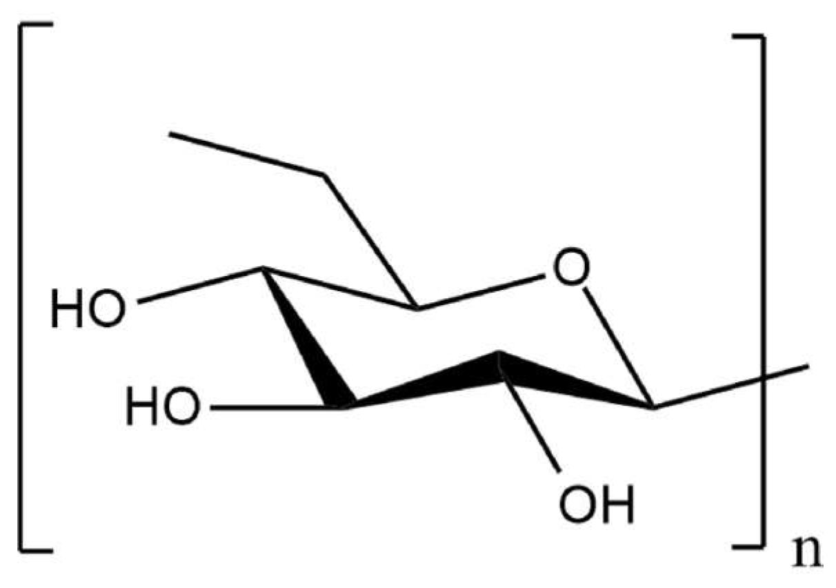
3.3. Scleroglucan
Scleroglucan is a microbial polysaccharide, a non-ionic branched glucan which consists in a backbone of (1,3)-β-linked D-glucopyranosyl residues with a single (1,6)-β-linked D-glucopyranosyl unit at every three main chain sugar residues. The molecular weight of scleroglucan is approximately 2–20 × 103 kDa [61] (Figure 3).
Sclerotium glucanicum and Sclerotium rolfsii are filamentous fungi that produce it extracellularly [62] through liquid submerged culture, using various carbon sources, such as sugarcane molasses or condensed corn soluble [63].
In general, culture media with a high ratio of carbon to limiting nutrients (often nitrogen) are favorable for scleroglucan production. Among the carbon sources, usually glucose and sucrose are used for biopolymer biosynthesis, although other substrates can also be successfully used, as previously mentioned. A concentration of glucose or sucrose of 30–35 g/L in the culture medium can lead to a maximum yield of 8.5–10 g/L of scleroglucan. Regarding the nitrogen source, it can be represented by a wide range of inorganic and organic compounds, such as inorganic salts of NH4+ and NO3−, or natural products, including yeast extract, corn steep liquor, soya or casein hydrolysate [62].
Scleroglucan provides significant advantages in terms of biocompatibility, pseudoplasticity, water solubility, resistance to hydrolysis, salt tolerance, moisture retention, and viscosity stability due to its unique chemical structure and higher molecular weight [64].
The potential applications of scleroglucan present in a variety of industrial fields have already been described: in cosmetics (skin and hair care products), together with xanthan, but also together with other polysaccharides, as hydrogel and water immobilizer [62], in drug delivery [65] as antitumor, antiviral against Herpes virus [66] and Rubella virus) [67], and antimicrobial compound in biomedical fields and in the pharmaceutical industry (tablet coatings, ophthalmic solutions, injectable antibiotic suspensions, calamine lotion), even showing immunostimulatory effects compared to other polysaccharides, so its potential contribution to the treatment of many diseases should be considered in therapeutic regimens [68].
Scleroglucan and some of its derivatives can be used particularly for the formulation of modified-release dosage forms, according to Coviello et al. [69]. It can be used in its native form for the preparation of sustained release tablets and ocular formulations [70].
Moreover, scleroglucan can be used as a matrix for drug delivery from tablets or films in the form of a carboxylated derivative.
Hydrogels made by crosslinking the polycarboxylated scleroglucan derivative with alkane dihalides were tested for diffusion and water uptake for this purpose [71].
3.4. Botryosphaeran
Botryosphaeran is defined as a β-(1→3,1→6)-glucan (1→3 backbone, 1→6 branched glucose, and gentiobiosis) and is produced by the fungus Botryosphaeria rhodina [72]. Botryosphaeran is an exopolysaccharide with a high molecular mass (>1 × 10³ kDa), that is soluble in water, consists of a main chain of (1→3)-linked β-D-glucose units, with a degree of branching of ~22% at carbon-6, with glucose and gentiobiose residues linked through β-(1→6)-bonds, and presents a triple helix conformation [73]. The chemical structure of botryosphaeran is shown in Figure 4.
Giese et al. [74] reported the optimum botryosphaeran production at 88 h of growth, using glucose as a carbon source and ammonium nitrate as a nitrogen source (C/N ratio of 30), at 28 °C under shaking conditions (180 rpm). Beside glucose, other carbon sources that can be used in the liquid fermentation process are sucrose and fructose [75].
Dekker et al. [76], as well as Dekker and Barbosa-Dekker et al. [77], provided an overview of the biological activities of botryosphaeran. It has been shown to have biological effects, including high anticlastogenic activities in reticulocytes from peripheral blood and erythrocytes from mouse bone marrow (in vitro) [78], as well as immunomodulatory activity [79].
Miranda-Nantes et al. [80] showed that botryosphaeran has antidiabetic (ability to reduce glycemia by 52%) and hypocholesterolemic (total and LDL cholesterol were reduced by 27% in hyperlipidemic rats) properties. Moreover, Silva-Sena et al. [81] concluded that botryosphaeran was effective in improving the lipidic profile and vascular lipid deposition in an in vivo model of atherosclerosis. In vitro, sulfonylated botryosphaeran displayed new biological functions, for example, anticoagulant [82] and antiviral activities [83].
It was demonstrated that botryosphaeran reduced significantly tumor development and corrected macrocytic anemia in vivo (rats) [72]. In vitro studies showed that botryosphaeran had a direct antiproliferative and pro-apoptotic action in MCF-7 breast cancer cells [84] and regulated the cell cycle by repressing G1 phase-related genes in human tumor lymphocytes (Jurkat cells) [85].
Kerche-Silva et al. [86] studied the cytotoxic, mutagenic, genotoxic, and protective effects of this biopolymer in chinese hamster lung fibroblasts (V79) and rat hepatocarcinoma cells (HTC) and reported that botryosphaeran was not genotoxic in either cell line, decreased the clastogenic effects of doxorubicin, H2O2, and benzo[α]pyrene, concluding that it may be a promising candidate for chemoprevention trials.
Fujiike et al. [87] investigated the in vitro anticancer effects of carboxymethylated botryosphaeran (CM-BOT) on breast cancer MCF-7 cells cultivated in multicellular tumor spheroids (MCTS). CM-was shown to inhibit cell proliferation in a concentration at 1000 µg/mL, which resulted in MCTSs with smaller diameters than controls, results that are very important in the search for bioactive compounds for use in breast cancer therapy or as adjuvants in reducing the adverse effects of mammary tumor chemotherapy.
Moreover, the effects of botryosphaeran on inflammation have been described. According to Silva et al. [88], this orally administered biopolymer has the property of reducing inflammation by activating leukocytes, but also by modulating migration, in addition to minimizing cold nociception, concluding that this fungal β-glucan may be a new possibility for complementary treatment of acute and chronic inflammation.
Therefore, botryosphaeran polysaccharide offers promising health applications in the development of new products related to pharmaceuticals and biomedicine.
3.5. Lentinan
The basidiomycete fungus Lentinus edodes, often known as the shiitake mushroom, was one of the first macrofungi cultivated at large scale in submerged culture [89].
Submerged culture of Lentinus edodes presents the advantage of a higher mycelia production in a shorter time with a lower risk of contamination [90]. During the fermentation process, the composition of the culture medium, cultivation temperature, and pH value are important factors in the production of L. edodes mycelia [91].
According to Bisko et al. [92] it has been shown that glucose and peptone are the most suitable sources of carbon and nitrogen for lentinan production. The modified glucose-peptone liquid medium (C/N ratio of 18), pH value adjusted to 6.0 and cultivation temperature of 25 °C resulted in increased biomass and lentinan polysaccharide yield in submerged culture.
Lentinan is a high-molecular-weight homopolysaccharide, consisting of five linearly linked 1-3-β-glucose residues and two 1-6-β-glucopyranoside branches [93], slightly soluble in hot water and insoluble in cold water, with a molecular mass that varies between 300 and 1000 kDa ([94,95,96,97] (Figure 5).
Lentinan is currently used in China and Japan in the treatment of various types of cancer, being administered orally or intravenously (at a dose of 1–2 mg for intravenous infusion). It is frequently combined with other conventional pharmaceutical drugs in the treatment of bowel, liver, stomach, ovarian, and lung cancers. The effect of lentinan in the treatment of gastric cancer was the most studied [98,99], followed by sarcoma, colon, and lung cancers.
Furthermore, the results obtained in the study reported by Oba et al. [100] showed that the addition of lentinan to the chemotherapy treatment produced a significant prolongation of patient survival rates compared with those treated only with chemotherapy.
In Japan, during cancer chemotherapy on solid tumors, patients to whom it was administered lentinan showed a considerably greater response rate (14.9%) than the patients without a treatment with lentinan. Moreover, lentinan combined with other chemotherapeutic drugs decreased the side effects of chemotherapy, including nausea, discomfort, hair loss, and weakened immunity. Other clinical investigations with positive results, involving 359 patients with gastric cancer who received lentinan in addition to chemotherapy, have been reported [101,102]. Therefore, chemotherapy plus lentinan showed promising results in the treatment of cancer.
Regarding the immunomodulatory properties of lentinan, they include dendritic cell (DC) activation by Dectin-1 binding, macrophage activation, an increase in cytotoxic T lymphocytes and natural killer (NK) activity, and enhancement of the Th1 versus Th2 response. Lentinan-functionalization of graphene oxide (GO) nanoparticles showed long-term sustained immune effects, potentiating antigen uptake in macrophages in vitro and decreasing the release rate of antigen (ovalbumin), resulting in higher levels of IgGs. In conclusion, GO-lentinan could be a carrier, ensuring a long-term immune memory response [103].
3.6. Grifolan
Over 37 biologically active polysaccharide fractions have been so far isolated as products of the fungus Grifola frondosa, depending on the extraction and purification procedures [104].
The D-fraction, containing so called GFP (G. frondosa polysaccharide) is the most active and consists of β-glucans alone or protein-bound (proteoglucans). Submerged or liquid fermentation is a preferable method for obtaining grifolan polysaccharide, as it provides higher mycelial productivity and more effective product quality control in a shorter time [105].
In the study of Shih et al. [106], the fermentation of Grifola frondosa was investigated in shake flasks and also in a fermenter operating in batch and fed-batch modes. In the case of shake-flasks, it was observed a high mycelial growth and exopolysaccharide production at a low pH value, using maltose and glucose as carbon sources, and yeast extract in combination with corn steep powder as nitrogen sources (13 days). The amount of accumulated mycelial biomass and exopolysaccharide was significantly increased through fed-batch fermentation by glucose feeding, which was performed when the glucose concentration of the medium was less than 0.5% (5 g/L). Before glucose feeding, these amounts were 3.97 g/L and 1.04 g/L, respectively, but after 13 days of cultivation, they had increased to 8.23 g/L and 3.88 g/L. The results reported in the case of batch fermentation showed that mycelial biomass and exopolysaccharide were 6.7 g/L, respectively, 3.3 g/L, at the end of 13 days of cultivation.
The structure of glucan is a 1,3, -1,6-β-D-glucan, which contains either as a monomer three β-1,6-glucose units present in the main chain and a branched β-1,3-glucose unit, or a reverse β-1,3-glucose unit (three β-1,3- and one branched β-1,6)-glucan.
Grifolan has a molecular weight of approximately 1000 kDa and glucose has been found as the major monosaccharide [104,107,108]. The chemical structure of grifolan is presented in Figure 6.
The pharmacological activities of G. frondosa glucans have been studied for more than 30 years. Anticancer effects are considered the most important in three directions: protection of healthy cells, prevention of metastasis, and inhibition of tumor growth, whether acting directly on tumors or through immunomodulatory mechanisms. The anticancer activity of grifolan is considered superior to that of lentinan, not by direct inhibition, but through higher immune-stimulating effects enhancing innate, and especially acquired immunity by inducing the production of cytokines and chemokines [109].
Antitumor effects have been demonstrated in vivo using mouse tumor models (mammary carcinoma and colon adenocarcinoma) by intraperitoneal and oral administration. Tumor inhibition by tumor-specific agents as well as systemic immunity stimulation were noticed. It is worth mentioning that water soluble grifolan is the single fungal β-glucan which could orally administered efficiently [110].
Another in vivo study on mice with implanted mammary carcinoma cells showed increased antitumor activity and a reduced immuno-suppressive effect of mitomycin treatment [111]. A sulfate derivative of a water-insoluble polysaccharide of the same origin inhibited in vivo and in vitro (in mice) ascites cell line tumor growth, accelerated the antitumor effect, and reduced immune damage when combined with cyclophosphamide [112].
Different studies showed that grifolan-reach extracts induced apoptosis in human breast [113], gastric cancer cells [114], and prostate cancer [115]. Other health-beneficial activities (liver and renal protection, antidiabetic, antilipidemic, antihypertensive, and antiradiation), as well as antiviral activities have been noticed [116].
3.7. Lasiodiplodan
The filamentous fungus Lasiodiplodia theobromae has become of increasing interest as producer of lasiodiplodan, an extracellular β-glucan polymer, with a high potential for industrial production.
The use of soybean molasses, sugarcane straw, or sugarcane bagasse as a low-cost carbon source, rice bran extract, and soybean bran extract as a nitrogen source, respectively, has been reported as promising substrates for lasiodiplodan production through submerged fermentation [117,118,119]. Furthermore, dextrose, glucose, mannitol, sucrose, fructose, maltose, and lactose have been tested as carbon sources, followed by potassium nitrate, ammonium sulfate, and ammonium chloride as nitrogen sources [120].
Its structure was identified as linear, water soluble β-(1→6)-D-glucan, containing glucose monomer units, presenting spherical structures such as granules with an ovoid shape (3.33 μm average diameter) (Figure 7). Depending mostly on the producing strain and fermentation conditions, the molecular weight of lasiodiplodan was found between 7 kDa and 2000 kDa [119].
Given its wide range of biological functions, including protective activity against induced DNA damage by doxorubicin, anticoagulant, and hypoglycemic activities, and a decrease of transaminase activity in rats without any hematologic or histologic changes that indicate toxicity in the vital organs [121,122], lasiodiplodan exopolysaccharide presents a significant commercial potential [123].
It inhibited cell proliferation in MCF-7 breast cancer cells, whereas its sulfonated derivative had anticoagulant and antithrombotic properties similar to heparin [124]. Chemical derivatization (acetylation, carboxymethylation, phosphorylation, and sulphonylation) proved to improve and extend the biological properties and pharmaceutical applications of lasiodiplodan (e.g., antimicrobial, antioxidant, antiproliferation of cancer cells) [125,126,127].
The results obtained in the study of Malfatti et al. [128] showed that lasiodiplodan can prevent the signals of neurotoxicity induced by D-penicillamine, reducing lipid peroxidation in the brain cortex and the typical automatism of convulsions. According to these findings, lasiodiplodan may be useful for both preventing neurotoxicity, as well as attenuating the damage produced by the convulsive episodes related to the GABAergic system.
Moreover, another study investigated the importance of this polysaccharide in wound healing. Nissola et al. [129] have developed a hydrogel based on lasiodiplodan as a bioactive ingredient, and it was observed that the formulation stimulated re-epithelialization, cell proliferation, and collagen production, suggesting its potential use in products for the topical treatment of skin wounds and burns.
4. Extraction and Analytical Methods of Fungal Exopolysaccharides
Exopolysaccharides can be separated from the fermentation broth using several techniques and procedures. Before the isolation and quantification of exopolysaccharides, the biomass from the fermentation broth must be removed through centrifugation. Exopolysaccharides are often separated from cell-free systems by precipitation with organic solvents (ethanol [130], acetone or isopropyl alcohol [131]) and subsequent cold storage [89]. For example, lentinan produced by the Lentinus edodes fungus can be extracted by ethanol precipitation [10]. Furthermore, lasiodiplodan secreted into the culture medium during submerged fermentation can be easily collected by ethanol precipitation; thus, their isolation is easier and cheaper compared to extractive processes for glucans from fungal fruiting bodies or yeast cell walls [132]. The precipitate (“crude EPS”) obtained by centrifugation may contain certain amounts of other substances, such as proteins, minerals, or low molecular mass carbohydrates, leading to the polysaccharide purification step [89]. Ion exchange chromatography (IEC) and gel permeation chromatography (GPC) are methods that can be used for the purification of exopolysaccharides [133]. Regarding the composition and structure of fungal exopolysaccharides, they can usually be analyzed by high-performance liquid chromatography (HPLC), gas-liquid chromatography (GLC), gas-liquid chromatography-mass spectrometry (GLC-MS), and 1D and 2D NMR spectroscopy [5].
5. Discussions
Fungal polysaccharides are important candidates for different therapies, including cancer, due to their bioactive properties [3]. Various fungal polysaccharides with different molecular chain lengths and chemical compositions are synthesized by different strains of microorganisms and have promising applications in medicine and the pharmaceutical industry.
It has been proven that these biopolymers possess immunomodulatory activity, as their use has led to a prolongation of survival time, renewal of immune parameters and improvement of quality of life in patients with different types of cancers compared to patients who received only chemotherapy (Table 1).
The antitumor property of polysaccharides is immunologically mediated via T and B cells on cancer cells, but also by stimulating innate immunity. Even though they are slower acting compared to conventional therapies (radiotherapy, chemotherapy), they show the ability to be more adaptable, durable, and specific [27], playing a crucial role in improving the human immune system. Such biopolymers as chemically modified derivatives proved to be valuable drug delivery agents, achieving rate and important target-controlled release.
As a first comment, the specific difference between an α-glucan (pullulan) and β-glucans consists in the fact that pullulan and its derivatives showed almost only very suitable properties as classical formulation ingredients and drug carriers, but not as specific bioactive products, as β-glucans.
Though pullulan has monographs in the United States [134], Europe [135], Japan [136], and Britain [137], its presence is only as a classical ingredient, far from the targeted drug delivery properties of derivatives, as numerous published studies showed. In this regard, they face the generally existing challenges for medicines containing nanoparticles of non-familiar origin to the human body. To become medicines, regulatory issues, essentially imposed on innovative medicines must be met. Therefore, new preclinical studies in vitro and in vivo, in accordance with those issues, especially regarding pharmacology, pharmacokinetics, and toxicology aspects, are necessary. Obviously, such formulations as medical devices for local therapy (e.g., wound healing) have a greater chance of being approved.
Regarding the bioactive β-glucans, despite the recognition of their efficacy as adjuvants in cancer therapy in Asia, they are not present in any worldwide known pharmacopoeias as individual drugs, but only included among dietary supplements, as products of complementary therapy.
6. Conclusions
The pharmaceutical importance of fungal polysaccharides appears obvious, especially regarding their proven great potential therapeutic effects in a major disease field, namely cancer. There are two main directions: direct cancer immunotherapy and antitumor activity, mostly associated with existing chemotherapeutics, and target selective drug delivery of chemotherapeutics.
There are obvious challenges that cannot be avoided but could be overcome. As innovative pharmaceuticals, the fungal polysaccharide biopolymers as active drug substances and their nanoparticle formulations should be reliable, reproducible, and comply with specific regulatory requirements, whose efficacy and safety must be proven in preclinical and clinical trials. Apart from the reliability and reproducibility depending on appropriate technological solutions, the challenges refer especially to in vivo required pharmacokinetic characterization (mostly metabolism and excretion), which generally hinders non-physiological biopolymers as nanoparticles to become authorized drugs or pharmaceutical ingredients. The enormous volume of studies that are going on, based on their great potential applications in major therapeutic domains, justifies the confidence in the outlook for highly effective medicines containing fungal exopolysaccharide biopolymers.
Author Contributions
Concept, R.M.S., E.S.L.; writing-original draft preparation, R.M.S., M.M. and L.I.C.; writing-review and editing, R.M.S., M.M. and E.S.L. All authors have read and agreed to the published version of the manuscript.
Funding
This work was supported by a grant of the Ministry of Research, Innovation and Digitization, CCCDI-UEFISCDI, project number PN-III-P2-2.1-PED-2021-3528/731PED-2022 within PNCDI III.
Data Availability Statement
Not applicable.
Conflicts of Interest
The authors declare no conflict of interest.
References
- van Dam, J.E.G.; van den Broek, L.A.M.; Boeriu, C.G. Polysaccharides in human health care. Nat. Prod. Commun. 2017, 12, 821–830. [Google Scholar] [CrossRef] [Green Version]
- Naranjo-Ortiz, M.A.; Gabaldon, T. Fungal evolution: Major ecological adaptations and evolutionary transitions. Biol. Rev. 2019, 94, 1443–1476. [Google Scholar] [CrossRef] [PubMed] [Green Version]
- Giavasis, I. Bioactive fungal polysaccharides as potential functional ingredients in food and nutraceuticals. Curr. Opin. Biotechnol. 2014, 26, 162–173. [Google Scholar] [CrossRef]
- Jaroszuk, M.O.; Wilkolazka, A.J.; Ścisel, J.J.; Szalapata, K.; Nowak, A.; Jaszek, M.; Ozimek, E.; Majewska, M. Extracellular polysaccharides from Ascomycota and Basidiomycota: Production conditions, biochemical characteristics, and biological properties. World J. Microbiol. Biotechnol. 2015, 31, 1823–1844. [Google Scholar] [CrossRef] [Green Version]
- Mahapatra, S.; Banerjee, D. Fungal exopolysaccharide: Production, composition and applications. Microbiol. Insights 2013, 6, 1–16. [Google Scholar] [CrossRef] [PubMed] [Green Version]
- Herrera, J.R.; Ortiz-Castellanos, L. Cell wall glucans of fungi. A review. Cell Surf. 2019, 5, 100022. [Google Scholar] [CrossRef]
- Złotko, K.; Wiater, A.; Waśko, A.; Pleszczyńska, M.; Paduch, R.; Jaroszuk-Ściseł, J.; Bieganowski, A. A report on fungal (1→3)-α-d-glucans: Properties, functions and application. Molecules 2019, 24, 3972. [Google Scholar] [CrossRef] [Green Version]
- Chimilovski, J.S.; Habu, S.; Teixeira, R.F.B.; Thomaz-Soccol, V.; Noseda, M.D.; Medeiros, A.B.P.; Pandey, A.; Soccol, C.R. Antitumour activity of Grifola frondosa exopolysaccharides produced by submerged fermentation using sugar cane and soy molasses as carbon sources. Food Technol. Biotechnol. 2011, 49, 359–363. [Google Scholar]
- Özcan, E.; Öner, E.T. Microbial of extracellular polysaccharide production from biomass sources. In Polysaccharides; Ramawat, K., Mérillon, J.M., Eds.; Springer: Berlin/Heidelberg, Germany, 2018; pp. 1–21. [Google Scholar] [CrossRef]
- Venkatachalam, G.; Arumugam, S.; Doble, M. Industrial production and applications of α/β linear and branched glucans. Indian Chem. Eng. 2020, 63, 533–547. [Google Scholar] [CrossRef]
- Barbosa, J.R.; de Carvalho, R.N., Jr. Occurrence and possible roles of polysaccharides in fungi and their influence on the development of new technologies. Carbohydr. Polym. 2020, 246, 116613. [Google Scholar] [CrossRef]
- Ganie, S.A.; Rather, L.J.; Li, Q. A review on anticancer applications of pullulan and pullulan derivative nanoparticles. Carbohydr. Polym. Technol. Appl. 2021, 2, 116613. [Google Scholar] [CrossRef]
- Sugumaran, K.R.; Ponnusami, V. Review on production, downstream processing and characterization of microbial pullulan. Carbohydr. Polym. 2017, 173, 573–591. [Google Scholar] [CrossRef]
- Singh, R.S.; Saini, G.K.; Kennedy, J.F. Pullulan: Microbial sources, production and applications. Carbohydr. Polym. 2008, 73, 515–531. [Google Scholar] [CrossRef] [PubMed]
- Key, S.W.C.; Dailin, D.J.; Selvamani, S.; Malek, R.A.; Sukmawati, D.; El Enshasy, H. Pullulan production in submerged cultivation: A review. J. Crit. Rev. 2020, 7, 3220–3231. [Google Scholar]
- Dailin, D.J.; Low, L.Z.M.I.; Kumar, K.; Malek, R.A.; Natasya, K.H.; Keat, H.C.; Sukmawati, D.; El Enshasy, H. Agro-industrial waste: A potential feedstock for pullulan production. Biosci. Biotechnol. Res. Asia. 2019, 16, 229–250. [Google Scholar] [CrossRef]
- Akdeniz Oktay, B.; Bozdemir, M.T.; Özbaş, Z.Y. Evaluation of some agro-industrial wastes as fermentation medium for pullulan production by Aureobasidium pullulans AZ-6. Curr. Microbiol. 2022, 79, 93. [Google Scholar] [CrossRef]
- Thirumavalavan, K.; Manikkadan, T.R.; Dhanasekar, R. Pullulan production from coconut by-products by Aureobasidium pullulans. Afr. J. Biotechnol. 2009, 8, 254–258. [Google Scholar]
- Kumar, A.S.; Mody, K.; Jha, B. Bacterial exopolysaccharides-a perception. J. Basic Microbiol. 2007, 47, 103–117. [Google Scholar] [CrossRef]
- West, T.P. Production of the polysaccharide pullulan by Aureobasidium pullulans cell immobilization. Polysaccharides 2022, 3, 544–555. [Google Scholar] [CrossRef]
- Kumar, D.; Saini, N.; Pandit, V.; Ali, S. An insight to pullulan: A biopolymer in pharmaceutical approaches. Int. J. Basic Appl. Sci. 2012, 1, 202–219. [Google Scholar] [CrossRef]
- Thangavelu, M.; Kulandhaivelu, S.V. Development and Characterization of Pullulan-Carboxymethyl cellulose blend film for packaging applications. Int. J. Polym. Sci. 2022, 2022, 9649726. [Google Scholar] [CrossRef]
- Singh, R.S.; Kaur, N.; Hassan, M.; Kennedy, J.F. Pullulan in biomedical research and development—A review. Int. J. Biol. Macromol. 2021, 166, 694–706. [Google Scholar] [CrossRef] [PubMed]
- Raychaudhuri, R.; Naik, S.; Shreya, A.B.; Kandpal, N.; Pandey, A.; Kalthur, G.; Mutalik, S. Pullulan based stimuli responsive and sub cellular targeted nanoplatforms for biomedical application: Synthesis, nanoformulations and toxicological perspective. Int. J. Biol. Macromol. 2020, 161, 1189–1205. [Google Scholar] [CrossRef] [PubMed]
- Rekha, M.R.; Chandra, P.S. Pullulan as a promising biomaterial for biomedical applications: A perspective. Trends Biomater. Artif. Organs 2007, 20, 111–116. [Google Scholar]
- Coltelli, M.B.; Danti, S.; de Clerck, K.; Lazzeri, A.; Morganti, P. Pullulan for advanced sustainable body- and skin-contact applications. J. Funct. Biomater. 2020, 11, 20. [Google Scholar] [CrossRef] [Green Version]
- Moscovici, M. Present and future medical applications of microbial exopolysaccharides. Front. Microbiol. 2015, 6, 1012. [Google Scholar] [CrossRef] [Green Version]
- Ferreira, L.M.; Velasquez, A.A.; Schaffazick, S.R.; Cruz, L. Pullulan: An advantageous natural polysaccharide excipient to formulate tablets of alendronate-loaded microparticles. Braz. J. Pharm. Sci. 2015, 51, 28–33. [Google Scholar] [CrossRef] [Green Version]
- Cheng, K.C.; Demirci, A.; Catchmark, J.M. Pullulan: Biosynthesis, production, and applications. Appl. Microbiol. Biotechnol. 2011, 92, 29–44. [Google Scholar] [CrossRef]
- Singh, R.S.; Kaur, N.; Kennedy, J.F. Pullulan and pullulan derivatives as promising biomolecules for drug and gene targeting. Carbohydr. Polym. 2015, 123, 190–207. [Google Scholar] [CrossRef]
- Li, H.; Xue, Y.; Jia, B.; Bai, Y.; Zuo, Y.; Wang, S.; Zhao, Y.; Yang, W.; Tang, H. The preparation of hyaluronic acid grafted pullulan polymers and their use in the formation of novel biocompatible wound healing film. Carbohydr. Polym. 2018, 188, 92–100. [Google Scholar] [CrossRef]
- Singh, S.; Kaur, N.; Rana, V.; Kennedy, J.F. Recent insights on applications of pullulan in tissue engineering. Carbohydr. Polym. 2016, 153, 455–462. [Google Scholar] [CrossRef] [PubMed]
- Singh, R.S.; Kaur, N.; Rana, V.; Kennedy, J.F. Pullulan: A novel molecule for biomedical applications. Carbohydr. Polym. 2017, 171, 102–121. [Google Scholar] [CrossRef]
- Mishra, B.; Vuppu, S.; Rath, K. The role of microbial pullulan, a biopolymer in pharmaceutical approaches: A review. J. Appl. Pharm. Sci. 2011, 1, 45–50. [Google Scholar]
- Wu, D.; Chen, Y.; Wen, S.; Wen, Y.; Wang, R.; Zhang, Q.; Qin, G.; Yi, H.; Wu, M.; Lu, L.; et al. Synergistically enhanced inhibitory effects of pullulan nanoparticle-mediated co-delivery of lovastatin and doxorubicin totriple-negative breast cancer cells. Nanoscale Res. Lett. 2019, 14, 1–12. [Google Scholar] [CrossRef]
- Wu, S.; Yang, X.; Yang, X. Methotrexate and 10-hydroxycamptothecine loaded pullulan nanoparticles with the targeting property for efficient cancer therapy. Mater. Technol. 2022, 37, 2777–2784. [Google Scholar] [CrossRef]
- Rekha, M.R.; Pal, K.; Bala, P.; Shetty, M.; Mittra, I.; Bhuvaneshwar, G.S.; Sharma, C.P. Pullulan-histone antibody nanoconjugates for the removal of chromatin fragments from systemic circulation. Biomaterials 2013, 34, 6328–6338. [Google Scholar] [CrossRef]
- Coseri, S.; Spatareanu, A.; Sacarescu, L.; Rimbu, C.; Suteu, D.; Spik, S.; Harabagiu, V. Green synthesis of the silver nanoparticles mediated by pullulan and 6-carboxypullulan. Carbohydr. Polym. 2015, 116, 9–17. [Google Scholar] [CrossRef]
- Boridy, S.; Takahashi, H.; Akiyoshi, K.; Maysinger, D. The binding of pullulan modified cholesteryl nanogels to Aβ oligomers and their suppression of cytotoxicity. Biomaterials 2009, 30, 5583–5591. [Google Scholar] [CrossRef]
- Kobayashi, H.; Katakura, O.; Morimoto, N.; Akiyoshi, K.; Kasugai, S. Effects of colesterol-bearing pullulan (CHP)-nanogels in combination with prostaglandin E1 wound healing. J. Biomed. Mater. Res. B Appl. Biomater. 2009, 91, 55–60. [Google Scholar] [CrossRef]
- Tabernero, A.; Cardea, S. Microbial exopolysaccharides as drug carriers. Polymers 2020, 12, 2142. [Google Scholar] [CrossRef]
- Iswariya, S.; Bhanukeerthi, A.V.; Velswamy, P.; Uma, T.S.; Perumal, P.T. Design and development of a piscine collagen blended pullulan hydrogel for skin tissue engineering. RSC Adv. 2016, 6, 57863–57871. [Google Scholar] [CrossRef]
- Mahmoud, Y.A.G.; El-Naggar, M.E.; Abdel-Megeed, A.; El-Newehy, M. Recent advancements in microbial polysaccharides: Synthesis and applications. Polymers 2021, 13, 4136. [Google Scholar] [CrossRef]
- Kumar, A.; Bharti, A.K.; Bezie, Y. Schizophyllum commune: A fungal cell-factory for production of valuable metabolites and enzymes. BioResources 2022, 17, 5420–5436. [Google Scholar] [CrossRef]
- Zhong, K.; Liu, L.; Tong, L.; Zhong, X.; Wang, Q.; Zhou, S. Rheological properties and antitumor produced activity of schizophyllan with solid-state fermentation. Int. J. Biol. Macromol. 2013, 62, 13–17. [Google Scholar] [CrossRef] [PubMed]
- Kumari, M.; Survase, S.A.; Singhal, R.S. Production of schizophyllan using Schizophyllum commune NRCM. Bioresour Technol. 2008, 99, 1036–1043. [Google Scholar] [CrossRef] [PubMed]
- Mohammadi, A.; Shojaosadati, S.A.; Tehrani, H.J.; Mousavi, S.M.; Saleh, T.; Khorasani, A.K. Schizophyllan production by newly isolated fungus Schizophyllum commune IBRC-M 30213: Optimization of culture medium using response surface methodology. Ann. Microbiol. 2018, 68, 47–62. [Google Scholar] [CrossRef]
- Teoh, Y.P.; Don, M.M. Optimization of parameters for mycelia growth by Schizophyllum commune and a kinetic model study of its growth morphology. J. Appl. Sci. 2012, 12, 1100–1105. [Google Scholar] [CrossRef] [Green Version]
- Mironczuk-Chodakowska, I.; Kujawowicz, K.; Witkowska, A.M. Beta-glucans from fungi: Biological and health-promoting potential in the COVID-19 Pandemic Era. Nutrients 2021, 13, 3960. [Google Scholar] [CrossRef]
- Abdel-Mohsen, A.M.; Abdel-Rahman, R.M.; Fouda, M.M.G.; Vojtova, L.; Uhrova, L.; Hassan, A.F.; Al-Deyab, S.S.; El-Shamy, I.E.; Jancar, J. Preparation, characterization and cytotoxicity of schizophyllan/silver nanoparticle composite. Carbohydr. Polym. 2014, 102, 238–245. [Google Scholar] [CrossRef]
- Saetang, N.; Ramaraj, R.; Unpaprom, Y. Optimization of ethanol precipitation of schizophyllan from Schizophyllum commune by applied statistical modelling. Biomass Convers. Biorefin. 2022, 1–13. [Google Scholar] [CrossRef]
- Zhang, Y.; Kong, H.; Fang, Y.; Nishinari, K.; Phillips, G.O. Schizophyllan: A review of its structure, properties, bioactivity and recent developments. Bioact. Carbohydr. Diet. Fibre 2013, 1, 53–71. [Google Scholar] [CrossRef]
- Zhao, S.; Gao, Q.; Rong, C.; Wang, S.; Zhao, Z.; Liu, Y. Immunomodulatory effects of edible and medicinal mushrooms and their bioactive immunoregulatory products. J. Fungi. 2020, 6, 269. [Google Scholar] [CrossRef]
- Mansour, A.; Daba, A.; Baddour, N.; El-Saadani, M.; Aleem, E. Schizophyllan inhibits the development of mammary and hepatic carcinomas induced by 7,12-dimethylbenz(α)anthracene and decreases cell proliferation: Comparison with tamoxifen. J. Cancer Res. Clin. Oncol. 2012, 138, 1579–1596. [Google Scholar] [CrossRef]
- Zhou, B.; Fu, Q.; Song, S.S.; Zheng, H.L.; Wei, Y.Z. Inhibitory effect of schizophyllan on rat glioma cells. Bangladesh J. Pharmacol. 2015, 10, 759–764. [Google Scholar] [CrossRef]
- Thongsiri, C.; Nagai-Yoshioka, Y.; Yamasaki, R.; Adachi, Y.; Usui, M.; Nakashima, K.; Nishihara, T.; Ariyoshi, W. Schizophyllum commune β-glucan: Effect on interleukin-10 expression induced by lipopolysaccharide from periodontopathic bacteria. Carbohydr. Polym. 2021, 253, 117285. [Google Scholar] [CrossRef]
- Lee, S.; Ki, C.S. Inflammatory responses of macrophage-like RAW264. 7 cells in a 3D hydrogel matrix to ultrasonicated schizophyllan. Carbohydr. Polym. 2020, 229, 115555. [Google Scholar] [CrossRef]
- Atiq, A.; Parhar, I. Anti-neoplastic potential of flavonoids and polysaccharide phytochemicals in glioblastoma. Molecules 2020, 25, 4895. [Google Scholar] [CrossRef]
- Matsumoto, T.; Numata, M.; Anada, T.; Mizu, M.; Koumoto, K.; Sakurai, K.; Nagasaki, T.; Shinkai, S. Chemically modified polysaccharide schizophyllan for antisense oligonucleotides delivery to enhance the cellular uptake efficiency. Biochim. Biophys. Acta 2004, 1670, 91–104. [Google Scholar] [CrossRef]
- Takedatsu, H.; Mitsuyama, K.; Mochizuki, S.; Kobayashi, T.; Sakurai, K.; Takeda, H.; Fujiyama, Y.; Koyama, Y.; Nishihira, J.; Sata, M. A new therapeutic approach using a schizophyllan-based drug delivery system for inflammatory bowel disease. Mol. Ther. 2012, 20, 1234–1241. [Google Scholar] [CrossRef] [Green Version]
- Bai, T.; Wang, T.; Li, Y.; Gao, N.L.; Zhang, L.; Chen, W.H.; Yin, X. Optimization of scleroglucan production by Sclerotium rolfsii by lowering pH during fermentation via oxalate metabolic pathway manipulation using CRISPR/Cas9. Fungal Biol. Biotechnol. 2021, 8, 1–9. [Google Scholar] [CrossRef]
- Survase, S.A.; Saudagar, S.; Bajaj, I.B.; Singhal, R.S. Scleroglucan: Fermentative production, downstream processing and applications. Food Technol. Biotechnol. 2007, 45, 107–118. [Google Scholar]
- Fosmer, A.; Gibbons, W.R.; Heisel, N.J. Reducing the cost of scleroglucan production by use of a condensed corn solubles medium. J. Biotech. Res. 2010, 2, 131. [Google Scholar]
- Zeng, W.; Wang, J.; Shan, X.; Yu, S.; Zhou, J. Efficient production of scleroglucan by Sclerotium rolfsii and insights into molecular weight modification by high-pressure homogenization. Front. Bioeng. Biotechnol. 2021, 9, 799. [Google Scholar] [CrossRef] [PubMed]
- Li, X.; Lu, Y.; Adams, G.G.; Zobel, H.; Ballance, S.; Wolf, B.; Harding, S.E. Characterisation of the molecular properties of scleroglucan as an alternative rigid rod molecule to xanthan gum for oropharyngeal dysphagia. Food Hydrocoll. 2020, 101, 105446. [Google Scholar] [CrossRef]
- Marchetti, M.; Pisani, S.; Petropaolo, V.; Seganti, L.; Nicoletti, R.; Degener, A.; Orsi, N. Antiviral effect of polysaccha-ride from Sclerotium glucanicum towards herpes simplex virus type I infection. Planta Med. 1996, 62, 303–307. [Google Scholar] [CrossRef] [PubMed]
- Mastromarino, P.; Petruzziello, R.; Macchia, S.; Rieti, S.; Nicoletti, R.; Orsi, N. Antiviral activity of natural and semi-synthetic polysaccharides on early steps of rubella virus infection. J. Antimicrob. Chemother. 1997, 39, 339–345. [Google Scholar] [CrossRef] [PubMed]
- Manjanna, K.M.; Shivakumar, B.; Pramodkumar, T.M. Natural exopolysaccharides as novel excipients in drug delivery: A review. Arch. Appl. Sci. Res. 2009, 1, 230–253. [Google Scholar]
- Coviello, T.; Palleschi, A.; Matricardi, P.; Bocchinfuso, G.; Grassi, M.; Alhaique, F. Scleroglucan: A versatile polysaccharide for modified drug delivery. Molecules 2005, 10, 6–33. [Google Scholar] [CrossRef] [Green Version]
- Rizk, S.; Duru, C.; Gaudy, D.; Jacob, M.; Ferrari, F.; Bertoni, M.; Caramella, C. Physico-chemical characterization andtabletting properties of scleroglucan. Int. J. Pharm. 1994, 112, 125–130. [Google Scholar] [CrossRef]
- Vinarta, S.C.; Francois, N.J.; Daraio, M.E.; Figueroa, L.I.C.; Farina, J.I. Sclerotium rolfsii scleroglucan: The promising behavior of a natural polysaccharide as a drug delivery vehicle, suspension stabilizer and emulsifier. Int. J. Biol. Macromol. 2007, 41, 314–323. [Google Scholar] [CrossRef]
- Geraldelli, D.; Ribeiro, M.C.; Madeiros, T.C.; Comiran, P.K.; Martins, K.O.; Oliveira, M.F.; Oliveira, G.A.; Dekker, R.F.H.; Barbosa-Dekker, A.M.; Alegranci, P.; et al. Botryosphaeran, a (1→3)(1→6)-β-D-glucan, reduces tumor development and cachexia syndrome in obese male rats by increasing insulin sensitivity and FOXO3a activity. Int. J. Biol. Macromol. 2020, 165, 985–994. [Google Scholar] [CrossRef] [PubMed]
- Giese, E.C.; Dekker, R.F.H.; Barbosa, A.M.; Silva, R. Triple helix conformation of botryosphaeran, a (1→3; 1→6)-β-D-glucan produced by Botryosphaeria rhodina MAMB-05. Carbohydr. Polym. 2008, 74, 953–956. [Google Scholar] [CrossRef]
- Giese, E.C.; Sumiya, A.F.G.; Borsato, D.; Dekker, R.F.H.; Barbosa, A.M. Evaluation of fermentative parameters for the production of botryosphaeran (a (1→3,1→6)- β-D-glucan) and mycelial biomass by Botryosphaeria rhodina MAMB-05. Orbital Electron. J. Chem. 2015, 7, 36–43. [Google Scholar] [CrossRef]
- Fonseca, P.R.M.S.; Dekker, R.F.H.; Barbosa, A.M.; Silveira, J.L.M.; Vasconcelos, A.F.D.; Monteiro, N.K.; Aranda-Silverio, G.; Corradi da Silva, M.L. Thermal and rheological properties of a family of botryosphaerans produced by Botryosphaeria rhodina MAMB-05. Molecules 2011, 16, 7488–7501. [Google Scholar] [CrossRef] [PubMed] [Green Version]
- Dekker, R.F.H.; Queiroz, E.A.I.F.; Cunha, M.A.A.; Barbosa-Dekker, A.M. Botryosphaeran - A fungal exopolysaccharide of the (1→3)(1→6)-β-d-Glucan kind: Structure and biological functions. In Extracellular Sugar-Based Biopolymers Matrices. Biologically-Inspired Systems; Cohen, E., Merzendorfer, H., Eds.; Springer: Berlin/Heidelberg, Germany, 2019; Volume 12; pp. 433–484. [Google Scholar]
- Dekker, R.F.H.; Barbosa-Dekker, A.M. Botryosphaeran. An unusual exocellular fungal (1→3)(1→6)-β-D-Glucan with notable biomedical applications. In Polysaccharides of Microbial Origin; Oliveira, J., Radhouani, H., Reis, R.L., Eds.; Springer International Publishing: Berlin/Heidelberg, Germany, 2021; pp. 1–17. [Google Scholar]
- Miranda, C.C.B.O.; Dekker, R.F.H.; Serpeloni, J.M.; Fonseca, E.A.L.; Colus, I.M.S.; Barbosa, A.M. Anticlastogenic activity exhibited by botryosphaeran, a new exopolysaccharide produced by Botryosphaeria rhodina MAMB-05. Int. J. Biol. Macromol. 2008, 42, 172–177. [Google Scholar] [CrossRef]
- Weng, B.B.C.; Lin, Y.C.; Hu, C.W.; Kao, M.Y.; Wang, S.H.; Lo, D.Y.; Lai, T.; Kan, L.S.; Chiou, R.Y.Y. Toxicological and immunomodulatory assessments of botryosphaeran (β-glucan) produced by Botryosphaeria rhodina RCYU 30101. Food Chem. Toxicol. 2011, 49, 910–916. [Google Scholar] [CrossRef] [PubMed]
- Miranda-Nantes, C.C.B.O.; Fonseca, E.A.I.; Zaia, C.T.B.V.; Dekker, R.F.H.; Khaper, N.; Castro, I.A.; Barbosa, A.M. Hypoglycemic and hypocholesterolemic effects of botryosphaeran from Botryosphaeria rhodina MAMB-05 in diabetes induced and hyperlipidemia conditions in rats. Mycobiology 2011, 39, 187–193. [Google Scholar] [CrossRef]
- Silva-Sena, G.G.; Malini, M.; Delarmelina, J.M.; Dutra, J.C.V.; Gervasio, S.V.; Leal, M.A.S.; Pereira, T.M.C.; Barbosa-Dekker, A.M.; Dekker, R.F.H.; de Paula, F.; et al. In vivo antimutagenic and antiatherogenic effects of the (1→3)(1→6)-β-D-glucan botryosphaeran. Mutat. Res. /Genet. Toxicol. Environ. 2018, 826, 6–14. [Google Scholar] [CrossRef]
- Brandi, J. Chemical modification of botryosphaeran: Structural characterization and anticoagulant activity of a water-soluble sulfonated (1→3)(1→6)-β-D-glucan. J. Microbiol. Biotechnol. 2011, 21, 1036–1042. [Google Scholar] [CrossRef]
- Sacchelli, B.A.L.; Faccin-Galhardi, L.C.; Ito, V.Y.; Lopes, J.L.; Dekker, R.F.H.; Barbosa-Dekker, A.M.; Orsato, A. Botryosphaeran and sulfonated derivatives as novel antiviral agents for herpes simplex and dengue fever. Int. J. Biol. Macromol. 2019, 138, 334–339. [Google Scholar] [CrossRef]
- Queiroz, A.I.F.; Fortes, Z.B.; Cunha, M.A.A.; Barbosa, A.M.; Khaper, N.; Dekker, R.F.H. Antiproliferative and pro-apoptotic effects of three fungal exocellular-glucans in MCF-7 breast cancer cells is mediated by oxidative stress, AMP-activated protein kinase (AMPK) and the Forkhead transcription factor, FOXO3a. Int. J. Biochem. Cell Biol. 2015, 67, 14–24. [Google Scholar] [CrossRef] [PubMed]
- Malini, M.; Souza, M.F.; Oliveira, M.T.; Antunes, L.M.G.; Figueiredo, S.G.; Barbosa, A.M.; Dekker, R.F.H.; Cólus, I.M.S. Modulation of gene expression and cell cycle by botryosphaeran, a (1→3) (1→6)-D-glucan in human lymphocytes. Int. J. Biol. Macromol. 2015, 77, 214–221. [Google Scholar] [CrossRef] [PubMed]
- Kerche-Silva, L.E.; Colus, I.M.S.; Malini, M.; Mori, M.P.; Dekker, R.F.H.; Barbosa-Dekker, A.M.B. In vitro protective effects of botryosphaeran, a (1→3;1→6)-d-glucan, against mutagens in normal and tumor rodent cells. Mutat. Res. 2017, 814, 29–36. [Google Scholar] [CrossRef] [PubMed]
- Fujiike, A.Y.; Lee, C.Y.A.L.; Rodrigues, F.S.T.; Oliveira, L.C.B.; Barbosa-Dekker, A.M.; Dekker, R.F.H.; Colus, I.M.S.; Serpeloni, J.M. Anticancer effects of carboxymethylated (1→3)(1→6)-β-D-glucan (botryosphaeran) on multicellular tumor spheroids of MCF-7 cells as a model of breast cancer. J. Toxicol. Environ. Health 2022, 85, 521–537. [Google Scholar] [CrossRef]
- Silva, N.A.; Pereira, B.G.; Santos, J.A.; Guarnier, F.A.; Barbosa-Dekker, A.M.; Dekker, R.F.H.; Kassuya, C.A.L.; Sara, S.B. Oral administration of botryosphaeran [(1→3) (1→6)-β-D-glucan] reduces inflammation through modulation of leukocytes and has limited effect on inflammatory nociception. Cell Biochem. Funct. 2022, 40, 578–588. [Google Scholar] [CrossRef]
- Jaros, D.; Kobsch, J.; Rohm, H. Exopolysaccharides from Basidiomycota: Formation, isolation and techno-functional properties. Eng. Life Sci. 2018, 18, 743–752. [Google Scholar] [CrossRef] [Green Version]
- Feng, Y.L.; Li, W.Q.; Wu, X.Q.; Cheng, J.W.; Ma, S.Y. Statistical optimization of media for mycelial growth and exo-polysaccharide production by Lentinus edodes and a kinetic model study of two growth morphologies. Biochem. Eng. J. 2010, 49, 104–112. [Google Scholar] [CrossRef]
- Qiuyang, L.; Yuguo, L.; Guoyuan, H.; Yuanzheng, L.; Dongmei, D. Effects of Tween 80 on the liquid fermentation of Lentinus Edodes. Food Sci. Biotechnol. 2018, 27, 1103–1109. [Google Scholar] [CrossRef]
- Bisko, N.; Lomberg, M.; Mustafin, K.; Al-Maali, G.; Suleimenova, Z.; Narmuratova, Z.; Mykchaylova, O.; Mytropolska, N.; Zhakipbekova, A. Effects of cultivation parameters on intracellular polysaccharide production in submerged culture of the edible medicinal mushroom Lentinula edodes. Czech Mycol. 2020, 72, 1–17. [Google Scholar] [CrossRef]
- Sobieralski, K.; Siwulski, M.; Lisiecka, J.; Jedryczka, M.; Golak, I.S.; Jozwiak, D.F. Fungi-derived β-glucans as a component of functional food. Acta Sci. Pol. Hortorum Cultus 2012, 11, 111–128. [Google Scholar]
- Ooi, V.E.C.; Liu, F. Immunomodulation and anti-cancer activity of polysaccharide-protein complexes. Curr. Med. Chem. 2000, 7, 715–729. [Google Scholar] [CrossRef] [PubMed] [Green Version]
- Laroche, C.; Michaud, P. New developments and prospective for (1,3) glucans. Recent Pat. Biotechnol. 2007, 1, 59–73. [Google Scholar] [CrossRef] [PubMed]
- Zhang, Y.; Li, S.; Wang, X.; Zhang, L.; Cheung, P.C.K. Advances in lentinan, isolation, structure, chain conformation and bioactives. Food Hydrocoll. 2011, 25, 196–206. [Google Scholar] [CrossRef]
- Jampilek, J.; Kralova, K. Advances in drug delivery nanosystems using graphene-based materials and carbon nanotubes. Materials 2021, 14, 1059. [Google Scholar] [CrossRef]
- Yagi, M.; Watanabe, S.; Yoshino, S.; Hazama, S.; Suga, T.; Nakazama, S. Provision for adverse effect of S-1 containing chemotherapy in patients with advanced digestive cancer--combination with superfine dispersed lentinan. Gan Kagaku Ryoho. 2010, 37, 457–462. [Google Scholar] [PubMed]
- Liu, W.; Gu, J.; Qi, J.; Zeng, X.N.; Ji, J.; Chen, Z.Z.; Sun, X.L. Lentinan exerts synergistic apoptotic effects with paclitaxel in A549 cells via activating ROS-TXNIP- NLRP3 inflammasome. J. Cell Mol. Med. 2015, 19, 1949–1955. [Google Scholar] [CrossRef]
- Oba, K.; Kobayashi, M.; Matsui, T.; Kodera, Y.; Sakamoto, J. Individual patient based meta-analysis of lentinan for unresectable/recurrent gastric cancer. Anticancer Res. 2009, 29, 2739–2745. [Google Scholar]
- Hori, T.; Ikehara, T.; Takatsuka, S.; Fukuoka, T.; Tendo, M.; Tezuka, K.; Dan, N.; Nishino, H.; Hirakawa, K. Combination chemotherapy of S-1/low-dose CDDP/lentinan for advanced gastric cancer. Gan Kagaku Ryoho. 2011, 38, 293–295. [Google Scholar] [PubMed]
- Ina, K.; Furuta, R.; Kataoka, T.; Kayukawa, S.; Yoshida, T.; Miwa, T.; Yamamura, Y.; Takeuchi, Y. Lentinan prolonged survival in patients with gastric cancer receiving S-1-based chemotherapy. World J. Clin. Oncol. 2011, 2, 339–343. [Google Scholar] [CrossRef]
- Vannucci, L.; Sima, P.; Vetvicka, V.; Křižan, J. Lentinan properties in anticancer therapy: A review on the last 12-year literature. Am. J. Immunol. 2017, 13, 50–61. [Google Scholar] [CrossRef] [Green Version]
- Seo, Y.R.; Patel, D.K.; Shin, W.C.; Sim, W.S.; Lee, O.H.; Lim, K.T. Structural elucidation and immune-enhancing effects of novel polysaccharide from Grifola frondosa. Biomed. Res. Int. 2019, 2019, 7528609. [Google Scholar] [CrossRef] [Green Version]
- Wu, J.Y.; Siu, K.C.; Geng, P. Bioactive ingredients and medicinal values of Grifola frondosa (Maitake). Foods 2021, 10, 95. [Google Scholar] [CrossRef] [PubMed]
- Shih, I.L.; Chou, B.W.; Chen, C.C.; Wu, J.Y.; Hsieh, C. Study of mycelial growth and bioactive polysaccharide production in batch and fed-batch culture of Grifola frondosa. Bioresour. Technol. 2008, 99, 785–793. [Google Scholar] [CrossRef]
- He, Y.; Li, X.; Hao, C.; Zeng, P.; Zhang, M.; Liu, Y.; Chang, Y.; Zhang, L. Grifola frondosa polysaccharide: A review of antitumor and other biological activity studies in China. Discov. Med. 2018, 25, 159–176. [Google Scholar] [PubMed]
- He, Y.; Zhang, L.; Wang, H. The biological activities of the antitumor drug Grifola frondosa polysaccharide. Prog. Mol. Biol. Transl. Sci. 2019, 163, 221–261. [Google Scholar] [CrossRef] [PubMed]
- Masuda, Y.; Inoue, H.; Ohta, H.; Miyake, A.; Konishi, M.; Nanba, H. Oral administration of soluble β-glucans extracted from Grifola frondosa induces systemic antitumor immune response and decreases immunosuppression in tumor-bearing mice. Int. J. Cancer 2013, 133, 108–119. [Google Scholar] [CrossRef] [PubMed]
- Kodama, N.; Murata, Y.; Asakawa, A.; Inui, A.; Hayashi, M.; Sakai, N.; Nanba, H. Maitake D-fraction enhances antitumor effects and reduces immunosuppression by mitomycin-C in tumor-bearing mice. Nutrition 2005, 21, 624–629. [Google Scholar] [CrossRef]
- Nie, X.; Shi, B.; Ding, Y.; Tao, W. Preparation of a chemically sulfated polysaccharide derived from Grifola frondosa and its potential biological activities. Int. J. Biol. Macromol. 2006, 39, 228–233. [Google Scholar] [CrossRef]
- Lema, D.R.; Iglesias, O.M.; Portela, C.F.A.; Blanco, A.R.; Ayerbes, M.V.; Díaz, A.D.; Pais, A.C.; Prego, C.; Figueroa, A. In vitro anti-proliferative and anti-invasive effect of polysaccharide-rich extracts from Trametes Versicolor and Grifola Frondosa in colon cancer cells. Int. J. Med. Sci. 2019, 16, 231–240. [Google Scholar] [CrossRef]
- Shomori, K.; Yamamoto, M.; Arifuku, I.; Teramachi, K.; Ito, H. Antitumor effects of a water-soluble extract from Maitake (Grifola frondosa) on human gastric cancer cell lines. Oncol. Rep. 2009, 22, 615–620. [Google Scholar] [CrossRef] [Green Version]
- Kou, L.; Du, M.; Liu, P.; Zhang, B.; Zhang, Y.; Yang, P.; Shang, M.; Wang, X. Anti-diabetic and anti-nephritic activities of Grifola frondosa mycelium polysaccharides in diet-streptozotocin-induced diabetic rats via modulation on oxidative stress. Appl. Biochem. Biotechnol. 2019, 187, 310–322. [Google Scholar] [CrossRef] [PubMed]
- Wasser, S.P. Medicinal mushrooms as a source of antitumor and immunomodulating polysaccharides. Appl. Microbiol. Biotechnol. 2002, 60, 258–274. [Google Scholar] [CrossRef] [PubMed]
- Mayell, M. Maitake extracts and their therapeutic potential—A Review. Altern. Med. Rev. 2001, 6, 48–60. [Google Scholar] [PubMed]
- Abdeshahian, P.; Ascencio, J.J.; Philippini, R.R.; Antunes, F.A.F.; Abdeshahian, M.; dos Santos, C.J.; da Silva, S.S. Fermentative production of lasiodiplodan by Lasiodiplodia theobromae CCT3966 from pretreated sugarcane straw. Sustainability 2021, 13, 9697. [Google Scholar] [CrossRef]
- Acosta, S.B.P.; Marchioro, M.L.K.; Santos, V.A.Q.; Calegari, G.C.; Lafay, C.B.B.; Barbosa-Dekker, A.M.; Dekker, R.F.H.; Da Cunha, M.A.A. Valorization of soybean molasses as fermentation substrate for the production of microbial exocellular β-Glucan. J. Polym. Environ. 2020, 28, 2149–2160. [Google Scholar] [CrossRef]
- Ascensio, J.J.; Philippini, R.R.; Gomes, F.M.; Pereira, F.M.; da Silva, S.S.; Kumar, V.; Chandel, A.K. Comparative highly efficient production of beta-glucan by Lasiodiplodia theobromae CCT 3966 and its multiscale characterization. Fermentation 2021, 7, 108. [Google Scholar] [CrossRef]
- Renganathan, P.; Karan, R.; Dhaarani, S.; Saravanan, K.R.; Premkumar, R. Effect of different media, temperature, pH, carbon source and nitrogen source on mycelial growth of Lasiodiplodia theobromae causing crown rot of banana. Ann. Rom. Soc. Cell Biol. 2020, 24, 1494–1506. [Google Scholar]
- Mello, M.B.; Machado, C.S.; Ribeiro, D.L.; Aissa, A.F.; Burim, R.V.; Alves da Cunha, M.A.; Barcelos, G.R.M.; Antunes, L.M.G.; Bianchi, M.L.P. Protective effects of the exopolysaccharide lasiodiplodan against DNA damage and inflammation induced by doxorubicin in rats: Cytogenetic and gene expression assays. Toxicology 2017, 376, 66–74. [Google Scholar] [CrossRef]
- Vasconcelos, A.F.D.; Dekker, R.F.H.; Barbosa, A.M.; Carbonero, E.R.; Silveira, J.L.M.; Glauser, B.; Pereira, M.S.; da Silva, C.M.L. Sulfonation and anticoagulant activity of fungal exocellular ß-(1→6)-D-glucan (lasiodiplodan). Carbohydr. Polym. 2013, 92, 1908–1914. [Google Scholar] [CrossRef] [Green Version]
- Turmina, J.A.; Carraro, E.; Cunha, M.A.A.; Dekker, R.F.; Barbosa, A.M.; Santos, F.S.; Silva, L.A.; Malfatti, C.R. Toxicological assessment of β-(1→6)-glucan (lasiodiplodan) in mice during a 28-day feeding study by gavage. Molecules 2012, 17, 14298–14309. [Google Scholar] [CrossRef]
- Alves da Cunha, M.A.; Turmina, J.A.; Ivanov, R.C.; Barroso, R.R.; Marques, P.T.; Fonseca, E.A.; Fortes, Z.B.; Dekker, R.F.H.; Khaper, N.; Barbosa, A.M. Lasiodiplodan, an exocellular (1→6)-D-glucan from Lasiodiplodia theobromae MMPI: Production on glucose, fermentation kinetics, rheology and anti-proliferative activity. J. Ind. Microbiol. Biotechnol. 2012, 39, 1179–1188. [Google Scholar] [CrossRef] [PubMed]
- Kagimura, F.Y.; da Cunha, M.A.A.; Theis, T.V.; Malfatti, C.R.M.; Dekker, R.F.H.; Barbosa, A.M.; Teixeira, S.D.; Salome, K. Carboxymethylation of (1→6)-β-glucan (lasiodiplodan): Preparation, characterization and antioxidant evaluation. Carbohydr. Polym. 2015, 127, 390–399. [Google Scholar] [CrossRef] [PubMed] [Green Version]
- Calegari, G.C.; Santos, V.A.Q.; Teixeira, S.; Barbosa, A.; Dekker, R.F.H.; Alves da Cunha, M.A. Sulfonation of (1→6)-β-D-Glucan (Lasiodiplodan) and its antioxidant and antimicrobial potential. J. Pharm. Pharmacol. 2017, 5, 850–863. [Google Scholar] [CrossRef] [Green Version]
- Luna, W.N.S.; Santos, V.A.Q.; Teixeira, S.D.; Barbosa-Dekker, A.M.; Dekker, R.F.H.; Alves da Cunha, M.A. O-Acetylated (1→6)-β-D-Glucan (Lasiodiplodan): Chemical derivatization, characterization and antioxidant activity. J. Pharm. Pharmacol. 2018, 6, 320–332. [Google Scholar] [CrossRef] [Green Version]
- Malfatti, C.R.M.; dos Santos, F.S.; Wouk, J.; da Silva, L.A.; Michel, R.G.; Snak, A.L.; Czervinski, T.; Alves da Cunha, M.A.; Barbosa, A.M.; Dekker, F.H. Intracerebroventricular administration of the (1-6)-β-d-glucan (lasiodiplodan) in male rats prevents d-penicillamine-induced behavioral alterations and lipoperoxidation in the cortex. Pharm. Biol. 2017, 55, 1289–1294. [Google Scholar] [CrossRef] [Green Version]
- Nissola, C.; Marchioro, M.L.K.; de Souza Leito Mello, E.V.; Guidi, A.C.; de Medeiros, D.C.; da Silva, C.G.; de Mello, J.C.P.; Pereira, E.A.; Barbosa-Dekker, A.M.; Cunha, M.A.A. Hydrogel containing (1→6)-β-D-glucan (lasiodiplodan) effectively promotes dermal wound healing. Int. J. Biol. Macromol. 2021, 183, 316–330. [Google Scholar] [CrossRef]
- Elisashvili, V.I.; Kachlishvili, E.T.; Wasser, S.P. Carbon and nitrogen source effects on basidiomycetes exopolysaccharide production. Appl. Biochem. Microbiol. 2009, 45, 531–535. [Google Scholar] [CrossRef]
- Maziero, R.; Cavazzoni, V.; Bononi, V.L.R. Screening of basidiomycetes for the production of exopolysaccharide and biomass in submerged culture. Rev. Microbiol. 1999, 30, 77–84. [Google Scholar] [CrossRef]
- Kagimura, F.Y.; da Cunha, M.A.A.; Barbosa, A.M.; Dekker, R.F.; Malfatti, C.R.M. Biological activities of derivatized d-glucans: A review. Int. J. Biol. Macromol. 2015, 72, 588–598. [Google Scholar] [CrossRef]
- Osińska-Jaroszuk, M.; Sulej, J.; Jaszek, M.; Jaroszuk-Ściseł, J. Applications of Fungal Polysaccharides. Encycl. Micol. 2021, 2, 613–628. [Google Scholar] [CrossRef]
- United States Pharmacopeia (2022). NF Monographs, Pullulan. Available online: https://www.uspnf.com/sites/default/files/usp_pdf/EN/USPNF/pf-48-1-toc-archive.pdf (accessed on 7 July 2022).
- European Directorate for the Quality of Medicines and Healthcare of the Council of Europe, European Pharmacopoeia (Ph. Eur.). Available online: https://www.edqm.eu/en/european-pharmacopoeia (accessed on 26 July 2022).
- The Japanese Pharmacopoeia Eighteenth Edition (JP18), October 2021, p. 1607, JP18: Japanese Pharmacopoeia Seventeenth Edition (nihs.go.jp). Available online: https://jpdb.nihs.go.jp/kyokuhou/files/000904449.pdf (accessed on 12 August 2022).
- British Pharmacopoeia Commission. Available online: https://www.pharmacopoeia.com/file/NOM---February-2015.pdf (accessed on 22 August 2022).
Figure 1.
The chemical structure of pullulan.

Figure 2.
The chemical structure of schizophyllan.

Figure 3.
The chemical structure of sleroglucan.

Figure 4.
The chemical structure of botryosphaeran.

Figure 5.
The chemical structure of lentinan.

Figure 6.
The chemical structure of grifolan.

Figure 7.
The chemical structure of lasiodiplodan.


Table 1.
Some fungal polysaccharides with pharmaceutical applications.
| Sources | Polymer | Monosaccharide Constituents | Types of Glycosidic Linkages | Pharmaceutical Applications | References |
|---|---|---|---|---|---|
| Aureobasidium pullulans | pullulan | d-Glucose | α (1,6), α (1,4) | targeted drug delivery, tissue engineering, wound healing, anticancer activity | [30,31,32,33,34,35] |
| Schizophyllum commune | schizophyllan | d-Glucose | β (1,3), β (1,6) | anti-head, neck and mammary cancers, antibacterial, antiparasitic, hypoglycemic properties, immunobiological activities | [51,52,53,54,55,56,57] |
| Sclerotium rolfsii | scleroglucan | d-Glucose | β (1,3), β (1,6) | antitumor, antiviral, antimicrobial activities, drug delivery systems, immunomodulatory effects | [65,66,67,68,69,70,71] |
| Botryosphaeria rhodina | botryosphaeran | d-Glucose | β (1,3), β (1,6) | antiproliferative and immunomodulatory activities, anti-diabetic and hypocholesterolemic properties | [78,79,80,81,84] [85,86,87] |
| Lentinus edodes | lentinan | d-Glucose | β (1,3), β (1,6) | immunomodulatory and anti-cancer properties, such as sarcoma, lung, colon, and gastric cancers, K36 murine lymphoma | [98,99,100,101,102,103] |
| Grifola frondosa | grifolan | d-Glucose | β (1,3), β (1,6) | breast, stomach, and colon cancer activity, immunomodulatory effect, anti-diabetic, antilipidemic, antiviral and anti-hypertensive properties | [109,110,111,112,113,114,115,116] |
| Lasiodiplodia theobromae | lasiodiplodan | d-Glucose | β (1,6) | antiproliferative activity in breast cancer MCF-7 cells, anticoagulant, hypoglycemic, antimicrobial, antioxidant, and wound healing effects | [122,124,125,126,127,129] |
Disclaimer/Publisher’s Note: The statements, opinions and data contained in all publications are solely those of the individual author(s) and contributor(s) and not of MDPI and/or the editor(s). MDPI and/or the editor(s) disclaim responsibility for any injury to people or property resulting from any ideas, methods, instructions or products referred to in the content. |
© 2023 by the authors. Licensee MDPI, Basel, Switzerland. This article is an open access article distributed under the terms and conditions of the Creative Commons Attribution (CC BY) license (https://creativecommons.org/licenses/by/4.0/).
Share and Cite
MDPI and ACS Style
Stoica, R.M.; Moscovici, M.; Lakatos, E.S.; Cioca, L.I. Exopolysaccharides of Fungal Origin: Properties and Pharmaceutical Applications. Processes 2023, 11, 335. https://doi.org/10.3390/pr11020335
AMA Style
Stoica RM, Moscovici M, Lakatos ES, Cioca LI. Exopolysaccharides of Fungal Origin: Properties and Pharmaceutical Applications. Processes. 2023; 11(2):335. https://doi.org/10.3390/pr11020335
Chicago/Turabian StyleStoica, Roxana Mădălina, Misu Moscovici, Elena Simina Lakatos, and Lucian Ionel Cioca. 2023. "Exopolysaccharides of Fungal Origin: Properties and Pharmaceutical Applications" Processes 11, no. 2: 335. https://doi.org/10.3390/pr11020335
Note that from the first issue of 2016, this journal uses article numbers instead of page numbers. See further details here.