Article

Effect of Sintering Time and Cl Doping Concentrations on Structural, Optical, and Luminescence Properties of ZnO Nanoparticles

by
Arshad Khan
1,2,3,*,
Soheir E. Mohamed
4,
Tayseer I. Al-Naggar
1,
Hasan B. Albargi
1,2,3,
Jari S. Algethami
3,5 and
Ayman M. Abdalla
1,2,3,*
1
Department of Physics, Faculty of Arts and Sciences, Najran University, Najran 11001, Saudi Arabia
2
Unit of Radiation Protection, Najran University, Najran 11001, Saudi Arabia
3
Advanced Materials and Nano-Research Centre (AMNRC), Najran University, Najran 11001, Saudi Arabia
4
Department of Radiological Sciences, College of Applied Medical Sciences, Najran University, Najran 11001, Saudi Arabia
5
Department of Chemistry, College of Science and Arts, Najran University, Najran 11001, Saudi Arabia
*
Authors to whom correspondence should be addressed.
Inorganics 2024, 12(2), 53; https://doi.org/10.3390/inorganics12020053
Submission received: 15 December 2023 / Revised: 17 January 2024 / Accepted: 30 January 2024 / Published: 4 February 2024

Abstract

:
Zinc oxide (ZnO) nanoparticles were synthesized hydrothermally using zinc acetate dihydrate and sodium thiosulfate pentahydrate precursors. The synthesized powders were sintered in air at 600 °C for different durations with a Cl-doping concentration of 25 mg/g. The optimal sintering time was found to be 5 h, resulting in the successful formation of the ZnO phase with small particle sizes of around 90 nm, nominal atomic fractions of Zn and O (~50%, ~50%), and increased luminescence intensity. The ideal concentration of Cl was discovered to be 25 mg/g of ZnO, which resulted in the highest luminescence intensity. The ZnO luminescence characteristics were observed in emission bands peaking at approximately 503 nm attributed to the transition from oxygen vacancies. A considerable improvement in the emission intensity was observed with increased Cl doping concentration, up to eight orders of magnitude, compared to pristine ZnO nanoparticles. However, the luminescence intensity decreased in samples with higher concentrations of Cl doping due to concentration quenching. These preliminary outcomes suggest that Cl-doped ZnO nanoparticles could be used for radiation detector development for radon monitoring and other related applications.

1. Introduction

ZnO has garnered significant attention from researchers and industries alike due to its multifunctionality. One of its most promising features is its direct band gap of 3.37 eV at room temperature, which enables it to be activated with various ions, making it a potential candidate for optoelectronic devices [1]. Additionally, ZnO possesses excellent transparency, intense luminescence at room temperature, low cost, non-toxicity, and remarkable chemical stability. These properties make it a versatile material with a wide range of applications in diverse fields such as photocatalysis [2,3,4,5], solar cells [5,6,7], gas sensing [8,9,10], radiation detection [11,12], radiation shielding [13,14], LEDs [15,16], and photosensors [17,18]. To tailor the properties of ZnO for specific applications, its structure is modified through ion activation from different groups. Incorporating various impurity ions into ZnO adjusts its electrical and optical properties, making it suitable for practical applications. Among these impurities, cation doping, which controls the electrical conductivity of ZnO, has gained considerable attention. Cations like Al, Ga, In, and Sb activation in ZnO enhance its electrical conductivity, making it more suitable for different applications [19,20,21].
Scientists are greatly interested in exploring the effects of halogens such as F, Cl, Br, and I on the properties of ZnO [22,23,24,25]. These halogen substitutions have been found to significantly alter the characteristics of ZnO, making it a promising material for various practical applications. For instance, it has been reported that halogen substitutions in ZnO nanogenerators can enhance their piezoelectric properties by inducing lattice strain along the crystallographic c-axis [26,27]. Additionally, Cl doping in ZnO nanowires has been found to significantly improve their electrical conductivity. Due to their high electronegativity, halogen ions incorporated into the ZnO structure can passivate O vacancies, which in turn significantly alter the material’s emission properties [28,29,30,31]. Furthermore, researchers have reported that Cl doping in ZnO nanostructures can enhance their photocatalytic activity [32,33].
ZnO nanostructures can be synthesized using various techniques such as wet chemistry, chemical vapor deposition, thermal evaporation, molecular beam epitaxy, and high-temperature vapor transport methods [34]. These methods result in the formation of ZnO in different shapes such as nanorods, nanowires, nanotubes, nanobelts, and nanospheres. The morphology and size of the ZnO nanostructures are highly dependent on the specific synthesis techniques and conditions used. These techniques play a crucial role in determining the structural, optical, and electrical properties of the ZnO nanostructures, which can be further utilized in various applications such as optoelectronics, sensors, and energy conversion devices.
ZnO:F films are known for their high transparency and low electrical conductivity [35]. In a previous study, ZnO:Cl was synthesized using a wet chemical technique, and its photoluminescent properties were examined [30]. The results showed that increasing the Cl concentration up to 10% enhanced the emission intensity, which was then further quenched. The observed green emission was attributed to singly ionized O vacancies in ZnO. Interestingly, a different study found that incorporating Cl ions in ZnO synthesized via a hydrothermal technique increased the intensity of blue luminescence while suppressing the green emission [36].
Our focus in this study is on synthesizing Cl-doped ZnO nanoparticles, as previous reports have shown that Cl doping can enhance the emission intensity significantly. We selected a sintering temperature of 600 °C and first sintered the Cl-doped ZnO nanoparticles for different amounts of time to find the optimum sintering time. We then activated the ZnO with various concentrations of Cl ions and sintered them at 600 °C with the optimum sintering time to find the optimum Cl concentrations. We measured the effect of sintering time and Cl concentrations on the structural, optical, and luminescence properties of ZnO using various techniques and discussed our findings. To preliminarily verify the effectiveness of Cl-doped ZnO nanoparticles for radiation detection applications, we used photoluminescence spectroscopy, as the emission intensity can indirectly verify its suitability for said purpose.

2. Results and Discussion

2.1. X-ray Diffraction Analysis

The powder X-ray diffraction (PXRD) was measured for all the synthesized nanoparticles with Cl concentration of 25 mg/g sintered at 600 °C for various durations, as shown in Figure 1a. As the initial powders contain sources of Zn and S ions, at a lower sintering time of 1 h, the dominant phase was observed to be cubic ZnS, which matches well with JCPDS card number 80-0020. With increasing sintering time, the S reacted with atmospheric O, producing SO2 gas and reducing the fraction of the ZnS phase while increasing the fraction of the ZnO phase.
At a sintering time of more than 4 h, an isolated ZnO phase was observed (see Figure 1a). The relationship of ZnS and ZnO phases with the sintering time is shown in Figure 1c. As seen in Figure 1c, at a sintering time of 5 h, the ZnS phase was eliminated, leading only to the formation of the ZnO phase. At sintering times of 5 and 6 h, the PXRD patterns were well matched with hexagonal ZnO phase JCPDS card number 36-1451, as shown in Figure 1. The results obtained are consistent with those reported by Tao et al. [30] for Cl-doped ZnO nanoparticles sintered at 700 °C and Cl-doped ZnO nanodisks grown on an Si substrate by Yousefi et al. [29]. The PXRD patterns of the Cl-doped ZnO nanoparticles sintered at 600 °C for a duration of 5 h are shown in Figure 1b. All these patterns were well matched with the reference ZnO phase JCPDS card number 36-1451, forming a hexagonal phase. The effect of Cl doping on the lattice parameters a and c and unit cell volumes of the ZnO nanoparticles are shown in Figure 2a,b, respectively. The lattice parameters and unit cell volumes increased with an increase in the concentration of Cl ions in the lattice structure of ZnO, showing a successful incorporation of Cl ions in ZnO. The ionic radii of Cl and O2− ions are 1.81 Å and 1.40 Å, respectively. Therefore, substituting O2− ions with Cl ions leads to the expansion of ZnO lattice structures, as observed in this study. The increase in lattice parameters and unit cell volume for the Cl-doped ZnO nanostructures synthesized by solvothermal technique was reported previously by Alshgari et al. [37] and attributed to the substitution of O2− ions by Cl ions.

2.2. Optical Properties

The optical band gap of the synthesized nanoparticles was calculated by measuring their diffuse reflectance spectra. To obtain the Kubelka–Munk (KM) function, the diffuse reflectance data were analyzed as [38]:
F R = 1 R 2 2 R = K S
In Equation (1), the symbol    R  represents the reflectance. It is worth noting that because no light can penetrate through a measured powder sample with a thickness of more than 1.5 mm, the thickness of the measured samples can be treated as infinitely thick. The values K and S in Equation (1) correspond to the absorption and scattering coefficients, respectively. Furthermore, Tauc’s relation, a well-known relationship, establishes a connection between the band gap (Eg) of materials and their absorption coefficient (α) as [38]:
α h ν = A h ν E g n
Equation (2) provides a mathematical representation of the relationship between the incident photon energy (), a proportionality constant (A), and a constant (n) that varies depending on the type of transition. For direct, allowed transitions, n has a value of 0.5. This equation is used to calculate the optical band gap values of ZnS and ZnO from their measured diffuse reflectance spectra, as it has been determined that ZnS and ZnO have direct band gaps. In the case of perfectly diffuse reflectance, the constant K is equal to 2α, and Tauc’s relation for direct band gap materials can be expressed as shown in Equation (3):
F R h ν 2 = A h ν E g
Figure 3a shows the  F R h ν 2   vs .     plot for 25 mg/g Cl-doped ZnS/ZnO nanoparticles sintered at 600 °C for different durations. The optical band gaps were determined by extrapolating the straight lines to the x-axis from the linearly rising part and obtained an optical band gap of 3.61 eV for a 1 h sintered sample. The optical band gaps for 25 mg/g Cl-doped ZnS/ZnO nanoparticles were found to be about 3.61 eV for durations lower than 5 h. This value is similar to the band gap value previously reported for ZnS nanoparticles [39]. The transformation from ZnS to ZnO oxide reduces the band gap, which is about 3.15 eV for 5 and 6 h of sintered powders at 600 °C.
Figure 3b shows the  F R h ν 2   vs .     plot for ZnO nanoparticles doped with various concentrations of Cl and sintered at 600 °C for a duration of 5 h. For pristine ZnO nanoparticles, an optical band gap of 3.19 eV was obtained, which was further reduced by increasing the Cl concentration in the ZnO. An optical band gap of 3.09 eV was estimated for a higher concentration of 55 mg/g of ZnO, as shown in Figure 3b. The band gap energy follows a slightly decreasing order owing to the density of defects between the conduction band and valence band of Cl-doped ZnO. The defects possibly arise from the Cl doping into ZnO. Therefore, non-metal (Cl)-doped ZnO nanostructures can decrease the optical band gap energy. The study conducted by Alshgari et al. [32] showed that the optical band gap decreased as the Cl doping concentration increased. However, the lower values obtained in their study in comparison to ours may be due to the different type and concentration of defects in the ZnO lattice. The correlation between the reflectance and PXRD analysis indicated a transformation from the ZnS to ZnO phase as the sintering time increased, and successful incorporation of Cl ions in the ZnO was also observed. Overall, our study provides valuable insights into the effects of Cl doping on the optical properties of ZnO and the role of sintering time in the synthesis of ZnO nanoparticles.

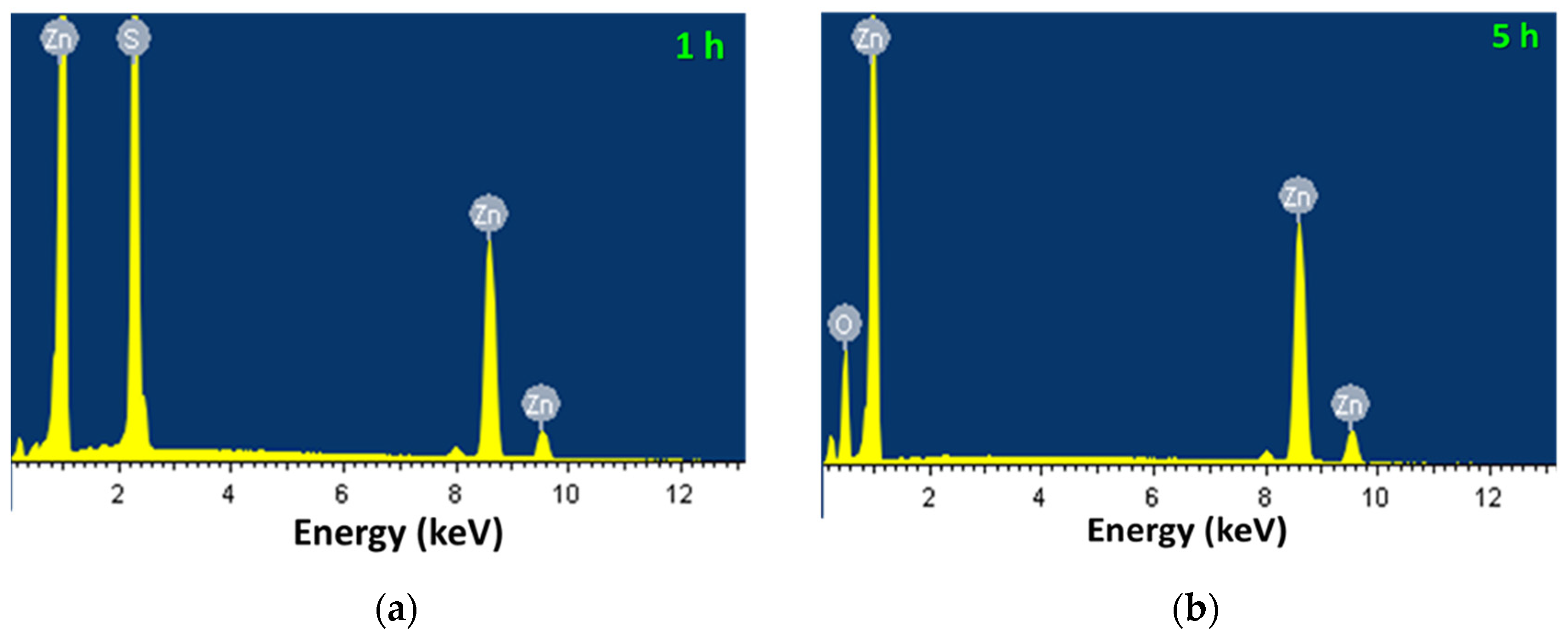
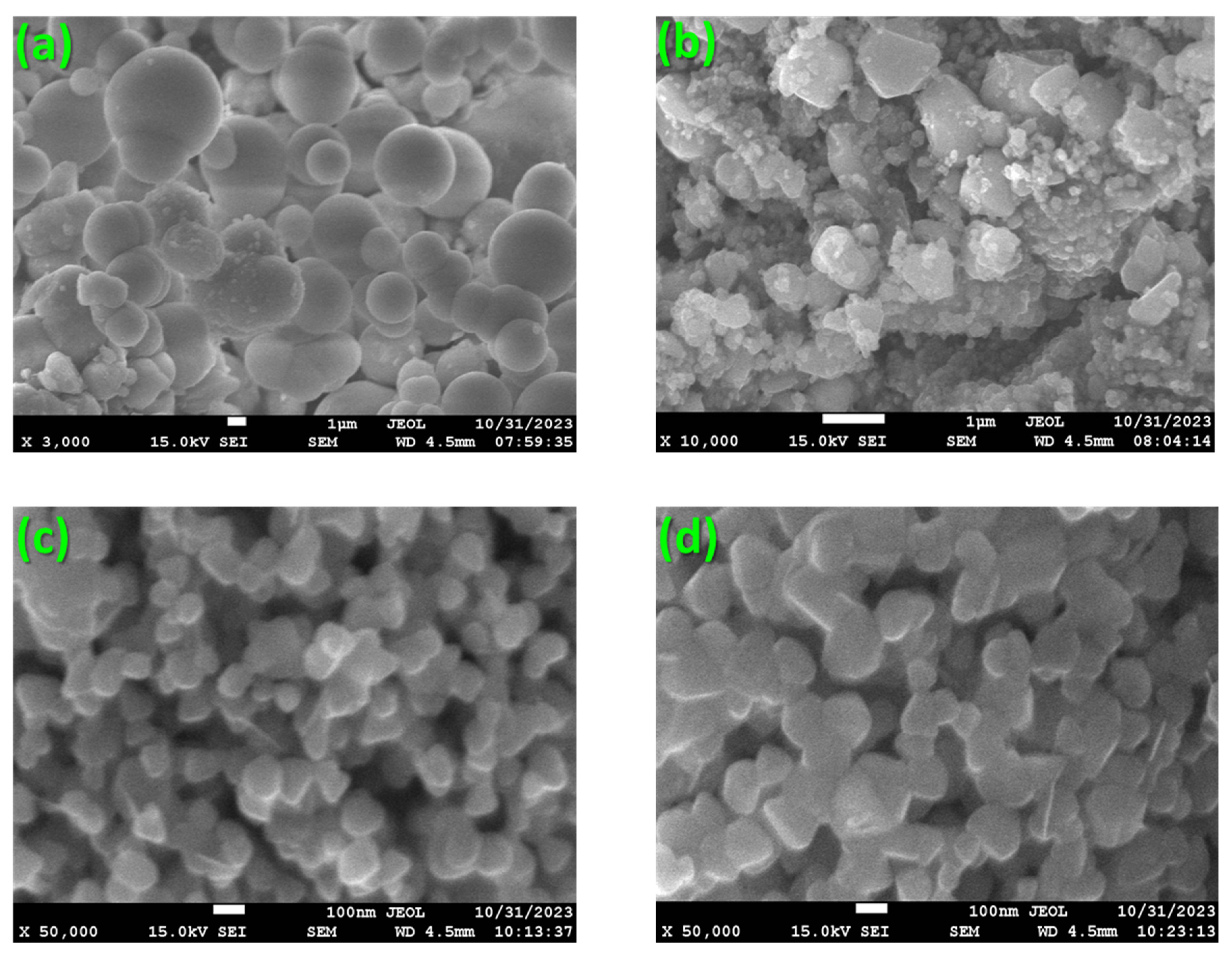
2.3. EDX and FE-SEM Analysis

An energy dispersive X-ray (EDX) analysis was carried out to calculate the atomic percentages of Zn, S, and O atoms in the synthesized nanoparticles. The EDX spectra for the nanoparticles that were sintered for 1 h and 5 h at 600 °C are illustrated in Figure 4a,b, respectively, while the percentage of each atomic element detected is given in Table 1. The nanoparticles which were sintered for 1 h at 600 °C showed the presence of only Zn and S atoms, indicating the formation of the ZnS phase. As the sintering time was increased, the percentage of detected S atoms decreased, while the amount of O atoms increased. When the sintering duration was increased to 5 h, all S atoms disappeared, while the atomic percentage of O reached 50%, which resulted in the formation of the ZnO phase exclusively. These EDX analysis findings are consistent with our PXRD analysis (see Figure 1). EDX measurements were not carried out for the pristine and different concentrations of Cl-doped ZnO nanoparticles because the sintering time and temperature used for the synthesis were the same as in the 5 h synthesized sample (refer to Figure 4b and Table 1).
In Figure 5a, the field emission scanning electron microscopy (FE-SEM) patterns of powders synthesized through hydrothermal means are displayed. Using ImageJ (1.54h), an open-source software for image analysis [40] the particle sizes of the captured particles were calculated and averaged. These particles had spherical shapes with an average size of approximately 2.8 µm and a standard deviation of 1.5 µm. Upon doping the powders with 25 mg/g Cl and sintering them at 600 °C for 1 h, the larger-sized spheres were destroyed, resulting in the creation of larger-sized faceted particles along with smaller nanoparticles, as shown in Figure 5b. The average particle size for the 25 mg/g Cl-doped ZnS/ZnO nanoparticles sintered at 600 °C for 1 h was found to be 433 nm with a standard deviation of 361 nm. With a further increase in sintering time, the particle sizes were decreased to 95 nm with a standard deviation of 14 nm for 25 mg/g Cl-doped ZnO nanoparticles sintered at 600 °C for 5 h, as demonstrated in Figure 5c. Increasing the sintering temperature to 6 h resulted in a slight increase in particle size to about 102 nm, as shown in Figure 5d. According to these results (FE-SEM and photoluminescence (Section 2.3 and Section 2.4), it was found that the optimal sintering time for ZnO nanoparticles is 5 h at a temperature of 600 °C. This led to the production of smaller nanoparticle sizes and high luminescence intensity. In a previous study by Tao et al. [30], it was found that sintering Cl-doped ZnO nanoparticles at 700 °C produced spherical nanoparticles with an average diameter of about 60 nm. Although the results of this study were similar in terms of particle size, the morphology of the nanoparticles was different. FE-SEM measurements were not carried out for the pristine and different concentrations of Cl-doped ZnO nanoparticles because the sintering time and temperature used for the synthesis were the same as in the 5 h synthesized sample, which was considered to have similar particle sizes and morphology.

2.4. Luminescence Properties

Figure 6a displays the photoluminescence excitation spectra of 25 mg/g Cl-doped ZnS that has been sintered at 600 °C for 2 h and 5 h. These spectra were measured at room temperature under 465 nm and 503 nm emissions, respectively. As depicted in Figure 6a, these excitations consisted of a broad band resulting from the overlapping of multiple excitation peaks. The most intense excitations for the dominant ZnS phase were discovered to be situated at around 350 nm, whereas for the ZnO phase, they were at 375 nm. Similar excitation bands to those shown in Figure 6a were observed in the 25 mg/g Cl-doped samples that were sintered for over 5 h at 600 °C. Additionally, the excitation bands in the ZnO samples, which were doped with varying concentrations of Cl and sintered for 5 h at 600 °C, displayed similarities to those in Figure 6a for the 5 h sintered sample.
The emissions spectra were measured under 350 nm excitation for 25 mg/g Cl-doped ZnS/ZnO nanostructures sintered at 600 °C for varying durations, as shown in Figure 6b. When the sintering duration was less than 4 h, the ZnS phase was dominant, and a broadband emission of ZnS was observed. As the sintering time increased, the emission band was red shifted due to the increased fraction of the ZnO phase and reduction in the ZnS phase. For a sintering duration of 5 h, the characteristic green emission of ZnO was obtained. The emission spectra under 375 nm excitations for the same samples are shown in Figure 6c. Except for the 1 h sintered ZnO sample, the characteristic green emission of ZnO was obtained, confirming the presence of the ZnO phase as observed in the PXRD and EDX analyses. The intensity of this emission increased up to a sintering time of 5 h and then decreased upon a further increase in sintering time. This effect could be attributed to an increase in defect states due to off-stoichiometry, as observed in the EDX analysis for the 6 h sintered sample (see Table 1). The photoluminescence emission spectra of pristine and various concentrations of Cl-doped ZnO nanoparticles were measured under 375 nm excitation, and the results are presented in Figure 6d. It was observed that all samples had a similar broadband emission, with a peak at approximately 503 nm. However, the incorporation of Cl significantly enhanced the emission intensity.
The dependence of integrated luminescence intensity of the emission bands presented in Figure 6c as a function of sintering time is shown in Figure 7a. These results showed that the highest luminescence light yield was achieved by sintering samples for 5 h at a temperature of 600 °C. The dependence of integrated luminescence intensity of the emission bands presented in Figure 6d as a function of Cl doping concentrations in the ZnO nanoparticles is shown in Figure 7b. These results showed that 25 mg/g Cl-doped ZnO samples had the highest luminescent light yield, which decreased upon a further increase in Cl concentration in ZnO, as illustrated in Figure 7b.
Moreover, the emission spectra of 25 mg/g Cl-doped ZnO nanoparticles sintered for 2 h and 5 h at 600 °C were deconvoluted using a Gaussian function. The emission spectrum of the sample sintered for 2 h was deconvoluted with the sum of two Gaussians, as shown in Figure 8a. The emission maximums of these two bands were found to be at 2.40 eV and 2.69 eV, respectively. On the other hand, the emission spectrum of the sample sintered for 5 h was also deconvoluted with the sum of two Gaussians, as shown in Figure 8b, with the emission maximums of these two bands found at 2.26 eV and 2.47 eV, respectively. The green emission bands observed in the range of 2.26–2.47 eV in the synthesized ZnS and ZnO nanoparticles were previously reported for ZnO phosphors, films, and nanocrystals [29,41,42,43]
Valence band maxima in Cl-doped ZnO are formed mainly from O-2p states, while occupied states near the conduction band minimum, around the Fermi level, are formed mainly from Cl-3s states, confirming the donor (n-type) levels [44]. It is essential to note that Cl ions replace O2− ions in the lattice structure, as reported earlier [37]. Three types of O vacancies, namely,  V O ,   V O . and   V O .. ,    can be formed, among which  V O .  is believed to be responsible for the observed green emission in ZnO [42]. The green emission is caused by the recombination of electrons in the  V O .  center with photoexcited holes in the valence band. Some studies suggest that the green emission peaking at around 500 nm is caused by the recombination of photoexcited electrons with deeply trapped holes in the  V O . [44]. As previously stated, Cl doping gives rise to the formation of a new defect level in the ZnO lattice with an excitation band at around 375 nm due to the excitation of electrons in the valence band to  C l O . :
C l O . + e     C l O ×
The  C l O ×  did not exhibit any new emission band in this study or in previous literature, indicating that it does not function as a luminescent center [30]. Theoretical investigations demonstrated that  C l O ×  acts as a donor in the ZnO lattice [44]. Therefore, it contributes the captured electron to  V O .. , forming  V O . :
C l O × + V O ..     C l O . + V O .
The concentration of  V O .  in ZnO increases significantly as a result of Cl doping. Consequently, nonradiative recombination decreases, and the radiative recombination of charge carriers increases, giving rise to the increase in photoluminescence intensity as oberved in this study and reported previously.
The ZnS lattice structures can form several defect states due to S/Zn vacancies and interstitials. Donor defects can be created by interstitial Zn atoms and S vacancies, while acceptor defects can be formed by interstitial S atoms and Zn vacancies. The energy levels of S interstitials/vacancies and Zn vacancies/interstitials are near the valance band maximum (VBM) and conduction band minimum (CBM), respectively [39] As a result of these defects, multiple transitions are possible, as previously reported [22,42]. The excitation bands depicted in Figure 6a for a 1 h sintered sample, which depicts a dominant ZnS phase, were broad, with several overlapped peaks resulting from the transitions from the defect states < 350 nm (i.e., below the band gap energies) and the characteristic states of Zn, with S atoms > 350 nm (i.e., above the band gap). Cl doping gives rise to the formation of a new defect level in the ZnO lattice, with an excitation band > 350 nm (see Figure 6a) due to the excitation of electrons in the valence band to  C l O . .
Based on the literature and the emission peaks observed in the synthesized ZnS and ZnO nanoparticles in this study, it was concluded that these bands were assigned to the transition from O vacancies. Furthermore, the band located at about 2.69 eV for the 2 h sintered sample (Figure 8a) was assigned to the transition from the divalent Zn interstitial defect to the CBM [44]. It is worth noting that previous studies have reported the presence of blue and green emission bands in pure and activated ZnO [28,29,30,44]. However, in our study of ZnO nanoparticles, we did not observe any blue emission band. This could be attributed to the emergence of different defect states in our samples as compared to those in previous studies.
The intense emission that was observed from the Cl-doped ZnO nanoparticles, which were optimally sintered at 600 °C, is promising for the future development of radiation detectors, particularly for radon monitoring in the environment. We are currently working on further improving the Cl-doped ZnO nanoparticles and will be using them to develop a radiation detector. The results of our study will be communicated in our follow-up report.

3. Materials and Methods

3.1. Materials

Zinc acetate dihydrate and sodium thiosulfate pentahydrate powders, both of which had a purity level of 99.9%, were acquired from Sigma Aldrich (St. Louis, MO, USA). Absolute ethanol, which had a purity level greater than 99.8%, was also obtained from Sigma Aldrich. These materials were utilized in their original form, without undergoing any further purification process.

3.2. Synthesis

A batch of ZnO nanoparticles was synthesized using the hydrothermal method followed by sintering at 600 °C in an air environment. To begin with, 77 g of zinc acetate dihydrate and 85.5 g of sodium thiosulfate pentahydrate powders were separately dissolved in 350 mL of distilled water. The two solutions were thoroughly mixed for an hour under vigorous stirring at room temperature. Next, 100 mL of the mixed solution was transferred to several Teflon-lined autoclaves of 150 mL capacity each. The autoclaves were then placed in a furnace at 200 °C for four hours and then cooled to room temperature. The synthesized powders were washed multiple times with distilled water and ethanol and then dried at 60 °C. Subsequently, 25 mg/g of NH4Cl was added to the synthesized powders to incorporate Cl ions as an activator, and sintering was carried out at different times (1, 2, 3, 4, 5, and 6 h) at a temperature of 600 °C in an air atmosphere. The sintered powders were subjected to several characterization techniques such as PXRD, EDX, SEM, reflectance spectroscopy, and photoluminescence to determine the optimal sintering time. Further, NH4Cl with different concentrations (0, 5, 15, 25, 35, 45, and 55 mg/g) was incorporated into the hydrothermally synthesized powders and sintered at the optimum time and temperature. These nanoparticles were characterized with different techniques to study the effect of Cl on the structural, morphological, optical, and luminescence properties.

3.3. Characterizations

To understand the crystal structure of the synthesized nanoparticles, PXRD measurements were conducted using an XPERT-MED (Malvern Panalytical, Malvern, UK) X-ray diffractometer. The samples were exposed to 0.154 nm Cu Kα1 radiation and measured in the 2θ range of 10° to 80° with a step size of 0.020° and a scanning rate of 2 s/step. The X-ray generator was operated at 40 kV and a tube current of 30 mA during the measurement process. In addition, the diffuse reflectance spectra of the synthesized ZnS nanoparticles were measured using a UV-3600 Plus Shimadzu UV–Vis–NIR spectrometer (Shimadzu corporation, Kyoto, Japan) ranging from 200 nm to 800 nm at room temperature. This helped to determine the optical properties of the nanoparticles. EDX analysis was also conducted to investigate the chemical composition of these nanoparticles. FE-SEM was used to provide detailed information on the size and morphology of the prepared samples. The EDX and FE-SEM analyses were conducted using a high-resolution JSM-6700F FE-SEM instrument (JEOL company, Tokyo, Japan) with an operating voltage of 10 kV. Lastly, the emission spectrum of the ZnS nanoparticles was recorded using a Hitachi F-7000 fluorescence spectrophotometer (Hitachi Ltd., Tokyo, Japan). To conduct the photoluminescence measurement, 20 mg of powder was precisely weighed for each sample and placed inside a cylindrical solid sample holder. The holder had a super quartz window, and the lid was screwed on tightly. The surface area of the illuminated surface was 1.57 cm2 for all the samples. During the measurements, both the excitations and emission slits were set at 1.5 nm, the acquisition speed was 200 nm/s, and the number of scans was 5 for all the samples. These conditions were maintained to ensure identical measurement conditions, which were necessary for comparative analysis.

4. Conclusions

Nanoparticles of ZnO doped with Cl were synthesized at a sintering temperature of 600 °C for 5 h, with a particle size of around 95 nm, as confirmed by FE-SEM analysis. The introduction of Cl led to an increase in the lattice parameters a and c and lattice volume, as Cl ions with slightly larger ionic diameters replaced O2− ions in the ZnO lattice. With increasing Cl concentrations, the optical band gap decreased, and a new defect level was created in the ZnO lattice, resulting in an excitation band at approximately 375 nm. The emission intensity was observed to increase significantly with a sintering time of up to 5 h and Cl concentration of 25 mg/g. The photoluminescence emission intensity for 25 mg/g Cl-doped ZnO nanoparticles was found to be eight times higher than the pristine sample. The green emission observed in the range of 2.26–2.47 eV is attributed to the transition from O defects.

Author Contributions

Conceptualization, A.K.; methodology, A.K. and A.M.A.; software, A.K.; validation, A.K., A.M.A. and J.S.A.; formal analysis, A.K.; investigation, A.K. and A.M.A.; data curation, A.K.; writing—original draft preparation, A.K.; writing—review and editing, A.K., S.E.M., T.I.A.-N., H.B.A., J.S.A. and A.M.A.; visualization, A.K. and A.M.A.; supervision, A.K.; project administration, A.K.; funding acquisition, A.K. All authors have read and agreed to the published version of the manuscript.

Funding

This research was funded by Deputy for Research and Innovation—Ministry of Education, Kingdom of Saudi Arabia through a grant (NU/IFC/2/-/12) under the Institutional Funding Committee at Najran University, Kingdom of Saudi Arabia.

Data Availability Statement

Data used to support the findings of this study are available from the corresponding author upon request.

Acknowledgments

The authors would like to acknowledge the support of the Deputy for Research and Innovation—Ministry of Education, Kingdom of Saudi Arabia for this research through a grant (NU/IFC/2/-/12) under the Institutional Funding Committee at Najran University, Kingdom of Saudi Arabia.

Conflicts of Interest

The authors declare no conflicts of interest.

References

  1. Klingshirn, C.F.; Waag, A.; Hoffman, A.; Geurts, J. Zinc Oxide: From Fundamental Properties Towards Novel Applications; Springer: Berlin/Heidelberg, Germany, 2013; Volume 53. [Google Scholar]
  2. Weerathunga, H.; Tang, C.; Brock, A.J.; Sarina, S.; Wang, T.; Liu, Q.; Zhu, H.Y.; Du, A.; Waclawik, E.R. Nanostructure Shape-Effects in ZnO Heterogeneous Photocatalysis. J. Colloid Interface Sci. 2022, 606, 588–599. [Google Scholar] [CrossRef]
  3. Le Pivert, M.; Martin, N.; Leprince-Wang, Y. Hydrothermally Grown ZnO Nanostructures for Water Purification via Photocatalysis. Crystals 2022, 12, 308. [Google Scholar] [CrossRef]
  4. Di Mauro, A.; Fragalà, M.E.; Privitera, V.; Impellizzeri, G. ZnO for Application in Photocatalysis: From Thin Films to Nanostructures. Mater. Sci. Semicond. Process. 2017, 69, 44–51. [Google Scholar] [CrossRef]
  5. Vattikuti, S.V.P.; Reddy, P.A.K.; Shim, J.; Byon, C. Visible-Light-Driven Photocatalytic Activity of SnO2-ZnO Quantum Dots Anchored on g-C3N4 Nanosheets for Photocatalytic Pollutant Degradation and H2 Production. ACS Omega 2018, 3, 7587–7602. [Google Scholar] [CrossRef] [PubMed]
  6. Kumar, V.; Prakash, J.; Pathak, D.; Sharma, D.P.; Purohit, L.P.; Swart, H.C. Ion Beam Engineering of Implanted ZnO Thin Films for Solar Cell and Lighting Applications. Chem. Eng. J. Adv. 2023, 15, 100501. [Google Scholar] [CrossRef]
  7. Zahoor, R.; Jalil, A.; Ilyas, S.Z.; Ahmed, S.; Hassan, A. Optoelectronic and Solar Cell Applications of ZnO Nanostructures. Results Surf. Interfaces 2021, 2, 100003. [Google Scholar] [CrossRef]
  8. Wibowo, A.; Marsudi, M.A.; Amal, M.I.; Ananda, M.B.; Stephanie, R.; Ardy, H.; Diguna, L.J. ZnO Nanostructured Materials for Emerging Solar Cell Applications. RSC Adv. 2020, 10, 42838–42859. [Google Scholar] [CrossRef]
  9. Wang, J.X.; Sun, X.W.; Yang, Y.; Huang, H.; Lee, Y.C.; Tan, O.K.; Vayssieres, L. Hydrothermally Grown Oriented ZnO Nanorod Arrays for Gas Sensing Applications. Nanotechnology 2006, 17, 4995. [Google Scholar] [CrossRef]
  10. Umar, A.; Ibrahim, A.A.; Kumar, R.; Algadi, H.; Albargi, H.; Alsairi, M.A.; Alhmami, M.A.M.; Zeng, W.; Ahmed, F.; Akbar, S. CdO–ZnO Nanorices for Enhanced and Selective Formaldehyde Gas Sensing Applications. Environ. Res. 2021, 200, 111377. [Google Scholar] [CrossRef]
  11. Islam, M.; Srivastava, A.K.; Basavaraja, B.M.; Sharma, A. “Nano-on-Micro” Approach Enables Synthesis of ZnO Nano-Cactus for Gas Sensing Applications. Sens. Int. 2021, 2, 100084. [Google Scholar] [CrossRef]
  12. Sahani, R.M.; Dixit, A. A Comprehensive Review on Zinc Oxide Bulk and Nano-Structured Materials for Ionizing Radiation Detection and Measurement Applications. Mater. Sci. Semicond. Process. 2022, 151, 107040. [Google Scholar] [CrossRef]
  13. Wen, X.; Zhang, Q.; Shao, Z. Magnetron Sputtering for ZnO:Ga Scintillation Film Production and Its Application Research Status in Nuclear Detection. Crystals 2019, 9, 263. [Google Scholar] [CrossRef]
  14. Saad, M.; AlMohiy, H.; Alshihri, A.A.; Alelyani, M.; Shalaby, R.M. Fabrication, Microstructural Modifications, Elastic Properties and Radiation Attenuation Performance of ZnO Nano-Sized Particles-Reinforced Pb-Based Alloys for Radiation Shielding Applications. Radiat. Eff. Defects Solids 2023, 178, 751–767. [Google Scholar] [CrossRef]
  15. Eskalen, H.; Kavun, Y.; Kavgacı, M. Preparation and Study of Radiation Shielding Features of ZnO Nanoparticle Reinforced Borate Glasses. Appl. Radiat. Isot. 2023, 198, 110858. [Google Scholar] [CrossRef] [PubMed]
  16. Xin, M. Effect of Eu Doping on the Structure, Morphology and Luminescence Properties of ZnO Submicron Rod for White LED Applications. J. Theor. Appl. Phys. 2018, 12, 177–182. [Google Scholar] [CrossRef]
  17. Das, S.; Ghorai, U.K.; Dey, R.; Ghosh, C.K.; Pal, M. White Light Phosphorescence from ZnO Nanoparticles for White LED Applications. New J. Chem. 2022, 46, 17585–17595. [Google Scholar] [CrossRef]
  18. Young, S.-J.; Yang, C.-C.; Lai, L.-T. Review—Growth of Al-, Ga-, and In-Doped ZnO Nanostructures via a Low-Temperature Process and Their Application to Field Emission Devices and Ultraviolet Photosensors. J. Electrochem. Soc. 2017, 164, B3013. [Google Scholar] [CrossRef]
  19. Su, Y.K.; Peng, S.M.; Ji, L.W.; Wu, C.Z.; Cheng, W.B.; Liu, C.H. Ultraviolet ZnO Nanorod Photosensors. Langmuir 2010, 26, 603–606. [Google Scholar] [CrossRef] [PubMed]
  20. Caglar, Y.; Caglar, M.; Ilican, S. XRD, SEM, XPS Studies of Sb Doped ZnO Films and Electrical Properties of Its Based Schottky Diodes. Optik 2018, 164, 424–432. [Google Scholar] [CrossRef]
  21. Adaikalam, K.; Valanarasu, S.; Ali, A.M.; Sayed, M.A.; Yang, W.; Kim, H.S. Photosensing Effect of Indium-Doped ZnO Thin Films and Its Heterostructure with Silicon. J. Asian Ceram. Soc. 2022, 10, 108–119. [Google Scholar] [CrossRef]
  22. Gabás, M.; Landa-Cánovas, A.; Luis Costa-Krämer, J.; Agulló-Rueda, F.; González-Elipe, A.R.; Díaz-Carrasco, P.; Hernández-Moro, J.; Lorite, I.; Herrero, P.; Castillero, P.; et al. Differences in N-Type Doping Efficiency between Al- and Ga-ZnO Films. J. Appl. Phys. 2013, 113, 163709. [Google Scholar] [CrossRef]
  23. Tao, Z.; Yu, X.; Fei, X.; Liu, J.; Zhao, Y.; Wu, H.; Yang, G.; Yang, S.; Yang, L. Synthesis and Optical Properties of Halogen-Doped ZnO Phosphor. Mater. Lett. 2008, 62, 3018–3020. [Google Scholar] [CrossRef]
  24. Muhammad, A.; Hassan, Z.; Mohammad, S.M.; Rajamanickam, S.; Shitu, I.G. Fabrication of Ultra-Violet Photodetector with Enhanced Optoelectronic Parameters Using Low-Cost F-Doped ZnO Nanostructures. Sens. Actuators A Phys. 2021, 332, 113092. [Google Scholar] [CrossRef]
  25. Ujan, Z.A.; Tahira, A.; Mahesar, A.A.; Markhand, A.H.; Bhatti, A.L.; Mugheri, A.Q.; Bhatti, M.A.; Shaikh, N.M.; Mari, R.H.; Nafady, A.; et al. The Crystal Disorder into ZnO with Addition of Bromine and It’s Outperform Role in the Photodegradation of Methylene Blue. J. Clust. Sci. 2022, 33, 281–291. [Google Scholar] [CrossRef]
  26. Bouaziz, L.; Si-Ahmed, K.; Özacar, M.; Trari, M.; Bessekhouad, Y. Sensor Prospect of Iodine-Doped ZnO Materials for Ethyl Paraben Detection. Microchem. J. 2022, 183, 108132. [Google Scholar] [CrossRef]
  27. Liu, C.; Yu, A.; Peng, M.; Song, M.; Liu, W.; Zhang, Y.; Zhai, J. Improvement in the Piezoelectric Performance of a ZnO Nanogenerator by a Combination of Chemical Doping and Interfacial Modification. J. Phys. Chem. C 2016, 120, 6971–6977. [Google Scholar] [CrossRef]
  28. Zhang, Y.; Liu, C.; Liu, J.; Xiong, J.; Liu, J.; Zhang, K.; Liu, Y.; Peng, M.; Yu, A.; Zhang, A.; et al. Lattice Strain Induced Remarkable Enhancement in Piezoelectric Performance of ZnO-Based Flexible Nanogenerators. ACS Appl. Mater. Interfaces 2016, 8, 1381–1387. [Google Scholar] [CrossRef]
  29. Yousefi, R.; Jamali-Sheini, F. Effect of Chlorine Ion Concentration on Morphology and Optical Properties of Cl-Doped ZnO Nanostructures. Ceram. Int. 2012, 38, 5821–5825. [Google Scholar] [CrossRef]
  30. Yousefi, R.; Zak, A.K.; Mahmoudian, M.R. Growth and Characterization of Cl-Doped ZnO Hexagonal Nanodisks. J. Solid State Chem. 2011, 184, 2678–2682. [Google Scholar] [CrossRef]
  31. Tao, Z.; Yu, X.; Fei, X.; Liu, J.; Yang, G.; Zhao, Y.; Yang, S.; Yang, L. Synthesis and Photoluminescence of Cl-Doped ZnO Nanospheres. Opt. Mater. 2008, 31, 1–5. [Google Scholar] [CrossRef]
  32. Wang, F.; Seo, J.H.; Li, Z.; Kvit, A.V.; Ma, Z.; Wang, X. Cl-Doped ZnO Nanowires with Metallic Conductivity and Their Application for High-Performance Photoelectrochemical Electrodes. ACS Appl. Mater. Interfaces 2014, 6, 1288–1293. [Google Scholar] [CrossRef]
  33. Jiamprasertboon, A.; Dixon, S.C.; Sathasivam, S.; Powell, M.J.; Lu, Y.; Siritanon, T.; Carmalt, C.J. Low-Cost One-Step Fabrication of Highly Conductive ZnO:Cl Transparent Thin Films with Tunable Photocatalytic Properties via Aerosol-Assisted Chemical Vapor Deposition. ACS Appl. Electron. Mater. 2019, 1, 1408–1417. [Google Scholar] [CrossRef]
  34. Jiamprasertboon, A.; Powell, M.J.; Dixon, S.C.; Quesada-Cabrera, R.; Alotaibi, A.M.; Lu, Y.; Zhuang, A.; Sathasivam, S.; Siritanon, T.; Parkin, I.P.; et al. Photocatalytic and Electrically Conductive Transparent Cl-Doped ZnO Thin Films: Via Aerosol-Assisted Chemical Vapour Deposition. J. Mater. Chem. A 2018, 6, 12682–12692. [Google Scholar] [CrossRef]
  35. Kahouli, M.; Barhoumi, A.; Bouzid, A.; Al-Hajry, A.; Guermazi, S. Structural and Optical Properties of ZnO Nanoparticles Prepared by Direct Precipitation Method. Superlattices Microstruct. 2015, 85, 7–23. [Google Scholar] [CrossRef]
  36. Zhao, W.; Li, H.; Liu, Z.; Wang, D.; Liu, S. Controlled Defects and Enhanced Electronic Extraction in Fluorine-Incorporated Zinc Oxide for High-Performance Planar Perovskite Solar Cells. Sol. Energy Mater. Sol. Cells 2018, 182, 263–271. [Google Scholar] [CrossRef]
  37. Gautam, K.; Singh, I.; Bhatnagar, P.K.; Peta, K.R. Role of Cl Doping on the Growth and Relaxation Dynamics of ZnO Nanorods Synthesized by Hydrothermal Method. Chem. Phys. Lett. 2016, 662, 196–200. [Google Scholar] [CrossRef]
  38. Alshgari, R.A.; Ujjan, Z.A.; Shah, A.A.; Bhatti, M.A.; Tahira, A.; Shaikh, N.M.; Kumar, S.; Ibupoto, M.H.; Elhawary, A.; Nafady, A.; et al. ZnO Nanostructures Doped with Various Chloride Ion Concentrations for Efficient Photocatalytic Degradation of Methylene Blue in Alkaline and Acidic Media. Molecules 2022, 27, 8726. [Google Scholar] [CrossRef] [PubMed]
  39. Abdullahi, S.S.; Güner, S.; Koseoglu, Y.; Musa, I.M.; Adamu, B.I.; Abdulhamid, M.I. Simple Method for The Determınatıon of Band Gap of a Nanopowdered Sample Usıng Kubelka Munk Theory. J. Niger. Assoc. Math. Phys. 2016, 35, 241–246. [Google Scholar]
  40. Abdalla, A.M.; Khan, A.; Almalki, S.; Kawaguchi, N.; Yanagida, T.; Alsareii, S.A.; Algethami, J.S. Evaluation of Structural, Optical, and Scintillation Characteristics of Ag Activated ZnS Nanoparticles. Radiat. Phys. Chem. 2023, 210, 110999. [Google Scholar] [CrossRef]
  41. Slassi, A. First-Principles and Boltzmann Equation Studies of the Cl-Doped ZnO Transparent Conducting Oxide. Optik 2015, 126, 4751–4756. [Google Scholar] [CrossRef]
  42. Pamungkas, M.A.; Putra, S.A.; Nurhuda, M. Bandgap Control in ZnO with Na and Cl Adatom: DFT Calculations. J. Phys. Conf. Ser. 2021, 1811, 012126. [Google Scholar] [CrossRef]
  43. Vanheusden, K.; Warren, W.L.; Seager, C.H.; Tallant, D.R.; Voigt, J.A.; Gnade, B.E. Mechanisms behind Green Photoluminescence in ZnO Phosphor Powders. J. Appl. Phys. 1996, 79, 7983–7990. [Google Scholar] [CrossRef]
  44. Wu, X.L.; Siu, G.G.; Fu, C.L.; Ong, H.C. Photouminescence and Cathodoluminescence Studies of Stoichiometric and Oxygen-Deficient ZnO Films. Appl. Phys. Lett. 2001, 78, 2285–2287. [Google Scholar] [CrossRef]
Figure 1. PXRD patterns of (a) 25 mg/g Cl-doped ZnS/ZnO nanoparticles sintered at 600 °C for various durations. (b) Cl-doped ZnO nanoparticles sintered at 600 °C for a duration of 5 h. (c) Phase fractions of ZnS and ZnO phases as a function of sintering time for 25 mg/g Cl-doped ZnS/ZnO nanoparticles sintered at 600 °C.
Inorganics 12 00053 g001
Figure 2. Dependence of lattice parameter a and c (a) and unit cell volume (b) on Cl concentrations for the ZnO nanoparticles.
Inorganics 12 00053 g002
Figure 3. The Tauc’s plot obtained from the diffuse reflectance data of (a) 25 mg/g Cl-doped ZnS/ZnO nanoparticles sintered at 600 °C for different durations and (b) ZnO nanoparticles doped with various concentrations of Cl and sintered at 600 °C for 5 h. For both graphs, the Y-axis is presented as normalized for better readability.
Inorganics 12 00053 g003
Figure 4. EDX spectra of 25 mg/g Cl-doped ZnS/ZnO nanoparticles sintered at 600 °C (a) for 1 h and (b) for 5 h.
Inorganics 12 00053 g004
Figure 5. FE-SEM photographs of (a) hydrothermally synthesized ZnS nanoparticles without any annealing and 25 mg/g Cl-doped ZnS/ZnO nanoparticles sintered at 600 °C (b) for 1 h, (c) for 5 h, (d) for 6 h.
Inorganics 12 00053 g005
Figure 6. (a) Excitation spectra of 1 h and 2 h sintered samples measured at 460 nm and 503 nm emissions, respectively. Emission spectra of the samples sintered for different durations measured under (b) 350 nm excitations and (c) 375 nm excitations. (d) Emission spectra of pristine and various concentrations of Cl-doped samples sintered for 5 h and measured under 375 nm excitations. The sintering temperature for all these samples was 600 °C, and the nominal Cl concentration in the samples in (ac) was 25 mg/g.
Inorganics 12 00053 g006
Figure 7. (a) Integrated luminescence intensity of the 25 mg/g Cl-doped ZnS/ZnO nanoparticles as a function of sintering time. (b) Integrated luminescence intensity as a function of Cl doping concentration for the ZnO nanoparticles sintered for five hours at 600 °C.
Inorganics 12 00053 g007
Figure 8. Gaussian function deconvoluted emission spectrum of (a) 2 h and (b) 5 h for 25 mg/g Cl-doped samples sintered at 600 °C measured under 340 nm and 375 nm excitations, respectively.
Inorganics 12 00053 g008
Table 1. Atomic percentages of Zn, S, and O atoms calculated from EDX analysis for 25 mg/g Cl-doped ZnS/ZnO nanoparticles sintered at 600 °C for different durations.
Samples1 h2 h3 h4 h5 h6 h
ElementsAt.%At.%At.%At.%At.%At.%
Zn52.0252.4247.0143.9250.0739.96
S47.9843.4440.1118.5100
O04.1512.8837.5649.9360.04
Disclaimer/Publisher’s Note: The statements, opinions and data contained in all publications are solely those of the individual author(s) and contributor(s) and not of MDPI and/or the editor(s). MDPI and/or the editor(s) disclaim responsibility for any injury to people or property resulting from any ideas, methods, instructions or products referred to in the content.

