
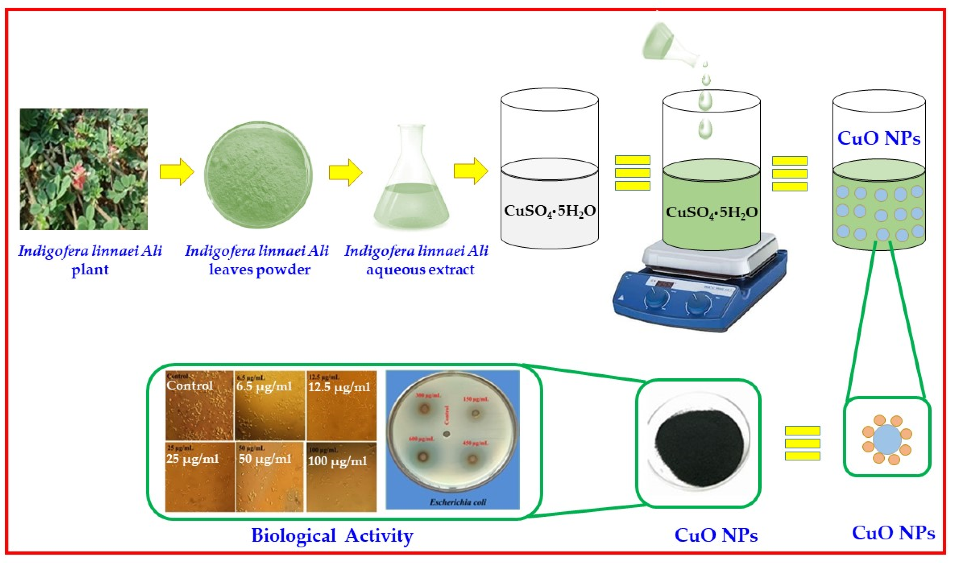
Copper Oxide Nanoparticles Synthesized from Indigofera linnaei Ali and This Plant’s Biological Applications
 ,
,  and
and
Abstract
:1. Introduction
2. Results
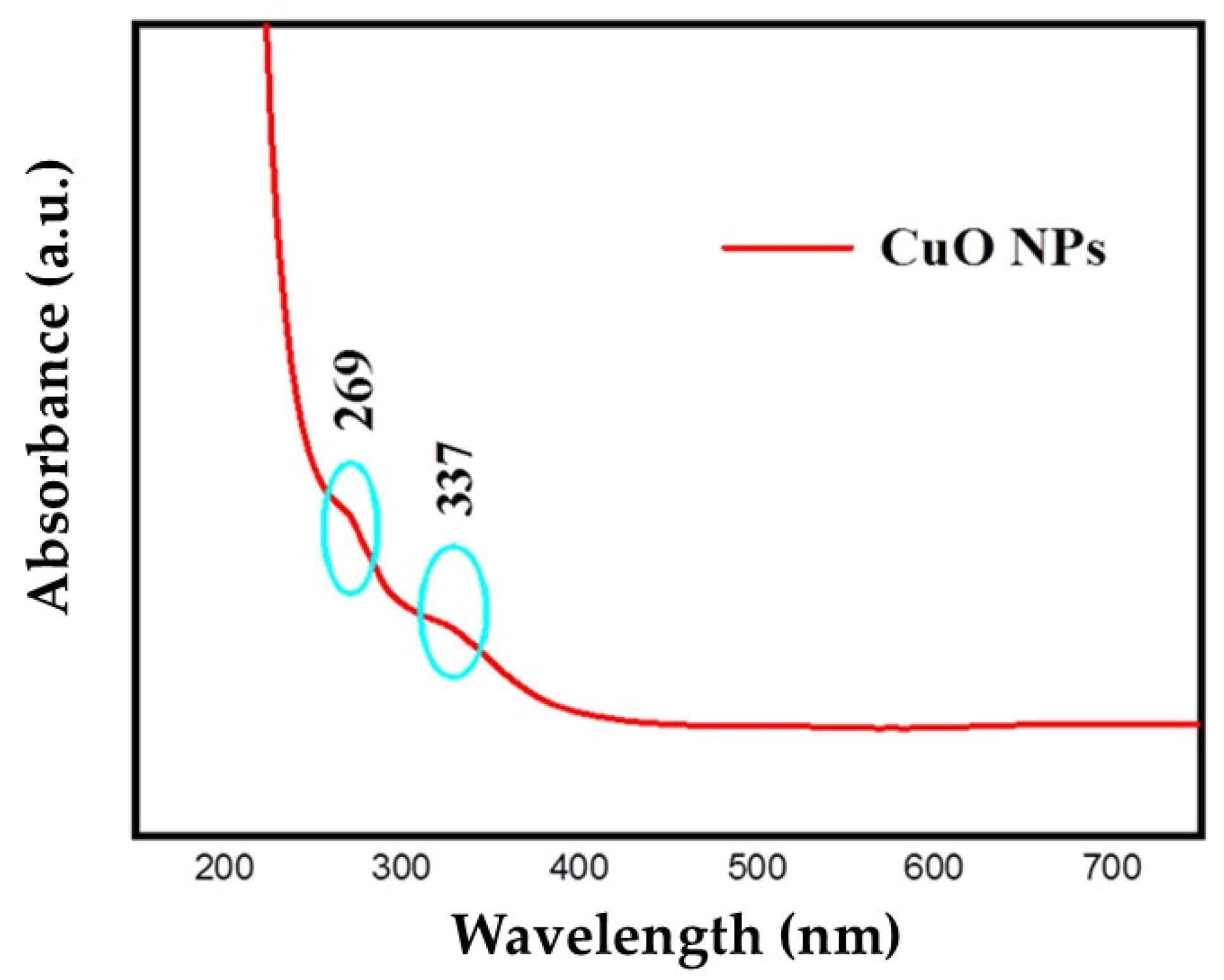
2.1. UV-Visible Spectrum Analysis
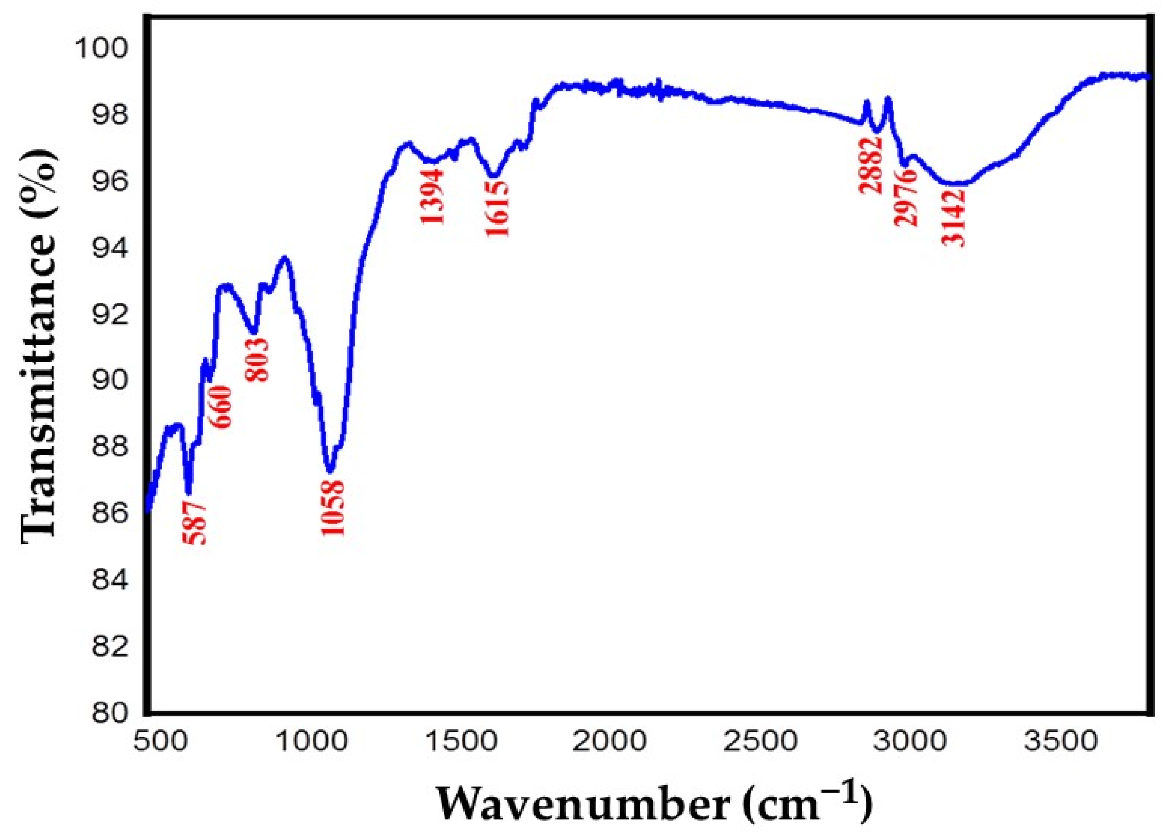
2.2. FT-IR Analysis
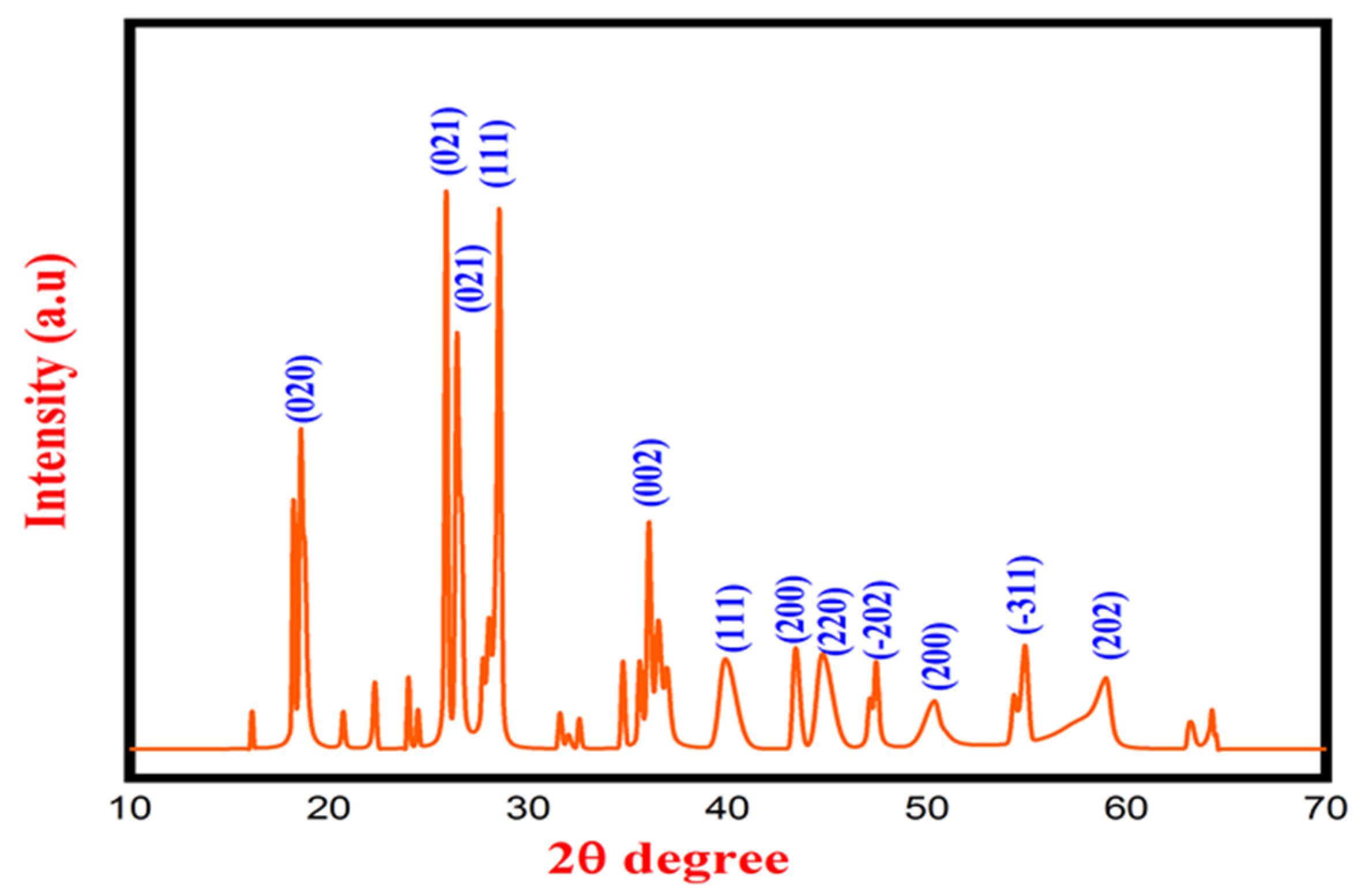
2.3. XRD Analysis
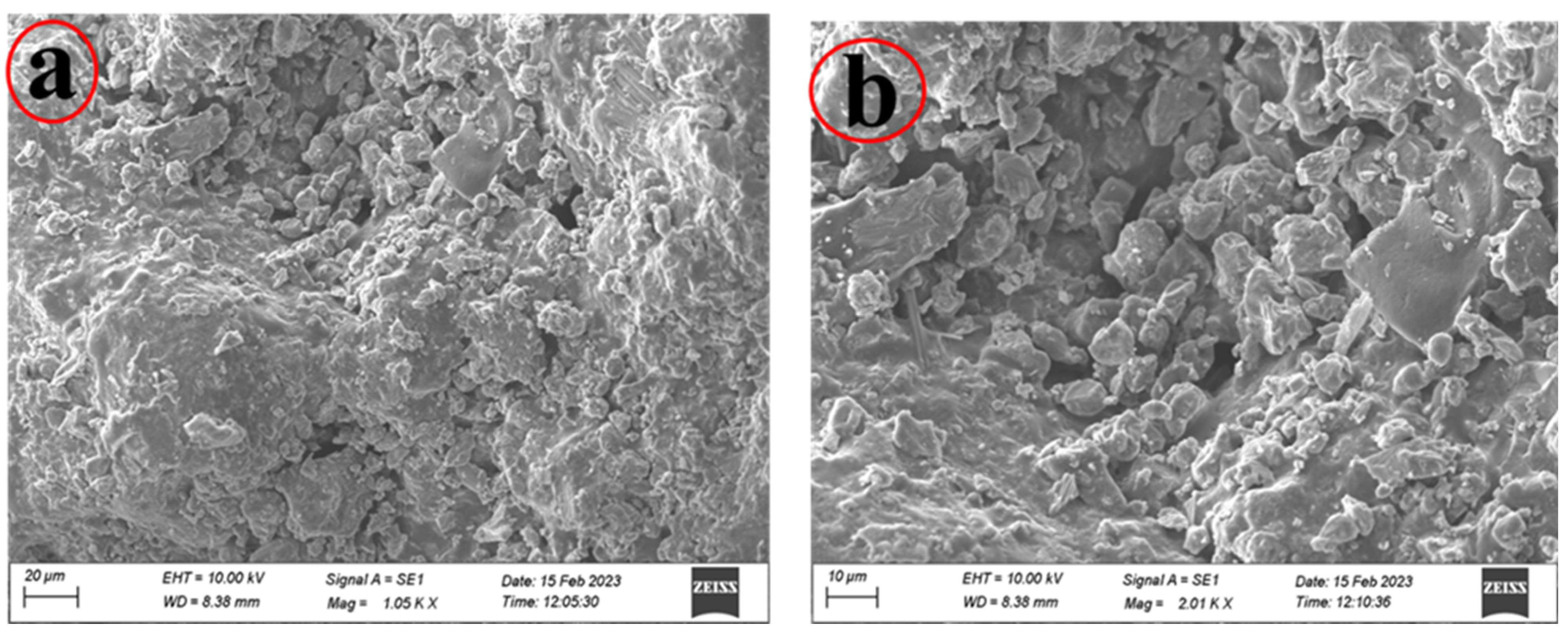
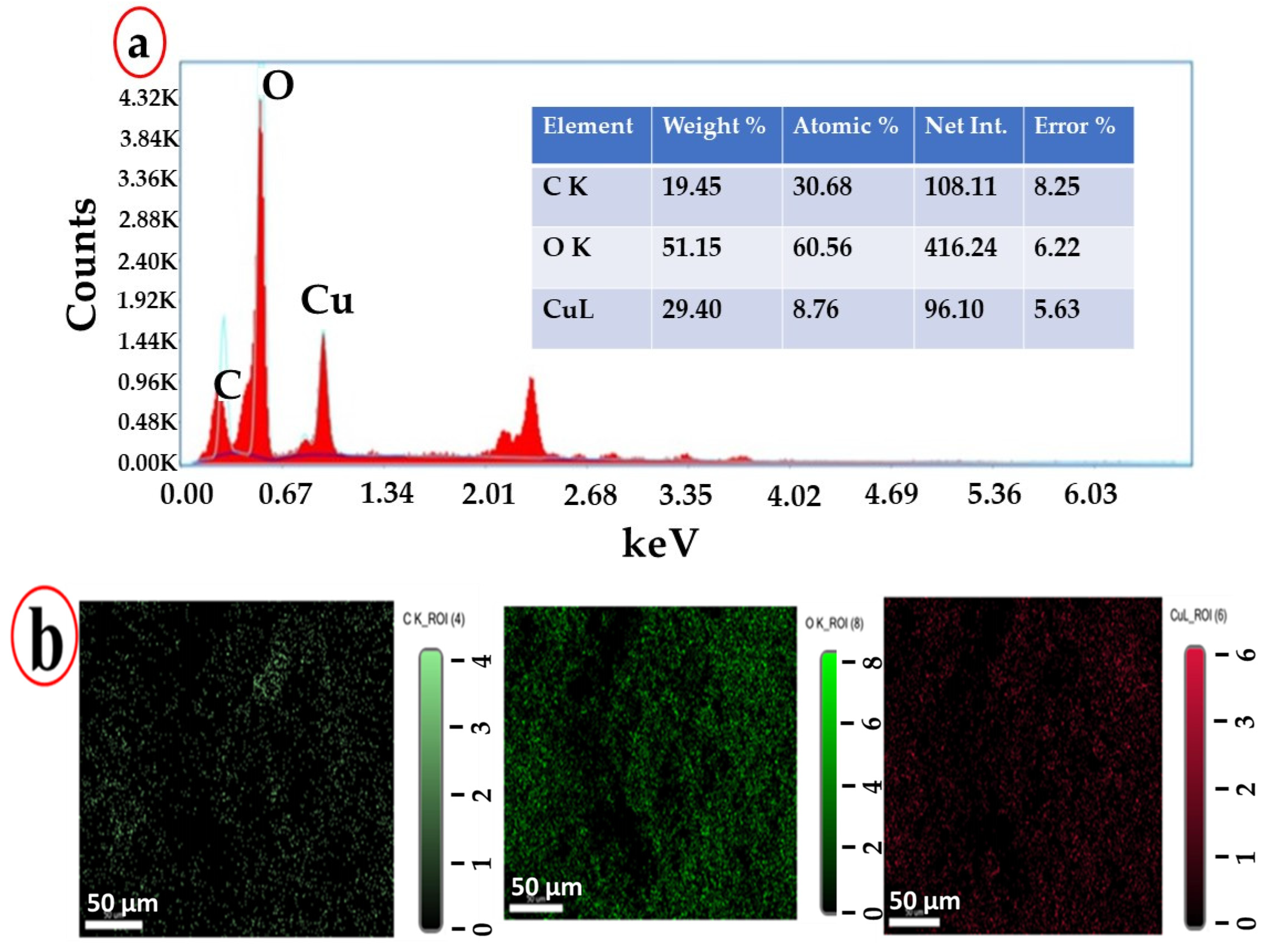
2.4. SEM and EDX Analysis
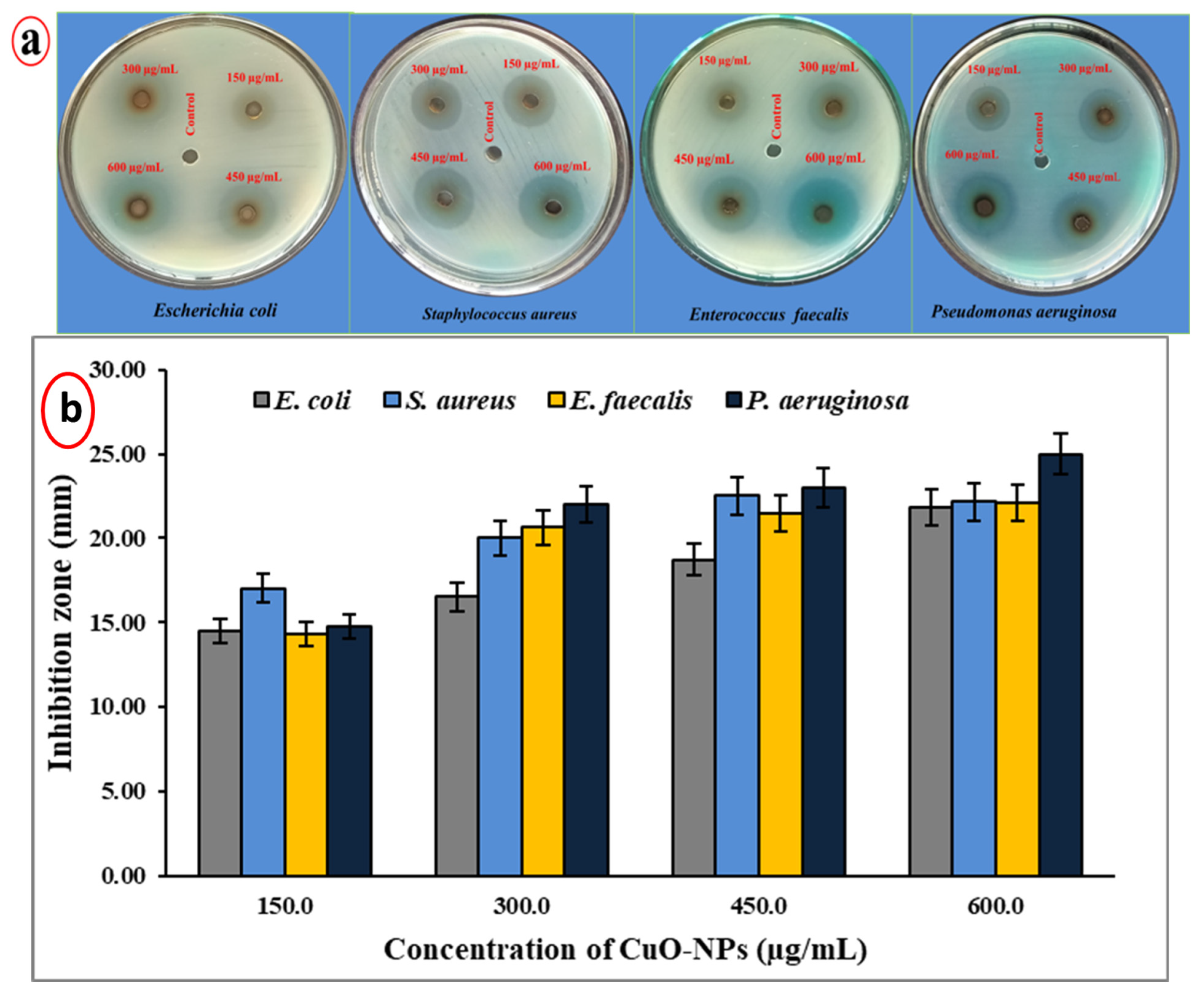
2.5. Antibacterial Activity
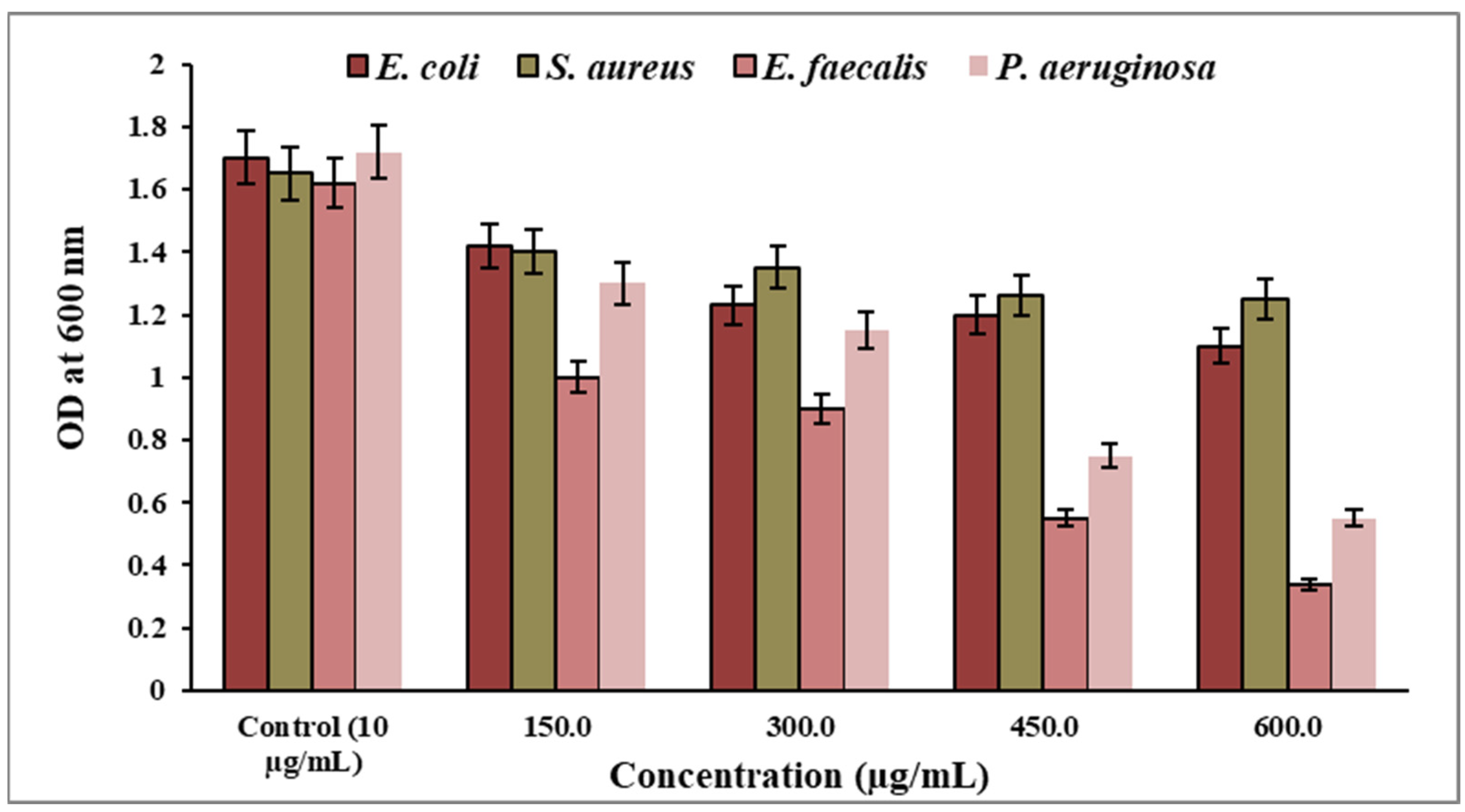
2.6. Assay for the Determination of Minimum Inhibitory Concentration (MIC)
2.7. Larvicidal Activity
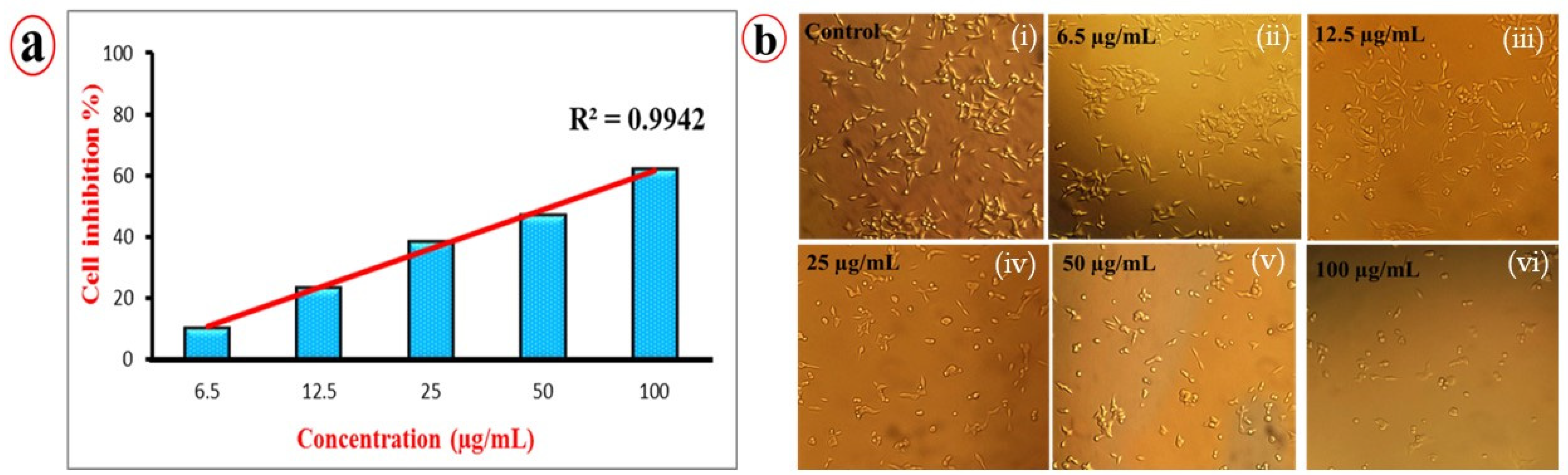
2.8. Anticancer Activity
3. Materials and Methods
3.1. Collection and Substantiation of Plants
3.2. Preparation of CuO NPs Using Aqueous Extract of Indigofera linnaei Ali Plants
3.3. Characterization of Synthesized Copper Nanoparticles
3.4. Evaluation of the Antibacterial Activity Using Agar Well Plate Method
3.5. Determination of Minimum Inhibitory Concentration (MIC)
3.6. Larvicidal Activity
3.7. Anticancer Activity Determined via MTT Assay
4. Conclusions
Author Contributions
Funding
Data Availability Statement
Acknowledgments
Conflicts of Interest
References
- Bouafia, A.; Meneceur, S.; Chami, S.; Laouini, S.E.; Daoudi, H.; Legmairi, S.; Mohammed Mohammed, H.A.; Aoun, N.; Menaa, F. Removal of hydrocarbons and heavy metals from petroleum water by modern green nanotechnology methods. Sci. Rep. 2023, 13, 5637. [Google Scholar] [CrossRef] [PubMed]
- Gu, J.; Chen, F.; Zheng, Z.; Bi, L.; Morovvati, H.; Goorani, S. Novel green formulation of copper nanoparticles by Foeniculum vulgare: Chemical characterization and determination of cytotoxicity, anti-human lung cancer and antioxidant effects. Inorg. Chem. Commun. 2023, 150, 110442. [Google Scholar] [CrossRef]
- Dejen, K.D.; Kibret, D.Y.; Mengesha, T.H.; Bekele, E.T.; Tedla, A.; Bafa, T.A.; Derib, F.T. Green synthesis and characterisation of silver nanoparticles from leaf and bark extract of Croton macrostachyus for antibacterial activity. Mater. Technol. 2023, 38, 2164647. [Google Scholar] [CrossRef]
- Prakash, M.; Kavitha, H.P.; Abinaya, S.; Vennila, J.P.; Lohita, D. Green synthesis of bismuth based nanoparticles and its applications-A review. Sustain. Chem. Pharm. 2022, 25, 100547. [Google Scholar] [CrossRef]
- Nthunya, L.N.; Mbakop, S.; Makgabutlane, B.; Matlou, G.; Mhlanga, S.; Richards, H. Fungal synthesis of copper nanoparticles and their applications in agri-food, environmental, and biomedical sectors. In Fungal Cell Factories for Sustainable Nanomaterials Productions and Agricultural Applications; Elsevier: Amsterdam, The Netherlands, 2023; pp. 91–114. [Google Scholar]
- Abed, A.S.; Khalaf, Y.H.; Mohammed, A.M. Green synthesis of gold nanoparticles as an effective opportunity for cancer treatment. Results Chem. 2023, 5, 100848. [Google Scholar] [CrossRef]
- Gemin, L.G.; de Lara, G.B.; Mógor, Á.F.; Mazaro, S.M.; Sant’Anna-Santos, B.F.; Mógor, G.; Amatussi, J.D.O.; Cordeiro, E.C.N.; Marques, H.M.C. Polysaccharides combined to copper and magnesium improve tomato growth, yield, anti-oxidant and plant defense enzymes. Sci. Hortic. 2023, 310, 111758. [Google Scholar] [CrossRef]
- Naz, S.; Gul, A.; Zia, M.; Javed, R. Synthesis, biomedical applications, and toxicity of CuO nanoparticles. Appl. Microbiol. Biotechnol. 2023, 107, 1039–1061. [Google Scholar] [CrossRef]
- Sukumar, S.; Rudrasenan, A.; Padmanabhan Nambiar, D. Green-synthesized rice-shaped copper oxide nanoparticles using Caesalpinia bonducella seed extract and their applications. ACS Omega 2020, 5, 1040–1051. [Google Scholar] [CrossRef]
- Siddiqui, V.U.; Ansari, A.; Chauhan, R.; Siddiqi, W.A. Green synthesis of copper oxide (CuO) nanoparticles by Punica granatum peel extract. Mater. Today Proc. 2021, 36, 751–755. [Google Scholar] [CrossRef]
- Ramadhan, V.; Ni’Mah, Y.; Yanuar, E.; Suprapto, S. Synthesis of copper nanoparticles using Ocimum tenuiflorum leaf extract as capping Agent. AIP Conf. Proc. 2019, 2202, 020067. [Google Scholar]
- Kolahalam, L.A.; Prasad, K.; Krishna, P.M.; Supraja, N.; Shanmugan, S. The exploration of bio-inspired copper oxide nanoparticles: Synthesis, characterization and in-vitro biological investigations. Heliyon 2022, 8, e09726. [Google Scholar] [CrossRef] [PubMed]
- Khani, R.; Roostaei, B.; Bagherzade, G.; Moudi, M. Green synthesis of copper nanoparticles by fruit extract of Ziziphus spina-christi (L.) Willd.: Application for adsorption of triphenylmethane dye and antibacterial assay. J. Mol. Liq. 2018, 255, 541–549. [Google Scholar] [CrossRef]
- Cuong, H.N.; Pansambal, S.; Ghotekar, S.; Oza, R.; Hai, N.T.T.; Viet, N.M.; Nguyen, V.-H. New frontiers in the plant extract mediated biosynthesis of copper oxide (CuO) nanoparticles and their potential applications: A review. Environ. Res. 2022, 203, 111858. [Google Scholar] [CrossRef] [PubMed]
- Verma, N.; Kumar, N. Synthesis and biomedical applications of copper oxide nanoparticles: An expanding horizon. ACS Biomater. Sci. Eng. 2019, 5, 1170–1188. [Google Scholar] [CrossRef] [PubMed]
- Adil, S.F.; Assal, M.E.; Khan, M.; Al-Warthan, A.; Siddiqui, M.R.H.; Liz-Marzán, L.M. Biogenic synthesis of metallic nanoparticles and prospects toward green chemistry. Dalton Trans. 2015, 44, 9709–9717. [Google Scholar] [CrossRef] [PubMed]
- Mohamed, E.A. Green synthesis of copper & copper oxide nanoparticles using the extract of seedless dates. Heliyon 2020, 6, e03123. [Google Scholar] [PubMed]
- Rehana, D.; Mahendiran, D.; Kumar, R.S.; Rahiman, A.K. Evaluation of antioxidant and anticancer activity of copper oxide nanoparticles synthesized using medicinally important plant extracts. Biomed. Pharmacother. 2017, 89, 1067–1077. [Google Scholar] [CrossRef] [PubMed]
- Akintelu, S.A.; Folorunso, A.S.; Folorunso, F.A.; Oyebamiji, A.K. Green synthesis of copper oxide nanoparticles for biomedical application and environmental remediation. Heliyon 2020, 6, e04508. [Google Scholar] [CrossRef]
- Kumar, R.S.; Rajkapoor, B.; Perumal, P. Antitumor and cytotoxic activities of methanol extract of Indigofera linnaei Ali. Asian Pac. J. Cancer Prev. 2011, 12, 613–618. [Google Scholar]
- Kumar, R.S.; Rajkapoor, B.; Perumal, P.; Kumar, S.V.; Geetha, A.S. Beneficial effects of methanolic extract of Indigofera linnaei Ali. on the inflammatory and nociceptive responses in rodent models. Braz. J. Pharm. Sci. 2016, 52, 113–123. [Google Scholar] [CrossRef]
- Kumar, R.S.; Kumar, S.V.; Rajkapoor, B.; Pravin, N.; Mahendiran, D. Chemopreventive effect of Indigofera linnaei extract against diethylnitrosamine induced hepatocarcinogenesis in rats. J. Appl. Pharm. Sci. 2016, 6, 199–209. [Google Scholar] [CrossRef]
- Nzilu, D.M.; Madivoli, E.S.; Makhanu, D.S.; Wanakai, S.I.; Kiprono, G.K.; Kareru, P.G. Green synthesis of copper oxide nanoparticles and its efficiency in degradation of rifampicin antibiotic. Sci. Rep. 2023, 13, 14030. [Google Scholar] [CrossRef] [PubMed]
- Karuppannan, S.K.; Ramalingam, R.; Khalith, S.M.; Dowlath, M.J.H.; Raiyaan, G.D.; Arunachalam, K.D. Characterization, antibacterial and photocatalytic evaluation of green synthesized copper oxide nanoparticles. Biocatal. Agric. Biotechnol. 2021, 31, 101904. [Google Scholar] [CrossRef]
- Manasa, D.; Chandrashekar, K.; Kumar, D.M.; Niranjana, M.; Navada, K.M. Mussaenda frondosa L. mediated facile green synthesis of copper oxide nanoparticles–characterization, photocatalytic and their biological investigations. Arab. J. Chem. 2021, 14, 103184. [Google Scholar] [CrossRef]
- Alhalili, Z. Green synthesis of copper oxide nanoparticles CuO NPs from Eucalyptus Globoulus leaf extract: Adsorption and design of experiments. Arab. J. Chem. 2022, 15, 103739. [Google Scholar] [CrossRef]
- Jayasimha, H.; Chandrappa, K.; Sanaulla, P.; Dileepkumar, V. Green synthesis of CuO nanoparticles: A promising material for photocatalysis and electrochemical sensor. Sens. Int. 2023, 7, 100254. [Google Scholar] [CrossRef]
- Din, M.I.; Arshad, F.; Rani, A.; Aihetasham, A.; Mukhtar, M.; Mehmood, H. Single step green synthesis of stable copper oxide nanoparticles as efficient photo catalyst material. Biomed. Mater. 2017, 9, 41–48. [Google Scholar]
- Thiruvengadam, M.; Chung, I.-M.; Gomathi, T.; Ansari, M.A.; Gopiesh Khanna, V.; Babu, V.; Rajakumar, G. Synthesis, characterization and pharmacological potential of green synthesized copper nanoparticles. Bioprocess Biosyst. Eng. 2019, 42, 1769–1777. [Google Scholar] [CrossRef]
- Holzwarth, U.; Gibson, N. The Scherrer equation versus the ‘Debye-Scherrer equation’. Nat. Nanotechnol. 2011, 6, 534. [Google Scholar] [CrossRef]
- Tamuly, C.; Saikia, I.; Hazarika, M.; Das, M.R. Reduction of aromatic nitro compounds catalyzed by biogenic CuO nanoparticles. RSC Adv. 2014, 4, 53229–53236. [Google Scholar] [CrossRef]
- Sharma, P.; Pant, S.; Poonia, P.; Kumari, S.; Dave, V.; Sharma, S. Green synthesis of colloidal copper nanoparticles capped with Tinospora cordifolia and its application in catalytic degradation in textile dye: An ecologically sound approach. J. Inorg. Organomet. Polym. Mater. 2018, 28, 2463–2472. [Google Scholar] [CrossRef]
- Ali, M.; Ijaz, M.; Ikram, M.; Ul-Hamid, A.; Avais, M.; Anjum, A.A. Biogenic synthesis, characterization and antibacterial potential evaluation of copper oxide nanoparticles against Escherichia coli. Nanoscale Res. Lett. 2021, 16, 148. [Google Scholar] [CrossRef] [PubMed]
- Wu, S.; Rajeshkumar, S.; Madasamy, M.; Mahendran, V. Green synthesis of copper nanoparticles using Cissus vitiginea and its antioxidant and antibacterial activity against urinary tract infection pathogens. Artif. Cells Nanomed. Biotechnol. 2020, 48, 1153–1158. [Google Scholar] [CrossRef] [PubMed]
- Ssekatawa, K.; Byarugaba, D.K.; Angwe, M.K.; Wampande, E.M.; Ejobi, F.; Nxumalo, E.; Maaza, M.; Sackey, J.; Kirabira, J.B. Phyto-mediated copper oxide nanoparticles for antibacterial, antioxidant and photocatalytic performances. Front. Bioeng. Biotechnol. 2022, 10, 820218. [Google Scholar] [CrossRef]
- Andualem, W.W.; Sabir, F.K.; Mohammed, E.T.; Belay, H.H.; Gonfa, B.A. Synthesis of copper oxide nanoparticles using plant leaf extract of Catha edulis and its antibacterial activity. J. Nanotechnol. 2020, 2020, 2932434. [Google Scholar] [CrossRef]
- Vinothkanna, A.; Mathivanan, K.; Ananth, S.; Ma, Y.; Sekar, S. Biosynthesis of copper oxide nanoparticles using Rubia cordifolia bark extract: Characterization, antibacterial, antioxidant, larvicidal and photocatalytic activities. Environ. Sci. Pollut. Res. 2023, 30, 42563–42574. [Google Scholar] [CrossRef]
- Rajagopal, G.; Nivetha, A.; Sundar, M.; Panneerselvam, T.; Murugesan, S.; Parasuraman, P.; Kumar, S.; Ilango, S.; Kunjiappan, S. Mixed phytochemicals mediated synthesis of copper nanoparticles for anticancer and larvicidal applications. Heliyon 2021, 7, e07360. [Google Scholar] [CrossRef]
- Tahir, A.; Quispe, C.; Herrera-Bravo, J.; Iqbal, H.; Anum, F.; Javed, Z.; Sehar, A.; Sharifi-Rad, J. Green synthesis, characterization and antibacterial, antifungal, larvicidal and anti-termite activities of copper nanoparticles derived from Grewia asiatica L. Bull. Natl. Res. Cent. 2022, 46, 188. [Google Scholar] [CrossRef]
- Mohanta, L.; Jena, B.S. One-pot facile biosynthesis of copper nanoparticles using Dillenia indica L. bark extract for in vitro anticancer activity against human lung and breast cancer cell lines. Res. Sq. 2022. [Google Scholar] [CrossRef]
- Letchumanan, D.; Sok, S.P.; Ibrahim, S.; Nagoor, N.H.; Arshad, N.M. Plant-based biosynthesis of copper/copper oxide nanoparticles: An update on their applications in biomedicine, mechanisms, and toxicity. Biomolecules 2021, 11, 564. [Google Scholar] [CrossRef]
- Yasin, A.; Fatima, U.; Shahid, S.; Mansoor, S.; Inam, H.; Javed, M.; Iqbal, S.; Alrbyawi, H.; Somaily, H.H.; Pashameah, R.A. Fabrication of copper oxide nanoparticles using passiflora edulis extract for the estimation of antioxidant potential and photocatalytic methylene blue dye degradation. Agronomy 2022, 12, 2315. [Google Scholar] [CrossRef]
- Mosmann, T. Rapid colorimetric assay for cellular growth and survival: Application to proliferation and cytotoxicity assays. J. Immunol. Methods 1983, 65, 55–63. [Google Scholar] [CrossRef] [PubMed]
- Monks, A.; Scudiero, D.; Skehan, P.; Shoemaker, R.; Paull, K.; Vistica, D.; Hose, C.; Langley, J.; Cronise, P.; Vaigro-Wolff, A. Feasibility of a high-flux anticancer drug screen using a diverse panel of cultured human tumor cell lines. JNCI J. Natl. Cancer Inst. 1991, 83, 757–766. [Google Scholar] [CrossRef] [PubMed]


| Insect | Con. (µg/mL) | Mortality (%) | LC50 (LCL-UCL) * (µg/mL) | LC90 (LCL-UCL) (µg/mL) | χ2 | df |
|---|---|---|---|---|---|---|
| C. quinquefasciatus | Control | 00.00 ± 00 | 55.716 (50.200–61.710) | 123.657 (104.960–156.663) | 11.587 | 13 |
| 20 | 10.00 ± 1.00 | |||||
| 40 | 21.66 ± 0.57 | |||||
| 60 | 48.33 ± 0.57 | |||||
| 80 | 71.66 ± 0.57 | |||||
| 100 | 90.00 ± 1.00 |
Disclaimer/Publisher’s Note: The statements, opinions and data contained in all publications are solely those of the individual author(s) and contributor(s) and not of MDPI and/or the editor(s). MDPI and/or the editor(s) disclaim responsibility for any injury to people or property resulting from any ideas, methods, instructions or products referred to in the content. |
© 2023 by the authors. Licensee MDPI, Basel, Switzerland. This article is an open access article distributed under the terms and conditions of the Creative Commons Attribution (CC BY) license (https://creativecommons.org/licenses/by/4.0/).
Share and Cite
Prathap, N.; Dravid, N.; Kaarmukhilnilavan, S.R.; Shivakumar, M.S.; Venkatesan, S.; Shaik, M.R.; Shaik, B. Copper Oxide Nanoparticles Synthesized from Indigofera linnaei Ali and This Plant’s Biological Applications. Inorganics 2023, 11, 462. https://doi.org/10.3390/inorganics11120462
Prathap N, Dravid N, Kaarmukhilnilavan SR, Shivakumar MS, Venkatesan S, Shaik MR, Shaik B. Copper Oxide Nanoparticles Synthesized from Indigofera linnaei Ali and This Plant’s Biological Applications. Inorganics. 2023; 11(12):462. https://doi.org/10.3390/inorganics11120462
Chicago/Turabian StylePrathap, Nadarajan, Nagarajan Dravid, Srinivasan R. Kaarmukhilnilavan, Muthugounder Subaramanian Shivakumar, Srinivasan Venkatesan, Mohammed Rafi Shaik, and Baji Shaik. 2023. "Copper Oxide Nanoparticles Synthesized from Indigofera linnaei Ali and This Plant’s Biological Applications" Inorganics 11, no. 12: 462. https://doi.org/10.3390/inorganics11120462